Embed Size (px)
Citation preview
ORIGINAL ARTICLES
MAGNETIC RESONANCE IMAGING ZYGAPOPHYSEAL JOINT
SPACE CHANGES (GAPPING) IN LOW BACK PAIN PATIENTS
FOLLOWING SPINAL MANIPULATION AND SIDE-POSTURE
POSITIONING: A RANDOMIZED CONTROLLED MECHANISMS
TRIAL WITH BLINDING☆
Gregory D. Cramer, DC, PhD,a Jerrilyn Cambron, DC, MPH, PhD,b Joe A. Cantu, DC, c
Jennifer M. Dexheimer, LMT, BS, d Judith D. Pocius, MS, e Douglas Gregerson, DC, f Michael Fergus, DC, g
Ray McKinnis, PhD,h and Thomas J. Grieve, DC, MPHi
a Professor andNational Univers
b Professor, DHealth Sciences,
c Radiologicald Clinical Res
National Universe Morphometry
University of Heaf Adjunct Profe
sity of Health Scig Assistant Pr
National Universh Statistical Coi Instructor, De
Health Sciences,☆ This is an op
CreativeCommon
ABSTRACT
Objective: The purpose of this study was to quantify lumbar zygapophyseal (Z) joint space separation (gapping) inlow back pain (LBP) subjects after spinal manipulative therapy (SMT) or side-posture positioning (SPP).Methods: This was a controlled mechanisms trial with randomization and blinding. Acute LBP subjects (N = 112;four n = 28 magnetic resonance imaging [MRI] protocol groups) had 2 MRI appointments (initial enrollment and after2 weeks of chiropractic treatment, receiving 2 MRI scans of the L4/L5 and L5/S1 Z joints at each MRI appointment.After the first MRI scan of each appointment, subjects were randomized (initial enrollment appointment) or assigned(after 2 weeks of chiropractic treatment appointment) into SPP (nonmanipulation), SMT (manipulation), or controlMRI protocol groups. After SPP or SMT, a second MRI was taken. The central anterior-posterior joint space wasmeasured. Difference between most painful side anterior-posterior measurements taken postintervention andpreintervention was the Z joint “gapping difference.” Gapping differences were compared (analysis of variance)among protocol groups. Secondary measures of pain (visual analog scale, verbal numeric pain rating scale) andfunction (Bournemouth questionnaire) were assessed.Results: Gapping differences were significant at the first (adjusted, P = .009; SPP, 0.66 ± 0.48 mm; SMT, 0.23 ±0.86; control, 0.18 ± 0.71) and second (adjusted, P = .0005; SPP, 0.65 ± 0.92 mm; SMT, 0.89 ± 0.71; control, 0.35 ±0.32) MRI appointments. Verbal numeric pain rating scale differences were significant at first MRI appointment (P =.04) with SMT showing the greatest improvement. Visual analog scale and Bournemouth questionnaire improved after2 weeks of care in all groups (both P b .0001).
Dean of Research, Department of Research,ity of Health Sciences, Lombard, IL.epartment of Research, National University ofLombard, IL.Consultant, Charlottesville, VA.earch Coordinator, Department of Research,ity of Health Sciences, Lombard, IL.Technician, Department of Research, Nationallth Sciences, Lombard, IL.ssor, Department of Research, National Univer-ences, Lombard, IL.ofessor, Department of Diagnostic Imaging,ity of Health Sciences, Lombard, IL.nsultant, Winfield, IL.partment of Research, National University ofLombard, IL.en-access article distributed under the terms of thesAttribution-NonCommercial-ShareAlikeLicense,
which permits non-commercial use, distribution, and reproduction inany medium, provided the original author and source are credited.
This project was made possible by grant number 2R01-AT000123 from the National Center for Complementary andAlternative Medicine. Its contents are solely the responsibility ofthe authors and do not necessarily represent the official views ofthe National Center for Complementary and Alternative Medicineor the National Institutes of Health.
Submit requests for reprint to: Gregory D. Cramer, DC, PhD,National University of Health Sciences, Department of Research,200 East Roosevelt Road, Lombard, IL 60148(e-mail: [email protected]).
Paper submitted September 8, 2012; in revised form February22, 2013; accepted March 25, 2013.
0161-4754/$36.00Copyright © 2013 The Authors. Published by Elsevier Inc. All
rights reserved.http://dx.doi.org/10.1016/j.jmpt.2013.04.003
203
204 Journal of Manipulative and Physiological TherapeuticsCramer et alMay 2013MRI Z Joint Changes
Conclusions: Side-posture positioning showed greatest gapping at baseline. After 2 weeks, SMT resulted in greatestgapping. Side-posture positioning appeared to have additive therapeutic benefit to SMT. (J Manipulative Physiol Ther2013;36:203-217)
Key Indexing Terms: Manipulation, Spinal; Zygapophyseal Joint; Chiropractic; Low Back Pain;Lumbar VertebraeA fundamental hypothesis of a beneficial effect ofchiropractic spinal manipulative therapy (SMT) isthat adhesions developing in hypomobile zygapo-
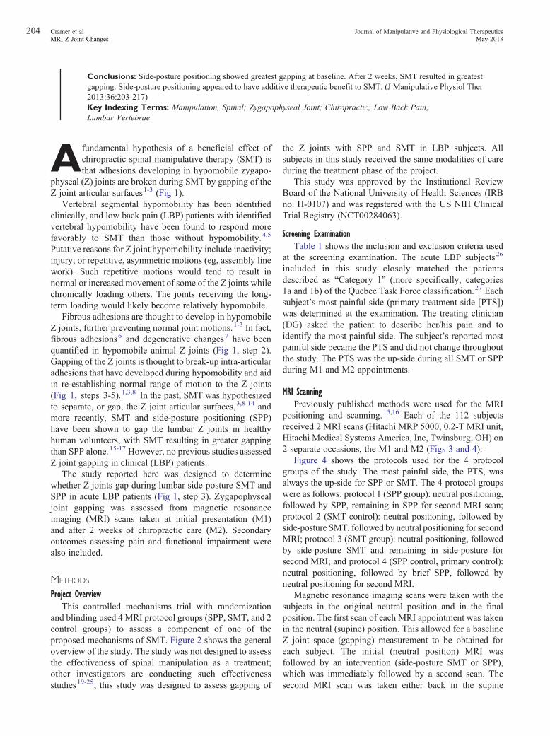
physeal (Z) joints are broken during SMT by gapping of theZ joint articular surfaces1-3 (Fig 1).
Vertebral segmental hypomobility has been identifiedclinically, and low back pain (LBP) patients with identifiedvertebral hypomobility have been found to respond morefavorably to SMT than those without hypomobility.4,5
Putative reasons for Z joint hypomobility include inactivity;injury; or repetitive, asymmetric motions (eg, assembly linework). Such repetitive motions would tend to result innormal or increased movement of some of the Z joints whilechronically loading others. The joints receiving the long-term loading would likely become relatively hypomobile.
Fibrous adhesions are thought to develop in hypomobileZ joints, further preventing normal joint motions.1-3 In fact,fibrous adhesions6 and degenerative changes7 have beenquantified in hypomobile animal Z joints (Fig 1, step 2).Gapping of the Z joints is thought to break-up intra-articularadhesions that have developed during hypomobility and aidin re-establishing normal range of motion to the Z joints(Fig 1, steps 3-5).1,3,8 In the past, SMT was hypothesizedto separate, or gap, the Z joint articular surfaces,3,8-14 andmore recently, SMT and side-posture positioning (SPP)have been shown to gap the lumbar Z joints in healthyhuman volunteers, with SMT resulting in greater gappingthan SPP alone.15-17 However, no previous studies assessedZ joint gapping in clinical (LBP) patients.
The study reported here was designed to determinewhether Z joints gap during lumbar side-posture SMT andSPP in acute LBP patients (Fig 1, step 3). Zygapophysealjoint gapping was assessed from magnetic resonanceimaging (MRI) scans taken at initial presentation (M1)and after 2 weeks of chiropractic care (M2). Secondaryoutcomes assessing pain and functional impairment werealso included.
METHODS
Project OverviewThis controlled mechanisms trial with randomization
and blinding used 4 MRI protocol groups (SPP, SMT, and 2control groups) to assess a component of one of theproposed mechanisms of SMT. Figure 2 shows the generaloverview of the study. The study was not designed to assessthe effectiveness of spinal manipulation as a treatment;other investigators are conducting such effectivenessstudies19-25; this study was designed to assess gapping of
the Z joints with SPP and SMT in LBP subjects. Allsubjects in this study received the same modalities of careduring the treatment phase of the project.
This study was approved by the Institutional ReviewBoard of the National University of Health Sciences (IRBno. H-0107) and was registered with the US NIH ClinicalTrial Registry (NCT00284063).
Screening ExaminationTable 1 shows the inclusion and exclusion criteria used
at the screening examination. The acute LBP subjects26
included in this study closely matched the patientsdescribed as “Category 1” (more specifically, categories1a and 1b) of the Quebec Task Force classification.27 Eachsubject's most painful side (primary treatment side [PTS])was determined at the examination. The treating clinician(DG) asked the patient to describe her/his pain and toidentify the most painful side. The subject's reported mostpainful side became the PTS and did not change throughoutthe study. The PTS was the up-side during all SMT or SPPduring M1 and M2 appointments.
MRI ScanningPreviously published methods were used for the MRI
positioning and scanning.15,16 Each of the 112 subjectsreceived 2 MRI scans (Hitachi MRP 5000, 0.2-T MRI unit,Hitachi Medical Systems America, Inc, Twinsburg, OH) on2 separate occasions, the M1 and M2 (Figs 3 and 4).
Figure 4 shows the protocols used for the 4 protocolgroups of the study. The most painful side, the PTS, wasalways the up-side for SPP or SMT. The 4 protocol groupswere as follows: protocol 1 (SPP group): neutral positioning,followed by SPP, remaining in SPP for second MRI scan;protocol 2 (SMT control): neutral positioning, followed byside-posture SMT, followed by neutral positioning for secondMRI; protocol 3 (SMT group): neutral positioning, followedby side-posture SMT and remaining in side-posture forsecond MRI; and protocol 4 (SPP control, primary control):neutral positioning, followed by brief SPP, followed byneutral positioning for second MRI.
Magnetic resonance imaging scans were taken with thesubjects in the original neutral position and in the finalposition. The first scan of each MRI appointment was takenin the neutral (supine) position. This allowed for a baselineZ joint space (gapping) measurement to be obtained foreach subject. The initial (neutral position) MRI wasfollowed by an intervention (side-posture SMT or SPP),which was immediately followed by a second scan. Thesecond MRI scan was taken either back in the supine
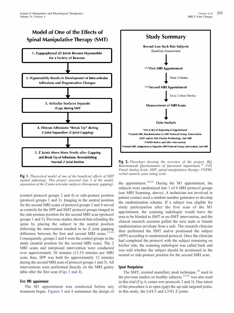
Fig 2. Flowchart showing the overview of the project. BQ,Bournemouth Questionnaire of functional impairment,18 VAS,Visual Analog Scale; SMT, spinal manipulative therapy; VNPRS,verbal numeric pain rating scale.
Fig 1. Theoretical model of one of the beneficial effects of SMT(spinal adjusting). This project assessed step 3 of the modelseparation of the Z joint articular surfaces (therapeutic gapping)
205Cramer et alJournal of Manipulative and Physiological TherapeuticsMRI Z Joint ChangesVolume 36, Number 4
,.
(control protocol groups 2 and 4) or side-posture position(protocol groups 1 and 3). Imaging in the neutral positionfor the second MRI scans of protocol groups 2 and 4 servedas controls for the SPP and SMT protocol groups imaged inthe side-posture position for the second MRI scan (protocolgroups 1 and 3). Previous studies showed that reloading thespine by placing the subject in the neutral positionfollowing the intervention resulted in no Z joint gappingdifference between the first and second MRI scans.15,16
Consequently, groups 2 and 4 were the control groups in thestudy (neutral position for the second MRI scan). The 2MRI scans and interposed intervention were conductedover approximately 30 minutes (12:19 minutes per MRIscan; thus, SPP was held for approximately 12 minutesduring the secondMRI scan of protocol groups 1 and 3). Allinterventions were performed directly on the MRI gantrytable after the first scan (Figs 3 and 4).
First MRI appointmentThe M1 appointment was conducted before any
treatment began. Figures 3 and 4 summarize the design of
the appointment.28,29 During the M1 appointment, thesubjects were randomized into 1 of 4 MRI protocol groups(see MRI Scanning, above). A technician not involved inpatient contact used a random number generator to developthe randomization scheme. If a subject was eligible forstudy participation after the first scan of the M1appointment, the scanning radiologist would leave thearea to be blinded to SMT or no-SMT intervention, and theclinical research assistant pulled the next male or femalerandomization envelope from a safe. The research clinicianthen performed the SMT and/or positioned the subject(SPP) according to randomized protocol. Once the clinicianhad completed the protocol with the subject remaining onhis/her side, the scanning radiologist was called back andwas told whether the subject should be positioned in theneutral or side-posture position for the second MRI scan.
Spinal ManipulationThe SMT, resisted mamillary push technique,30 used in
the previous studies on healthy subjects,15,16 was also usedin this trial (Fig 4, center row protocols 2 and 3). One intentof the procedure is to open (gap) the up-side targeted joints,in this study, the L4/L5 and L5/S1 Z joints.
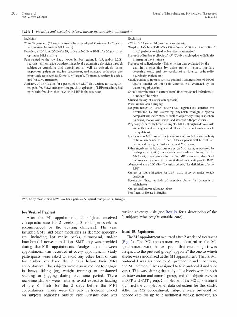
Table 1. Inclusion and exclusion criteria during the screening examination
Inclusion Exclusion
21 to 69 years old (21 years to ensure fully developed Z joints and b70 yearsto tolerate side-posture MRI scans)
Females, ≤160 lb or BMI of ≤28; males ≤200 lb or BMI of ≤30 (to ensureoptimum MRI quality)
Pain related to the low back (lower lumbar region, L4/L5, and/or L5/S1region)—this criterion was determined by the examining physician throughsubjective complaint and description as well as objectively usinginspection, palpation, motion assessment, and standard orthopedic andneurologic tests such as Kemp's, Milgram's, Yeoman's, straight-leg raise,and Valsalva maneuver.
A history of LBP lasting for a period of ≤6 wk;26 also defined as having ≥1mo pain free between current and previous episodes of LBP; must have hadmore pain free days than days with LBP in the past year.
b21 or ≥70 years old (see inclusion criteria)Weighs N160 lb or BMI N28 (if female) or N200 lb or BMI N30 (if
male) (subject weighed at baseline examination)Presence of lumbar scoliosis of N5° (Cobb's angle) (due to difficulty
in imaging the Z joints)Presence of radiculopathy (This criterion was evaluated by the
examining physician by using patient history, standardscreening tests, and the results of a detailed orthopedic/neurologic evaluation.)
Cauda equina symptoms such as perianal numbness, loss of bowel,and/or bladder control (This criterion was evaluated by theexamining physician.)
Spine deformity such as current spinal fractures, spinal infections, ortumors of the spine
Current history of severe osteoporosisPrior lumbar spine surgeryNo pain related to L4/L5 and/or L5/S1 region (This criterion was
determined by the examining physician through subjectivecomplaint and description as well as objectively using inspection,palpation, motion assessment, and standard orthopedic tests.)
Pregnancy or currently breastfeeding (for MRI, although no known risk,and in the event an x-ray is needed to screen for contraindications tomanipulation)
Intolerance to MRI procedures (including claustrophobia and inabilityto lie on one's side for 15 min). Claustrophobia will be evaluatedbefore and during the first and second MRI scans.
Other significant pathology discovered on MRI scans, as observed byreading radiologist. (This criterion was evaluated during the firstMRI visit, immediately after the first MRI scan was taken. Suchpathologies may constitute contraindications to chiropractic SMT.)
Absence of acute LBP (See “Inclusion criteria,” for definitions of acuteLBP.)
Current or future litigation for LBP (work injury or motor vehicleaccident)
Psychiatric illness or lack of cognitive ability (ie, dementia orAlzheimer)
Current and known substance abuseNot fluent or literate in English
BMI, body mass index; LBP, low back pain; SMT, spinal manipulative therapy.
206 Journal of Manipulative and Physiological TherapeuticsCramer et alMay 2013MRI Z Joint Changes
Two Weeks of TreatmentAfter the M1 appointment, all subjects received
chiropractic care for 2 weeks (1-3 visits per week asrecommended by the treating clinician). The careincluded SMT and other modalities as deemed appropri-ate, including hot moist packs, ultrasound, and/orinterferential nerve stimulation. SMT only was providedduring the MRI appointments. Analgesic use betweenappointments was recorded at every appointment. Studyparticipants were asked to avoid any other form of carefor his/her low back the 2 days before their MRIappointments. The subjects were also asked not to engagein heavy lifting (eg, weight training) or prolongedwalking or jogging during the same period. Theserecommendations were made to avoid excessive loadingof the Z joints for the 2 days before the MRIappointments. These were the only restrictions placedon subjects regarding outside care. Outside care was
tracked at every visit (see Results for a description of the3 subjects who sought outside care).
Second MRI AppointmentThe M2 appointment occurred after 2 weeks of treatment
(Fig 2). The M2 appointment was identical to the M1appointment with the exception that each subject wasassigned to the protocol group “opposite” the one to whichshe/he was randomized at the M1 appointment. That is, M1protocol 1 was assigned to M2 protocol 2 and vice versa,and M1 protocol 3 was assigned to M2 protocol 4 and viceversa. This way, during the study, all subjects were in bothan intervention and control group, and all subjects were inan SPP and SMT group. Completion of the M2 appointmentsignified the completion of data collection for this study.After the M2 appointment, subjects were provided asneeded care for up to 2 additional weeks; however, no
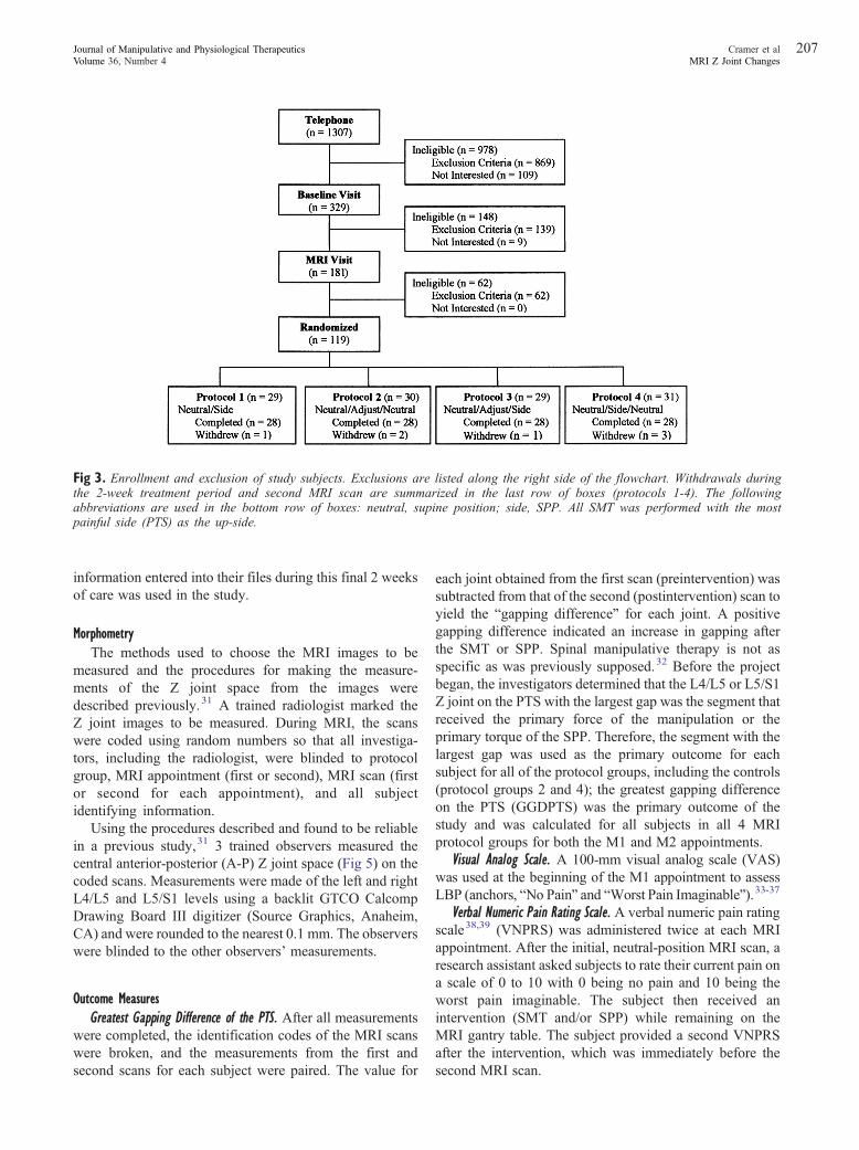
Fig 3. Enrollment and exclusion of study subjects. Exclusions are listed along the right side of the flowchart. Withdrawals duringthe 2-week treatment period and second MRI scan are summarized in the last row of boxes (protocols 1-4). The followingabbreviations are used in the bottom row of boxes: neutral, supine position; side, SPP. All SMT was performed with the mostpainful side (PTS) as the up-side.
207Cramer et alJournal of Manipulative and Physiological TherapeuticsMRI Z Joint ChangesVolume 36, Number 4
information entered into their files during this final 2 weeksof care was used in the study.
MorphometryThe methods used to choose the MRI images to be
measured and the procedures for making the measure-ments of the Z joint space from the images weredescribed previously.31 A trained radiologist marked theZ joint images to be measured. During MRI, the scanswere coded using random numbers so that all investiga-tors, including the radiologist, were blinded to protocolgroup, MRI appointment (first or second), MRI scan (firstor second for each appointment), and all subjectidentifying information.
Using the procedures described and found to be reliablein a previous study,31 3 trained observers measured thecentral anterior-posterior (A-P) Z joint space (Fig 5) on thecoded scans. Measurements were made of the left and rightL4/L5 and L5/S1 levels using a backlit GTCO CalcompDrawing Board III digitizer (Source Graphics, Anaheim,CA) and were rounded to the nearest 0.1 mm. The observerswere blinded to the other observers' measurements.
Outcome MeasuresGreatest Gapping Difference of the PTS. After all measurements
were completed, the identification codes of the MRI scanswere broken, and the measurements from the first andsecond scans for each subject were paired. The value for
each joint obtained from the first scan (preintervention) wassubtracted from that of the second (postintervention) scan toyield the “gapping difference” for each joint. A positivegapping difference indicated an increase in gapping afterthe SMT or SPP. Spinal manipulative therapy is not asspecific as was previously supposed.32 Before the projectbegan, the investigators determined that the L4/L5 or L5/S1Z joint on the PTS with the largest gap was the segment thatreceived the primary force of the manipulation or theprimary torque of the SPP. Therefore, the segment with thelargest gap was used as the primary outcome for eachsubject for all of the protocol groups, including the controls(protocol groups 2 and 4); the greatest gapping differenceon the PTS (GGDPTS) was the primary outcome of thestudy and was calculated for all subjects in all 4 MRIprotocol groups for both the M1 and M2 appointments.
Visual Analog Scale. A 100-mm visual analog scale (VAS)was used at the beginning of the M1 appointment to assessLBP (anchors, “No Pain” and “Worst Pain Imaginable”).33-37
Verbal Numeric Pain Rating Scale. A verbal numeric pain ratingscale38,39 (VNPRS) was administered twice at each MRIappointment. After the initial, neutral-position MRI scan, aresearch assistant asked subjects to rate their current pain ona scale of 0 to 10 with 0 being no pain and 10 being theworst pain imaginable. The subject then received anintervention (SMT and/or SPP) while remaining on theMRI gantry table. The subject provided a second VNPRSafter the intervention, which was immediately before thesecond MRI scan.
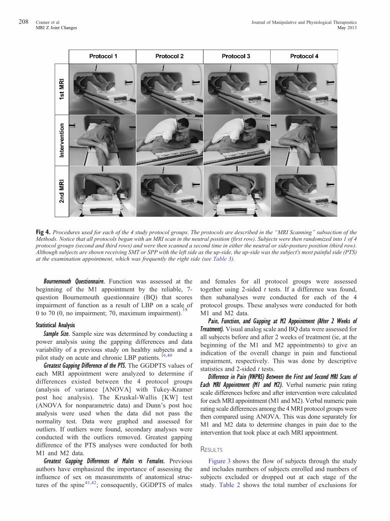
Fig 4. Procedures used for each of the 4 study protocol groups. The protocols are described in the “MRI Scanning” subsection of theMethods. Notice that all protocols began with an MRI scan in the neutral position (first row). Subjects were then randomized into 1 of 4protocol groups (second and third rows) and were then scanned a second time in either the neutral or side-posture position (third row)Although subjects are shown receiving SMT or SPP with the left side as the up-side, the up-side was the subject's most painful side (PTSat the examination appointment, which was frequently the right side (see Table 3).
208 Journal of Manipulative and Physiological TherapeuticsCramer et alMay 2013MRI Z Joint Changes
Bournemouth Questionnaire. Function was assessed at thebeginning of the M1 appointment by the reliable, 7-question Bournemouth questionnaire (BQ) that scoresimpairment of function as a result of LBP on a scale of0 to 70 (0, no impairment; 70, maximum impairment).
18
Statistical AnalysisSample Size. Sample size was determined by conducting a
power analysis using the gapping differences and datavariability of a previous study on healthy subjects and apilot study on acute and chronic LBP patients.16,40
Greatest Gapping Difference of the PTS. The GGDPTS values ofeach MRI appointment were analyzed to determine ifdifferences existed between the 4 protocol groups(analysis of variance [ANOVA] with Tukey-Kramerpost hoc analysis). The Kruskal-Wallis [KW] test(ANOVA for nonparametric data) and Dunn's post hocanalysis were used when the data did not pass thenormality test. Data were graphed and assessed foroutliers. If outliers were found, secondary analyses wereconducted with the outliers removed. Greatest gappingdifference of the PTS analyses were conducted for bothM1 and M2 data.
Greatest Gapping Differences of Males vs Females. Previousauthors have emphasized the importance of assessing theinfluence of sex on measurements of anatomical struc-tures of the spine41,42; consequently, GGDPTS of males
.)
and females for all protocol groups were assessedtogether using 2-sided t tests. If a difference was found,then subanalyses were conducted for each of the 4protocol groups. These analyses were conducted for bothM1 and M2 data.
Pain, Function, and Gapping at M2 Appointment (After 2 Weeks ofTreatment). Visual analog scale and BQ data were assessed forall subjects before and after 2 weeks of treatment (ie, at thebeginning of the M1 and M2 appointments) to give anindication of the overall change in pain and functionalimpairment, respectively. This was done by descriptivestatistics and 2-sided t tests.
Difference in Pain (VNPRS) Between the First and Second MRI Scans ofEach MRI Appointment (M1 and M2). Verbal numeric pain ratingscale differences before and after intervention were calculatedfor eachMRI appointment (M1 andM2). Verbal numeric painrating scale differences among the 4MRI protocol groupswerethen compared using ANOVA. This was done separately forM1 and M2 data to determine changes in pain due to theintervention that took place at each MRI appointment.
RESULTS
Figure 3 shows the flow of subjects through the studyand includes numbers of subjects enrolled and numbers ofsubjects excluded or dropped out at each stage of thestudy. Table 2 shows the total number of exclusions for
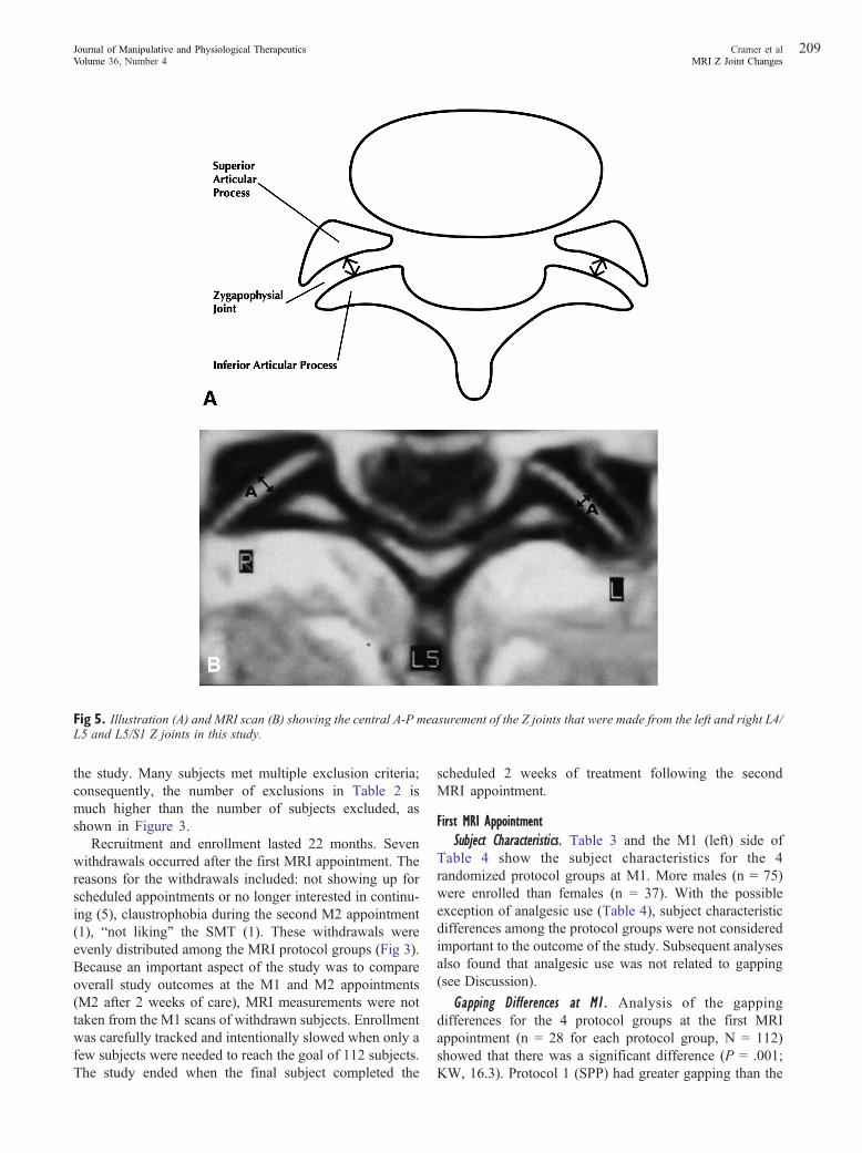
Fig 5. Illustration (A) and MRI scan (B) showing the central A-P measurement of the Z joints that were made from the left and right L4/L5 and L5/S1 Z joints in this study.
209Cramer et alJournal of Manipulative and Physiological TherapeuticsMRI Z Joint ChangesVolume 36, Number 4
the study. Many subjects met multiple exclusion criteria;consequently, the number of exclusions in Table 2 ismuch higher than the number of subjects excluded, asshown in Figure 3.
Recruitment and enrollment lasted 22 months. Sevenwithdrawals occurred after the first MRI appointment. Thereasons for the withdrawals included: not showing up forscheduled appointments or no longer interested in continu-ing (5), claustrophobia during the second M2 appointment(1), “not liking” the SMT (1). These withdrawals wereevenly distributed among the MRI protocol groups (Fig 3).Because an important aspect of the study was to compareoverall study outcomes at the M1 and M2 appointments(M2 after 2 weeks of care), MRI measurements were nottaken from the M1 scans of withdrawn subjects. Enrollmentwas carefully tracked and intentionally slowed when only afew subjects were needed to reach the goal of 112 subjects.The study ended when the final subject completed the
scheduled 2 weeks of treatment following the secondMRI appointment.
First MRI AppointmentSubject Characteristics. Table 3 and the M1 (left) side of
Table 4 show the subject characteristics for the 4randomized protocol groups at M1. More males (n = 75)were enrolled than females (n = 37). With the possibleexception of analgesic use (Table 4), subject characteristicdifferences among the protocol groups were not consideredimportant to the outcome of the study. Subsequent analysesalso found that analgesic use was not related to gapping(see Discussion).
Gapping Differences at M1. Analysis of the gappingdifferences for the 4 protocol groups at the first MRIappointment (n = 28 for each protocol group, N = 112)showed that there was a significant difference (P = .001;KW, 16.3). Protocol 1 (SPP) had greater gapping than the
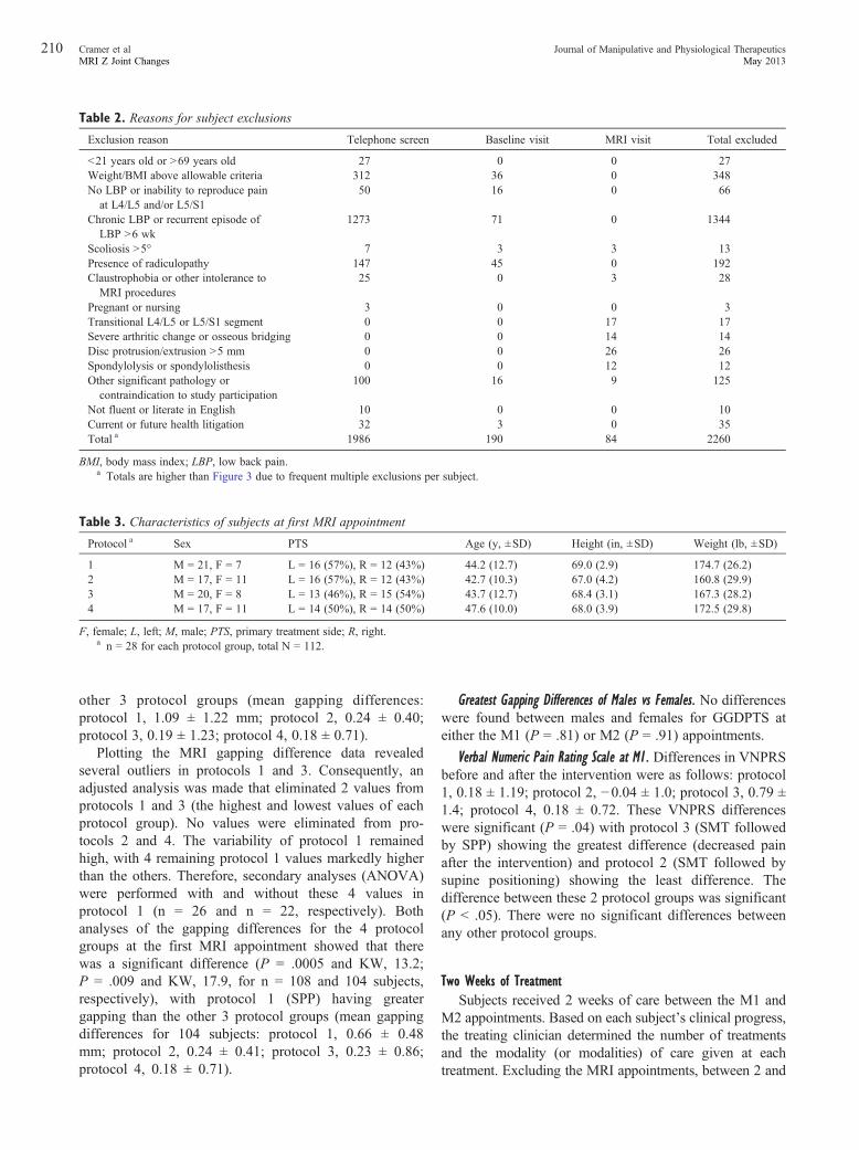
Table 2. Reasons for subject exclusions
Exclusion reason Telephone screen Baseline visit MRI visit Total excluded
b21 years old or N69 years old 27 0 0 27Weight/BMI above allowable criteria 312 36 0 348No LBP or inability to reproduce pain
at L4/L5 and/or L5/S150 16 0 66
Chronic LBP or recurrent episode ofLBP N6 wk
1273 71 0 1344
Scoliosis N5° 7 3 3 13Presence of radiculopathy 147 45 0 192Claustrophobia or other intolerance to
MRI procedures25 0 3 28
Pregnant or nursing 3 0 0 3Transitional L4/L5 or L5/S1 segment 0 0 17 17Severe arthritic change or osseous bridging 0 0 14 14Disc protrusion/extrusion N5 mm 0 0 26 26Spondylolysis or spondylolisthesis 0 0 12 12Other significant pathology or
contraindication to study participation100 16 9 125
Not fluent or literate in English 10 0 0 10Current or future health litigation 32 3 0 35Total a 1986 190 84 2260
BMI, body mass index; LBP, low back pain.a Totals are higher than Figure 3 due to frequent multiple exclusions per subject.
Table 3. Characteristics of subjects at first MRI appointment
Protocol a Sex PTS Age (y, ±SD) Height (in, ±SD) Weight (lb, ±SD)
1 M = 21, F = 7 L = 16 (57%), R = 12 (43%) 44.2 (12.7) 69.0 (2.9) 174.7 (26.2)2 M = 17, F = 11 L = 16 (57%), R = 12 (43%) 42.7 (10.3) 67.0 (4.2) 160.8 (29.9)3 M = 20, F = 8 L = 13 (46%), R = 15 (54%) 43.7 (12.7) 68.4 (3.1) 167.3 (28.2)4 M = 17, F = 11 L = 14 (50%), R = 14 (50%) 47.6 (10.0) 68.0 (3.9) 172.5 (29.8)
F, female; L, left; M, male; PTS, primary treatment side; R, right.a n = 28 for each protocol group, total N = 112.
210 Journal of Manipulative and Physiological TherapeuticsCramer et alMay 2013MRI Z Joint Changes
other 3 protocol groups (mean gapping differences:protocol 1, 1.09 ± 1.22 mm; protocol 2, 0.24 ± 0.40;protocol 3, 0.19 ± 1.23; protocol 4, 0.18 ± 0.71).
Plotting the MRI gapping difference data revealedseveral outliers in protocols 1 and 3. Consequently, anadjusted analysis was made that eliminated 2 values fromprotocols 1 and 3 (the highest and lowest values of eachprotocol group). No values were eliminated from pro-tocols 2 and 4. The variability of protocol 1 remainedhigh, with 4 remaining protocol 1 values markedly higherthan the others. Therefore, secondary analyses (ANOVA)were performed with and without these 4 values inprotocol 1 (n = 26 and n = 22, respectively). Bothanalyses of the gapping differences for the 4 protocolgroups at the first MRI appointment showed that therewas a significant difference (P = .0005 and KW, 13.2;P = .009 and KW, 17.9, for n = 108 and 104 subjects,respectively), with protocol 1 (SPP) having greatergapping than the other 3 protocol groups (mean gappingdifferences for 104 subjects: protocol 1, 0.66 ± 0.48mm; protocol 2, 0.24 ± 0.41; protocol 3, 0.23 ± 0.86;protocol 4, 0.18 ± 0.71).
Greatest Gapping Differences of Males vs Females. No differenceswere found between males and females for GGDPTS ateither the M1 (P = .81) or M2 (P = .91) appointments.
Verbal Numeric Pain Rating Scale at M1. Differences in VNPRSbefore and after the intervention were as follows: protocol1, 0.18 ± 1.19; protocol 2, −0.04 ± 1.0; protocol 3, 0.79 ±1.4; protocol 4, 0.18 ± 0.72. These VNPRS differenceswere significant (P = .04) with protocol 3 (SMT followedby SPP) showing the greatest difference (decreased painafter the intervention) and protocol 2 (SMT followed bysupine positioning) showing the least difference. Thedifference between these 2 protocol groups was significant(P b .05). There were no significant differences betweenany other protocol groups.
Two Weeks of TreatmentSubjects received 2 weeks of care between the M1 and
M2 appointments. Based on each subject's clinical progress,the treating clinician determined the number of treatmentsand the modality (or modalities) of care given at eachtreatment. Excluding the MRI appointments, between 2 and
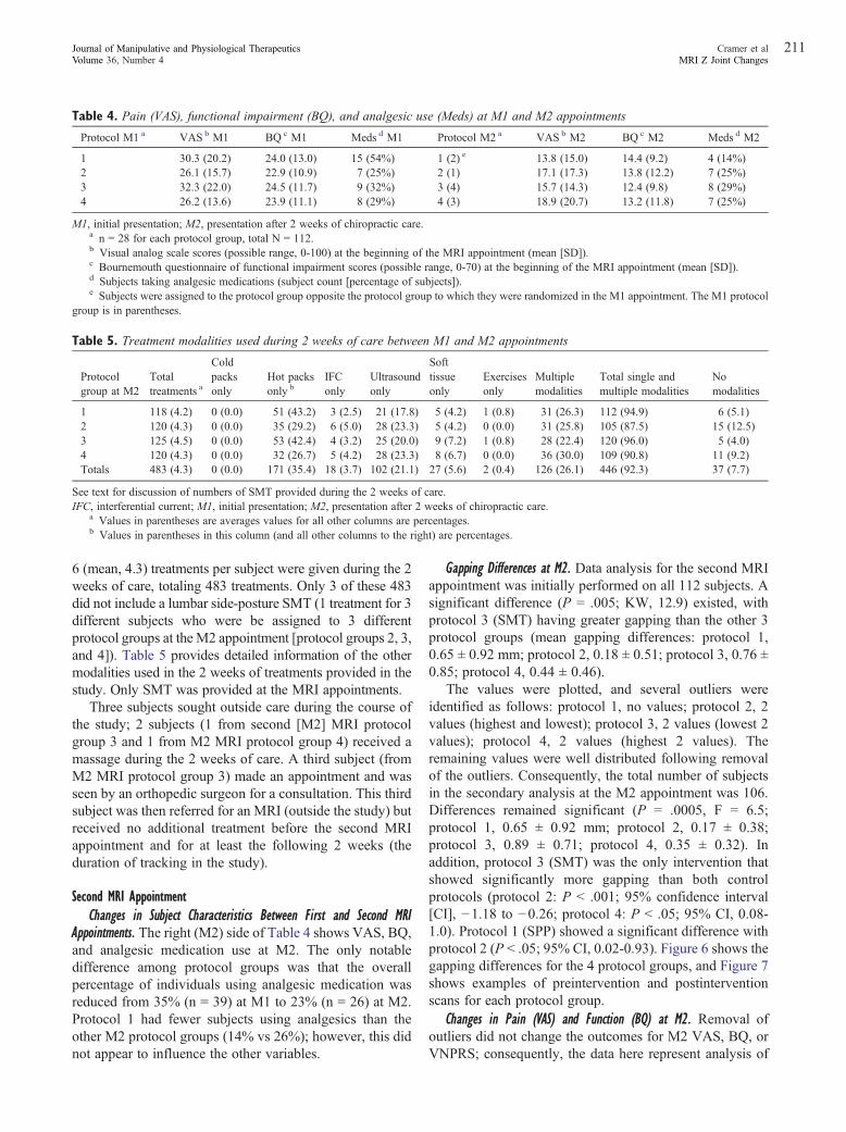
Table 4. Pain (VAS), functional impairment (BQ), and analgesic use (Meds) at M1 and M2 appointments
Protocol M1 a VAS b M1 BQ c M1 Meds d M1 Protocol M2 a VAS b M2 BQ c M2 Meds d M2
1 30.3 (20.2) 24.0 (13.0) 15 (54%) 1 (2) e 13.8 (15.0) 14.4 (9.2) 4 (14%)2 26.1 (15.7) 22.9 (10.9) 7 (25%) 2 (1) 17.1 (17.3) 13.8 (12.2) 7 (25%)3 32.3 (22.0) 24.5 (11.7) 9 (32%) 3 (4) 15.7 (14.3) 12.4 (9.8) 8 (29%)4 26.2 (13.6) 23.9 (11.1) 8 (29%) 4 (3) 18.9 (20.7) 13.2 (11.8) 7 (25%)
M1, initial presentation; M2, presentation after 2 weeks of chiropractic care.a n = 28 for each protocol group, total N = 112.b Visual analog scale scores (possible range, 0-100) at the beginning of the MRI appointment (mean [SD]).c Bournemouth questionnaire of functional impairment scores (possible range, 0-70) at the beginning of the MRI appointment (mean [SD]).d Subjects taking analgesic medications (subject count [percentage of subjects]).e Subjects were assigned to the protocol group opposite the protocol group to which they were randomized in the M1 appointment. The M1 protoco
group is in parentheses.
Table 5. Treatment modalities used during 2 weeks of care between M1 and M2 appointments
Protocolgroup at M2
Totaltreatments a
Coldpacksonly
Hot packsonly b
IFConly
Ultrasoundonly
Softtissueonly
Exercisesonly
Multiplemodalities
Total single andmultiple modalities
Nomodalities
1 118 (4.2) 0 (0.0) 51 (43.2) 3 (2.5) 21 (17.8) 5 (4.2) 1 (0.8) 31 (26.3) 112 (94.9) 6 (5.1)2 120 (4.3) 0 (0.0) 35 (29.2) 6 (5.0) 28 (23.3) 5 (4.2) 0 (0.0) 31 (25.8) 105 (87.5) 15 (12.5)3 125 (4.5) 0 (0.0) 53 (42.4) 4 (3.2) 25 (20.0) 9 (7.2) 1 (0.8) 28 (22.4) 120 (96.0) 5 (4.0)4 120 (4.3) 0 (0.0) 32 (26.7) 5 (4.2) 28 (23.3) 8 (6.7) 0 (0.0) 36 (30.0) 109 (90.8) 11 (9.2)Totals 483 (4.3) 0 (0.0) 171 (35.4) 18 (3.7) 102 (21.1) 27 (5.6) 2 (0.4) 126 (26.1) 446 (92.3) 37 (7.7)
See text for discussion of numbers of SMT provided during the 2 weeks of care.IFC, interferential current; M1, initial presentation; M2, presentation after 2 weeks of chiropractic care.
a Values in parentheses are averages values for all other columns are percentages.b Values in parentheses in this column (and all other columns to the right) are percentages.
211Cramer et alJournal of Manipulative and Physiological TherapeuticsMRI Z Joint ChangesVolume 36, Number 4
6 (mean, 4.3) treatments per subject were given during the 2weeks of care, totaling 483 treatments. Only 3 of these 483did not include a lumbar side-posture SMT (1 treatment for 3different subjects who were be assigned to 3 differentprotocol groups at the M2 appointment [protocol groups 2, 3,and 4]). Table 5 provides detailed information of the othermodalities used in the 2 weeks of treatments provided in thestudy. Only SMT was provided at the MRI appointments.
Three subjects sought outside care during the course ofthe study; 2 subjects (1 from second [M2] MRI protocolgroup 3 and 1 from M2 MRI protocol group 4) received amassage during the 2 weeks of care. A third subject (fromM2 MRI protocol group 3) made an appointment and wasseen by an orthopedic surgeon for a consultation. This thirdsubject was then referred for an MRI (outside the study) butreceived no additional treatment before the second MRIappointment and for at least the following 2 weeks (theduration of tracking in the study).
Second MRI AppointmentChanges in Subject Characteristics Between First and Second MRI
Appointments. The right (M2) side of Table 4 shows VAS, BQ,and analgesic medication use at M2. The only notabledifference among protocol groups was that the overallpercentage of individuals using analgesic medication wasreduced from 35% (n = 39) at M1 to 23% (n = 26) at M2.Protocol 1 had fewer subjects using analgesics than theother M2 protocol groups (14% vs 26%); however, this didnot appear to influence the other variables.
l
Gapping Differences at M2. Data analysis for the second MRIappointment was initially performed on all 112 subjects. Asignificant difference (P = .005; KW, 12.9) existed, withprotocol 3 (SMT) having greater gapping than the other 3protocol groups (mean gapping differences: protocol 1,0.65 ± 0.92 mm; protocol 2, 0.18 ± 0.51; protocol 3, 0.76 ±0.85; protocol 4, 0.44 ± 0.46).
The values were plotted, and several outliers wereidentified as follows: protocol 1, no values; protocol 2, 2values (highest and lowest); protocol 3, 2 values (lowest 2values); protocol 4, 2 values (highest 2 values). Theremaining values were well distributed following removalof the outliers. Consequently, the total number of subjectsin the secondary analysis at the M2 appointment was 106.Differences remained significant (P = .0005, F = 6.5;protocol 1, 0.65 ± 0.92 mm; protocol 2, 0.17 ± 0.38;protocol 3, 0.89 ± 0.71; protocol 4, 0.35 ± 0.32). Inaddition, protocol 3 (SMT) was the only intervention thatshowed significantly more gapping than both controlprotocols (protocol 2: P b .001; 95% confidence interval[CI], −1.18 to −0.26; protocol 4: P b .05; 95% CI, 0.08-1.0). Protocol 1 (SPP) showed a significant difference withprotocol 2 (P b .05; 95% CI, 0.02-0.93). Figure 6 shows thegapping differences for the 4 protocol groups, and Figure 7shows examples of preintervention and postinterventionscans for each protocol group.
Changes in Pain (VAS) and Function (BQ) at M2. Removal ofoutliers did not change the outcomes for M2 VAS, BQ, orVNPRS; consequently, the data here represent analysis of

Fig 6. Gapping differences (in millimeters, Y-axis) between the A-P Z joint space measurements of the first and second MRI scans(value from the first scan was subtracted from the value of thesecond scan) at the M2 appointment. The GGDPTS are presentedhere. Protocol 3 (SMT protocol groups) showed more gapping(therapeutic gapping) than the other protocol groups, followed byprotocol 1 (SPP protocol group).
212 Journal of Manipulative and Physiological TherapeuticsCramer et alMay 2013MRI Z Joint Changes
all 112 subjects. Visual analog scale changed from 28.8 ±18.16 mm (range, 1-82 mm) at the first MRI appointment to16.4 ± 16.88 mm (range, 0-81 mm) at the second MRIappointment, a difference of 12.4 mm (P b .0001).Bournemouth questionnaire changed from 23.8 ± 11.56(range, 3-54) at the first MRI appointment to 13.4 ± 10.69(range, 0-48) at the second MRI appointment, a differenceof 10.4 (P b .0001).
Verbal Numeric Pain Rating Scale at M2. Verbal numeric painrating scale preintervention values were low (little painreported) at M2, and differences in VNPRS before and afterthe intervention at M2 were not significant (P = .41).
No changes were made to the protocols, and no adverseevents, harms, or unintended effects were reported duringthe course of the study.
DISCUSSION
Approximately twice as many males (n = 75) wereenrolled as females (n = 37). This representation is differentthan is usually found in chiropractic practices, which have aslightly higher percentage of female patients.43-45 Theincidence of LBP in the general population is also slightlyhigher in females.46 The higher number of males in thisstudy was a reflection of the numbers of males (n = 798)and females (n = 497) who responded to the recruitingadvertisements (12 subjects declined before any data wererecorded). Although recruitment for this study was based onmethods used successfully in previous clinical trials,47,48
future studies will include more advertisements that targetfemales and run on media and programming with a higherfemale demographic. Because no difference was found ingapping between male and female subjects at either the M1or M2 appointments, the higher percentage of males mostlikely did not affect the outcome of the study.
First MRI AppointmentSubjects were allowed to take analgesic medications,
both over-the-counter and previously prescribed prescrip-tion medication, and medication use between appoint-ments was documented at every visit. Although a muchhigher percentage of M1 protocol 1 subjects were takinganalgesic medication, those protocol 1 subjects takinganalgesic medications had only 0.16 mm more gappingthan those not taking analgesic medications; the GGDPTSfor patients not taking and taking analgesic medicationswas 1.00 and 1.16 mm, respectively. Removal of protocolgroup 1 subjects taking analgesic medication did not alterthe results.
First MRI appointment protocol 1 (SPP only) subjectsshowed more gapping than the other protocol groups,including protocol 3 (side-posture SMT followed by SPP).Paraspinal muscles may have relaxed more during pro-longed SPP in protocol 1, whereas SMT may have resultedin transient increased muscle tightness in protocol 3. Thisrelationship reversed at the second MRI appointment (M2)with protocol 3 showing more gapping than protocol 1. Theincreased gapping of M2 protocol 3 subjects may indicatethat the paraspinal muscles were more relaxed after 2 weeksof treatment (including SMT) at the M2 appointmentallowing more Z joint gapping with SMT.
Protocol 3 (SMT followed by SPP) was the onlyprotocol group to show significant improvement in pain(VNPRS), whereas those subjects receiving side-postureSMT and then placed on their backs (protocol 2) hadalmost no change in pain following the intervention.These results indicate that the lumbar side-postureposition may have therapeutic benefit in acute LBP,increasing gapping in patients in acute pain (protocol 1)and enhancing pain reduction following SMT (protocol3). This is consistent with the common recommendationby clinicians and the literature that lying on the side is ofbenefit for LBP patients.49-54 The increased gapping thatoccurred with prolonged SPP (ie, SPP of approximately12 minutes) could conceivably promote the break-up ofintra-articular Z joint adhesions.6 In addition, like SMTalone, SMT followed by SPP may also reduce pain bystimulating mechanoreceptors in the Z joint capsules55-57
and paraspinal muscles.58,59 Stimulation of such Z jointmechanoreceptors has been hypothesized as a mechanismof reducing pain via a gating mechanism in the spinalcord. This hypothesis is supported by animal studiesshowing that pressure on the Z joint (including thecapsule) decreases activity of spinal cord dorsal hornneurons responding to nociceptive stimulation.60
Two Weeks of TreatmentThe treatments in this study could be described as
“structured pragmatic” in nature. Because the purpose of thestudy was to assess Z joint gapping at initial LBP
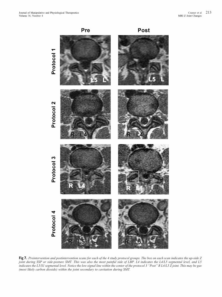
Fig 7. Preintervention and postintervention scans for each of the 4 study protocol groups. The box on each scan indicates the up-side Zjoint during SSP or side-posture SMT. This was also the most painful side of LBP. L4 indicates the L4/L5 segmental level, and L5indicates the L5/S1 segmental level. Notice the low signal line within the center of the protocol 3 “Post” R L4/L5 Z joint. This may be gas(most likely carbon dioxide) within the joint secondary to cavitation during SMT.
213Cramer et alJournal of Manipulative and Physiological TherapeuticsMRI Z Joint ChangesVolume 36, Number 4
214 Journal of Manipulative and Physiological TherapeuticsCramer et alMay 2013MRI Z Joint Changes
presentation and after clinical improvement at 2 weeks, thepurpose of the treatment was to provide usual chiropracticcare that would result in the maximum improvementfollowing 2 weeks of treatment. Spinal manipulativetherapy was provided in almost all treatment appointments(only 3 of 483 treatments did not receive SMT). The studyclinician was given the latitude to determine the number oftreatments per week, within the parameters of 1 to 3treatments, and choose from a “menu” of other modalitiesthat could be used.
The goals of the treatments were successfully achieved.The results show that the subjects all received approxi-mately the same number of treatments, SMT, and similarnumbers and types of “other modalities.” In addition, after 2weeks of care, the subjects showed approximately the sameimprovement as measured by pain and functional impair-ment. Therefore, the study was able to measure Z jointgapping in a homogenous cohort at initial presentation andafter similar improvement of LBP following 2 weeks ofcare. Recall, this was not a study assessing effectiveness ofcare (although all subjects significantly improved following2 weeks of care) but was designed to assess Z joint gappingin acute LBP patients.
Second MRI AppointmentConsistent with previous studies,15,16 the Z joints
resumed their normal spacing once they received the loadof the supine position, explaining why protocol 4 (brief SPPfollowed by neutral/supine position [SPP control group])and protocol 2 (SMT followed by neutral/supine position[SMT control group]) showed little gapping.
Spinal manipulative therapy followed by SPP (protocol3) resulted in the greatest amount of Z joint gapping in LBPsubjects at the M2 appointment, followed by SPP (protocol1). These data indicate that SMT produced more gappingafter subjects received 2 weeks of treatment. One wouldanticipate even greater differences between protocol 1(SPP) and protocol 3 (SMT) mean gapping differences ifthe subjects had been assessed with a third MRIappointment after 4 weeks of care. The subjects wouldthen have been more similar in pain and function to thehealthy subjects assessed in previous studies, whereprotocol 3 showed 0.7 mm more gapping than protocol1.15,16 The increased gapping after 2 weeks of care in thisstudy could have been due to reduction of intra-articularadhesions (and potentially other connective tissue adhe-sions, including adhesions within the fascia) and reducedmuscle tension of the paraspinal muscles surrounding the Zjoints. Future studies should assess changes in muscleactivity, as measured by electromyography, at the M1 andM2 appointments.
The gapping changes were also accompanied with anoverall reduction of pain and improved functional impair-ment. The protocol 3 (SMT followed by SPP) findings alsoindicate that keeping a person in the side-posture position
for several minutes following SMT may have therapeuticbenefit. Future work in animals and humans should furtherassess the unique effects of SPP alone and SMT followedby SPP.
This study provides additional evidence that normal Zjoints (ie, Z joints within normal anatomical limits; recallthat subjects with anomalous Z joints were excluded fromthis study) gap with SMT and SPP, which is different fromconclusions of previous authors61 who believed that Zjoints that were within normal anatomical limits do not gap.These previous authors strongly indicated that chiropractorswere misinforming their patients when describing Z jointgapping as a mechanism of SMT. The study conducted bythe other investigators placed the cadaveric spines in a moreextended posture, which significantly reduces Z jointrotation, and, consequently, Z joint gapping. The standardSMT of this study is administered with the Z joints in aflexed position, which allows for rotation62-64 and gapping.The results of this and previous studies15,16 indicate thattypical Z joints do gap with SMT and SPP. The evidencethat Z joints do gap can lead to a different approach topatient care than an assumption that they do not gap. Whencombined with other studies showing that adhesionsdevelop in hypomobile Z joints6 and LBP patients withclinical hypomobility respond favorably to SMT,4,5 theresults of this study further buttress the theory provided inFigure 1. This theory begins with the a priori assumptionthat Z joints become hypomobile for a variety of reasons(eg, sedentary lifestyle, injury, repetitive asymmetricaltasks at work) and that hypomobile Z joints developadhesions, which further reduces motion; SMT gaps the Zjoint surfaces, thus breaking up Z joint adhesions andreestablishing spinal motion.
LIMITATIONS
This study was conducted on the lumbar spine. Additionalresearch assessing the cervical and thoracic regions is neededto determine the effects of positioning and gapping of the Zjoints in these regions of the vertebral column.
As discussed previously, future studies should targetfemale subjects for recruitment to obtain a more equaldistribution of male and female subjects.
The ideal design would have been 4 MRI appointments:1 before commencement of treatment (M1 in this study), 1after 1 week of treatment, 1 after 2 weeks of treatment (M2in this study), and 1 after 4 weeks of treatment. This wouldnot only have allowed for assessment of gapping atadditional time points in the LBP continuum but wouldalso have allowed each subject to be in each of the 4protocol groups. However, 4 MRI appointments could notbe justified from cost and patient burden standpoints. Inaddition, future research assessing gapping differences insubjects with chronic LBP should be performed. Future
215Cramer et alJournal of Manipulative and Physiological TherapeuticsMRI Z Joint ChangesVolume 36, Number 4
work could also include a medication only control treatmentprotocol group.
CONCLUSION
In this study of acute LBP subjects, SPP subjects(protocol 1) showed the greatest Z joint gapping at thebaseline MRI appointment. After 2 weeks of standardchiropractic treatment, SMT followed by SPP (protocol 3)resulted in the greatest amount of Z joint gapping, followedby SPP alone (protocol 1); these results are consistent withthose of previous studies on healthy subjects.15,16 The side-posture position appeared to have additive therapeuticbenefit to SMT, with acute LBP subjects receiving SMTand remaining in side-posture experiencing the greatestreduction of pain, independent of Z joint gapping, at thefirst appointment and the greatest amount of Z joint gappingafter 2 weeks of care.
Practical Applications• Zygapophyseal joint gapping is hypothesized to berelated to a therapeutic benefit of SMT (“thera-peutic gapping”).
• Previous studies of healthy subjects found that Zjoints receiving SMT gapped more than thosereceiving SPP alone.
• In this study of acute LBP subjects, SPP showedthe greatest Z joint gapping at the baseline MRIappointment.
• After 2 weeks of standard chiropractic treatment,SMT followed by SPP resulted in the greatestamount of Z joint gapping, followed by SPP alone.
• The side-posture position appeared to haveadditive benefit to SMT regarding pain reductionand Z joint gapping.
ACKNOWLEDGMENT
We gratefully acknowledge the technical support ofJoshua Healy, DC, and the observers who made themorphometric measurements: Frank Balester, MSOM,LAc; Tyra Horner, DC; and Derek Simpson, DC, ND.
FUNDING SOURCES AND POTENTIAL CONFLICTS OF INTEREST
This project was made possible by grant number R01-AT000123 from the National Center for Complementaryand Alternative Medicine. Its contents are solely theresponsibility of the authors and do not necessarilyrepresent the official views of the National Center forComplementary and Alternative Medicine or the National
Institutes of Health. No conflict of interest was reported byany of the authors.
REFERENCES
1. Janse J. In: Hildebrandt RW, editor. Principles and practice ofchiropractic: an anthology. Wheaton: Kjellberg & Sons; 1976.p. 326.
2. Triano JJ. Interaction of spinal biomechanics and physiology.In: Haldeman S, editor. Principles and practice of chiropractic.2nd ed. East Norwalk, Conn: Appleton & Lange; 1992. p.225-57.
3. Evans DW. Mechanisms and effects of spinal high-velocity,low-amplitude thrust manipulation: previous theories. JManipulative Physiol Ther 2002;25:251-62.
4. Flynn T, Fritz J, Whitman J, et al. A clinical prediction rule forclassifying patients with low back pain who demonstrateshort-term improvement with spinal manipulation. Spine(Phila Pa 1976) 2002;27:2835-43.
5. Fritz JM, Whitman JM, Childs JD. Lumbar spine segmentalmobility assessment: an examination of validity for determin-ing intervention strategies in patients with low back pain. ArchPhys Med Rehabil 2005;86:1745-52.
6. Cramer GD, Henderson CN, Little JW, Daley C, Grieve TJ.Zygapophyseal joint adhesions after induced hypomobility. JManipulative Physiol Ther 2010;33:508-18.
7. Cramer GD, Fournier JT, Henderson CN, Wolcott CC.Degenerative changes following spinal fixation in a smallanimal model. J Manipulative Physiol Ther 2004;27:141-54.
8. Sandoz R. Some physical mechanisms and effects of spinaladjustments. Ann Swiss Chiropr Assoc 1976;6:91-141.
9. Cassidy J, Kirkaldy-Willis W. Manipulation. In: Kirkaldy-Willis W, Burton V, editors. Managing low back pain. 3rd ed.New York: Churchill Livingstone; 1992. p. 283-96.
10. Engel R, Bogduk N. The menisci of the lumbar zygapophysialjoints. J Anat 1982;135(Pt 4):795-809.
11. Giles LG, Taylor JR. Human zygapophyseal joint capsule andsynovial fold innervation. Br J Rheumatol 1987;26:93-8.
12. Kirkaldy-Willis W. The pathology and pathogenesis of lowback pain. In: Kirkaldy-Willis WH, Burton V, editors.Managing low back pain. 3rd ed. New York: ChurchillLivingstone; 1992. p. 283-96.
13. Kos J, Wolf J. Les menisques intervertebraux et le rolepossible dans les blocages vertebraux (translation). J OrthopSports Phys Ther 1972;1:8-9.
14. Sandoz R. Some reflex phenomena associated with spinalderangements and adjustments. Ann Swiss Chiropr Assoc 1981;7:45-65.
15. Cramer GD, Tuck NR, Knudsen JT, et al. Effects of side-posture positioning and side-posture adjusting on the lumbarzygapophysial joints as evaluated by magnetic resonanceimaging: a before and after study with randomization. JManipulative Physiol Ther 2000;23:380-94.
16. Cramer GD, Gregerson DM, Knudsen JT, Hubbard BB, UstasLM, Cantu JA. The effects of side-posture positioning and spinaladjusting on the lumbar Z joints: a randomized controlled trialwith sixty-four subjects. Spine (Phila Pa 1976) 2002;27:2459-66.
17. Cramer GD, Ross K, Pocius J, et al. Evaluating therelationship among cavitation, zygapophyseal joint gapping,and spinal manipulation: an exploratory case series. JManipulative Physiol Ther 2011;34:2-14.
18. Bolton JE, Breen AC. The Bournemouth Questionnaire: ashort-form comprehensive outcome measure. I. Psychometric
216 Journal of Manipulative and Physiological TherapeuticsCramer et alMay 2013MRI Z Joint Changes
properties in back pain patients. J Manipulative Physiol Ther1999;22:503-10.
19. Bronfort G, Evans RL, Maiers M, Anderson AV. Spinalmanipulation, epidural injections, and self-care for sciatica: apilot study for a randomized clinical trial. J ManipulativePhysiol Ther 2004;27:503-8.
20. Bronfort G, Goldsmith CH, Nelson CF, Boline PD, AndersonAV. Trunk exercise combined with spinal manipulative orNSAID therapy for chronic low back pain: a randomized,observer-blinded clinical trial. J Manipulative Physiol Ther1996;19:570-82.
21. Bronfort G, Haas M, Evans R, Kawchuk G, Dagenais S.Evidence-informed management of chronic low back painwith spinal manipulation and mobilization. Spine J 2008;8:213-25.
22. Bronfort G, Haas M, Evans R, Leiniger B, Triano J.Effectiveness of manual therapies: the UK evidence report.Chiropr Osteopath 2010;18:3.
23. Bronfort G, Haas M, Evans RL, Bouter LM. Efficacy of spinalmanipulation and mobilization for low back pain and neckpain: a systematic review and best evidence synthesis. Spine J2004;4:335-56.
24. Haas M, Goldberg B, Aickin M, Ganger B, Attwood M. Apractice-based study of patients with acute and chronic lowback pain attending primary care and chiropractic physicians:two-week to 48-month follow-up. J Manipulative PhysiolTher 2004;27:160-9.
25. Haas M, Groupp E, Kraemer DF. Dose-response forchiropractic care of chronic low back pain. Spine J 2004;4:574-83.
26. Frymoyer J. Back pain and sciatica. N Engl J Med 1988;318:291-300.
27. Spitzer WO, Le Blanc FE, Dupuis M, et al. Quebec TaskForce on Spinal Disorders. Scientific approach to theassessment and management of activity-related spinal disor-ders: a monograph for clinicians. Spine (Phila Pa 1976) 1987;12(7 Suppl):S1-S59.
28. Schulz KF, Altman DG, Moher D. CONSORT 2010statement: updated guidelines for reporting parallelgroup randomized trials. Ann Intern Med 2010;152:726-32.
29. Moher D, Hopewell S, Schulz KF, et al. CONSORT 2010explanation and elaboration: updated guidelines for reportingparallel group randomised trials. BMJ 2010;340:c869.
30. Peterson D, Bergmann T. Chiropractic technique. 3rd ed. NewYork: Churchill Livingstone; 2002. p. 810.
31. Cramer GD, Cantu JA, Pocius JD, Cambron JA, McKinnisRA. Reliability of zygapophysial joint space measurementsmade from magnetic resonance imaging scans of acute lowback pain subjects: comparison of 2 statistical methods. JManipulative Physiol Ther 2010;33:220-5.
32. Ross JK, Bereznick DE, McGill SM. Determining cavitationlocation during lumbar and thoracic spinal manipulation: isspinal manipulation accurate and specific? Spine 2004;29:1452-7.
33. Dixon JS, Bird HA. Reproducibility along a 10 cm verticalvisual analogue scale. Ann Rheum Dis 1981;40:87-9.
34. Carlsson AM. Assessment of Chronic Pain. I. Aspects of thereliability and validity of the visual analog scale. Pain 1983;16:87-101.
35. Machin D, Lewith GT, Wylson S. Pain measurement inrandomized clinical trials: a comparison of two pain scales.Clin J Pain 1988;4:161-8.
36. Love A, Leboeuf C, Crisp TC. Chiropractic chronic low backpain sufferers and self-report assessment methods. Part I. Areliability study of the Visual Analogue Scale, the Pain
Drawing, and the McGill Pain Questionnaire. J ManipulativePhysiol Ther 1989;12:21-5.
37. Giles LG, Muller R. Chronic spinal pain syndromes: a clinicalpilot trial comparing acupuncture, a nonsteroidal anti-inflammatory drug, and spinal manipulation. J ManipulativePhysiol Ther 1999;22:376-81.
38. Von Korff M, Jensen MP, Karoly P. Assessing global painseverity by self-report in clinical and health services research.Spine (Phila Pa 1976) 2000;25:3140-51.
39. Dworkin RH, Turk DC, Farrar JT, et al. Core outcomemeasures for chronic pain clinical trials: IMMPACT recom-mendations. Pain 2005;113:9-19.
40. Cramer GD, Wolcott CC, Cantu J, et al. The effects of side-posture adjusting on the lumbar zygapophysial joints of lowback pain patients as evaluated by magnetic resonanceimaging: a preliminary study. J Chiropr Educ 2004;18:4.
41. FujiwaraA, LimTH,AnHS, et al. The effect of disc degenerationand facet joint osteoarthritis on the segmental flexibility of thelumbar spine. Spine (Phila Pa 1976) 2000;25:3036-44.
42. Nachemson AL, Schultz AB, Berkson MH. Mechanicalproperties of human lumbar spine motion segments. Influenceof age, sex, disc level, and degeneration. Spine (Phila Pa1976) 1979;4:1-8.
43. Coulter ID, Hurwitz EL, Adams AH, Genovese BJ, Hays R,Shekelle PG. Patients using chiropractors in North America:who are they, and why are they in chiropractic care? Spine(Phila Pa 1976) 2002;27:291-6 [discussion 7-8].
44. Mootz RD, Cherkin DC, Odegard CE, Eisenberg DM, BarassiJP, Deyo RA. Characteristics of chiropractic practitioners,patients, and encounters in Massachusetts and Arizona. JManipulative Physiol Ther 2005;28:645-53.
45. Ndetan HT, Bae S, Evans MW, Rupert RL, Singh KP.Characterization of health status and modifiable risk behavioramong United States adults using chiropractic care ascompared with general medical care. J Manipulative PhysiolTher 2009;32:414-22.
46. Waterman BR, Belmont PJ, Schoenfeld AJ. Low back pain inthe United States: incidence and risk factors for presentation inthe emergency setting. Spine J 2012;12:63-70.
47. Gudavalli MR, Cambron JA, McGregor M, et al. Arandomized clinical trial and subgroup analysis to compareflexion-distraction with active exercise for chronic low backpain. Eur Spine J 2006;15:1070-82.
48. Cambron JA, Dexheimer JM, Chang M, Cramer GD.Recruitment methods and costs for a randomized, placebo-controlled trial of chiropractic care for lumbar spinal stenosis:a single-site pilot study. J Manipulative Physiol Ther 2010;33:56-61.
49. Gracovetsky SA. The resting spine. A conceptual approach tothe avoidance of spinal reinjury during rest. Phys Ther 1987;67:549-53.
50. Finneson BE. Low back pain. 2nd ed. Hagerstown: LippincottWilliams & Wilkins; 1981. p. 597.
51. Normand MC, Descarreaux M, Poulin C, et al. Biomechanicaleffects of a lumbar support in a mattress. J Can Chiropr Assoc2005;49:96-101.
52. Burkhart L. Proper sleep ergonomics. J Am Chiropr Assoc2011;48:1-2.
53. NASS. Back Pain Basics. Burr Ridge, IL: North AmericanSpine Society; 2007. Available from: http://www.knowyourback.org/documents/back_pain_basics_web.pdf.
54. DeVocht JW, Wilder DG, Bandstra ER, Spratt KF. Biome-chanical evaluation of four different mattresses. Appl Ergon2006;37:297-304.
55. Ianuzzi A, Khalsa PS. Comparison of human lumbar facetjoint capsule strains during simulated high-velocity, low-
217Cramer et alJournal of Manipulative and Physiological TherapeuticsMRI Z Joint ChangesVolume 36, Number 4
amplitude spinal manipulation versus physiological motions.Spine J 2005;5:277-90.
56. Ianuzzi A, Pickar JG, Khalsa PS. Relationships between jointmotion and facet joint capsule strain during cat and humanlumbar spinal motions. J Manipulative Physiol Ther 2011;34:420-31.
57. McLain RF, Pickar JG. Mechanoreceptor endings in humanthoracic and lumbar facet joints. Spine (Phila Pa 1976) 1998;23:168-73.
58. Pickar JG, Wheeler JD. Response of muscle proprioceptors tospinal manipulative-like loads in the anesthetized cat. JManipulative Physiol Ther 2001;24:2-11.
59. Sung PS, Kang YM, Pickar JG. Effect of spinal manipulationduration on low threshold mechanoreceptors in lumbarparaspinal muscles. Spine 2004;30:115-22.
60. Gillette RG, Kramis RC, Roberts WJ. Suppression ofactivity in spinal nocireceptive “low back” neurons byparavertebral somatic stimuli in the cat. Neurosci Lett 1998;241:45-8.
61. McFadden KD, Taylor JR. Axial rotation in the lumbarspine and gaping of the zygapophyseal joints. Spine 1990;15:295-9.
62. White AA, Panjabi MM. Clinical biomechanics of the spine.2nd ed. Philadelphia: JB Lippincott; 1990.
63. Williams PL, Banniser LH, Berry MM, et al. Gray's anatomy.Edinburgh: Churchill Livingstone; 1995.
64. Kapandji IA. The physiology of the joints. Anno-tated diagrams of the mechanics of the humanjoints. 6th ed. Edinburgh: Churchill Livingstone;2008.