Embed Size (px)
Citation preview
PICTORIAL REVIEW
Magnetic resonance imaging of pineal region tumours
Adam S. Fang & Steven P. Meyers
Received: 18 October 2012 /Revised: 19 March 2013 /Accepted: 28 March 2013 /Published online: 3 May 2013# The Author(s) 2013. This article is published with open access at Springerlink.com
AbstractObjectives Pineal lesions can present as a heterogeneous col-lection of benign and malignant disease conditions. Pineallesions include germ cell tumours, neoplasms arising fromthe pineal parenchyma, as well as other pineal region masses.Methods A variety of cases of pineal lesions are presented.The important clinical features and typical imaging findingsof each pineal lesion are described with emphasis on theirmorphological appearance and signal intensity characteris-tics on magnetic resonance imaging (MRI).Conclusion Knowledge of the imaging characteristics andclinical features of varying pineal lesions can assist innarrowing the differential diagnosis for more accurate andrational therapeutic planning.Teaching Points• Pineal parenchymal tumours show an “explosion” ofnormal pineal calcifications towards the periphery.
• Pineoblastomas often have restricted diffusion, with ap-parent diffusion coefficient (ADC) values lower thangerminomas.
• Pineal teratomas and pineal lipomas display fat signalcharacteristics and fat saturation on MRI.
• Pineal lesions in patients with known malignancy shouldraise suspicion of metastatic involvement.
• Pineal cysts and arachnoid cysts show MRI signal char-acteristics similar to cerebrospinal fluid (CSF).
Keywords Pineal gland . Pineal tumors . Pineal neoplasm .
Pineal parenchyma tumors . MRI
Introduction
A wide range of lesions can arise in the pineal region, suchas tumours of the pineal parenchyma, germ cell tumours,
metastasis and cell types adjacent to the pineal gland(Table 1). Pineal lesions account for 1 % of intracranialtumours in adults and 3–8 % in children [1, 2].
The normal pineal gland appears as a small reddish-brown structure with a pine cone-like shape. The normalsize ranges between 10 and 14 mm [3]. It is commonlylocated along the midline above the superior colliculi andinferior to the splenium of the corpus callosum. It is at-tached to the superior aspect of the posterior border of thethird ventricle. The pineal gland plays a major role inregulating the wake/sleep cycle, photoperiodic (seasonal)functions and release of hormones, such as luteinising andfollicular stimulating hormone, through the secretion ofmelatonin.
Two primary cell types make up the pineal gland. Thepineocyte is the principal parenchyma cell and comprises95 % of the pineal gland. The other 5 % are supportingcells referred to as astrocytes. Together, these two celltypes are arranged in lobules, which are separated by afibrovascular stroma. Calcifications commonly occurwithin the pineal gland and are often associated withincreasing age [4].
Clinical presentation of pineal lesions
Signs and symptoms related to pineal lesions are generallysecondary to mass effect on adjacent structures. Compres-sion of the tectal plate can result in obstruction of the sylvianaqueduct and obstructive hydrocephalus. Cerebellar,corticospinal or sensory disturbance can result from directcompression of the midbrain [5]. Infiltrative lesions of thepineal gland may interfere with normal pineal gland func-tion and lead to precocious puberty, less commonlyhypogonadism and diabetes insipidus. Non-specific symp-toms, such as seizures, headaches, nausea and vomiting, canoccur due to increased intracranial pressure (ICP). If thepineal lesion invokes pressure on the reflex nuclei of thequadrigeminal plate, patients may develop Parinaud’s syn-drome (also known as dorsal midbrain syndrome), which
A. S. Fang (*) : S. P. MeyersDepartment of Imaging Sciences, University of Rochester MedicalCenter School of Medicine and Dentistry, 601 Elmwood Ave,Box 648, Rochester, NY 14642, USAe-mail: [email protected]
Insights Imaging (2013) 4:369–382DOI 10.1007/s13244-013-0248-6
leads to difficulty of upward vertical gaze, mydriasis, bleph-arospasm and impaired ocular convergence. In rarecases, vascular anomalies and pineal tumours canhaemorrhage into the pineal gland, referred to as pinealapoplexy, and present with severe headache of suddenonset [6].
Pineal parenchyma lesions
Pineal parenchymal lesions account for less than 15 % of allpineal masses and less than 0.2 % of intracranial neoplasms[5, 7]. These lesions commonly arise from pineocytes ortheir precursors and are classified by the World HealthOrganization (WHO) into the following entities: low-gradepineocytoma, pineal parenchymal tumour of intermediatedifferentiation (PPTID), papillary tumour of pineal region(PTPR) and highly malignant pineoblastoma [8].
Pineocytoma
Pineocytomas account for 14–60 % of pineal parenchymalneoplasms [8]. They are composed of well-differentiatedpineocytes and are considered as a slow-growing grade Ior II tumours based on the WHO classification. The usualage of presentation is during adulthood (mean age is38 years); however, they can occur throughout life. There
is an equal prevalence among men and women. The 5-yearsurvival rate is 86–100 % [8].
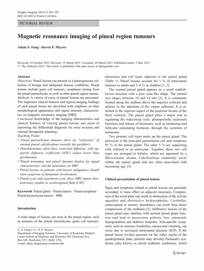
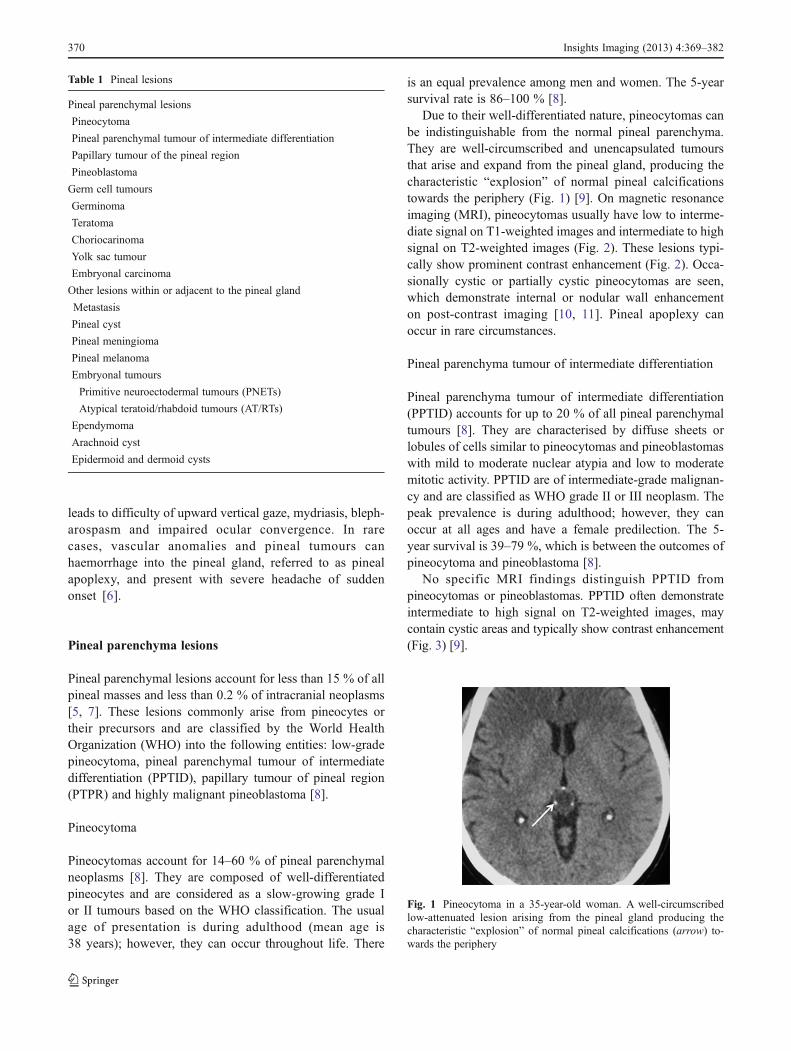
Due to their well-differentiated nature, pineocytomas canbe indistinguishable from the normal pineal parenchyma.They are well-circumscribed and unencapsulated tumoursthat arise and expand from the pineal gland, producing thecharacteristic “explosion” of normal pineal calcificationstowards the periphery (Fig. 1) [9]. On magnetic resonanceimaging (MRI), pineocytomas usually have low to interme-diate signal on T1-weighted images and intermediate to highsignal on T2-weighted images (Fig. 2). These lesions typi-cally show prominent contrast enhancement (Fig. 2). Occa-sionally cystic or partially cystic pineocytomas are seen,which demonstrate internal or nodular wall enhancementon post-contrast imaging [10, 11]. Pineal apoplexy canoccur in rare circumstances.
Pineal parenchyma tumour of intermediate differentiation
Pineal parenchyma tumour of intermediate differentiation(PPTID) accounts for up to 20 % of all pineal parenchymaltumours [8]. They are characterised by diffuse sheets orlobules of cells similar to pineocytomas and pineoblastomaswith mild to moderate nuclear atypia and low to moderatemitotic activity. PPTID are of intermediate-grade malignan-cy and are classified as WHO grade II or III neoplasm. Thepeak prevalence is during adulthood; however, they canoccur at all ages and have a female predilection. The 5-year survival is 39–79 %, which is between the outcomes ofpineocytoma and pineoblastoma [8].
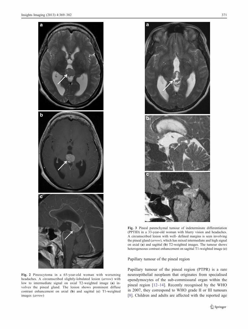
No specific MRI findings distinguish PPTID frompineocytomas or pineoblastomas. PPTID often demonstrateintermediate to high signal on T2-weighted images, maycontain cystic areas and typically show contrast enhancement(Fig. 3) [9].
Fig. 1 Pineocytoma in a 35-year-old woman. A well-circumscribedlow-attenuated lesion arising from the pineal gland producing thecharacteristic “explosion” of normal pineal calcifications (arrow) to-wards the periphery
Table 1 Pineal lesions
Pineal parenchymal lesions
Pineocytoma
Pineal parenchymal tumour of intermediate differentiation
Papillary tumour of the pineal region
Pineoblastoma
Germ cell tumours
Germinoma
Teratoma
Choriocarinoma
Yolk sac tumour
Embryonal carcinoma
Other lesions within or adjacent to the pineal gland
Metastasis
Pineal cyst
Pineal meningioma
Pineal melanoma
Embryonal tumours
Primitive neuroectodermal tumours (PNETs)
Atypical teratoid/rhabdoid tumours (AT/RTs)
Ependymoma
Arachnoid cyst
Epidermoid and dermoid cysts
370 Insights Imaging (2013) 4:369–382
Papillary tumour of the pineal region
Papillary tumour of the pineal region (PTPR) is a rareneuroepithelial neoplasm that originates from specialisedependymocytes of the sub-commissural organ within thepineal region [12–14]. Recently recognised by the WHOin 2007, they correspond to WHO grade II or III tumours[8]. Children and adults are affected with the reported age
Fig. 2 Pineocytoma in a 65-year-old woman with worseningheadaches. A circumscribed slightly-lobulated lesion (arrow) withlow to intermediate signal on axial T2-weighted image (a) in-volves the pineal gland. The lesion shows prominent diffusecontrast enhancement on axial (b) and sagittal (c) T1-weightedimages (arrow)
Fig. 3 Pineal parenchymal tumour of indeterminate differentiation(PPTID) in a 33-year-old woman with blurry vision and headaches.A circumscribed lesion with well- defined margins is seen involvingthe pineal gland (arrow), which has mixed intermediate and high signalon axial (a) and sagittal (b) T2-weighted images. The tumour showsheterogeneous contrast enhancement on sagittal T1-weighted image (c)
Insights Imaging (2013) 4:369–382 371
range of presentation between 5 and 66 years (mean age is29 years) with a slight female predominance. The 5-yearsurvival rate is 73 % and local recurrence is common evenafter complete resection and radiation therapy [12].
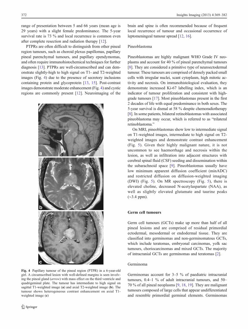
PTPRs are often difficult to distinguish from other pinealregion tumours, such as choroid plexus papillomas, papillarypineal parenchymal tumours, and papillary ependymomas,and often require immunohistochemical techniques for furtherdiagnosis [13]. PTPRs are well-circumscribed and can dem-onstrate slightly-high to high signal on T1- and T2-weightedimages (Fig. 4) due to the presence of secretory inclusionscontaining protein and glycoprotein [13, 15]. Post-contrastimages demonstrate moderate enhancement (Fig. 4) and cysticregions are commonly present [12]. Neuroimaging of the
brain and spine is often recommended because of frequentlocal recurrence of tumour and occasional occurrence ofleptomeningeal tumour spread [12, 16].
Pineoblastoma
Pineoblastomas are highly malignant WHO Grade IV neo-plasms and account for 40 % of pineal parenchymal tumours[8]. They are considered a primitive type of neuroectodermaltumour. These tumours are comprised of densely packed smallcells with irregular nuclei, scant cytoplasm, high mitotic ac-tivity and necrosis. On immunohistological evaluation, theydemonstrate increased Ki-67 labelling index, which is anindicator of tumour proliferation and consistent with high-grade tumours [17]. Most pineoblastomas present in the first2 decades of life with equal predominance in both sexes. The5-year survival is dismal at 58 % despite chemoradiotherapy[8]. In some patients, bilateral retinoblastomas with associatedpineoblastoma may occur, which is referred to as “trilateralretinoblastoma.”
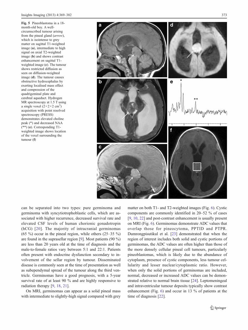
On MRI, pineoblastomas show low to intermediate signalon T1-weighted images, intermediate to high signal on T2-weighted images and demonstrate contrast enhancement(Fig. 5). Given their highly malignant nature, it is notuncommon to see haemorrhage and necrosis within thelesion, as well as infiltration into adjacent structures withcerebral spinal fluid (CSF) seeding and dissemination withinthe subarachnoid space [9]. Pineoblastomas usually havelow minimum apparent diffusion coefficient (minADC)and restricted diffusion on diffusion-weighted imaging(DWI) (Fig. 5). On MR spectroscopy (Fig. 5), there iselevated choline, decreased N-acetylaspartate (NAA), aswell as slightly elevated glutamate and taurine peaks(∼3.4 ppm).
Germ cell tumours
Germ cell tumours (GCTs) make up more than half of allpineal lesions and are comprised of residual primordialectodermal, mesodermal or endodermal tissue. They areclassified into germinomas and non-germinomatous GCTs,which include teratomas, embryonal carcinomas, yolk sactumours, choriocarcinomas and mixed GCTs. The majorityof intracranial GCTs are germinomas and teratomas [2].
Germinoma
Germinomas account for 3–5 % of paediatric intracranialtumours, 0.4–1 % of adult intracranial tumours, and 50–70 % of all pineal neoplasms [9, 18, 19]. They are malignanttumours composed of large cells that appear undifferentiatedand resemble primordial germinal elements. Germinomas
Fig. 4 Papillary tumour of the pineal region (PTPR) in a 6-year-oldgirl. A circumscribed lesion with well-defined margins is seen involv-ing the pineal gland (arrow) with mass effect on the third ventricle andquadrigeminal plate. The tumour has intermediate to high signal onsagittal T1-weighted image (a) and axial T2-weighted image (b). Thetumour shows heterogeneous contrast enhancement on axial T1-weighted image (c)
372 Insights Imaging (2013) 4:369–382
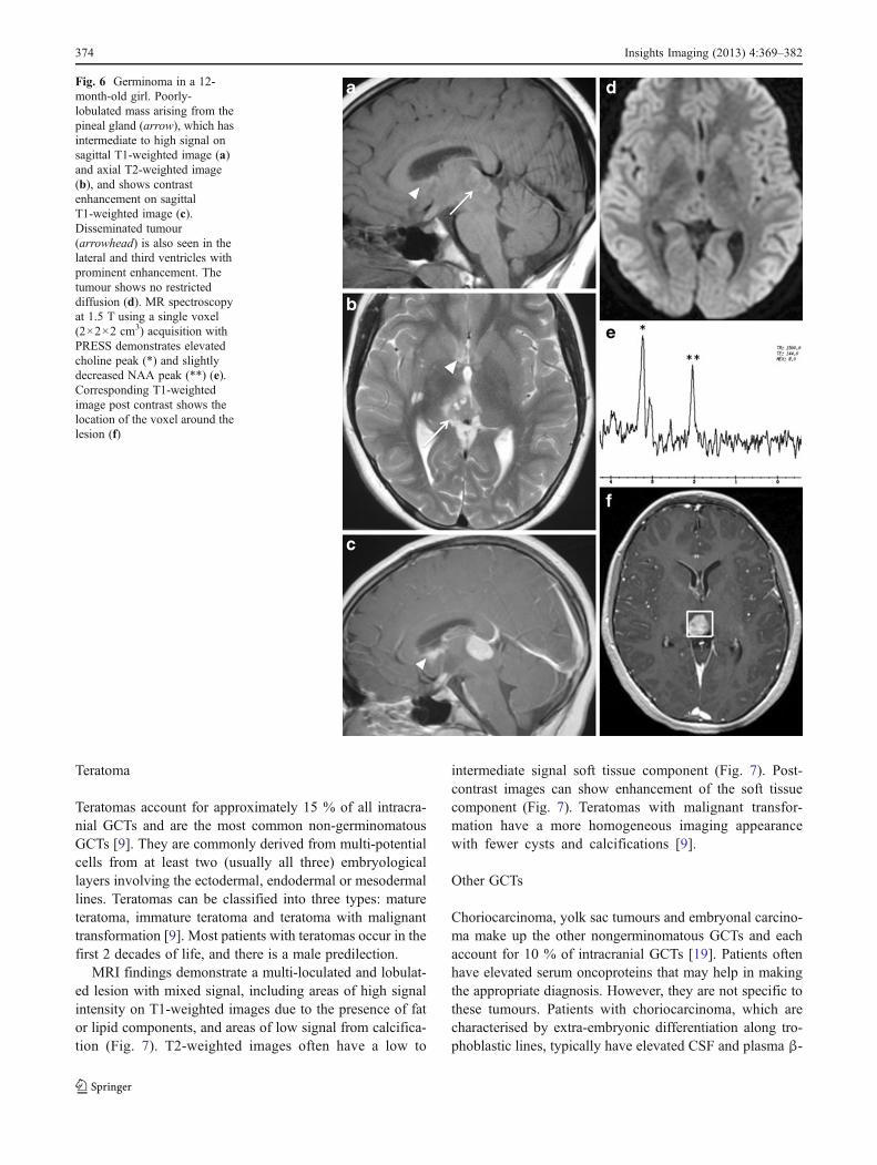
can be separated into two types: pure germinoma andgerminoma with syncytiotrophoblastic cells, which are as-sociated with higher recurrence, decreased survival rate andelevated CSF levels of human chorionic gonadotropin(hCG) [20]. The majority of intracranial germinomas(65 %) occur in the pineal region, while others (25–35 %)are found in the suprasellar region [9]. Most patients (90 %)are less than 20 years old at the time of diagnosis and themale-to-female ratios vary between 5:1 and 22:1. Patientsoften present with endocrine dysfunction secondary to in-volvement of the sellar region by tumour. Disseminateddisease is commonly seen at the time of presentation as wellas subependymal spread of the tumour along the third ven-tricle. Germinomas have a good prognosis, with a 5-yearsurvival rate of at least 90 % and are highly responsive toradiation therapy [9, 18, 21].
On MRI, germinomas can appear as a solid pineal masswith intermediate to slightly-high signal compared with grey
matter on both T1- and T2-weighted images (Fig. 6). Cysticcomponents are commonly identified in 20–52 % of cases[9, 10, 22] and post-contrast enhancement is usually presenton MRI (Fig. 6). Germinomas demonstrate ADC values thatoverlap those for pineocytoma, PPTID and PTPR.Dumrongpisutikul et al. [23] demonstrated that when theregion of interest includes both solid and cystic portions ofgerminomas, the ADC values are often higher than those ofthe more densely cellular pineal cell tumours, particularlypineoblastomas, which is likely due to the abundance ofcytoplasm, presence of cystic components, less tumour cel-lularity and lesser nuclear/cytoplasmic ratio. However,when only the solid portions of germinomas are included,normal, decreased or increased ADC values can be demon-strated relative to normal brain tissue [24]. Leptomeningealand intraventricular tumour deposits typically show contrastenhancement (Fig. 6) and occur in 13 % of patients at thetime of diagnosis [22].
Fig. 5 Pineoblastoma in a 18-month-old boy. A well-circumscribed tumour arisingfrom the pineal gland (arrow),which is isointense to greymatter on sagittal T1-weightedimage (a), intermediate to highsignal on axial T2-weightedimage (b) and shows contrastenhancement on sagittal T1-weighted image (c). The tumourshows restricted diffusion asseen on diffusion-weightedimage (d). The tumour causesobstructive hydrocephalus byexerting localised mass effectand compression of thequadrigeminal plate andcerebral aqueduct. HydrogenMR spectroscopy at 1.5 T usinga single voxel (2×2×2 cm3)acquisition with point resolvedspectroscopy (PRESS)demonstrates elevated cholinepeak (*) and decreased NAA(**) (e). Corresponding T1-weighted image shows locationof the voxel surrounding thetumour (f)
Insights Imaging (2013) 4:369–382 373
Teratoma
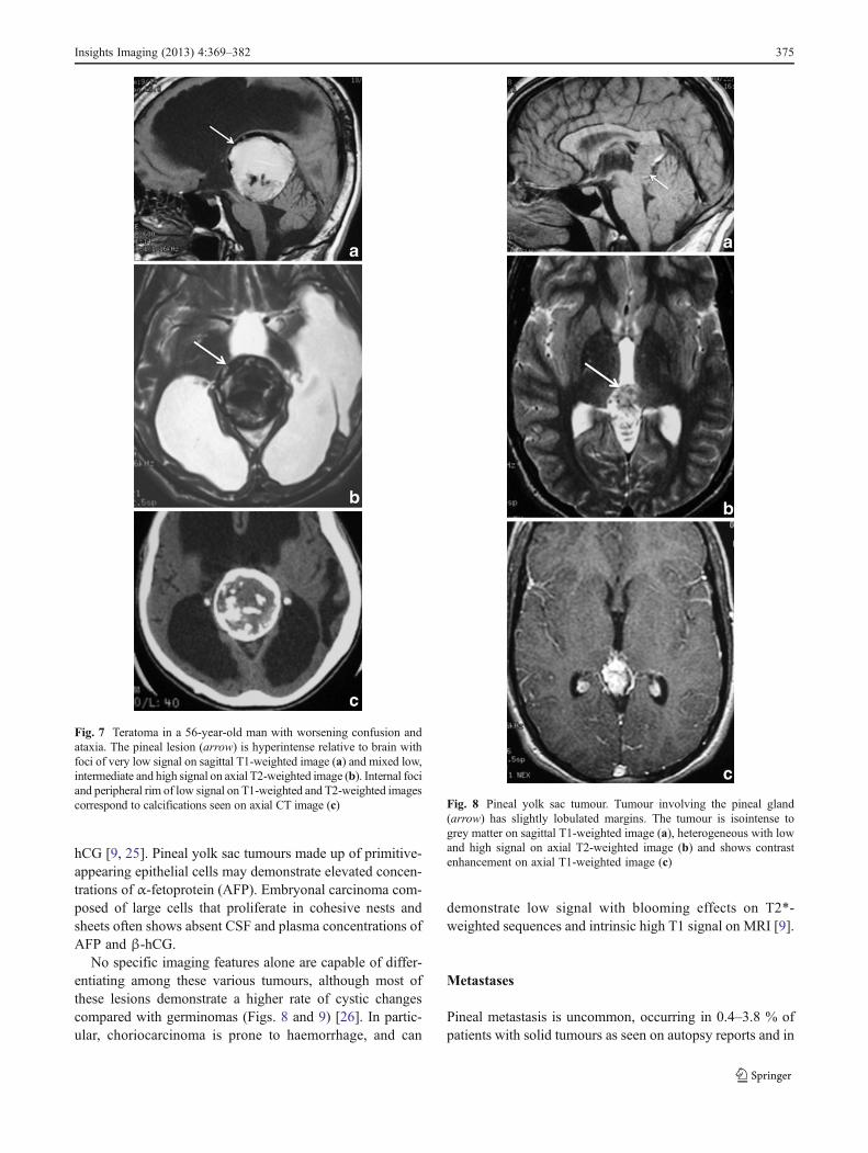
Teratomas account for approximately 15 % of all intracra-nial GCTs and are the most common non-germinomatousGCTs [9]. They are commonly derived from multi-potentialcells from at least two (usually all three) embryologicallayers involving the ectodermal, endodermal or mesodermallines. Teratomas can be classified into three types: matureteratoma, immature teratoma and teratoma with malignanttransformation [9]. Most patients with teratomas occur in thefirst 2 decades of life, and there is a male predilection.
MRI findings demonstrate a multi-loculated and lobulat-ed lesion with mixed signal, including areas of high signalintensity on T1-weighted images due to the presence of fator lipid components, and areas of low signal from calcifica-tion (Fig. 7). T2-weighted images often have a low to
intermediate signal soft tissue component (Fig. 7). Post-contrast images can show enhancement of the soft tissuecomponent (Fig. 7). Teratomas with malignant transfor-mation have a more homogeneous imaging appearancewith fewer cysts and calcifications [9].
Other GCTs
Choriocarcinoma, yolk sac tumours and embryonal carcino-ma make up the other nongerminomatous GCTs and eachaccount for 10 % of intracranial GCTs [19]. Patients oftenhave elevated serum oncoproteins that may help in makingthe appropriate diagnosis. However, they are not specific tothese tumours. Patients with choriocarcinoma, which arecharacterised by extra-embryonic differentiation along tro-phoblastic lines, typically have elevated CSF and plasma β-
Fig. 6 Germinoma in a 12-month-old girl. Poorly-lobulated mass arising from thepineal gland (arrow), which hasintermediate to high signal onsagittal T1-weighted image (a)and axial T2-weighted image(b), and shows contrastenhancement on sagittalT1-weighted image (c).Disseminated tumour(arrowhead) is also seen in thelateral and third ventricles withprominent enhancement. Thetumour shows no restricteddiffusion (d). MR spectroscopyat 1.5 T using a single voxel(2×2×2 cm3) acquisition withPRESS demonstrates elevatedcholine peak (*) and slightlydecreased NAA peak (**) (e).Corresponding T1-weightedimage post contrast shows thelocation of the voxel around thelesion (f)
374 Insights Imaging (2013) 4:369–382
hCG [9, 25]. Pineal yolk sac tumours made up of primitive-appearing epithelial cells may demonstrate elevated concen-trations of α-fetoprotein (AFP). Embryonal carcinoma com-posed of large cells that proliferate in cohesive nests andsheets often shows absent CSF and plasma concentrations ofAFP and β-hCG.
No specific imaging features alone are capable of differ-entiating among these various tumours, although most ofthese lesions demonstrate a higher rate of cystic changescompared with germinomas (Figs. 8 and 9) [26]. In partic-ular, choriocarcinoma is prone to haemorrhage, and can
demonstrate low signal with blooming effects on T2*-weighted sequences and intrinsic high T1 signal on MRI [9].
Metastases
Pineal metastasis is uncommon, occurring in 0.4–3.8 % ofpatients with solid tumours as seen on autopsy reports and in
Fig. 7 Teratoma in a 56-year-old man with worsening confusion andataxia. The pineal lesion (arrow) is hyperintense relative to brain withfoci of very low signal on sagittal T1-weighted image (a) and mixed low,intermediate and high signal on axial T2-weighted image (b). Internal fociand peripheral rim of low signal on T1-weighted and T2-weighted imagescorrespond to calcifications seen on axial CT image (c) Fig. 8 Pineal yolk sac tumour. Tumour involving the pineal gland
(arrow) has slightly lobulated margins. The tumour is isointense togrey matter on sagittal T1-weighted image (a), heterogeneous with lowand high signal on axial T2-weighted image (b) and shows contrastenhancement on axial T1-weighted image (c)
Insights Imaging (2013) 4:369–382 375
5 % of patients referred to surgical management of pinealtumours [27, 28]. Lung cancer is the most frequent cancer tometastasise to the pineal gland; however, other tumours suchas breast, kidney, esophagus, stomach, colon, melanoma andmyeloma have also been reported [28]. Metastatic disease isthought to be related to hematogenous spread due to theabsence of a blood-brain barrier in the pineal gland. In themajority of cases, metastasis to the pineal gland is asymptom-atic. However, a few patients have presented with clinicalfeatures, such as headache, encephalopathy, Parinaud’s syn-drome and hydrocephalus [28]. Leptomeningeal seeding is acommon finding with pineal metastases, occurring in 67 % ofpatients, and may be secondary to the proximity of the pinealgland to the CSF pathways in the third ventricle andquadrigeminal cistern [28].
Cells types adjacent to the pineal gland
Pineal cyst
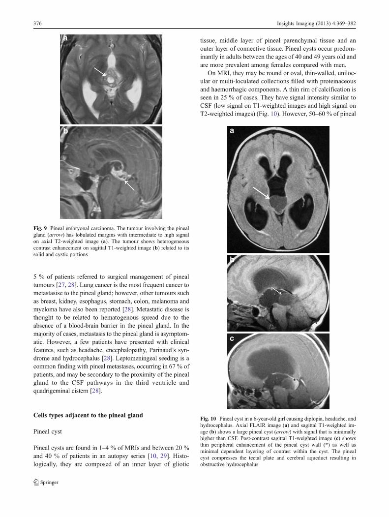
Pineal cysts are found in 1–4 % of MRIs and between 20 %and 40 % of patients in an autopsy series [10, 29]. Histo-logically, they are composed of an inner layer of gliotic
tissue, middle layer of pineal parenchymal tissue and anouter layer of connective tissue. Pineal cysts occur predom-inantly in adults between the ages of 40 and 49 years old andare more prevalent among females compared with men.
On MRI, they may be round or oval, thin-walled, uniloc-ular or multi-loculated collections filled with proteinaceousand haemorrhagic components. A thin rim of calcification isseen in 25 % of cases. They have signal intensity similar toCSF (low signal on T1-weighted images and high signal onT2-weighted images) (Fig. 10). However, 50–60 % of pineal
Fig. 9 Pineal embryonal carcinoma. The tumour involving the pinealgland (arrow) has lobulated margins with intermediate to high signalon axial T2-weighted image (a). The tumour shows heterogeneouscontrast enhancement on sagittal T1-weighted image (b) related to itssolid and cystic portions
Fig. 10 Pineal cyst in a 6-year-old girl causing diplopia, headache, andhydrocephalus. Axial FLAIR image (a) and sagittal T1-weighted im-age (b) shows a large pineal cyst (arrow) with signal that is minimallyhigher than CSF. Post-contrast sagittal T1-weighted image (c) showsthin peripheral enhancement of the pineal cyst wall (*) as well asminimal dependent layering of contrast within the cyst. The pinealcyst compresses the tectal plate and cerebral aqueduct resulting inobstructive hydrocephalus
376 Insights Imaging (2013) 4:369–382
cysts are slightly hyperintense to CSF [10]. The signal ofthese cysts often does not fully suppress on fluid-attenuatedinversion-recovery (FLAIR) (Fig. 10). The cyst contentstypically show no restricted diffusion. Post-contrast images(Fig. 10) demonstrate a thin rim of wall enhancement usu-ally <2-mm thickness that is seen as a result of fragmenta-tion of the pineal parenchyma, which occurs as the cystenlarges [10, 11]. The cyst can show with internal enhance-ment on delayed images.
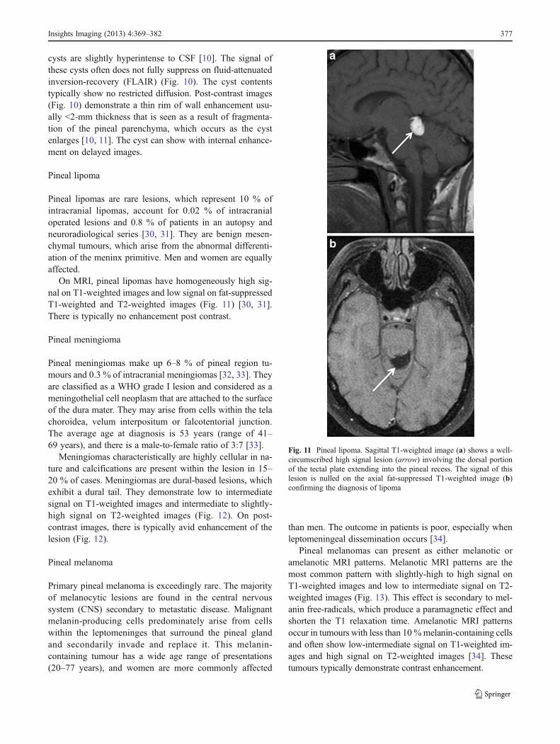
Pineal lipoma
Pineal lipomas are rare lesions, which represent 10 % ofintracranial lipomas, account for 0.02 % of intracranialoperated lesions and 0.8 % of patients in an autopsy andneuroradiological series [30, 31]. They are benign mesen-chymal tumours, which arise from the abnormal differenti-ation of the meninx primitive. Men and women are equallyaffected.
On MRI, pineal lipomas have homogeneously high sig-nal on T1-weighted images and low signal on fat-suppressedT1-weighted and T2-weighted images (Fig. 11) [30, 31].There is typically no enhancement post contrast.
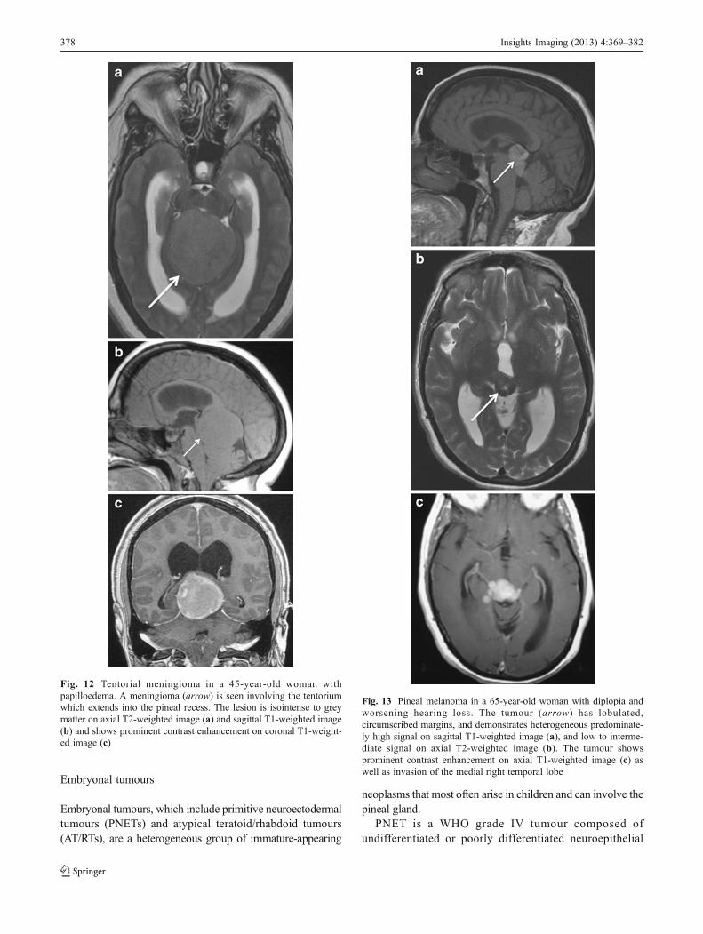
Pineal meningioma
Pineal meningiomas make up 6–8 % of pineal region tu-mours and 0.3 % of intracranial meningiomas [32, 33]. Theyare classified as a WHO grade I lesion and considered as ameningothelial cell neoplasm that are attached to the surfaceof the dura mater. They may arise from cells within the telachoroidea, velum interpositum or falcotentorial junction.The average age at diagnosis is 53 years (range of 41–69 years), and there is a male-to-female ratio of 3:7 [33].
Meningiomas characteristically are highly cellular in na-ture and calcifications are present within the lesion in 15–20 % of cases. Meningiomas are dural-based lesions, whichexhibit a dural tail. They demonstrate low to intermediatesignal on T1-weighted images and intermediate to slightly-high signal on T2-weighted images (Fig. 12). On post-contrast images, there is typically avid enhancement of thelesion (Fig. 12).
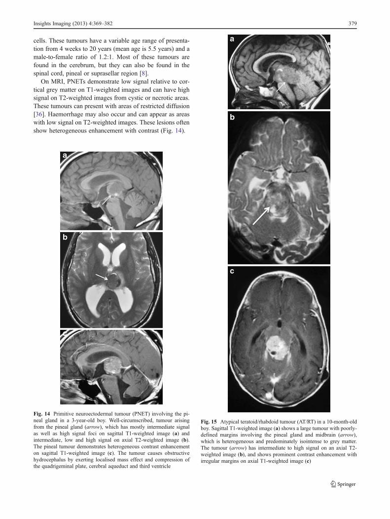
Pineal melanoma
Primary pineal melanoma is exceedingly rare. The majorityof melanocytic lesions are found in the central nervoussystem (CNS) secondary to metastatic disease. Malignantmelanin-producing cells predominately arise from cellswithin the leptomeninges that surround the pineal glandand secondarily invade and replace it. This melanin-containing tumour has a wide age range of presentations(20–77 years), and women are more commonly affected
than men. The outcome in patients is poor, especially whenleptomeningeal dissemination occurs [34].
Pineal melanomas can present as either melanotic oramelanotic MRI patterns. Melanotic MRI patterns are themost common pattern with slightly-high to high signal onT1-weighted images and low to intermediate signal on T2-weighted images (Fig. 13). This effect is secondary to mel-anin free-radicals, which produce a paramagnetic effect andshorten the T1 relaxation time. Amelanotic MRI patternsoccur in tumours with less than 10%melanin-containing cellsand often show low-intermediate signal on T1-weighted im-ages and high signal on T2-weighted images [34]. Thesetumours typically demonstrate contrast enhancement.
Fig. 11 Pineal lipoma. Sagittal T1-weighted image (a) shows a well-circumscribed high signal lesion (arrow) involving the dorsal portionof the tectal plate extending into the pineal recess. The signal of thislesion is nulled on the axial fat-suppressed T1-weighted image (b)confirming the diagnosis of lipoma
Insights Imaging (2013) 4:369–382 377
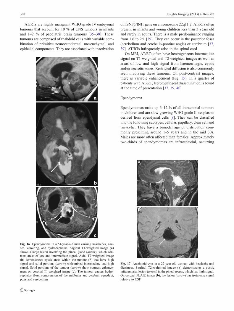
Embryonal tumours
Embryonal tumours, which include primitive neuroectodermaltumours (PNETs) and atypical teratoid/rhabdoid tumours(AT/RTs), are a heterogeneous group of immature-appearing
neoplasms that most often arise in children and can involve thepineal gland.
PNET is a WHO grade IV tumour composed ofundifferentiated or poorly differentiated neuroepithelial
Fig. 12 Tentorial meningioma in a 45-year-old woman withpapilloedema. A meningioma (arrow) is seen involving the tentoriumwhich extends into the pineal recess. The lesion is isointense to greymatter on axial T2-weighted image (a) and sagittal T1-weighted image(b) and shows prominent contrast enhancement on coronal T1-weight-ed image (c)
Fig. 13 Pineal melanoma in a 65-year-old woman with diplopia andworsening hearing loss. The tumour (arrow) has lobulated,circumscribed margins, and demonstrates heterogeneous predominate-ly high signal on sagittal T1-weighted image (a), and low to interme-diate signal on axial T2-weighted image (b). The tumour showsprominent contrast enhancement on axial T1-weighted image (c) aswell as invasion of the medial right temporal lobe
378 Insights Imaging (2013) 4:369–382
cells. These tumours have a variable age range of presenta-tion from 4 weeks to 20 years (mean age is 5.5 years) and amale-to-female ratio of 1.2:1. Most of these tumours arefound in the cerebrum, but they can also be found in thespinal cord, pineal or suprasellar region [8].
On MRI, PNETs demonstrate low signal relative to cor-tical grey matter on T1-weighted images and can have highsignal on T2-weighted images from cystic or necrotic areas.These tumours can present with areas of restricted diffusion[36]. Haemorrhage may also occur and can appear as areaswith low signal on T2-weighted images. These lesions oftenshow heterogeneous enhancement with contrast (Fig. 14).
Fig. 14 Primitive neuroectodermal tumour (PNET) involving the pi-neal gland in a 3-year-old boy. Well-circumscribed, tumour arisingfrom the pineal gland (arrow), which has mostly intermediate signalas well as high signal foci on sagittal T1-weighted image (a) andintermediate, low and high signal on axial T2-weighted image (b).The pineal tumour demonstrates heterogeneous contrast enhancementon sagittal T1-weighted image (c). The tumour causes obstructivehydrocephalus by exerting localised mass effect and compression ofthe quadrigeminal plate, cerebral aqueduct and third ventricle
Fig. 15 Atypical teratoid/rhabdoid tumour (AT/RT) in a 10-month-oldboy. Sagittal T1-weighted image (a) shows a large tumour with poorly-defined margins involving the pineal gland and midbrain (arrow),which is heterogeneous and predominately isointense to grey matter.The tumour (arrow) has intermediate to high signal on an axial T2-weighted image (b), and shows prominent contrast enhancement withirregular margins on axial T1-weighted image (c)
Insights Imaging (2013) 4:369–382 379
AT/RTs are highly malignant WHO grade IV embryonaltumours that account for 10 % of CNS tumours in infantsand 1–2 % of paediatric brain tumours [35–38]. Thesetumours are comprised of rhabdoid cells with variable com-bination of primitive neuroectodermal, mesenchymal, andepithelial components. They are associated with inactivation
of hSNF5/INI1 gene on chromosome 22q11.2. AT/RTs oftenpresent in infants and young children less than 3 years oldand rarely in adults. There is a male predominance rangingfrom 1.6 to 2:1 [39]. They can occur in the posterior fossa(cerebellum and cerebello-pontine angle) or cerebrum [37,39]. AT/RTs infrequently arise in the spinal cord.
On MRI, AT/RTs often have heterogeneous intermediatesignal on T1-weighted and T2-weighted images as well asareas of low and high signal from haemorrhagic, cysticand/or necrotic zones. Restricted diffusion is also commonlyseen involving these tumours. On post-contrast images,there is variable enhancement (Fig. 15). In a quarter ofpatients with AT/RT, leptomeningeal dissemination is foundat the time of presentation [37, 39, 40].
Ependymoma
Ependymomas make up 6–12 % of all intracranial tumoursin children and are slow-growing WHO grade II neoplasmsderived from ependymal cells [8]. They can be classifiedinto the following subtypes: cellular, papillary, clear cell andtanycytic. They have a bimodal age of distribution com-monly presenting around 1–5 years and in the mid 30s.Males are more often affected than females. Approximatelytwo-thirds of ependymomas are infratentorial, occurring
Fig. 16 Ependymoma in a 54-year-old man causing headaches, nau-sea, vomiting, and hydrocephalus. Sagittal T1-weighted image (a)shows a large lesion involving the pineal gland (arrow), which con-tains areas of low and intermediate signal. Axial T2-weighted image(b) demonstrates cystic areas within the tumour (*) that have highsignal and solid portions (arrow) with mixed intermediate and highsignal. Solid portions of the tumour (arrow) show contrast enhance-ment on coronal T1-weighted image (c). The tumour causes hydro-cephalus from compression of the midbrain and cerebral aqueduct,pons and cerebellum
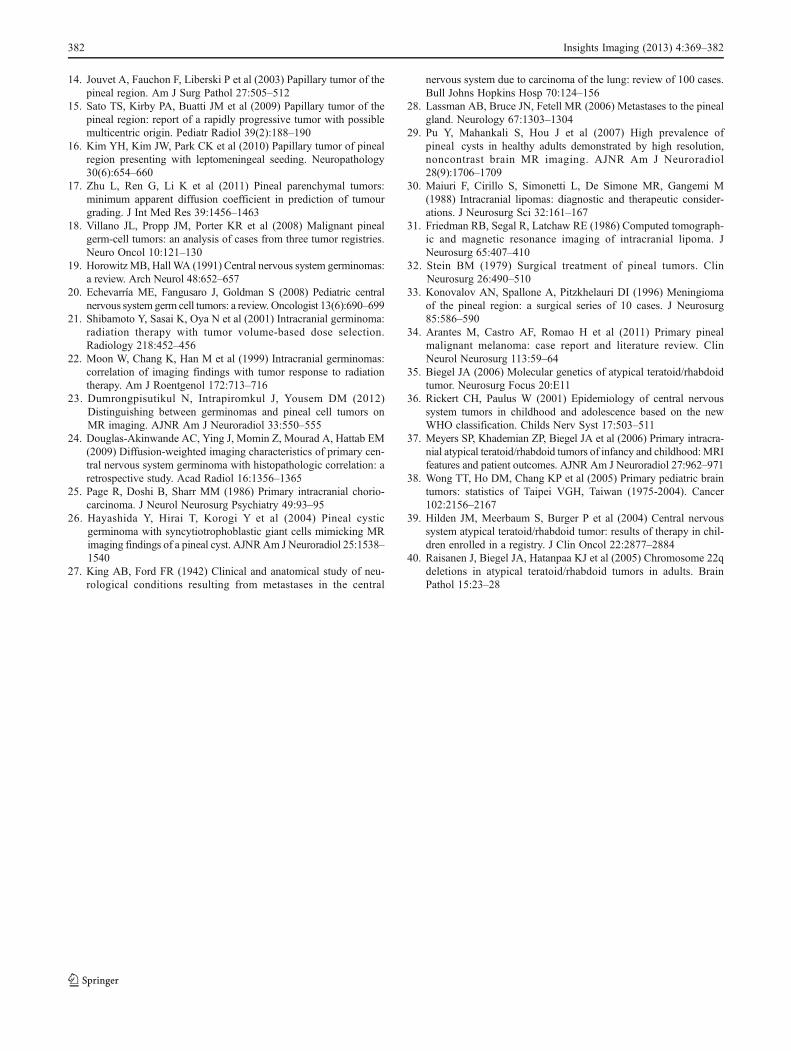
Fig. 17 Arachnoid cyst in a 27-year-old woman with headache anddizziness. Sagittal T2-weighted image (a) demonstrates a cysticinfratentorial lesion (arrow) in the pineal recess, which has high signal.On coronal FLAIR image (b), the lesion (arrow) has isointense signalrelative to CSF
380 Insights Imaging (2013) 4:369–382
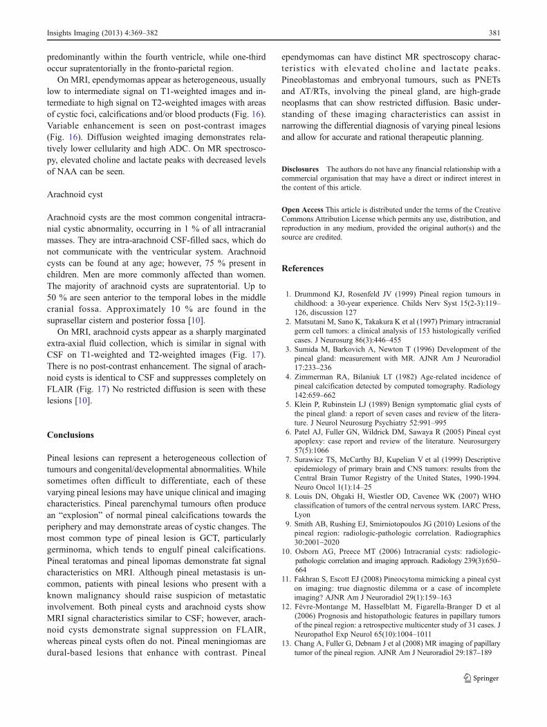
predominantly within the fourth ventricle, while one-thirdoccur supratentorially in the fronto-parietal region.
On MRI, ependymomas appear as heterogeneous, usuallylow to intermediate signal on T1-weighted images and in-termediate to high signal on T2-weighted images with areasof cystic foci, calcifications and/or blood products (Fig. 16).Variable enhancement is seen on post-contrast images(Fig. 16). Diffusion weighted imaging demonstrates rela-tively lower cellularity and high ADC. On MR spectrosco-py, elevated choline and lactate peaks with decreased levelsof NAA can be seen.
Arachnoid cyst
Arachnoid cysts are the most common congenital intracra-nial cystic abnormality, occurring in 1 % of all intracranialmasses. They are intra-arachnoid CSF-filled sacs, which donot communicate with the ventricular system. Arachnoidcysts can be found at any age; however, 75 % present inchildren. Men are more commonly affected than women.The majority of arachnoid cysts are supratentorial. Up to50 % are seen anterior to the temporal lobes in the middlecranial fossa. Approximately 10 % are found in thesuprasellar cistern and posterior fossa [10].
On MRI, arachnoid cysts appear as a sharply marginatedextra-axial fluid collection, which is similar in signal withCSF on T1-weighted and T2-weighted images (Fig. 17).There is no post-contrast enhancement. The signal of arach-noid cysts is identical to CSF and suppresses completely onFLAIR (Fig. 17) No restricted diffusion is seen with theselesions [10].
Conclusions
Pineal lesions can represent a heterogeneous collection oftumours and congenital/developmental abnormalities. Whilesometimes often difficult to differentiate, each of thesevarying pineal lesions may have unique clinical and imagingcharacteristics. Pineal parenchymal tumours often producean “explosion” of normal pineal calcifications towards theperiphery and may demonstrate areas of cystic changes. Themost common type of pineal lesion is GCT, particularlygerminoma, which tends to engulf pineal calcifications.Pineal teratomas and pineal lipomas demonstrate fat signalcharacteristics on MRI. Although pineal metastasis is un-common, patients with pineal lesions who present with aknown malignancy should raise suspicion of metastaticinvolvement. Both pineal cysts and arachnoid cysts showMRI signal characteristics similar to CSF; however, arach-noid cysts demonstrate signal suppression on FLAIR,whereas pineal cysts often do not. Pineal meningiomas aredural-based lesions that enhance with contrast. Pineal
ependymomas can have distinct MR spectroscopy charac-teristics with elevated choline and lactate peaks.Pineoblastomas and embryonal tumours, such as PNETsand AT/RTs, involving the pineal gland, are high-gradeneoplasms that can show restricted diffusion. Basic under-standing of these imaging characteristics can assist innarrowing the differential diagnosis of varying pineal lesionsand allow for accurate and rational therapeutic planning.
Disclosures The authors do not have any financial relationship with acommercial organisation that may have a direct or indirect interest inthe content of this article.
Open Access This article is distributed under the terms of the CreativeCommons Attribution License which permits any use, distribution, andreproduction in any medium, provided the original author(s) and thesource are credited.
References
1. Drummond KJ, Rosenfeld JV (1999) Pineal region tumours inchildhood: a 30-year experience. Childs Nerv Syst 15(2-3):119–126, discussion 127
2. Matsutani M, Sano K, Takakura K et al (1997) Primary intracranialgerm cell tumors: a clinical analysis of 153 histologically verifiedcases. J Neurosurg 86(3):446–455
3. Sumida M, Barkovich A, Newton T (1996) Development of thepineal gland: measurement with MR. AJNR Am J Neuroradiol17:233–236
4. Zimmerman RA, Bilaniuk LT (1982) Age-related incidence ofpineal calcification detected by computed tomography. Radiology142:659–662
5. Klein P, Rubinstein LJ (1989) Benign symptomatic glial cysts ofthe pineal gland: a report of seven cases and review of the litera-ture. J Neurol Neurosurg Psychiatry 52:991–995
6. Patel AJ, Fuller GN, Wildrick DM, Sawaya R (2005) Pineal cystapoplexy: case report and review of the literature. Neurosurgery57(5):1066
7. Surawicz TS, McCarthy BJ, Kupelian V et al (1999) Descriptiveepidemiology of primary brain and CNS tumors: results from theCentral Brain Tumor Registry of the United States, 1990-1994.Neuro Oncol 1(1):14–25
8. Louis DN, Ohgaki H, Wiestler OD, Cavenee WK (2007) WHOclassification of tumors of the central nervous system. IARC Press,Lyon
9. Smith AB, Rushing EJ, Smirniotopoulos JG (2010) Lesions of thepineal region: radiologic-pathologic correlation. Radiographics30:2001–2020
10. Osborn AG, Preece MT (2006) Intracranial cysts: radiologic-pathologic correlation and imaging approach. Radiology 239(3):650–664
11. Fakhran S, Escott EJ (2008) Pineocytoma mimicking a pineal cyston imaging: true diagnostic dilemma or a case of incompleteimaging? AJNR Am J Neuroradiol 29(1):159–163
12. Fèvre-Montange M, Hasselblatt M, Figarella-Branger D et al(2006) Prognosis and histopathologic features in papillary tumorsof the pineal region: a retrospective multicenter study of 31 cases. JNeuropathol Exp Neurol 65(10):1004–1011
13. Chang A, Fuller G, Debnam J et al (2008) MR imaging of papillarytumor of the pineal region. AJNR Am J Neuroradiol 29:187–189
Insights Imaging (2013) 4:369–382 381
14. Jouvet A, Fauchon F, Liberski P et al (2003) Papillary tumor of thepineal region. Am J Surg Pathol 27:505–512
15. Sato TS, Kirby PA, Buatti JM et al (2009) Papillary tumor of thepineal region: report of a rapidly progressive tumor with possiblemulticentric origin. Pediatr Radiol 39(2):188–190
16. Kim YH, Kim JW, Park CK et al (2010) Papillary tumor of pinealregion presenting with leptomeningeal seeding. Neuropathology30(6):654–660
17. Zhu L, Ren G, Li K et al (2011) Pineal parenchymal tumors:minimum apparent diffusion coefficient in prediction of tumourgrading. J Int Med Res 39:1456–1463
18. Villano JL, Propp JM, Porter KR et al (2008) Malignant pinealgerm-cell tumors: an analysis of cases from three tumor registries.Neuro Oncol 10:121–130
19. Horowitz MB, HallWA (1991) Central nervous system germinomas:a review. Arch Neurol 48:652–657
20. Echevarría ME, Fangusaro J, Goldman S (2008) Pediatric centralnervous system germ cell tumors: a review. Oncologist 13(6):690–699
21. Shibamoto Y, Sasai K, Oya N et al (2001) Intracranial germinoma:radiation therapy with tumor volume-based dose selection.Radiology 218:452–456
22. Moon W, Chang K, Han M et al (1999) Intracranial germinomas:correlation of imaging findings with tumor response to radiationtherapy. Am J Roentgenol 172:713–716
23. Dumrongpisutikul N, Intrapiromkul J, Yousem DM (2012)Distinguishing between germinomas and pineal cell tumors onMR imaging. AJNR Am J Neuroradiol 33:550–555
24. Douglas-Akinwande AC, Ying J, Momin Z, Mourad A, Hattab EM(2009) Diffusion-weighted imaging characteristics of primary cen-tral nervous system germinoma with histopathologic correlation: aretrospective study. Acad Radiol 16:1356–1365
25. Page R, Doshi B, Sharr MM (1986) Primary intracranial chorio-carcinoma. J Neurol Neurosurg Psychiatry 49:93–95
26. Hayashida Y, Hirai T, Korogi Y et al (2004) Pineal cysticgerminoma with syncytiotrophoblastic giant cells mimicking MRimaging findings of a pineal cyst. AJNRAm J Neuroradiol 25:1538–1540
27. King AB, Ford FR (1942) Clinical and anatomical study of neu-rological conditions resulting from metastases in the central
nervous system due to carcinoma of the lung: review of 100 cases.Bull Johns Hopkins Hosp 70:124–156
28. Lassman AB, Bruce JN, Fetell MR (2006) Metastases to the pinealgland. Neurology 67:1303–1304
29. Pu Y, Mahankali S, Hou J et al (2007) High prevalence ofpineal cysts in healthy adults demonstrated by high resolution,noncontrast brain MR imaging. AJNR Am J Neuroradiol28(9):1706–1709
30. Maiuri F, Cirillo S, Simonetti L, De Simone MR, Gangemi M(1988) Intracranial lipomas: diagnostic and therapeutic consider-ations. J Neurosurg Sci 32:161–167
31. Friedman RB, Segal R, Latchaw RE (1986) Computed tomograph-ic and magnetic resonance imaging of intracranial lipoma. JNeurosurg 65:407–410
32. Stein BM (1979) Surgical treatment of pineal tumors. ClinNeurosurg 26:490–510
33. Konovalov AN, Spallone A, Pitzkhelauri DI (1996) Meningiomaof the pineal region: a surgical series of 10 cases. J Neurosurg85:586–590
34. Arantes M, Castro AF, Romao H et al (2011) Primary pinealmalignant melanoma: case report and literature review. ClinNeurol Neurosurg 113:59–64
35. Biegel JA (2006) Molecular genetics of atypical teratoid/rhabdoidtumor. Neurosurg Focus 20:E11
36. Rickert CH, Paulus W (2001) Epidemiology of central nervoussystem tumors in childhood and adolescence based on the newWHO classification. Childs Nerv Syst 17:503–511
37. Meyers SP, Khademian ZP, Biegel JA et al (2006) Primary intracra-nial atypical teratoid/rhabdoid tumors of infancy and childhood:MRIfeatures and patient outcomes. AJNR Am J Neuroradiol 27:962–971
38. Wong TT, Ho DM, Chang KP et al (2005) Primary pediatric braintumors: statistics of Taipei VGH, Taiwan (1975-2004). Cancer102:2156–2167
39. Hilden JM, Meerbaum S, Burger P et al (2004) Central nervoussystem atypical teratoid/rhabdoid tumor: results of therapy in chil-dren enrolled in a registry. J Clin Oncol 22:2877–2884
40. Raisanen J, Biegel JA, Hatanpaa KJ et al (2005) Chromosome 22qdeletions in atypical teratoid/rhabdoid tumors in adults. BrainPathol 15:23–28
382 Insights Imaging (2013) 4:369–382