Embed Size (px)
Citation preview
Clin
ical
FOCU
SMoran
Eye Center
2 0 1 8
INSIDE: Migraine in Your Exam Lane | Photophobia Frontiers | Treating IIH
MAKING CONNECTIONS:
The Neuro-Ophthalmology Issue

Moran’s neuro-ophthalmology team, left to right: Alison Crum, MD; Bradley J. Katz, MD, PhD;
Judith E.A. Warner, MD; Kathleen B. Digre, MD; Donnell J. Creel, PhD; Meagan Seay, DO.
absolutely right. As I consider each member of the team, I
can’t think of a stronger, more cohesive group of clinicians
and researchers equipped to tackle some of the most
challenging questions in the field today.
This issue of Clinical Focus is full of their insights, from
frank discussions about migraine to tips on communicating
with patients. I couldn’t be more excited to share this issue
with you and hope that it informs, inspires, and enlightens
in our common goal to provide the very best care possible
to all patients who seek our help.
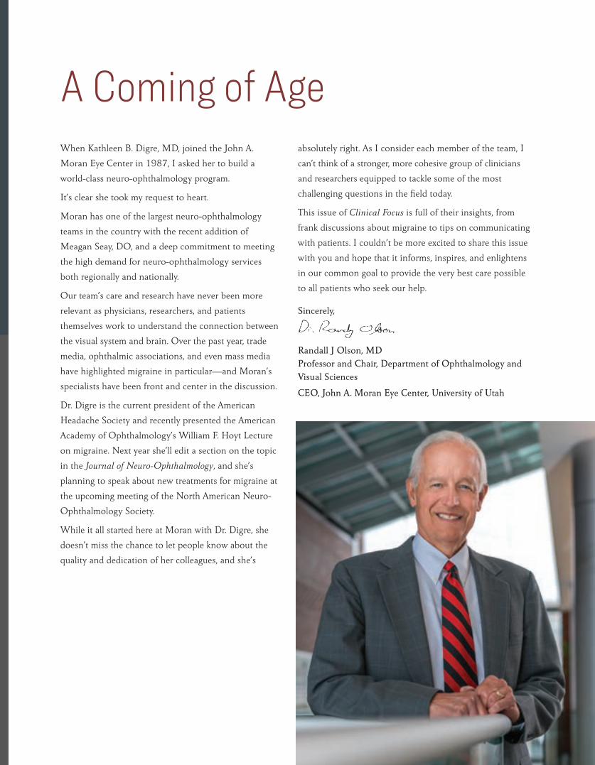
Sincerely,
Randall J Olson, MDProfessor and Chair, Department of Ophthalmology and Visual Sciences
CEO, John A. Moran Eye Center, University of Utah
When Kathleen B. Digre, MD, joined the John A.
Moran Eye Center in 1987, I asked her to build a
world-class neuro-ophthalmology program.
It’s clear she took my request to heart.
Moran has one of the largest neuro-ophthalmology
teams in the country with the recent addition of
Meagan Seay, DO, and a deep commitment to meeting
the high demand for neuro-ophthalmology services
both regionally and nationally.
Our team’s care and research have never been more
relevant as physicians, researchers, and patients
themselves work to understand the connection between
the visual system and brain. Over the past year, trade
media, ophthalmic associations, and even mass media
have highlighted migraine in particular—and Moran’s
specialists have been front and center in the discussion.
Dr. Digre is the current president of the American
Headache Society and recently presented the American
Academy of Ophthalmology’s William F. Hoyt Lecture
on migraine. Next year she’ll edit a section on the topic
in the Journal of Neuro-Ophthalmology, and she’s
planning to speak about new treatments for migraine at
the upcoming meeting of the North American Neuro-
Ophthalmology Society.
While it all started here at Moran with Dr. Digre, she
doesn’t miss the chance to let people know about the
quality and dedication of her colleagues, and she’s
A Coming of Age
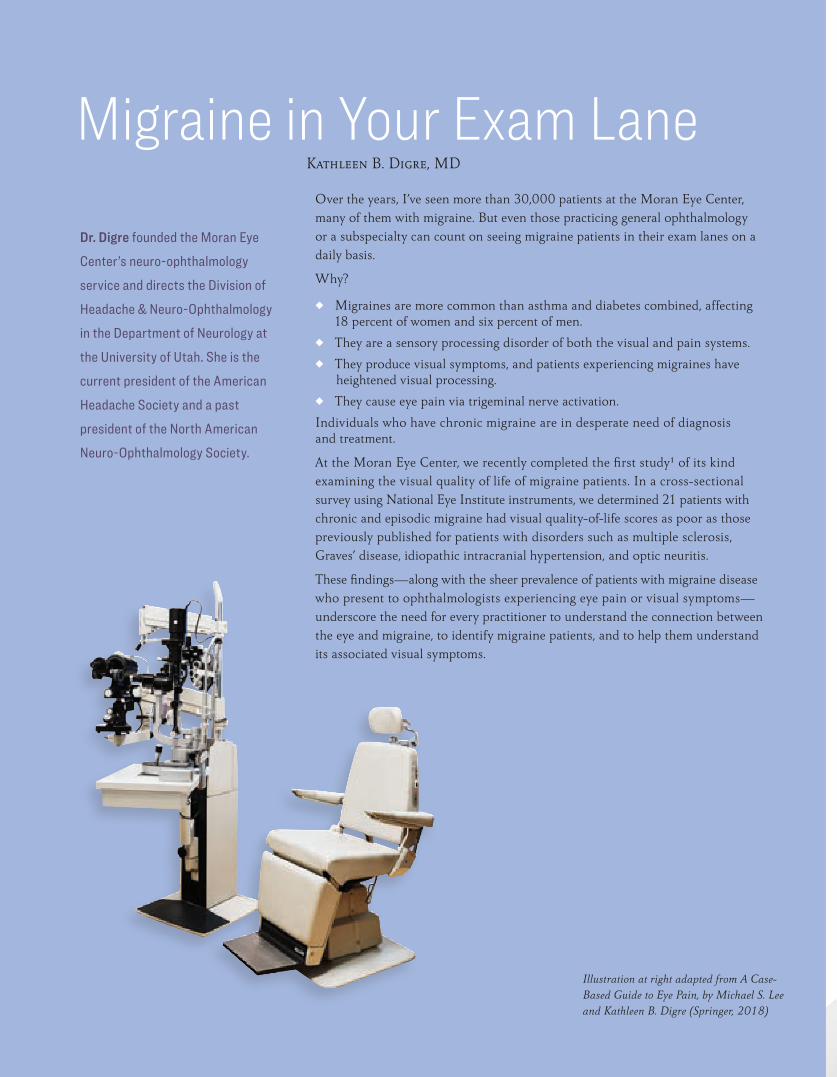
Migraine in Your Exam LaneKathleen B. Digre, MD
Dr. Digre founded the Moran Eye Center’s neuro-ophthalmology service and directs the Division of Headache & Neuro-Ophthalmology in the Department of Neurology at the University of Utah. She is the current president of the American Headache Society and a past president of the North American Neuro-Ophthalmology Society.
Over the years, I’ve seen more than 30,000 patients at the Moran Eye Center, many of them with migraine. But even those practicing general ophthalmology or a subspecialty can count on seeing migraine patients in their exam lanes on a daily basis.
Why?
◆ Migraines are more common than asthma and diabetes combined, affecting 18 percent of women and six percent of men.◆ They are a sensory processing disorder of both the visual and pain systems.◆ They produce visual symptoms, and patients experiencing migraines have heightened visual processing.◆ They cause eye pain via trigeminal nerve activation.
Individuals who have chronic migraine are in desperate need of diagnosis and treatment.
At the Moran Eye Center, we recently completed the first study¹ of its kind examining the visual quality of life of migraine patients. In a cross-sectional survey using National Eye Institute instruments, we determined 21 patients with chronic and episodic migraine had visual quality-of-life scores as poor as those previously published for patients with disorders such as multiple sclerosis, Graves’ disease, idiopathic intracranial hypertension, and optic neuritis.
These findings—along with the sheer prevalence of patients with migraine disease who present to ophthalmologists experiencing eye pain or visual symptoms— underscore the need for every practitioner to understand the connection between the eye and migraine, to identify migraine patients, and to help them understand its associated visual symptoms.
Illustration at right adapted from A Case-Based Guide to Eye Pain, by Michael S. Lee and Kathleen B. Digre (Springer, 2018)
Corneal nerve afferents
To paralimbic region and somatosensory corex
THIRD ORDER NEURON
Thalamus
Trigeminal ganglion
Trigeminal nucleus caudalis
Pterygopalatine ganglion
Superior salivatory nucleus
Amygdala
✸
✸
✸
➣
➣
➣
➣➣➣
✸
➣
➣
FIRST ORDER NEURONSECOND
ORDER NEURON
To paralimbic region and somatosensory cortex
C1
C2
C3
.
.
.
..
.✸
✸
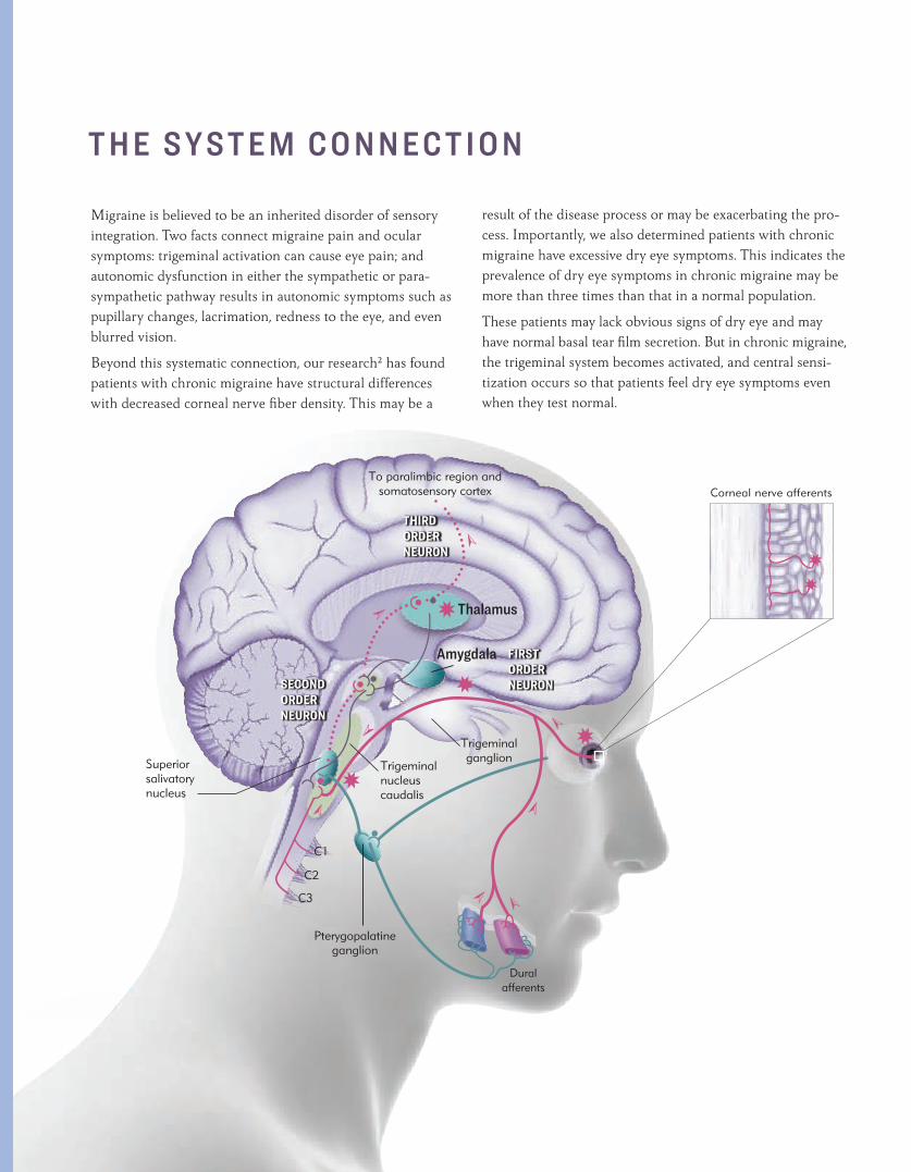
Migraine is believed to be an inherited disorder of sensory integration. Two facts connect migraine pain and ocular symptoms: trigeminal activation can cause eye pain; and autonomic dysfunction in either the sympathetic or para-sympathetic pathway results in autonomic symptoms such as pupillary changes, lacrimation, redness to the eye, and even blurred vision.
Beyond this systematic connection, our research² has found patients with chronic migraine have structural differences with decreased corneal nerve fiber density. This may be a
result of the disease process or may be exacerbating the pro-cess. Importantly, we also determined patients with chronic migraine have excessive dry eye symptoms. This indicates the prevalence of dry eye symptoms in chronic migraine may be more than three times than that in a normal population.
These patients may lack obvious signs of dry eye and may have normal basal tear film secretion. But in chronic migraine, the trigeminal system becomes activated, and central sensi-tization occurs so that patients feel dry eye symptoms even when they test normal.
T H E S YS T E M C O N N E C T I O N
Dural afferents
.
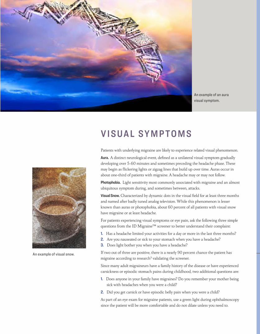
Patients with underlying migraine are likely to experience related visual phenomenon.
Aura. A distinct neurological event, defined as a unilateral visual symptom gradually developing over 5-60 minutes and sometimes preceding the headache phase. These may begin as flickering lights or zigzag lines that build up over time. Auras occur in about one-third of patients with migraine. A headache may or may not follow.
Photophobia. Light sensitivity most commonly associated with migraine and an almost ubiquitous symptom during, and sometimes between, attacks.
Visual Snow. Characterized by dynamic dots in the visual field for at least three months and named after badly tuned analog television. While this phenomenon is lesser known than auras or photophobia, about 60 percent of all patients with visual snow have migraine or at least headache.
For patients experiencing visual symptoms or eye pain, ask the following three simple questions from the ID Migraine™ screener to better understand their complaint:
1. Has a headache limited your activities for a day or more in the last three months?2. Are you nauseated or sick to your stomach when you have a headache? 3. Does light bother you when you have a headache?
If two out of three are positive, there is a nearly 90 percent chance the patient has migraine according to research³ validating the screener.
Since many adult migraineurs have a family history of the disease or have experienced carsickness or episodic stomach pains during childhood, two additional questions are:
1. Does anyone in your family have migraines? Do you remember your mother being sick with headaches when you were a child?
2. Did you get carsick or have episodic belly pain when you were a child?
As part of an eye exam for migraine patients, use a green light during ophthalmoscopy since the patient will be more comfortable and do not dilate unless you need to.
V I S UA L S Y M P TO M S
An example of visual snow.
An example of an aura visual symptom.
Ophthalmologists can confirm and treat migraine, partnering with a neurologist for continued care.
As the first line of treatment, consider treating for dry eye. This may be helpful in slowing down the trigeminal stimulation that keeps the migraine going—and it certainly won’t hurt.
Treatments for visual snow include simple reassurance that the patient’s experience is legitimate. Blue-yellow filters and FL-41 lens filters can also be helpful, while medications including lamotrigine, nortriptyline, carbamazepine, and sertraline have mixed results.
For photophobia associated with migraine, the FL-41 lens filters have also proven remarkably effective, and medications that may help include botulinum toxin injections and use of anticonvulsant drugs.
No matter what the approach, a better understanding of migraine and the experiences of the patients who will inevitably seek our help behooves us all.
¹ Hanson LL, Ahmed Z, Katz BJ, Warner JEA, Drum AV, Zhang Y, Baggaley S, Pippitt K, Cortez MM, Digre KB. Patients With Migraine Have Substantial Reductions in Mea-sures of Visual Quality of Life. Head-ache, 2018 Jun 7. Doi: 10.1111/head. 13330. Epub ahead of print.
Visual Quality of Life in Migraine
V I E W I T N O W
Corneal Nerves in Migraine Patients
V I E W I T N O W
Find these videos at morancore.utah.edu
T R E AT M E N T O P T I O N S
³ Lipton RB, Dodick D, Sadovsky R, Kolodner K, Endicott J, Hettiarachchi J, Harrison W. A self-ad ministered screener for migraine in primary care: The ID Migraine™ validation study. Neurology 2003, 61:375–38.
² Kinard KI, Smith AG, Singleton JR, Lessard MK, Katz BJ, Warner JEA, Crum AV, Mifflin MD, Brennan KC, Digre KB. Chronic migraine is associated with reduced corneal nerve fiber density and symptoms of dry eye. Headache. 2015;55:543–549.
Photophobia FrontiersWhat’s the science behind photophobia?Until recently, it hasn’t been clear how light is transduced into a painful stimulus. Now, we know that transducer appears to be melanopsin-containing intrinsically photosensitive retinal ganglion cells (ipRGCs). These cells are the beginning of a “photophobia circuit” that transforms light that is too bright into a painful sensation.
How is photophobia diagnosed?Photophobia is associated with a number of ophthalmic and neurologic conditions. However, in the presence of a normal eye examination, the most common causes are migraine, benign essential blepharospasm, traumatic brain injury, and dry eyes.
At right is a helpful diagnostic approach.
How does FL-41 tint eyewear help patients?If you look at the transmission spectrum of the FL-41 tint, it maximally blocks light around 480 nm. It turns out that this is the same wavelength that maximally stimulates ipRGCs. So, we can make patients feel better by using optical filters that block this wavelength of light.
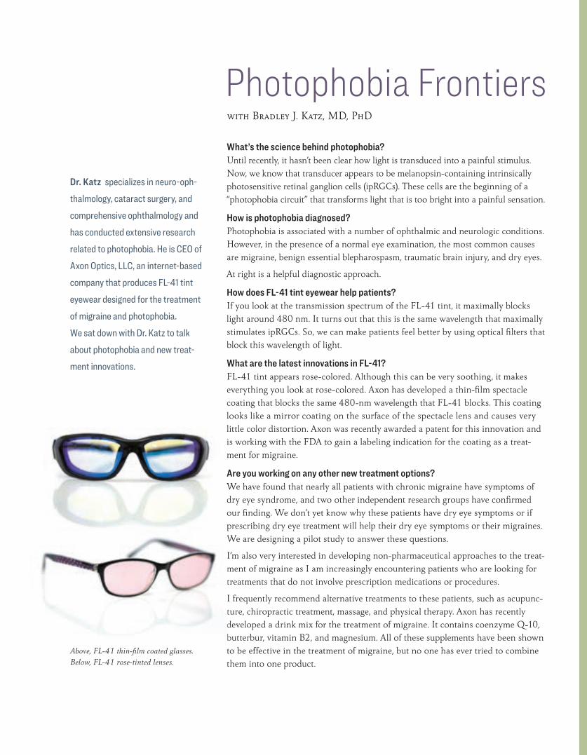
What are the latest innovations in FL-41?FL-41 tint appears rose-colored. Although this can be very soothing, it makes everything you look at rose-colored. Axon has developed a thin-film spectacle coating that blocks the same 480-nm wavelength that FL-41 blocks. This coating looks like a mirror coating on the surface of the spectacle lens and causes very little color distortion. Axon was recently awarded a patent for this innovation and is working with the FDA to gain a labeling indication for the coating as a treat-ment for migraine.
Are you working on any other new treatment options?We have found that nearly all patients with chronic migraine have symptoms of dry eye syndrome, and two other independent research groups have confirmed our finding. We don’t yet know why these patients have dry eye symptoms or if prescribing dry eye treatment will help their dry eye symptoms or their migraines. We are designing a pilot study to answer these questions.
I’m also very interested in developing non-pharmaceutical approaches to the treat-ment of migraine as I am increasingly encountering patients who are looking for treatments that do not involve prescription medications or procedures.
I frequently recommend alternative treatments to these patients, such as acupunc-ture, chiropractic treatment, massage, and physical therapy. Axon has recently developed a drink mix for the treatment of migraine. It contains coenzyme Q-10, butterbur, vitamin B2, and magnesium. All of these supplements have been shown to be effective in the treatment of migraine, but no one has ever tried to combine them into one product.
Dr. Katz specializes in neuro-oph-thalmology, cataract surgery, and comprehensive ophthalmology and has conducted extensive research related to photophobia. He is CEO of Axon Optics, LLC, an internet-based company that produces FL-41 tint eyewear designed for the treatment of migraine and photophobia. We sat down with Dr. Katz to talk about photophobia and new treat-ment innovations.
with Bradley J. Katz, MD, PhD
Above, FL-41 thin-film coated glasses. Below, FL-41 rose-tinted lenses.
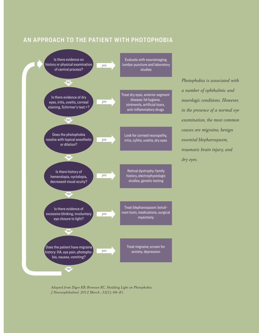
Adapted from Digre KB; Brennan KC. Shedding Light on Photophobia. J Neuroophthalmol. 2012 March ; 32(1): 68–81.
Photophobia is associated with
a number of ophthalmic and
neurologic conditions. However,
in the presence of a normal eye
examination, the most common
causes are migraine, benign
essential blepharospasm,
traumatic brain injury, and
dry eyes.
Is there evidence on history or physical examination
of central process?
Evaluate with neuroimaging, lumbar puncture and laboratory
studies
Is there evidence of dry eyes, iritis, uveitis, corneal
staining, Schirmer's test:+?
Does the photophobia resolve with topical anesthetic
or dilation?
Is there history of hemeralopia, nyctalopia, decreased visual acuity?
Does the patient have migraine history: HA, eye pain, photopho-
bia, nausea, vomiting?
Is there evidence of excessive blinking, involuntary
eye closure to light?
Treat dry eyes, anterior segment disease: lid hygiene,
ointments, artificial tears, anti-inflammatory drugs
Look for corneal neuropathy, iritis, cylitis, uveitis, dry eyes
Retinal dystrophy: family history, electrophysiologic
studies, genetic testing
Treat blepharospasm: botuli-num toxin, medications, surgical
myectomy
Treat migraine; screen for anxiety, depression
yes
yes
yes
yes
yes
yes
no
no
no
no
no
no
AN APPROACH TO THE PATIENT WITH PHOTOPHOBIA
Surgical Idiopathic Intracranial Hypertension Treatment (SIGHT) Trial From 2010 to 2013, Moran Eye Center physicians participated in a watershed study that advanced our ability to treat women with idiopathic intracranial hypertension (IIH) who have mild to moderate vision loss.
As part of the National Institutes of Health (NIH)-funded study,¹ Drs. Kathleen Digre, Bradley Katz, and Judith Warner discovered an inexpensive glaucoma drug—acetazolamide—paired with a weight-reduction plan, can improve and even restore vision for women with IIH.
Today, my colleagues and I are part of a second NIH trial to determine the best treatment for IIH sufferers with moderate to severe vision loss. This time, we will evaluate surgical interventions by randomizing patients to the best therapy:
◆ medical therapy: weight loss, acetazolamide only◆ medical therapy with ventriculoperitoneal cerebrospinal fluid shunting (VPS)◆ medical therapy with optic nerve sheath fenestration (ONSF)
We hope this study will lead to important new insights into a drastically frus-trating and debilitating visual condition. Along with vision loss, IIH patients experience headaches, blackouts, and constant ringing in their ears. Diagnosis and treatment can pose ongoing challenges for general ophthalmologists. For instance, the medications required for treatment are needed in such high dosages that I routinely get calls from pharmacists who want to verify numbers.
Although we now know that medications and weight loss can alleviate mild to moderate vision loss, we have seen IIH patients with moderate to severe loss go blind, regardless of the highest dosages. Symptoms can come on suddenly, and it’s not unusual to have patients from surrounding states flown to us by medical helicopter for treatment. In extreme cases, we admit them to the hospital for a lumbar drain to alleviate pressure before considering further treatment. Know-ing that IIH patients as young as 18 may face 60-80 years of blindness without surgery to reduce fluid and relieve pressure, we may recommend either ONSF or VPS, depending on symptoms.
But are we helping or harming patients in the long run? Can these surgeries treat the patient better than medicine alone?
This study marks an important “first” that will allow us to more confidently offer IIH patients the best option available.
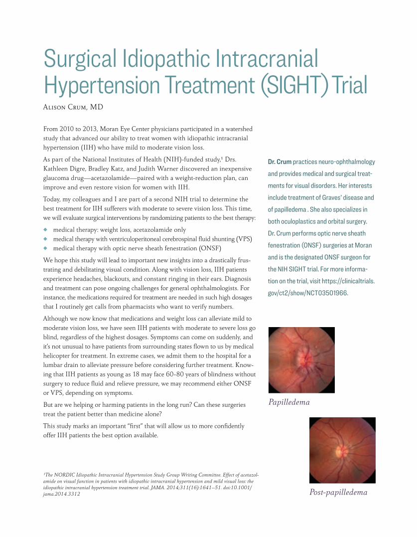
Dr. Crum practices neuro-ophthalmology and provides medical and surgical treat-ments for visual disorders. Her interests include treatment of Graves’ disease and of papilledema . She also specializes in both oculoplastics and orbital surgery. Dr. Crum performs optic nerve sheath fenestration (ONSF) surgeries at Moran and is the designated ONSF surgeon for the NIH SIGHT trial. For more informa-tion on the trial, visit https://clinicaltrials.gov/ct2/show/NCT03501966.
Alison Crum, MD
¹The NORDIC Idiopathic Intracranial Hypertension Study Group Writing Committee. Effect of acetazol-amide on visual function in patients with idiopathic intracranial hypertension and mild visual loss: the idiopathic intracranial hypertension treatment trial. JAMA. 2014;311(16):1641–51. doi:10.1001/jama.2014.3312
Papilledema
Post-papilledema
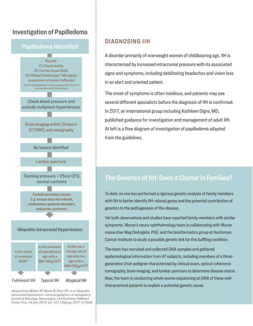
DIAGNOSING IIH
A disorder primarily of overweight women of childbearing age, IIH is
characterized by increased intracranial pressure with its associated
signs and symptoms, including debilitating headaches and vision loss
in an alert and oriented patient.
The onset of symptoms is often insidious, and patients may see
several different specialists before the diagnosis of IIH is confirmed.
In 2017, an international group including Kathleen Digre, MD,
published guidance for investigation and management of adult IIH.
At left is a flow diagram of investigation of papilledema adapted
from the guidelines.
The Genetics of IIH: Does it Cluster in Families?
To date, no one has performed a rigorous genetic analysis of family members with IIH to better identify IIH-related genes and the potential contribution of genetics to the pathogenesis of this disease.
Yet both observations and studies have reported family members with similar symptoms. Moran’s neuro-ophthalmology team is collaborating with Moran researcher Meg DeAngelis, PhD, and the bioinformatics group at Huntsman Cancer Institute to study a possible genetic link for this baffling condition.
The team has recruited and collected DNA samples and gathered epidemiological information from 47 subjects, including members of a three-generation Utah pedigree characterized by clinical exam, optical coherence tomography, brain imaging, and lumbar puncture to determine disease status. Now, the team is conducting whole exome sequencing on DNA of these well-characterized patients to explain a potential genetic cause.
Adapted from Mollan SP, Davies B, Silver NC, et al. Idiopathic intracranial hypertension: consensus guidelines on management. Journal of Neurology, Neurosurgery, and Psychiatry. Published Online First: 14 June 2018. doi: 10.1136/jnnp-2017-317440
Papilledema Identified
Idiopathic Intracranial Hypertension
Brain imaging within 24 hours (CT/MRI) and venography
Lumbar puncture
Check blood pressure and exclude malignant hypertension
No lesions identified
Exclude secondary causesE.g. venous sinus thrombosis,
medications, systemic disorders, endocrine, syndromic
Record: (1) Visual acuity,
(2) Formal visual fields, (3) Dilated fundoscopy* NB regular
assessment of vision if affected *Confirm papilledema with experienced clinician if
any doubt or with fundoscopy
Opening pressure > 25cm CFS; normal contents
Is the vision at imminent
RISK?
Is this a woman of reproductive
age with a BMI>30kg/m2?
Is this not a female, not of reproductive
age with a BMI<30kg/m2?
Fulminant IIH Typical IIH Atypical IIH
Investigation of Papilledema
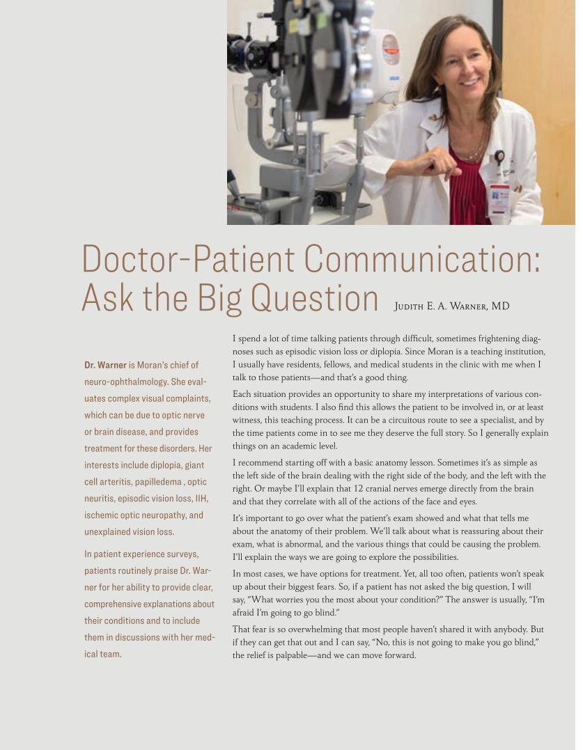
Doctor-Patient Communication: Ask the Big Question Judith E. A. Warner, MD
Dr. Warner is Moran’s chief of neuro-ophthalmology. She eval-uates complex visual complaints, which can be due to optic nerve or brain disease, and provides treatment for these disorders. Her interests include diplopia, giant cell arteritis, papilledema , optic neuritis, episodic vision loss, IIH, ischemic optic neuropathy, and unexplained vision loss.
In patient experience surveys, patients routinely praise Dr. War-ner for her ability to provide clear, comprehensive explanations about their conditions and to include them in discussions with her med-ical team.
I spend a lot of time talking patients through difficult, sometimes frightening diag-noses such as episodic vision loss or diplopia. Since Moran is a teaching institution, I usually have residents, fellows, and medical students in the clinic with me when I talk to those patients—and that’s a good thing.
Each situation provides an opportunity to share my interpretations of various con-ditions with students. I also find this allows the patient to be involved in, or at least witness, this teaching process. It can be a circuitous route to see a specialist, and by the time patients come in to see me they deserve the full story. So I generally explain things on an academic level.
I recommend starting off with a basic anatomy lesson. Sometimes it’s as simple as the left side of the brain dealing with the right side of the body, and the left with the right. Or maybe I’ll explain that 12 cranial nerves emerge directly from the brain and that they correlate with all of the actions of the face and eyes.
It’s important to go over what the patient’s exam showed and what that tells me about the anatomy of their problem. We’ll talk about what is reassuring about their exam, what is abnormal, and the various things that could be causing the problem. I’ll explain the ways we are going to explore the possibilities.
In most cases, we have options for treatment. Yet, all too often, patients won’t speak up about their biggest fears. So, if a patient has not asked the big question, I will say, “What worries you the most about your condition?” The answer is usually, “I’m afraid I’m going to go blind.”
That fear is so overwhelming that most people haven’t shared it with anybody. But if they can get that out and I can say, “No, this is not going to make you go blind,” the relief is palpable—and we can move forward.
Ophthalmologists and optometrists routinely ask me which electrophysiology test can best detect the site of their patients’ visual symptoms.
The answer depends on symptoms and suspected diagnoses.
As a neuro-ophthalmology team, we consider thousands of symptoms, suspected syndromes, and eye diseases. Each patient is unique, and we use a myriad of electrophysiological testing methods to explore suspected diagnoses and determine whether dysfunction is retinal, optic nerve, or cortical.
Sometimes ruling out a site can be helpful. Using multifocal electroretinograms (mfERGs) can determine if a visual field defect is due to retinal dysfunction. Normal mfERGs point to optic pathways or the cortex as the site of pathology.
Electrophysiology in the pediatric population is even more critical and informative than in adults since infants and children often do not recognize visual symptoms. Their parents may notice signs that indicate vision issues—such as not looking at an object, always looking sideways, or struggling to find toys on the floor.
Even children with severe visual dysfunction may seem normal if acuity remains good enough for them to get around. For example, night blindness, if present from birth, may be normal to children because they have no basis for comparison. Similarly, slow loss of peripheral visual field, such as from a brain tumor, might pass unnoticed by an infant or child.
With electrophysiology we can test the eyes, then the pathways in the brain, and do so without having to rely on verbal feedback.
Electrophysiology as a Diagnostic Tool Donnell J. Creel, PhD
Dr. Creel is Moran’s director of the electrophysiology service and research professor of ophthalmol-ogy and visual sciences, focusing on the neurobiology of disease.
Dr. Creel’s internet chapters posted on Webvision.med.utah.edu on Visually Evoked Potentials and Clinical Electroretinograms (ERGs) are the most accessed sites on these topics. His video on the multifocal electroretinogram (mfERG), the most recent advance in ERG technology, is available on the Neuro-Ophthalmology Virtual Education Library (NOVEL) at novel.utah.edu.
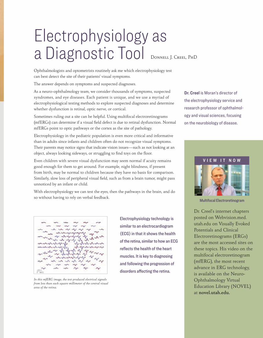
Multifocal Electroretinogram
V I E W I T N O W
Electrophysiology technology is similar to an electrocardiogram (ECG) in that it shows the health of the retina, similar to how an ECG reflects the health of the heart muscles. It is key to diagnosing and following the progression of disorders affecting the retina.
In this mfERG image, the test produced electrical signals from less than each square millimeter of the central visual area of the retina.
200 nV
200 ms0
$
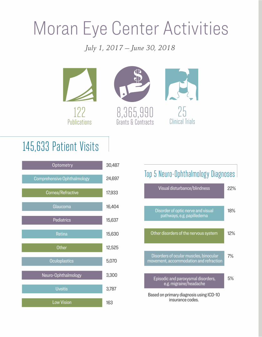
Moran Eye Center Activities
122 Publications
8,365,990 Grants & Contracts
25 Clinical Trials
July 1, 2017 – June 30, 2018
145,633 Patient Visits
Comprehensive Ophthalmology 24,697
Cornea/Refractive 17,933
Neuro-Ophthalmology 3,300
Oculoplastics 5,070
Glaucoma 16,404
Optometry 30,487
Pediatrics 15,637
Retina 15,630
Uveitis 3,787
Low Vision 163
Top 5 Neuro-Ophthalmology Diagnoses
Disorder of optic nerve and visual pathways, e.g. papilledema
Based on primary diagnosis using ICD-10 insurance codes.
Episodic and paroxysmal disorders, e.g. migraine/headache
Disorders of ocular muscles, binocular movement, accommodation and refraction
Visual disturbance/blindness 22%
18%
5%
7%
Other disorders of the nervous system 12%
Other 12,525
Moran Eye Center Activities
15
South Jordan Health Center 5126 West Daybreak Parkway 5200 West 11400 South
Moran Eye Center Clinic at Intermountain Riverton Hospital 3773 West 12600 South Suite 301
Parkway Health Center 145 West University Parkway
Midvalley Health Center 243 East 6100 South
Layton
Farmington
Farmington Health Center 165 North University Ave
Primary Children’s Hospital
University of Utah Hospital
Moran Eye Center 65 Mario Capecchi Drive
Park City
Redstone Health Center 1743 West Redstone Center Drive Suite 115
Redwood Health Center 1525 West 2100 South
South Salt Lake
Murray
Salt Lake City
Westridge Health Center 3730 West 4700 South
Sandy
West Jordan
South Jordan
Riverton
Orem
Tooele
West Valley City
100 South
400 South500 South
Redw
ood R
oadStansbury Health Center
220 Millpond Road Suite 100
Stat
e Stre
et
700 E
ast
2100 South
Foothill Dr
3500 South3300 South
4700 South Stat
e Stre
et
Redw
ood R
oad
10600 South
11400 South
12600 South
Bang
erte
r Hig
hway
80
215
15
215
15
215
80
Draper
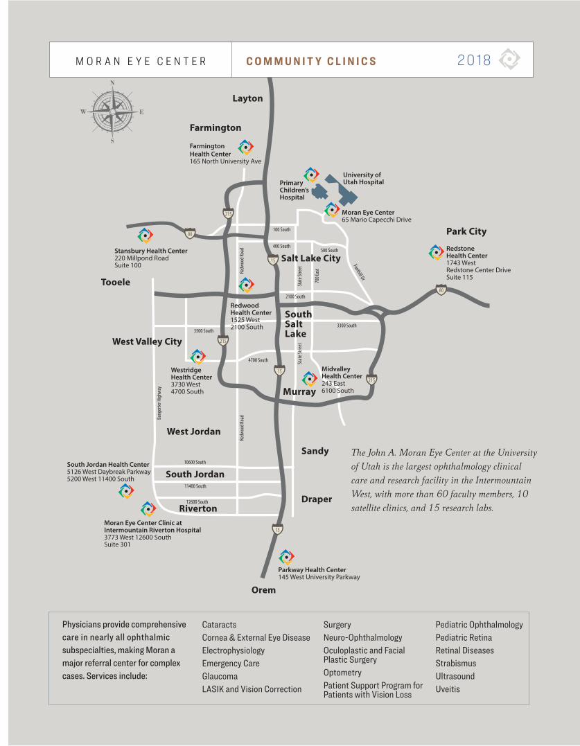
Physicians provide comprehensive care in nearly all ophthalmic subspecialties, making Moran a major referral center for complex cases. Services include:
CataractsCornea & External Eye DiseaseElectrophysiologyEmergency CareGlaucomaLASIK and Vision Correction
SurgeryNeuro-OphthalmologyOculoplastic and Facial Plastic SurgeryOptometryPatient Support Program for Patients with Vision Loss
Pediatric OphthalmologyPediatric RetinaRetinal DiseasesStrabismusUltrasoundUveitis
C O M M U N I T Y C L I N I C S 2 0 18M O R A N E Y E C E N T E R
The John A. Moran Eye Center at the University of Utah is the largest ophthalmology clinical care and research facility in the Intermountain West, with more than 60 faculty members, 10 satellite clinics, and 15 research labs.
65 Mario Capecchi Drive, Salt Lake City, Utah 84132
NON-PROFIT ORG. U.S. POSTAGE PAID
Permit No. 1529 Salt Lake City, Utah
moran.eye.centermoraneyecenter.org Moran Eye Center @moraneyecenter
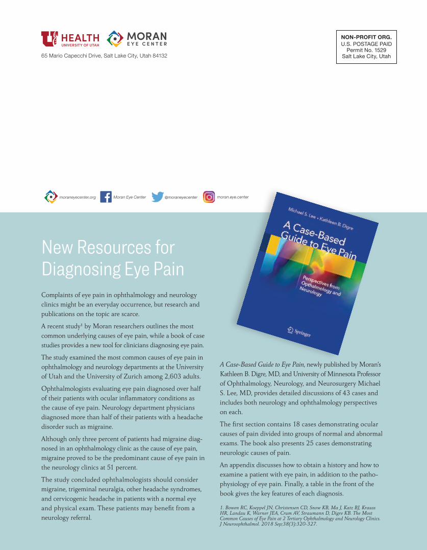
New Resources for Diagnosing Eye PainComplaints of eye pain in ophthalmology and neurology clinics might be an everyday occurrence, but research and publications on the topic are scarce.
A recent study¹ by Moran researchers outlines the most common underlying causes of eye pain, while a book of case studies provides a new tool for clinicians diagnosing eye pain.
The study examined the most common causes of eye pain in ophthalmology and neurology departments at the University of Utah and the University of Zurich among 2,603 adults.
Ophthalmologists evaluating eye pain diagnosed over half of their patients with ocular inflammatory conditions as the cause of eye pain. Neurology department physicians diagnosed more than half of their patients with a headache disorder such as migraine.
Although only three percent of patients had migraine diag-nosed in an ophthalmology clinic as the cause of eye pain, migraine proved to be the predominant cause of eye pain in the neurology clinics at 51 percent.
The study concluded ophthalmologists should consider migraine, trigeminal neuralgia, other headache syndromes, and cervicogenic headache in patients with a normal eye and physical exam. These patients may benefit from a neurology referral.
A Case-Based Guide to Eye Pain, newly published by Moran’s Kathleen B. Digre, MD, and University of Minnesota Professor of Ophthalmology, Neurology, and Neurosurgery Michael S. Lee, MD, provides detailed discussions of 43 cases and includes both neurology and ophthalmology perspectives on each.
The first section contains 18 cases demonstrating ocular causes of pain divided into groups of normal and abnormal exams. The book also presents 25 cases demonstrating neurologic causes of pain.
An appendix discusses how to obtain a history and how to examine a patient with eye pain, in addition to the patho-physiology of eye pain. Finally, a table in the front of the book gives the key features of each diagnosis.
1. Bowen RC, Koeppel JN, Christensen CD, Snow KB, Ma J, Katz BJ, Krauss HR, Landau K, Warner JEA, Crum AV, Straumann D, Digre KB. The Most Common Causes of Eye Pain at 2 Tertiary Ophthalmology and Neurology Clinics. J Neuroophthalmol. 2018 Sep;38(3):320-327.