Embed Size (px)
Citation preview
Making the Right Diagnosis
Symposium: Joint Preservation Hip Surgery – How to Avoid and Treat Complications and Failures
Wednesday, February 16th, 2011
Bryan T. Kelly, MDCo-DirectorCenter for Hip Pain and Preservation
Bryan T. Kelly, MD
Hospital for Special Surgery
Disclosure: I DO NOT have a financial interest in any commercial products or service presented in this lecture AND
DO NOT INTEND to discuss off label or investigational use of products or
services.

Types of financial relationships and the companies with whom I have relationships are as follows:
Pivot Medical, Inc.: Consultant
Smith & Nephew: Educational Consultant
A2 Surgical: Consultant
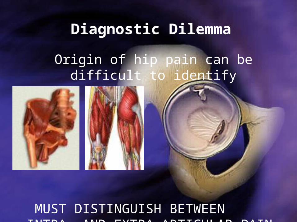
Diagnostic Dilemma
Origin of hip pain can be difficult to identify
MUST DISTINGUISH BETWEEN INTRA- AND EXTRA-ARTICULAR PAIN
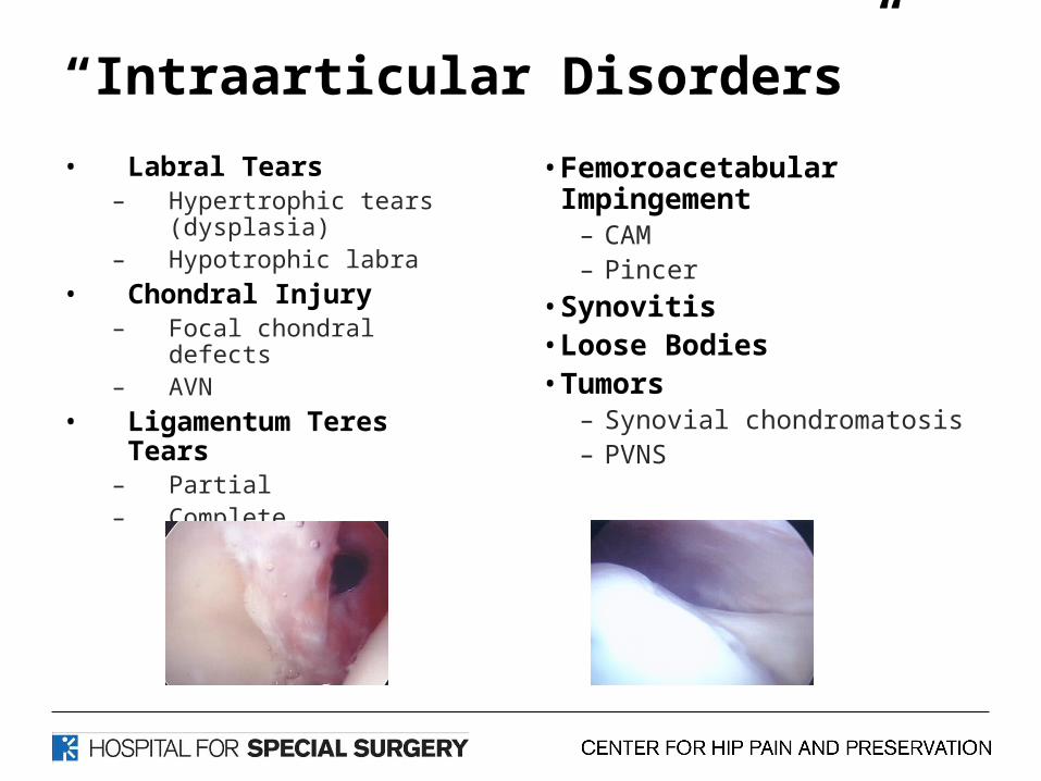
“Intraarticular Disorders”
• Labral Tears– Hypertrophic tears
(dysplasia)– Hypotrophic labra
• Chondral Injury– Focal chondral defects– AVN
• Ligamentum Teres Tears– Partial– Complete
• Femoroacetabular Impingement
– CAM– Pincer
• Synovitis• Loose Bodies • Tumors
– Synovial chondromatosis– PVNS
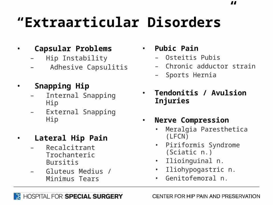
“Extraarticular Disorders”
• Capsular Problems– Hip Instability – Adhesive Capsulitis
• Snapping Hip– Internal Snapping Hip– External Snapping Hip
• Lateral Hip Pain– Recalcitrant Trochanteric
Bursitis – Gluteus Medius / Minimus
Tears
• Pubic Pain– Osteitis Pubis – Chronic adductor strain– Sports Hernia
• Tendonitis / Avulsion Injuries
• Nerve Compression• Meralgia Paresthetica (LFCN)• Piriformis Syndrome (Sciatic n.)• Ilioinguinal n.• Iliohypogastric n.• Genitofemoral n.

History & Physical Exam
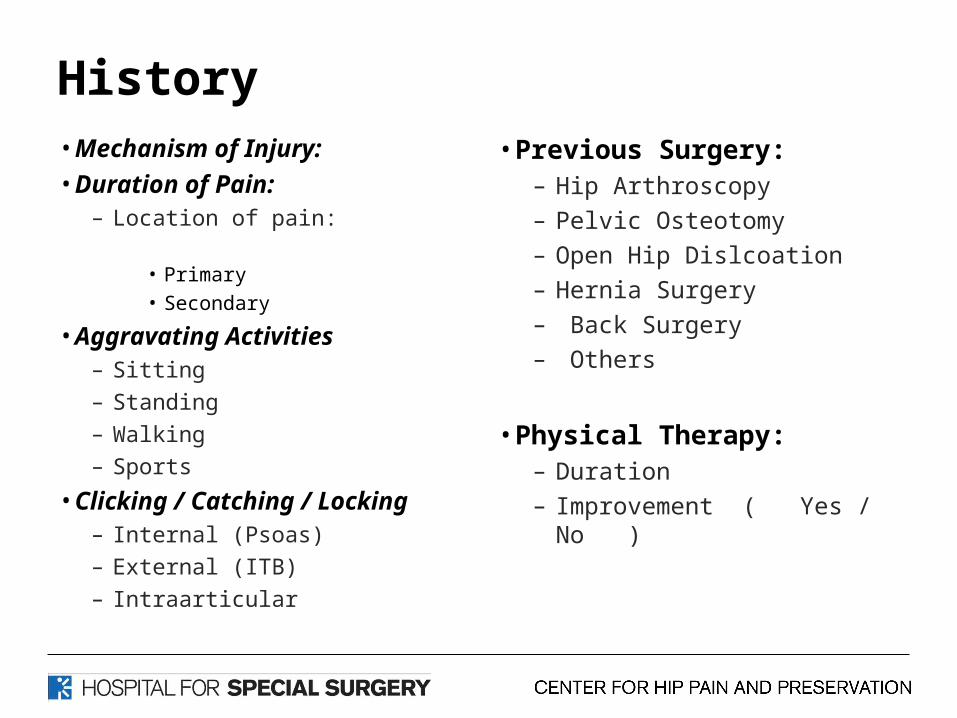
History• Mechanism of Injury:
• Duration of Pain:– Location of pain:
• Primary• Secondary
• Aggravating Activities
– Sitting– Standing– Walking– Sports
• Clicking / Catching / Locking– Internal (Psoas) – External (ITB)– Intraarticular
• Previous Surgery:– Hip Arthroscopy
– Pelvic Osteotomy
– Open Hip Dislcoation
– Hernia Surgery
– Back Surgery
– Others
• Physical Therapy:– Duration
– Improvement ( Yes / No )
Minimum Clinical Exam
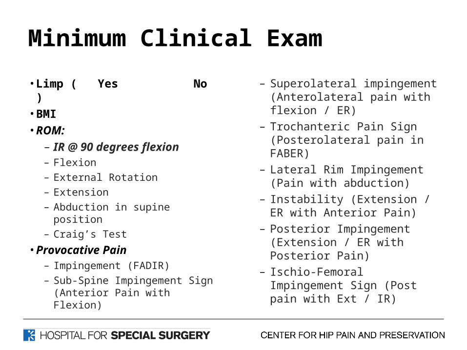
• Limp ( Yes No )• BMI• ROM:
– IR @ 90 degrees flexion– Flexion
– External Rotation
– Extension
– Abduction in supine position
– Craig’s Test
• Provocative Pain– Impingement (FADIR)
– Sub-Spine Impingement Sign (Anterior Pain with Flexion)
– Superolateral impingement (Anterolateral pain with flexion / ER)
– Trochanteric Pain Sign (Posterolateral pain in FABER)
– Lateral Rim Impingement (Pain with abduction)
– Instability (Extension / ER with Anterior Pain)
– Posterior Impingement (Extension / ER with Posterior Pain)
– Ischio-Femoral Impingement Sign (Post pain with Ext / IR)
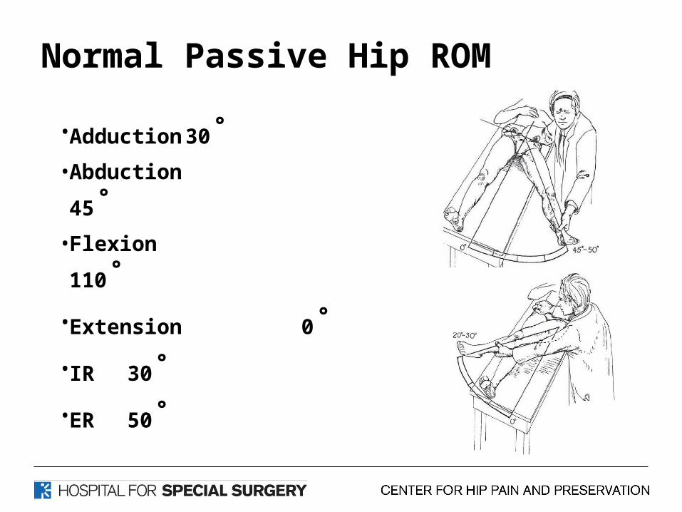
Normal Passive Hip ROM
• Adduction 30˚• Abduction 45˚• Flexion 110˚• Extension 0˚• IR 30˚• ER 50˚
How do you assess ROM
• IR Block Test
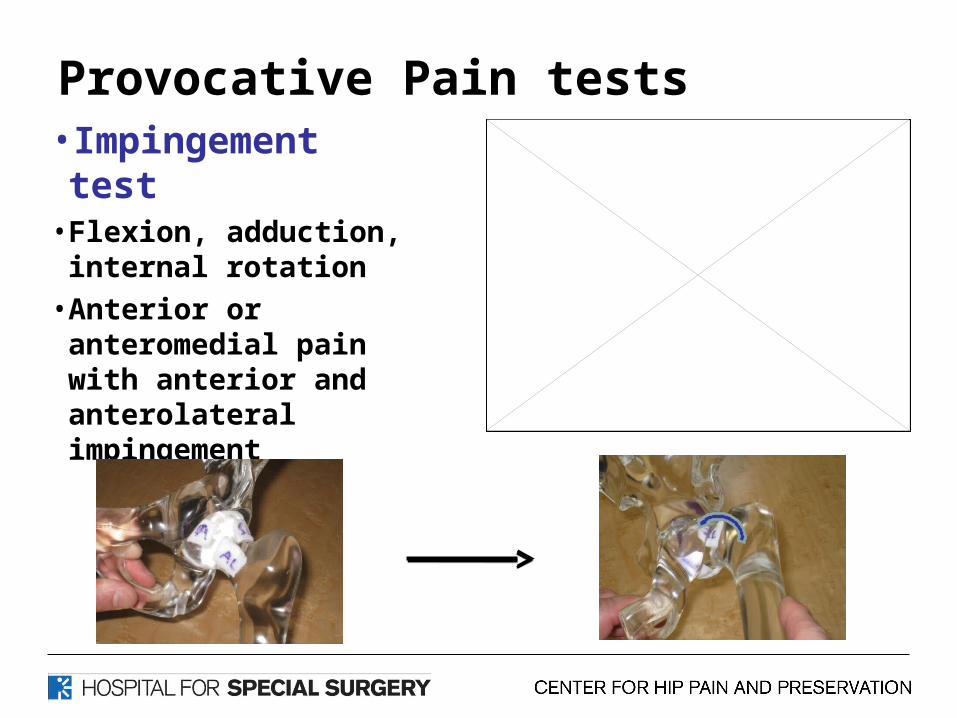
Provocative Pain tests• Impingement test• Flexion, adduction,
internal rotation
• Anterior or anteromedial pain with anterior and anterolateral impingement
Provocative Pain tests
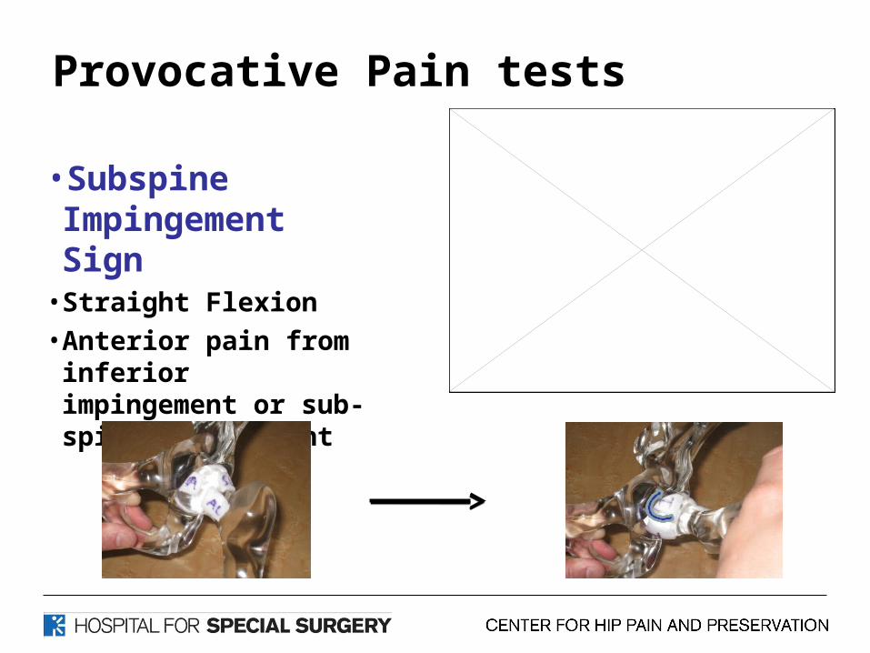
•Subspine Impingement Sign
• Straight Flexion
• Anterior pain from inferior impingement or sub-spine impingement
Provocative Pain tests
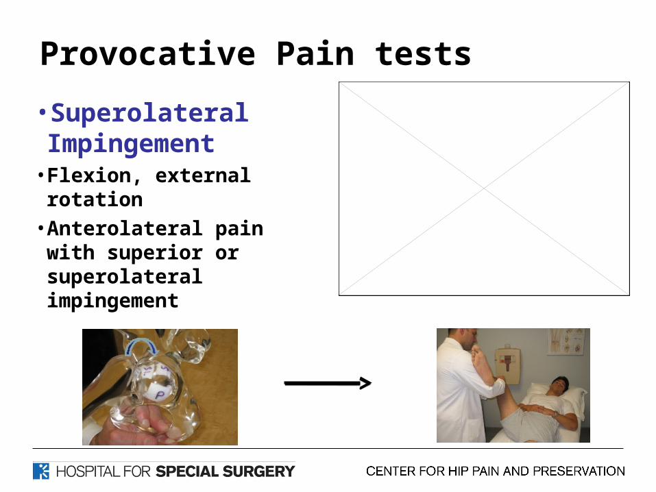
•Superolateral Impingement
• Flexion, external rotation
• Anterolateral pain with superior or superolateral impingement
Provocative Pain tests
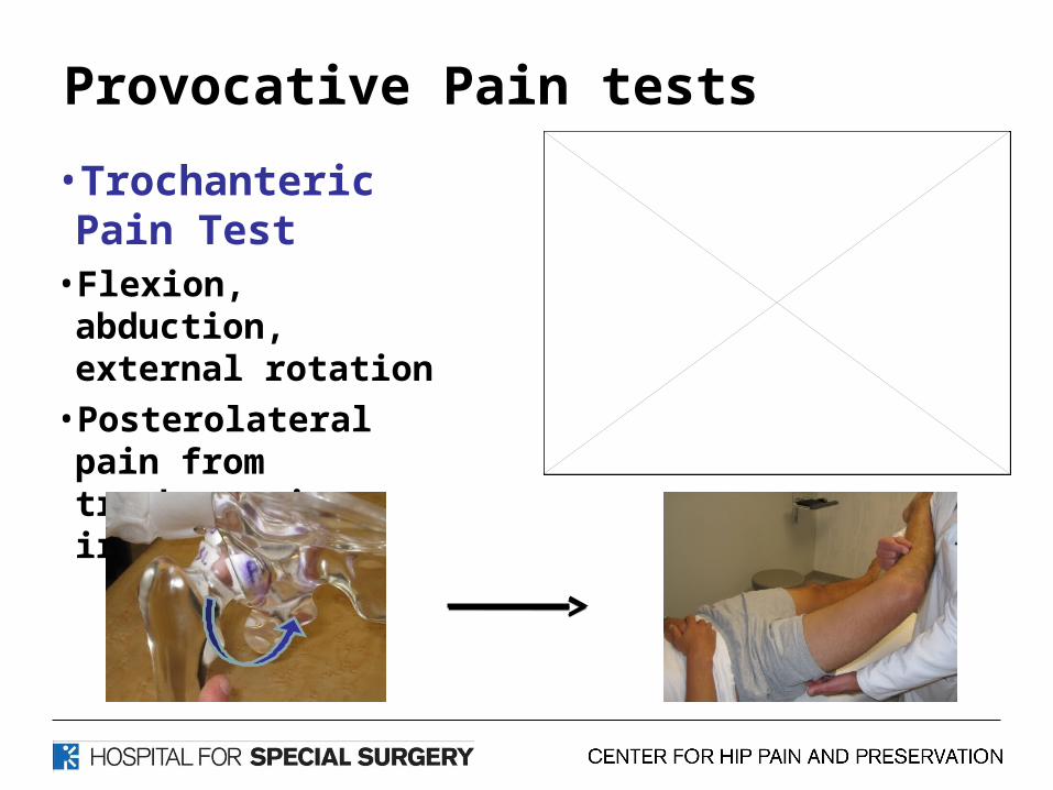
•Trochanteric Pain Test
• Flexion, abduction, external rotation
• Posterolateral pain from trochanteric irritation
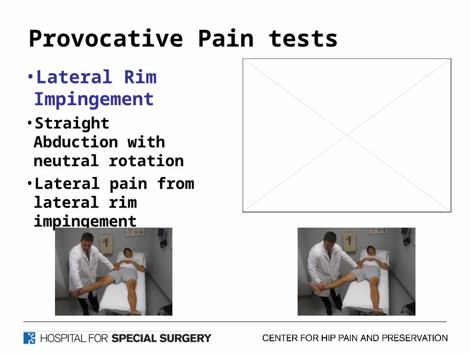
Provocative Pain tests
•Lateral Rim Impingement
• Straight Abduction with neutral rotation
• Lateral pain from lateral rim impingement
Provocative Pain tests
• Instability Test• Extension, external rotation• Anterior hip pain
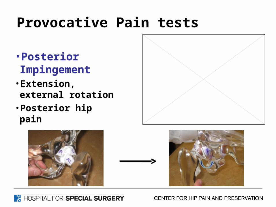
Provocative Pain tests
•Posterior Impingement
• Extension, external rotation
• Posterior hip pain
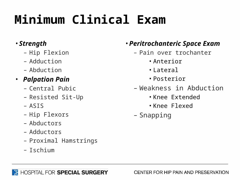
Minimum Clinical Exam
• Strength– Hip Flexion
– Adduction
– Abduction
• Palpation Pain– Central Pubic
– Resisted Sit-Up
– ASIS
– Hip Flexors
– Abductors
– Adductors
– Proximal Hamstrings
– Ischium
• Peritrochanteric Space Exam– Pain over trochanter
• Anterior
• Lateral
• Posterior
– Weakness in Abduction• Knee Extended
• Knee Flexed
– Snapping

COMPREHENSIVE EXAMINATION OF THE ADULT HIP
• Five points for five body positions
– STANDING– SITTING– SUPINE – LATERAL– PRONE
• ADDITIONAL TESTS AS NEEDED
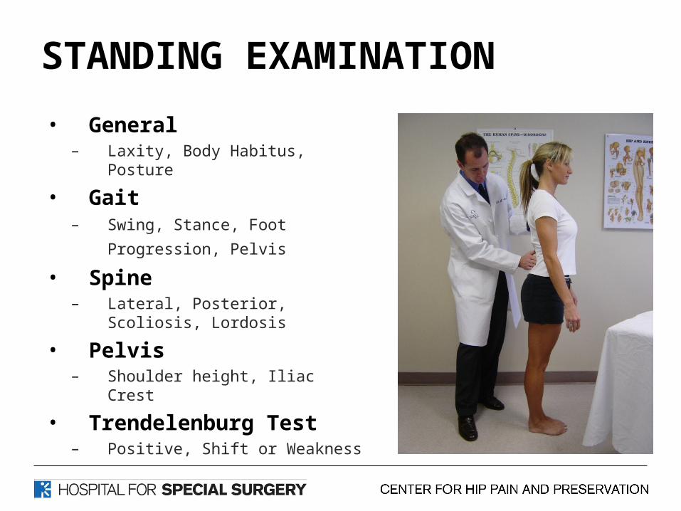
STANDING EXAMINATION
• General– Laxity, Body Habitus, Posture
• Gait– Swing, Stance, Foot Progression,
Pelvis • Spine
– Lateral, Posterior, Scoliosis, Lordosis
• Pelvis– Shoulder height, Iliac Crest
• Trendelenburg Test– Positive, Shift or Weakness

STANDING EXAMINATION• Gait
a. Trendelenburgb. Abductor lurchc. Antalgicd. Foot progression angle
a. Excessive External Rotationb. Excessive Internal Rotation
e. Short Leg Limp
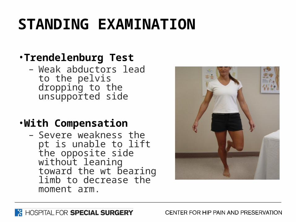
STANDING EXAMINATION
• Trendelenburg Test– Weak abductors lead to
the pelvis dropping to the unsupported side
• With Compensation– Severe weakness the pt is
unable to lift the opposite side without leaning toward the wt bearing limb to decrease the moment arm.

SEATED EXAMINATION
• Neurologic – DTRS, Sensory, Motor,
Straight Leg Raise
• Circulation– DP, PT, Popliteal
• Skin• Lymphatic• IR/ER
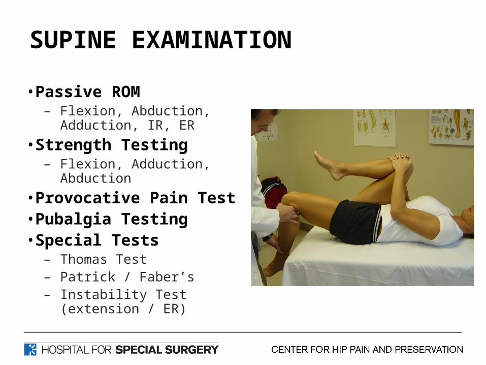
SUPINE EXAMINATION
• Passive ROM – Flexion, Abduction,
Adduction, IR, ER
• Strength Testing– Flexion, Adduction,
Abduction
• Provocative Pain Test• Pubalgia Testing• Special Tests
– Thomas Test– Patrick / Faber’s– Instability Test (extension /
ER)

LATERAL EXAMINATION
• Palpation GT, ABDUCTORS, SI, ISCHIAL BURSAE
• Obers Test FLEXION, EXTENSION
• Passive / Active ROM MEDIUS / MAX • FADDIR IMPINGEMENT
• Lateral Rim Impingement
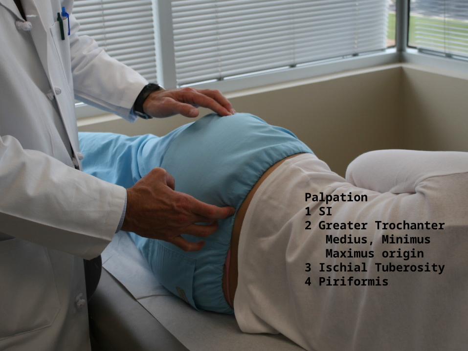
Palpation1 SI2 Greater Trochanter Medius, Minimus Maximus origin3 Ischial Tuberosity4 Piriformis
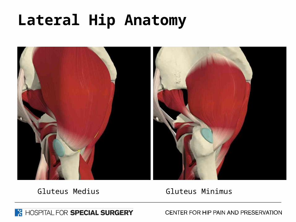
Lateral Hip Anatomy
Gluteus Medius Gluteus Minimus
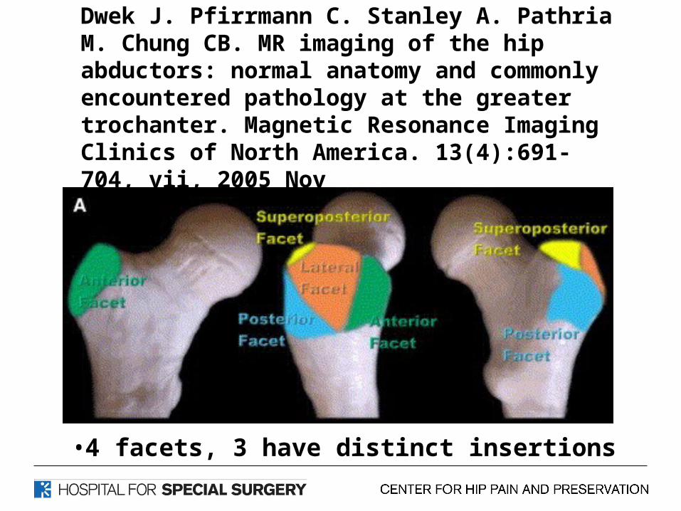
Dwek J. Pfirrmann C. Stanley A. Pathria M. Chung CB. MR imaging of the hip abductors: normal anatomy and commonly encountered pathology at the greater trochanter. Magnetic Resonance Imaging Clinics of North America. 13(4):691-704, vii, 2005 Nov
•4 facets, 3 have distinct insertions
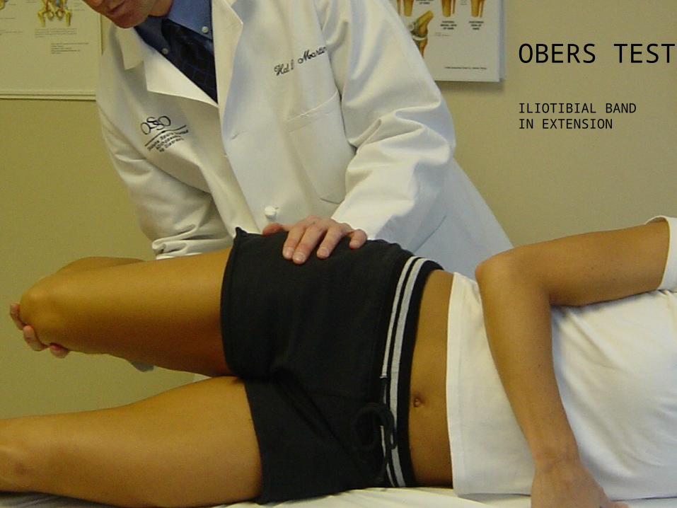
OBERS TEST
ILIOTIBIAL BAND IN EXTENSION
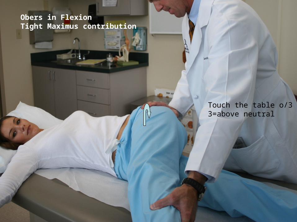
Touch the table o/33=above neutral
Obers in FlexionTight Maximus contribution
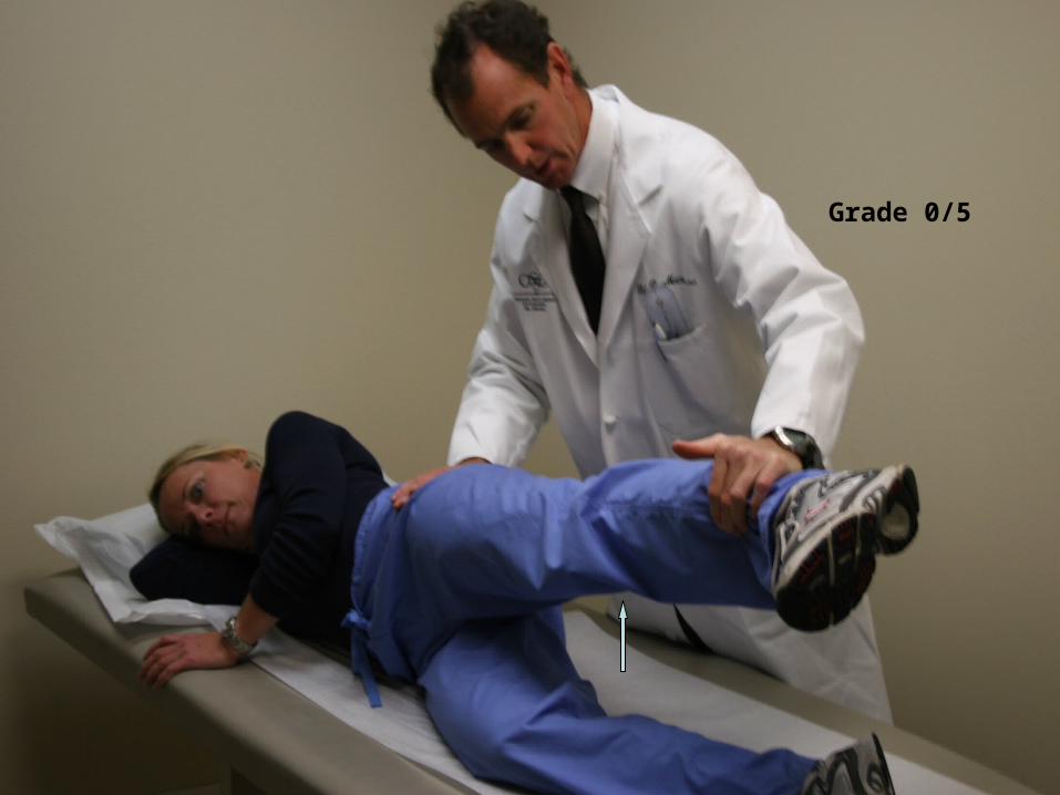
Grade 0/5
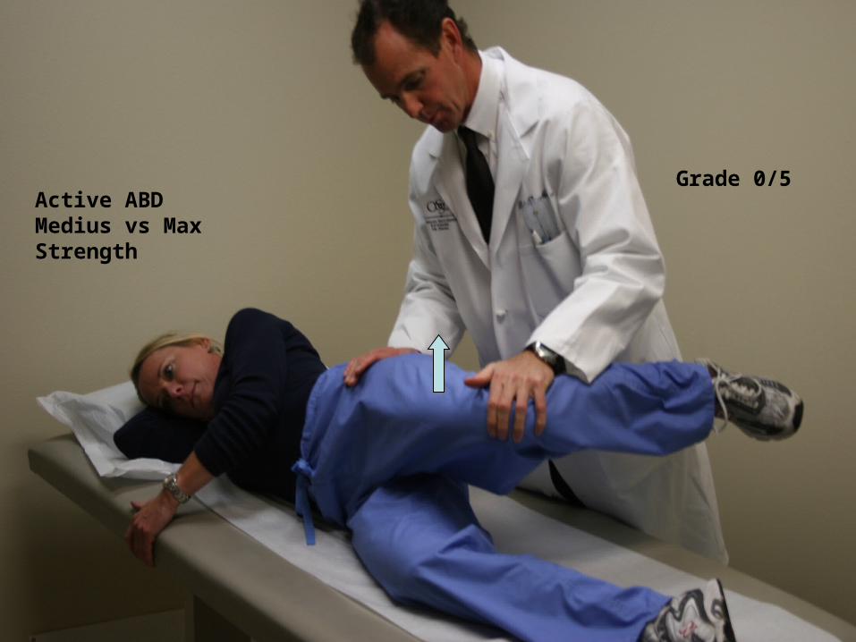
Grade 0/5Active ABDMedius vs MaxStrength

PRONE EXAMINATION
• Craig’s Test– Femoral anteversion
• Ely’s – Rectus Femoris Contracture
• Hyperextension – Lumbar Spine
• Palpation – Paravertebral muscles, spinous process
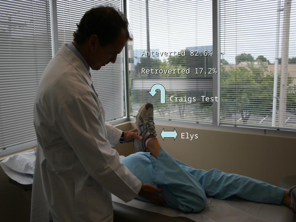
Anteverted 82.6%Anteverted 82.6% Retroverted 17.2%Retroverted 17.2%
Elys
Craigs Test
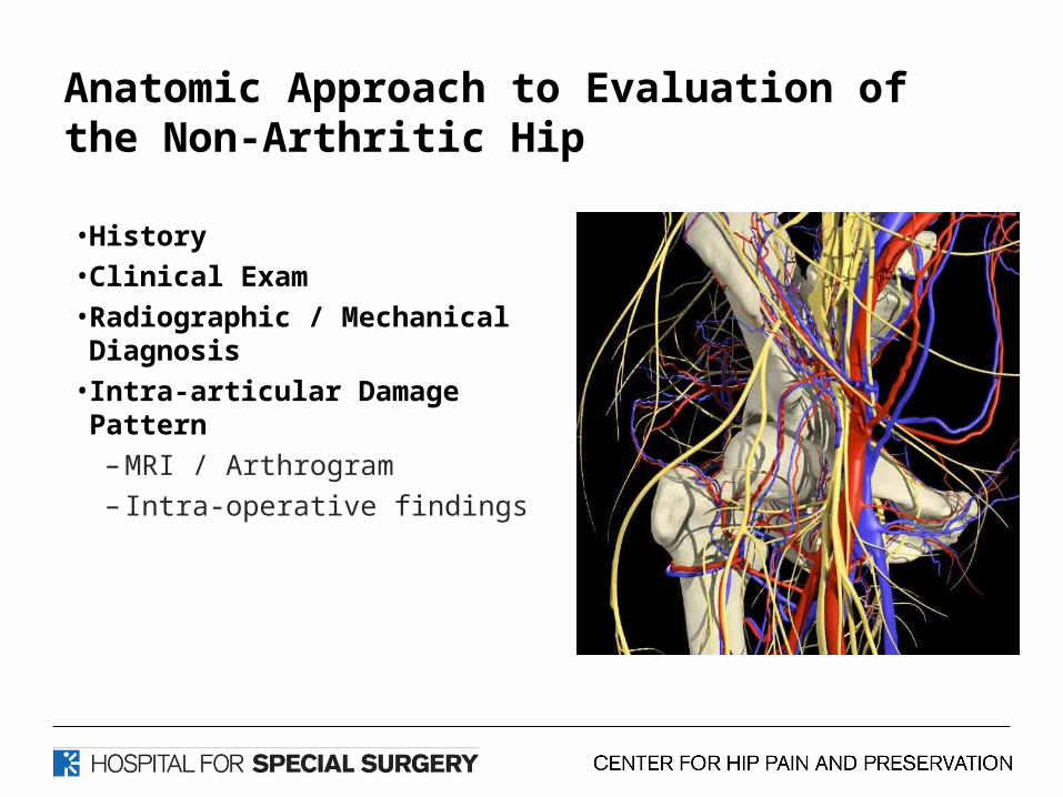
Anatomic Approach to Evaluation of the Non-Arthritic Hip
• History • Clinical Exam• Radiographic / Mechanical Diagnosis
• Intra-articular Damage Pattern– MRI / Arthrogram– Intra-operative findings
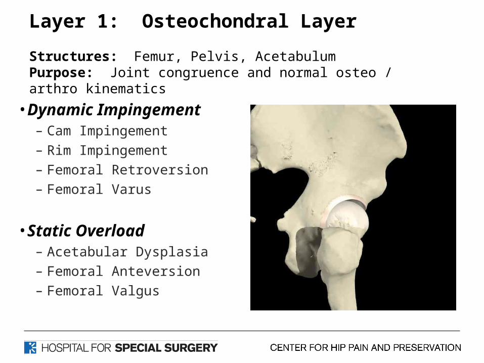
Layer 1: Osteochondral Layer
Structures: Femur, Pelvis, AcetabulumPurpose: Joint congruence and normal osteo / arthro kinematics
• Dynamic Impingement– Cam Impingement– Rim Impingement– Femoral Retroversion– Femoral Varus
• Static Overload– Acetabular Dysplasia– Femoral Anteversion– Femoral Valgus

Radiographic Indices: Mechanical Diagnosis
>15o [nml <10o]
Retroversion(15-20o anteversion)
<15o
[nml >25o]
>140 or <1207.2mm
Nml=11.6
Alpha Angle >50o
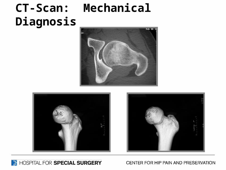
CT-Scan: Mechanical Diagnosis
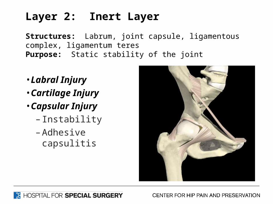
Layer 2: Inert Layer
Structures: Labrum, joint capsule, ligamentous complex, ligamentum teresPurpose: Static stability of the joint
• Labral Injury• Cartilage Injury• Capsular Injury
– Instability– Adhesive capsulitis
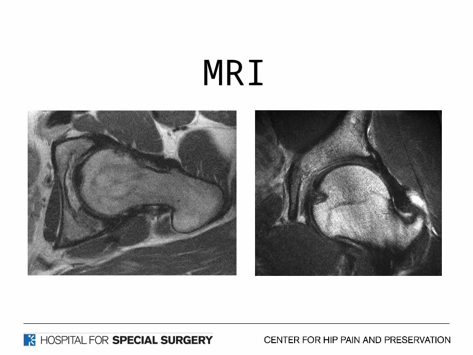
MRI
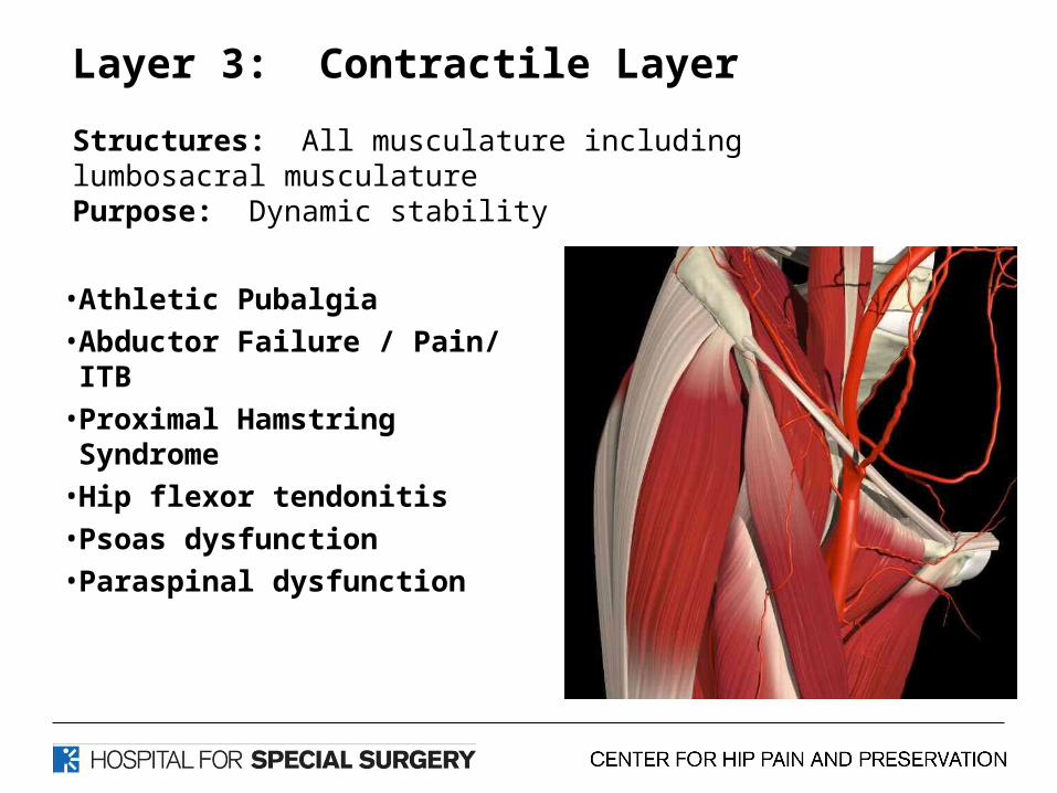
Layer 3: Contractile Layer
Structures: All musculature including lumbosacral musculaturePurpose: Dynamic stability
• Athletic Pubalgia
• Abductor Failure / Pain/ ITB
• Proximal Hamstring Syndrome
• Hip flexor tendonitis
• Psoas dysfunction
• Paraspinal dysfunction

Layer 4: Neuromechanical Layer
Structures: TLS Plexus, Lumbopelvic structures, LE structuresPurpose: Neuromuscular linking and functional control of the entire segment as it functions within its environment
• Nerve compression syndromes
• Pain syndromes• Neuromuscular dysfunction
• Spine referral patterns
Patient Selection
Hip loaded pelvis usually rotates over fixed femur
creating anterior and medial forces with rotary moments
Neuromuscular Research Neuromuscular Research LaboratoryLaboratory
University of PittsburghUniversity of Pittsburgh
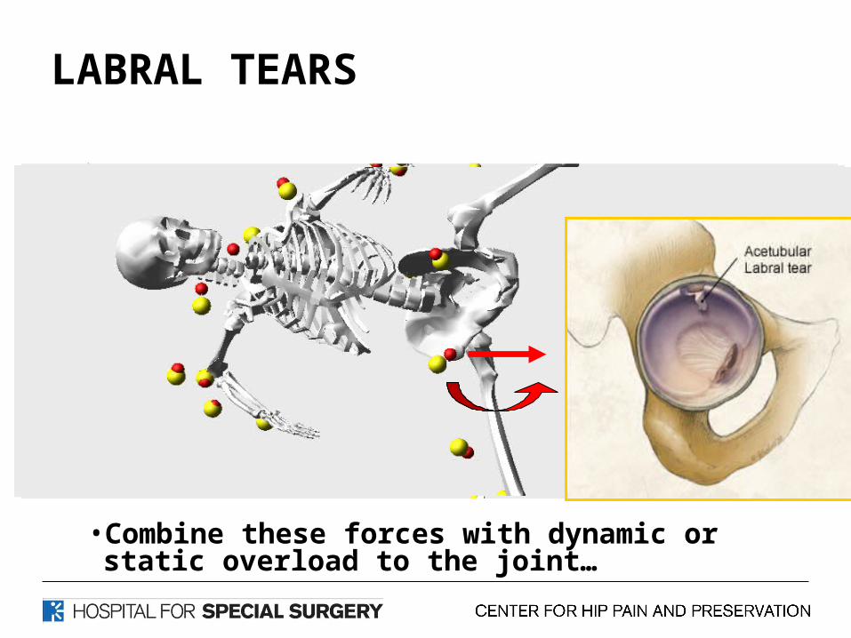
LABRAL TEARS
• Combine these forces with dynamic or static overload to the joint…
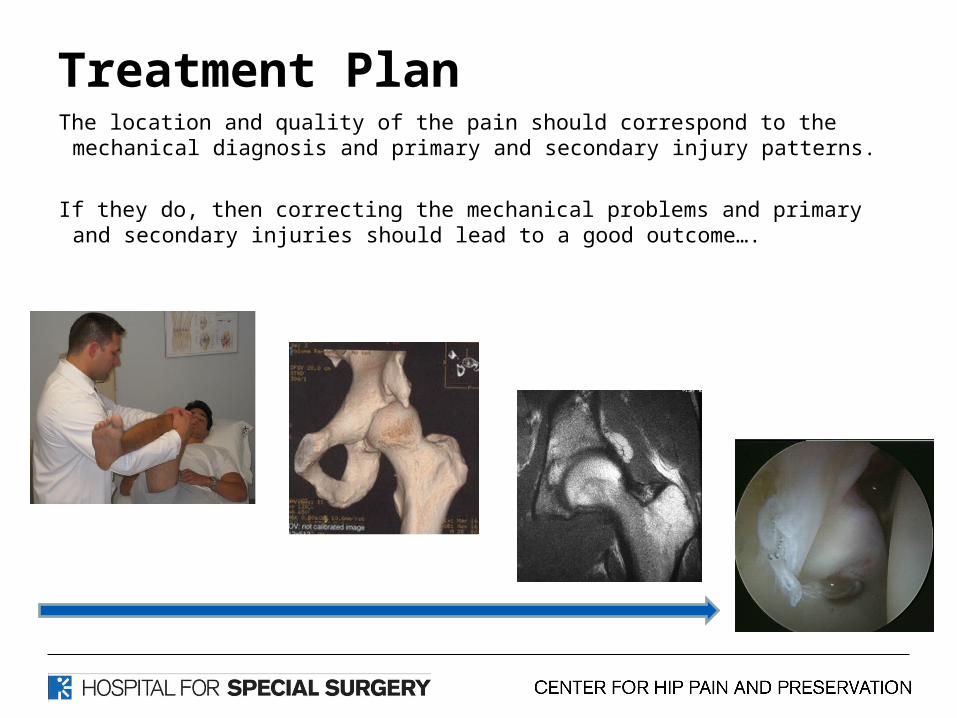
Treatment PlanThe location and quality of the pain should correspond to the
mechanical diagnosis and primary and secondary injury patterns.
If they do, then correcting the mechanical problems and primary and secondary injuries should lead to a good outcome….
Thank You


![Comparison of early complications between the use of a ......the hip, including a sliding hip screw/side plate device and multiple cannulated parallel lag screws [4]. However, even](https://img.pdfslide.net/doc/110x75/60ca56e9ff5bd26b28009709/comparison-of-early-complications-between-the-use-of-a-the-hip-including.jpg)