Embed Size (px)
Citation preview

Malignant disease of the cervix
Dr.Omar aldabbas
Assisstant-prof.,MUTA university
OBGYN specialist

Epidemiology
This is a disease of sexually active women.How frequent is this disease in nuns? “it is
rarely seen in nuns”. On the other hand, nuns have an increased risk of devloping Endometrial
cancer . There is increasing evidence that infection by
certain strains of human papilloma virus (HPV) is a factor.
HPV 16, 18, and others 10-30% of sexually active women have HPV
infection by the age of 30 years. The proportion increases if the women or her
partner have several sexual partners.
Epidemiology
The strains HPV 16 and 18 are particularly implicated in the development of cervical abnormality.
Smoking and immune suppression are added factors. Incidence of cervical cancer is increasing or decreasing?
Why? After the introduction of cervical smear, the incidence of cervical cancer is decreasing (It decreased by half or more than half!)
Now, it is the 3rd common genital cancer after endometrial and ovarian cancer.
The most common cancers in women differ from country to another:
Some countries: ovary> endometrium> cervixOthers, like the USA: Endometrium> ovary > cervix
Histological types
The cancer is either:.1Squamous cell carcinoma (85-90%)
(Because it arises from the ectocervix)
.2Adenocarcinoma (arises from the endocervix)
.3Mixed carcinoma (adenosquamous)
The development of invasive disease
If CIN is left untreated, after a variable length of time (may extend over years), it may invade through the basement membrane of the epithelium into the stroma beneath.
The estimated risk of invasion for CIN III is about 1.8 % per year (~ 2% per year) Will all CIN progress to invasive cancer? No! She
could have CIN – 2% per year is the risk

Micro-invasive carcinoma of the cervix (one type)
Is a part of the spectrum of cervical neoplasia is a stage between CIN and frankly invasive carcinoma.
This is considered to be a stage 1A of cervical cancer. Dx: On histopathology, the lesion is less than 5 mm in depth and less
than 7 mm in width. There is no vascular or lymphatic space involvement (cells have not
entered the vascular or lymphatic space).
Micro-invasive carcinoma of the cervix
The risk of spread to local lymph nodes is very rare (nearly nil to have LNs involvement).
The disease is usually asymptomatic. On examination, the cervix looks normal. The pap smear will show dysplastic cells. The diagnosis can only be confirmed after a cone
biopsy of the cervix (not based on clinical or cytology) only a cone biopsy

Micro-invasive carcinoma of the cervix: What is the treatment in this condition?
Cone excision is considered to be the only treatment needed if the margins of the cone are free from any lesion.
If not free margins: Total abdominal hysterectomy is indicated in those who are elderly and completed their family.
All patients should be followed with periodic physical and Pap smear examinations.
The 5-year survival rate following appropriate therapy is nearly 100%.

Invasive carcinoma of the cervix: anything that is not
The commonest symptoms are intermenstrual, post-coital and post-menopausal bleeding.
Patients may complain of a profuse, offensive vaginal discharge, which may be blood stained.
Pain is a late symptom of the disease Some cases may present as bleeding during pregnancy (in
antepartum hemorrhage, local causes, one of them is carcinoma of the cervix)
Invasive carcinoma of the cervix
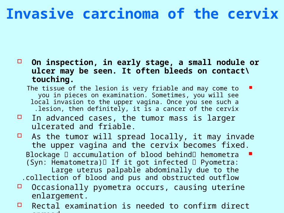
On inspection, in early stage, a small nodule or ulcer may be seen. It often bleeds on contact\touching.
The tissue of the lesion is very friable and may come to you in pieces on examination. Sometimes, you will see local invasion to the upper vagina. Once you see such a lesion,
then definitely, it is a cancer of the cervix . In advanced cases, the tumor mass is larger ulcerated and friable. As the tumor will spread locally, it may invade the upper vagina and the
cervix becomes fixed.Blockage accumulation of blood behind hemometra (Syn:
Hematometra) If it got infected Pyometra: Large uterus palpable abdominally due to the collection of blood and pus and obstructed outflow .
Occasionally pyometra occurs, causing uterine enlargement. Rectal examination is needed to confirm direct spread. Cystosopic examination is also needed to exclude invasion into the vase
of the UB

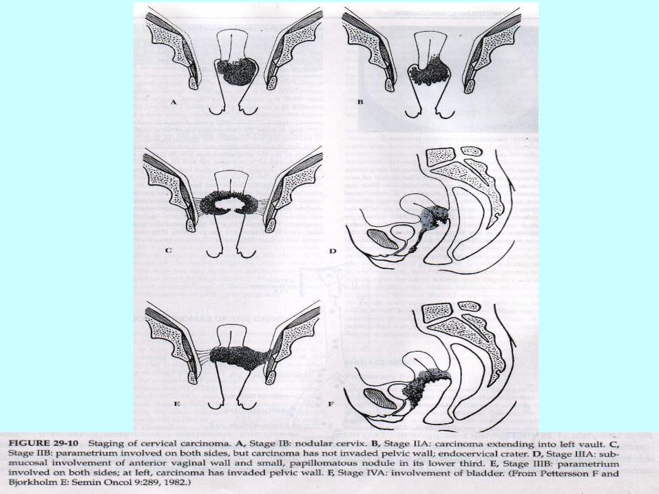
The lesion is filling the whole cervix but haven’t yet invaded the wall of the vagina. So, it can be considered stage IB

Staging of cervical cancer
Stage 0: CIN III ( carcinoma in situ) (not really a cancer… non-invasive)
Stage 1A: micro-invasive carcinoma.Stage 1B: invasive cancer limited to the cervix.Stage IIA: invasion to the upper third of the vagina.Stage IIB: invasion to the parametrium, but not to the lateral
pelvic wall.Stage IIIA: invasion to the lower third of the vagina.Stage IIIB: tumor reaching the lateral pelvic wall and/or
obstructing the ureter on IVU.Stage IVA: involvement of the bladder or rectum (once they
are involved, then it is stage IVA) (Lesion is limited to the cervix, but there is invasion of the rectum, what stage? IVA)
Stage IVB: extra pelvic spread, e.g. liver or lung metastasis.

Treatment
Either by surgery, radiotherapy or combination of the two.Depends on how good a surgeon you have or how good a
radiotherapy center you has!1B and IIA: Choice between surgery or radiotherapy. If you have a
good surgeon with low morbidity, then go to surgery. If you have a good radiotherapy, then go to radiotherapy. Combined treatment
carries a better prognosis (preoperative radiotherapy then surgery or surgery then radiotherapy)
All the stages after that, the answer is radiotherapy .Surgery is by gynecologist oncologist (a gynecologist who took a
subspecialty in gynecological oncology) In stage I and stage IIA, surgery or radiotherapy can be used,
but surgery offers lower morbidity. In later stages, radiotherapy is the mainstay of treatment.
Surgery
The standard surgery is Wertheim’s (VER-TIME -Ernst Wertheim - Austrian gynecologist) hysterectomy.
This involves removal of the uterus, paracervical tissues, upper vagina and pelvic lymphadenectomy.
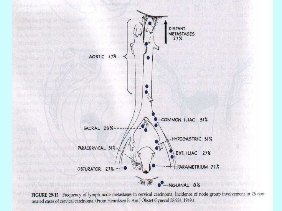
This includes external iliac, internal iliac, common iliac, obturator and presacral nodes.
Omentum removed ? In young women, ovaries may be conserved.

Radiotherapy: either external or internal
External beam therapy to the whole pelvis to shrink the central tumor and to treat the possible sites of regional metastasis.
The idea is reaching the invasions Internal sources placed in the upper vagina and
within the cervical canal to provide a very high dose to the central tumor.
If you are only aiming at the tumor itself Addition of chemotherapy during radiotherapy
increases the cure rate by 10 %.
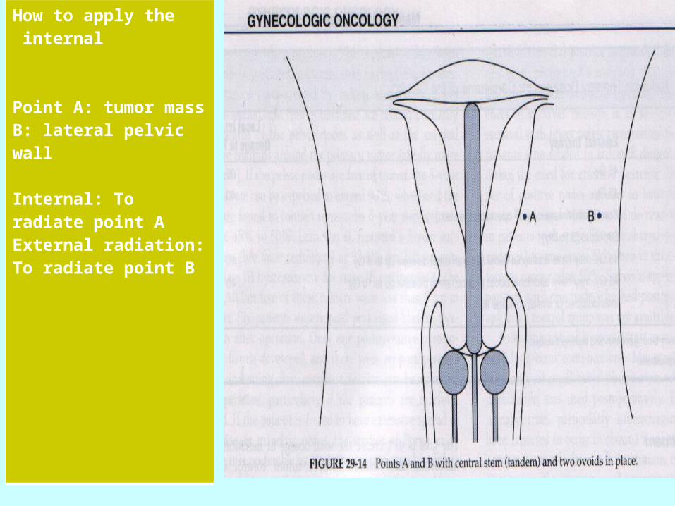
How to apply the internal
Point A: tumor massB: lateral pelvic wall
Internal: To radiate point AExternal radiation: To radiate point B
Combined treatment
Radiotherapy should be used after surgery if the lymph nodes histopathology showed involvement of the lymph nodes
Pre-operative radiotherapy can also be used in stage II to reduce the size of the tumor prior to surgery
Chemotherapy can also be used prior to surgery
Cancer of cervix and pregnancy
In early pregnancy (if discovered), treatment by external radiotherapy\radiation should be started. Abortion of a dead fetus will follow, then internal radiotherapy can be used.
In late pregnancy, Hysterectomy or caesarean section should be done, to be followed by appropriate treatment.
You should never ever allow vaginal delivery. The best idea is to perform CS then perform a
hysterectomy .

Pelvic Exenteration
This can be considered in recurrent pelvic disease or when the rectum or the bladder are involved with no clinical evidence of distant metastases.
It is a major surgery which involve the removal of the bladder (anterior exenteration) or the rectum ( posterior exenteration) or total exenteration.
The ureters are implanted in the ilium with terminal colostomy. The mortality and morbidity in this operation is high.
Palliative surgery in stage IV, the patient will have very heavy bleeding… The only way to stop it is surgery to excise the mass. It is of 3 types, either anterior, remove uterus and bladder, posterior, remove uterus and rectum. Total, remove bladder, rectum and uterus.
Ureter: go to small bowel.. Bowel: Colostomy
Exenteration: Removal of internal organs and tissues, usually radical removal of the contents of a body cavity. Syn:
evisceration(1).


![[2016 데이터 그랜드 컨퍼런스] 4 4(인공지능).마인즈랩 인공지능과 virtual assisstant-2016_datagrandconference](https://img.pdfslide.net/doc/110x75/587e14231a28abbc2e8b4dd7/2016-4-4-.jpg)