Embed Size (px)
Citation preview
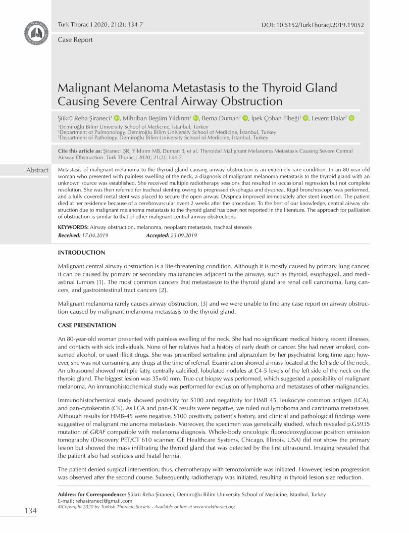
Turk Thorac J 2020; 21(2): 134-7
Case Report
Malignant Melanoma Metastasis to the Thyroid Gland Causing Severe Central Airway Obstruction
INTRODUCTION
Malignant central airway obstruction is a life-threatening condition. Although it is mostly caused by primary lung cancer, it can be caused by primary or secondary malignancies adjacent to the airways, such as thyroid, esophageal, and medi-astinal tumors [1]. The most common cancers that metastasize to the thyroid gland are renal cell carcinoma, lung can-cers, and gastrointestinal tract cancers [2].
Malignant melanoma rarely causes airway obstruction, [3] and we were unable to find any case report on airway obstruc-tion caused by malignant melanoma metastasis to the thyroid gland.
CASE PRESENTATION
An 80-year-old woman presented with painless swelling of the neck. She had no significant medical history, recent illnesses, and contacts with sick individuals. None of her relatives had a history of early death or cancer. She had never smoked, con-sumed alcohol, or used illicit drugs. She was prescribed sertraline and alprazolam by her psychiatrist long time ago; how-ever, she was not consuming any drugs at the time of referral. Examination showed a mass located at the left side of the neck. An ultrasound showed multiple fatty, centrally calcified, lobulated nodules at C4-5 levels of the left side of the neck on the thyroid gland. The biggest lesion was 35×40 mm. True-cut biopsy was performed, which suggested a possibility of malignant melanoma. An immunohistochemical study was performed for exclusion of lymphoma and metastases of other malignancies.
Immunohistochemical study showed positivity for S100 and negativity for HMB 45, leukocyte common antigen (LCA), and pan-cytokeratin (CK). As LCA and pan-CK results were negative, we ruled out lymphoma and carcinoma metastases. Although results for HMB-45 were negative, S100 positivity, patient’s history, and clinical and pathological findings were suggestive of malignant melanoma metastasis. Moreover, the specimen was genetically studied, which revealed p.G593S mutation of GRAF compatible with melanoma diagnosis. Whole-body oncologic fluorodeoxyglucose positron emission tomography (Discovery PET/CT 610 scanner, GE Healthcare Systems, Chicago, Illinois, USA) did not show the primary lesion but showed the mass infiltrating the thyroid gland that was detected by the first ultrasound. Imaging revealed that the patient also had scoliosis and hiatal hernia.
The patient denied surgical intervention; thus, chemotherapy with temozolomide was initiated. However, lesion progression was observed after the second course. Subsequently, radiotherapy was initiated, resulting in thyroid lesion size reduction.
DOI: 10.5152/TurkThoracJ.2019.19052
Şükrü Reha Şiraneci1 , Mihriban Begüm Yıldırım1 , Berna Duman2 , İpek Çoban Elbeği3 , Levent Dalar2 1Demiroğlu Bilim University School of Medicine, İstanbul, Turkey 2Department of Pulmonology, Demiroğlu Bilim University School of Medicine, İstanbul, Turkey3Department of Pathology, Demiroğlu Bilim University School of Medicine, İstanbul, Turkey
Address for Correspondence: Şükrü Reha Şiraneci, Demiroğlu Bilim University School of Medicine, İstanbul, Turkey E-mail: [email protected] ©Copyright 2020 by Turkish Thoracic Society - Available online at www.turkthoracj.org
134
Cite this article as: Şiraneci ŞR, Yıldırım MB, Duman B, et al. Thyroidal Malignant Melanoma Metastasis Causing Severe Central Airway Obstruction. Turk Thorac J 2020; 21(2): 134-7.
Metastasis of malignant melanoma to the thyroid gland causing airway obstruction is an extremely rare condition. In an 80-year-old woman who presented with painless swelling of the neck, a diagnosis of malignant melanoma metastasis to the thyroid gland with an unknown source was established. She received multiple radiotherapy sessions that resulted in occasional regression but not complete resolution. She was then referred for tracheal stenting owing to progressed dysphagia and dyspnea. Rigid bronchoscopy was performed, and a fully covered metal stent was placed to secure the open airway. Dyspnea improved immediately after stent insertion. The patient died at her residence because of a cerebrovascular event 2 weeks after the procedure. To the best of our knowledge, central airway ob-struction due to malignant melanoma metastasis to the thyroid gland has been not reported in the literature. The approach for palliation of obstruction is similar to that of other malignant central airway obstructions.
KEYWORDS: Airway obstruction, melanoma, neoplasm metastasis, tracheal stenosis
Abstract
Received: 17.04.2019 Accepted: 23.09.2019
Radiotherapy was repeated owing to a new retrosternal lesion seen on a control PET-computed tomography (CT) scan obtained 1 year after the first radiotherapy. After the second radiotherapy, whole-body PET-CT was performed, revealing a partial reduction in the size of the thyroid lesion and near-complete regression of the retrosternal lesion. The patient was then discharged with scheduled routine follow-ups. One year later, follow-up magnetic resonance imaging and PET-CT showed thyroid lesion progression.
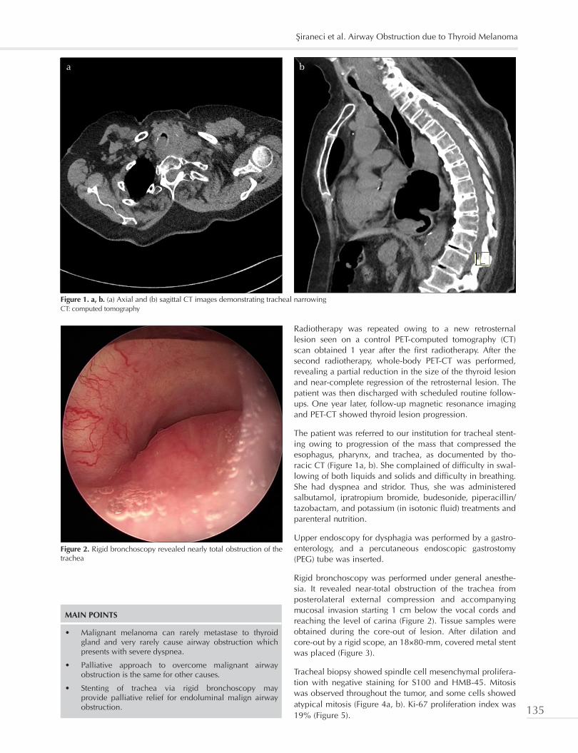
The patient was referred to our institution for tracheal stent-ing owing to progression of the mass that compressed the esophagus, pharynx, and trachea, as documented by tho-racic CT (Figure 1a, b). She complained of difficulty in swal-lowing of both liquids and solids and difficulty in breathing. She had dyspnea and stridor. Thus, she was administered salbutamol, ipratropium bromide, budesonide, piperacillin/tazobactam, and potassium (in isotonic fluid) treatments and parenteral nutrition.
Upper endoscopy for dysphagia was performed by a gastro-enterology, and a percutaneous endoscopic gastrostomy (PEG) tube was inserted.
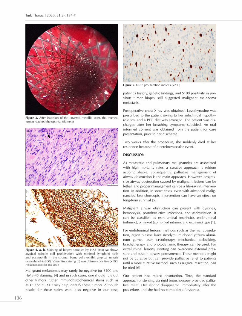
Rigid bronchoscopy was performed under general anesthe-sia. It revealed near-total obstruction of the trachea from posterolateral external compression and accompanying mucosal invasion starting 1 cm below the vocal cords and reaching the level of carina (Figure 2). Tissue samples were obtained during the core-out of lesion. After dilation and core-out by a rigid scope, an 18×80-mm, covered metal stent was placed (Figure 3).
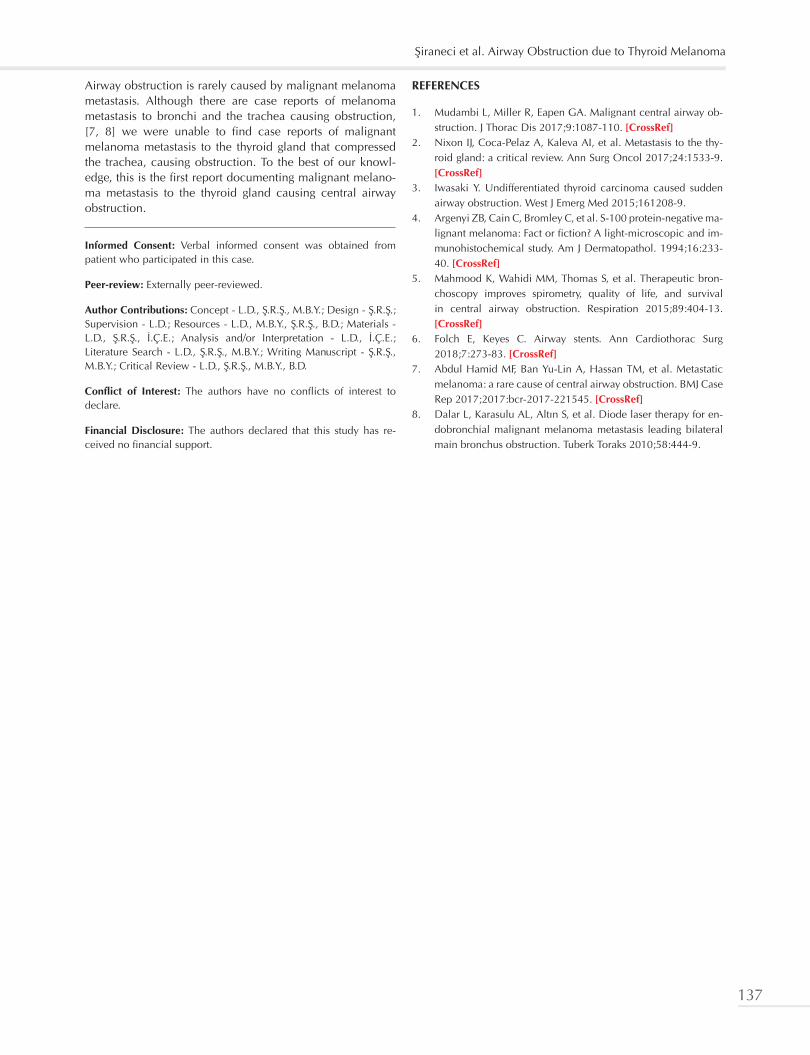
Tracheal biopsy showed spindle cell mesenchymal prolifera-tion with negative staining for S100 and HMB-45. Mitosis was observed throughout the tumor, and some cells showed atypical mitosis (Figure 4a, b). Ki-67 proliferation index was 19% (Figure 5).
MAIN POINTS
• Malignant melanoma can rarely metastase to thyroid gland and very rarely cause airway obstruction which presents with severe dyspnea.
• Palliative approach to overcome malignant airway obstruction is the same for other causes.
• Stenting of trachea via rigid bronchoscopy may provide palliative relief for endoluminal malign airway obstruction.
Figure 1. a, b. (a) Axial and (b) sagittal CT images demonstrating tracheal narrowingCT: computed tomography
a b
Figure 2. Rigid bronchoscopy revealed nearly total obstruction of the trachea
Şiraneci et al. Airway Obstruction due to Thyroid Melanoma
135
Malignant melanomas may rarely be negative for S100 and HMB-45 staining, [4] and in such cases, one should rule out other tumors. Other immunohistochemical stains such as MITF and SOX10 may help identify these tumors. Although results for these stains were also negative in our case,
patient’s history, genetic findings, and S100 positivity in pre-vious tumor biopsy still suggested malignant melanoma metastasis.
Postoperative chest X-ray was obtained. Levothyroxine was prescribed to the patient owing to her subclinical hypothy-roidism, and a PEG diet was arranged. The patient was dis-charged after her breathing symptoms subsided. An oral informed consent was obtained from the patient for case presentation, prior to her discharge.
Two weeks after the procedure, she suddenly died at her residence because of a cerebrovascular event.
DISCUSSION
As metastatic and pulmonary malignancies are associated with high mortality rates, a curative approach is seldom accomplishable; consequently, palliative management of airway obstruction is the main approach. However, progres-sive airway obstruction caused by malignant lesions can be lethal, and proper management can be a life-saving interven-tion. In addition, in some cases, even with advanced malig-nancies, bronchoscopic intervention can have an effect on long-term survival [5].
Malignant airway obstruction can present with dyspnea, hemoptysis, postobstructive infections, and asphyxiation. It can be classified as extraluminal (extrinsic), endoluminal (intrinsic), or mixed (combined intrinsic and extrinsic) type [1].
For endoluminal lesions, methods such as thermal coagula-tion, argon plasma laser, neodymium-doped yttrium alumi-num garnet laser, cryotherapy, mechanical debulking, brachytherapy, and photodynamic therapy can be used. For extraluminal lesions, stenting can overcome external pres-sure and sustain airway permanence. These methods might not be curative but can provide palliative relief to patients until a more curative method, such as surgical resection, can be tried [6].
Our patient had mixed obstruction. Thus, the standard approach of stenting via rigid bronchoscopy provided pallia-tive relief. Her stridor disappeared immediately after the procedure, and she had no complaint of dyspnea.
Figure 3. After insertion of the covered metallic stent, the tracheal lumen reached the optimal diameter
Figure 5. Ki-67 proliferation indices (×200)
Figure 4. a, b. Staining of biopsy samples by H&E stain (a) shows atypical spindle cell proliferation with minimal lymphoid cells and eosinophils in the stroma. Some cells exhibit atypical mitosis (arrowhead) (×200). Vimentin staining (b) was diffusely positive (×100)H&E: hematoxylin and eosin
a
b
Turk Thorac J 2020; 21(2): 134-7
136
Airway obstruction is rarely caused by malignant melanoma metastasis. Although there are case reports of melanoma metastasis to bronchi and the trachea causing obstruction, [7, 8] we were unable to find case reports of malignant melanoma metastasis to the thyroid gland that compressed the trachea, causing obstruction. To the best of our knowl-edge, this is the first report documenting malignant melano-ma metastasis to the thyroid gland causing central airway obstruction.
Informed Consent: Verbal informed consent was obtained from patient who participated in this case.
Peer-review: Externally peer-reviewed.
Author Contributions: Concept - L.D., Ş.R.Ş., M.B.Y.; Design - Ş.R.Ş.; Supervision - L.D.; Resources - L.D., M.B.Y., Ş.R.Ş., B.D.; Materials - L.D., Ş.R.Ş., İ.Ç.E.; Analysis and/or Interpretation - L.D., İ.Ç.E.; Literature Search - L.D., Ş.R.Ş., M.B.Y.; Writing Manuscript - Ş.R.Ş., M.B.Y.; Critical Review - L.D., Ş.R.Ş., M.B.Y., B.D.
Conflict of Interest: The authors have no conflicts of interest to declare.
Financial Disclosure: The authors declared that this study has re-ceived no financial support.
REFERENCES
1. Mudambi L, Miller R, Eapen GA. Malignant central airway ob-struction. J Thorac Dis 2017;9:1087-110. [CrossRef]
2. Nixon IJ, Coca-Pelaz A, Kaleva AI, et al. Metastasis to the thy-roid gland: a critical review. Ann Surg Oncol 2017;24:1533-9. [CrossRef]
3. Iwasaki Y. Undifferentiated thyroid carcinoma caused sudden airway obstruction. West J Emerg Med 2015;161208-9.
4. Argenyi ZB, Cain C, Bromley C, et al. S-100 protein-negative ma-lignant melanoma: Fact or fiction? A light-microscopic and im-munohistochemical study. Am J Dermatopathol. 1994;16:233-40. [CrossRef]
5. Mahmood K, Wahidi MM, Thomas S, et al. Therapeutic bron-choscopy improves spirometry, quality of life, and survival in central airway obstruction. Respiration 2015;89:404-13. [CrossRef]
6. Folch E, Keyes C. Airway stents. Ann Cardiothorac Surg 2018;7:273-83. [CrossRef]
7. Abdul Hamid MF, Ban Yu-Lin A, Hassan TM, et al. Metastatic melanoma: a rare cause of central airway obstruction. BMJ Case Rep 2017;2017:bcr-2017-221545. [CrossRef]
8. Dalar L, Karasulu AL, Altın S, et al. Diode laser therapy for en-dobronchial malignant melanoma metastasis leading bilateral main bronchus obstruction. Tuberk Toraks 2010;58:444-9.
Şiraneci et al. Airway Obstruction due to Thyroid Melanoma
137