Embed Size (px)
Citation preview
CentralBringing Excellence in Open Access
Journal of Cancer Biology & Research
Cite this article: Sills M, Norton S, Landers R, O’Donoghue G (2016) Malignant Melanoma of the Breast: A Case Report. J Cancer Biol Res 4(1): 1075.
*Corresponding author
Sarah Norton, Department of Surgery, University Hospital Waterford, Waterford, Ireland. Tel: +35351 84000, Email:
Submitted: 15 November 2015
Accepted: 15 January 2016
Published: 18 January 2016
Copyright© 2016 Norton et al.
OPEN ACCESS
Keywords•Melanoma•Breast
Case Report
Malignant Melanoma of the Breast: A Case ReportMatthew Sills, Sarah Norton*, Robert Landers and Gerrard O’DonoghueDepartment of Surgery, University Hospital Waterford, Ireland
Abstract
Background: Malignant melanoma of the areola of the breast is rare. Only a few cases have been reported in the literature. Diagnostic dilemmas may occur due to the location of these malignant lesions.
Case: A pigmented lesion of the left nipple in a young female patient that was increasing in size, with an associated colour change is described. A preoperative ultrasound was performed and the lesion was excised. Initial histology showed it to be an infiltrating malignant melanoma in a pre-existing naevus. The tumour was in vertical growth phase, filling the papillary dermis and extending into the superficial reticular dermis (Clarke’s level IV) to a depth of 1.4mm. It was noted that the exact depth of tumour was difficult to measure due to the presence of the associated naevus. In-situ tumour cells involved the radial margin at initial resection. The mitotic rate was 1/mm2.
Post-operatively, CT TAP and CT brain were performed which were negative for metastatic disease. A bone scan showed abnormal sternomanubrial activity. For this reason, a PET scan was performed which was negative for metastases. Following Breast and supra-regional Dermatopathology MDM discussion the patient underwent a non-nipple sparing WLE and SLNM. Final histology demonstrated no further disease in re-excision specimen and lymph nodes were reported as having dark carbon-like pigment within them with no malignancy seen. The carbon-like pigment was consistent with an ipsilateral tattoo on the patients’ hand.
Conclusion: Malignant melanoma of the breast is a rare but important differential diagnosis for pigmented lesions. The clinical presentation includes a change in size, pigmentation, ulceration and bleeding in a pre-existing naevus. Once diagnosed, prognosis depends on tumour size, Clarke’s level, Breslow Thickness, location, ulceration and metastases. SLNB with lymphoscintigraphic mapping is a minimally invasive procedure and is a useful tool for predicting survival and prognosis in these patients.
ABBREVIATIONSCT TAP: Computed Tomography Thorax Abdomen and
Pelvis; CT: Computed Tomography; PET: Positron Emission Tomography; MDM: Multidisciplinary Team Meeting; WLE: Wide Local Excision; SLNM: Sentinel Lymph Node Mapping; SLNB: Sentinel Lymph Node Biopsy
INTRODUCTIONThe incidence of malignant melanoma is on the rise,
accounting for 1-2% of all cancers and 75% of all skin cancer deaths [1]. In Ireland from 1994-2012, the incidence of melanoma has increased annually by 3% and 5% in females and males, respectively. Mortality rates are also on the incline, with a 2.2% and 6% annual rise in melanoma related deaths in females and males, respectively [2]. The majority of malignant melanoma cases in the breast are metastatic deposits from cutaneous
malignant melanoma. However, several cases of primary malignant melanoma of the breast have been reported [3-5], as is described in this case report. The clinical presentation of breast melanoma includes a change in size, pigmentation, ulceration and bleeding in a pre-existing naevus. Prognosis depends on tumour size, Clarke’s level, Breslow Thickness, location, ulceration and metastases [6].
CASE PRESENTATIONA 27 year old female presented to the University Hospital
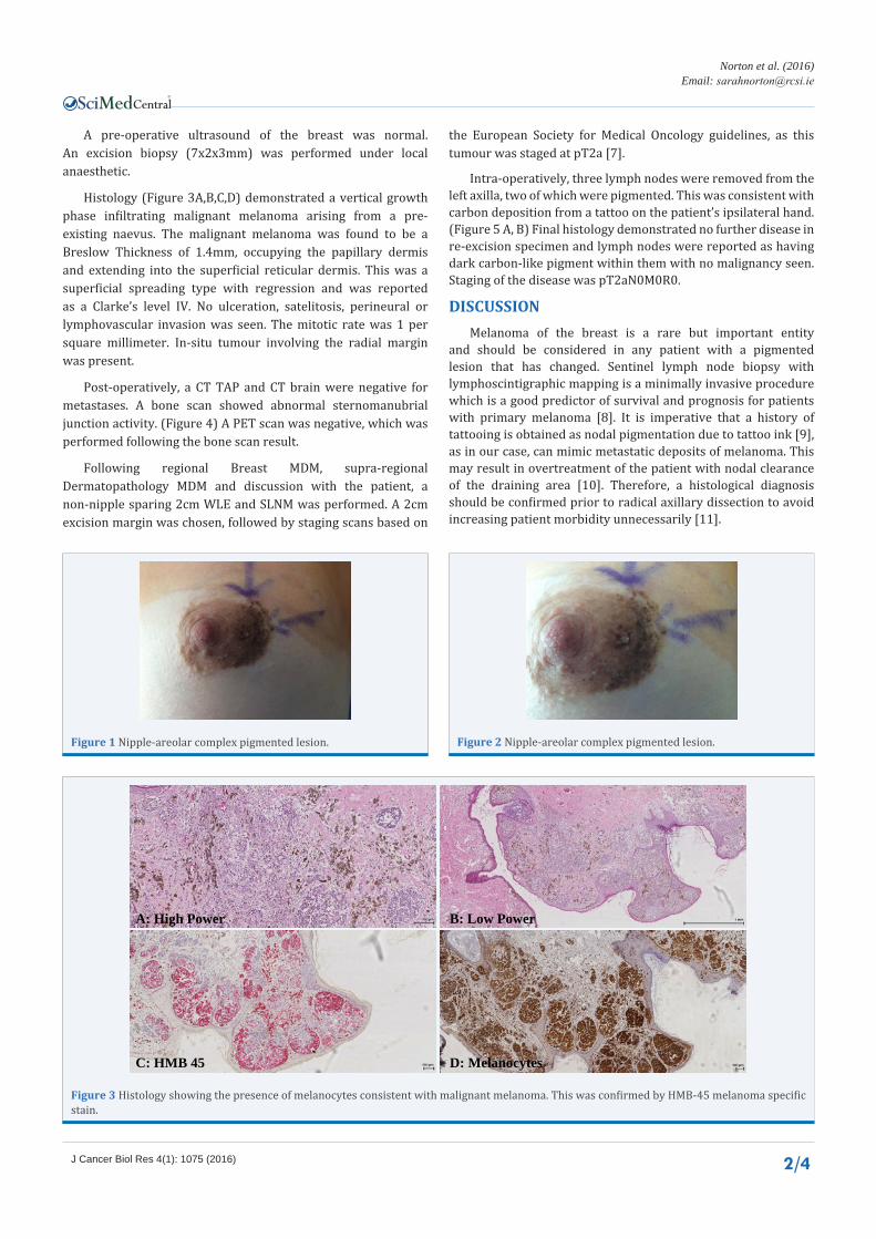
Waterford Breast Care service with a lesion on the left nipple. It was reported by the patient that this lesion had changed colour and was increasing in size. There was no relevant past medical or surgical history of note. Family history included a breast cancer diagnosis in a paternal cousin. On examination of the patient a 1cm left breast areolar complex lesion with an adjacent satellite deposit was present (Figure 1,2) .
CentralBringing Excellence in Open Access
Norton et al. (2016)Email:
J Cancer Biol Res 4(1): 1075 (2016) 2/4
A pre-operative ultrasound of the breast was normal. An excision biopsy (7x2x3mm) was performed under local anaesthetic.
Histology (Figure 3A,B,C,D) demonstrated a vertical growth phase infiltrating malignant melanoma arising from a pre-existing naevus. The malignant melanoma was found to be a Breslow Thickness of 1.4mm, occupying the papillary dermis and extending into the superficial reticular dermis. This was a superficial spreading type with regression and was reported as a Clarke’s level IV. No ulceration, satelitosis, perineural or lymphovascular invasion was seen. The mitotic rate was 1 per square millimeter. In-situ tumour involving the radial margin was present.
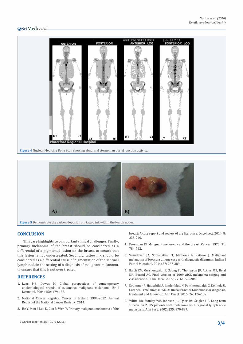
Post-operatively, a CT TAP and CT brain were negative for metastases. A bone scan showed abnormal sternomanubrial junction activity. (Figure 4) A PET scan was negative, which was performed following the bone scan result.
Following regional Breast MDM, supra-regional Dermatopathology MDM and discussion with the patient, a non-nipple sparing 2cm WLE and SLNM was performed. A 2cm excision margin was chosen, followed by staging scans based on
the European Society for Medical Oncology guidelines, as this tumour was staged at pT2a [7].
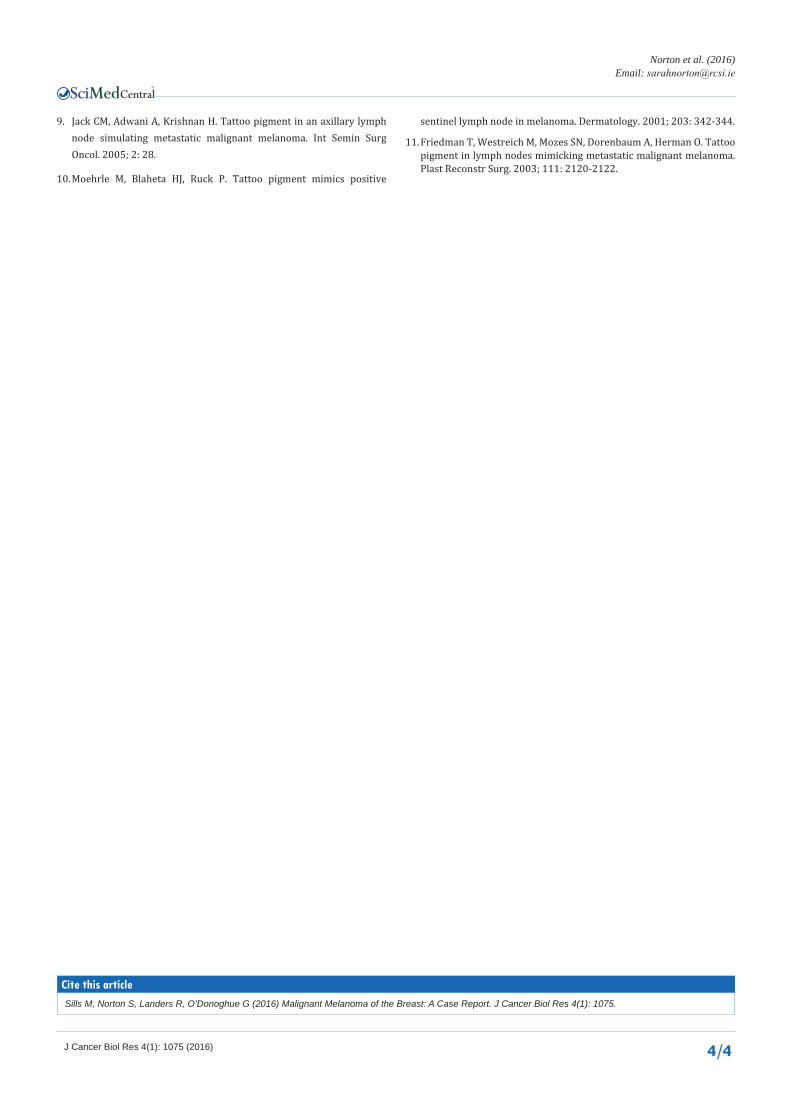
Intra-operatively, three lymph nodes were removed from the left axilla, two of which were pigmented. This was consistent with carbon deposition from a tattoo on the patient’s ipsilateral hand. (Figure 5 A, B) Final histology demonstrated no further disease in re-excision specimen and lymph nodes were reported as having dark carbon-like pigment within them with no malignancy seen. Staging of the disease was pT2aN0M0R0.
DISCUSSIONMelanoma of the breast is a rare but important entity
and should be considered in any patient with a pigmented lesion that has changed. Sentinel lymph node biopsy with lymphoscintigraphic mapping is a minimally invasive procedure which is a good predictor of survival and prognosis for patients with primary melanoma [8]. It is imperative that a history of tattooing is obtained as nodal pigmentation due to tattoo ink [9], as in our case, can mimic metastatic deposits of melanoma. This may result in overtreatment of the patient with nodal clearance of the draining area [10]. Therefore, a histological diagnosis should be confirmed prior to radical axillary dissection to avoid increasing patient morbidity unnecessarily [11].
Figure 1 Nipple-areolar complex pigmented lesion. Figure 2 Nipple-areolar complex pigmented lesion.
A: High Power B: Low Power
C: HMB 45 D: Melanocytes
Figure 3 Histology showing the presence of melanocytes consistent with malignant melanoma. This was confirmed by HMB-45 melanoma specific stain.
CentralBringing Excellence in Open Access
Norton et al. (2016)Email:
J Cancer Biol Res 4(1): 1075 (2016) 3/4
Figure 4 Nuclear Medicine Bone Scan showing abnormal sternoman ubrial junction activity.
A) B)
Figure 5 Demonstrate the carbon deposit from tattoo ink within the lymph nodes.
CONCLUSIONThis case highlights two important clinical challenges. Firstly,
primary melanoma of the breast should be considered as a differential of a pigmented lesion on the breast, to ensure that this lesion is not undertreated. Secondly, tattoo ink should be considered as a differential cause of pigmentation of the sentinel lymph nodein the setting of a diagnosis of malignant melanoma, to ensure that this is not over treated.
REFERENCES1. Lens MB, Dawes M. Global perspectives of contemporary
epidemiological trends of cutaneous malignant melanoma. Br J Dermatol. 2004; 150: 179-185.
2. National Cancer Registry. Cancer in Ireland 1994-2012: Annual Report of the National Cancer Registry. 2014.
3. He Y, Mou J, Luo D, Gao B, Wen Y. Primary malignant melanoma of the
breast: A case report and review of the literature. Oncol Lett. 2014; 8: 238-240.
4. Pressman PI. Malignant melanoma and the breast. Cancer. 1973; 31: 784-792.
5. Vasudevan JA, Somanathan T, Mathews A, Kattoor J. Malignant melanoma of breast: a unique case with diagnostic dilemmas. Indian J Pathol Microbiol. 2014; 57: 287-289.
6. Balch CM, Gershenwald JE, Soong SJ, Thompson JF, Atkins MB, Byrd DR, Buzaid AC. Final version of 2009 AJCC melanoma staging and classification. J Clin Oncol. 2009; 27: 6199-6206.
7. Drummer R, Hauschild A, Lindenblatt N, Pentheroudakis G, Keilholz U. Cutaneous melanoma: ESMO Clinical Practice Guidelines for diagnosis, treatment and follow-up. Ann Oncol. 2015; 26: 126-132.
8. White RR, Stanley WE, Johnson JL, Tyler DS, Seigler HF. Long-term survival in 2,505 patients with melanoma with regional lymph node metastasis. Ann Surg. 2002; 235: 879-887.
CentralBringing Excellence in Open Access
Norton et al. (2016)Email:
J Cancer Biol Res 4(1): 1075 (2016) 4/4
Sills M, Norton S, Landers R, O’Donoghue G (2016) Malignant Melanoma of the Breast: A Case Report. J Cancer Biol Res 4(1): 1075.
Cite this article
9. Jack CM, Adwani A, Krishnan H. Tattoo pigment in an axillary lymph node simulating metastatic malignant melanoma. Int Semin Surg Oncol. 2005; 2: 28.
10. Moehrle M, Blaheta HJ, Ruck P. Tattoo pigment mimics positive
sentinel lymph node in melanoma. Dermatology. 2001; 203: 342-344.
11. Friedman T, Westreich M, Mozes SN, Dorenbaum A, Herman O. Tattoo pigment in lymph nodes mimicking metastatic malignant melanoma. Plast Reconstr Surg. 2003; 111: 2120-2122.