Embed Size (px)
Citation preview
Malondialdehyde and 4-Hydroxynonenal Protein Adductsin Plasma and Liver of Rats with Iron OverloadKarl Houglum, Michael Filip, Joseph L. Witztum, and Mario ChojkierDepartment of Medicine, Veterans Administration Medical Center, and University of California, San Diego, California 92161
Abstract
In hepatic iron overload, iron-catalyzed lipid peroxidation hasbeen implicated in the mechanisms of hepatocellular injury.Lipid peroxidation may produce reactive aldehydes such asmalondialdehyde (MDA) and 4-hydroxynonenal (4-HNE),which may form aldehyde-protein adducts. We investiptedwhether lipid peroxidation occurred in rats fed a diet contain-ing 3% carbonyl iron for 5-13 wk, and if this resulted in theformation of MDA-and 4-HNE- protein adducts. Chronic ironfeeding resulted in hepatic iron overload (> 10-fold) and con-comitantly induced a 2-fold increase in hepatic lipid peroxida-tion. Using an antiserum specific for MDA-lysine protein ad-ducts, we demonstrated by immunohistochemistry the pres-ence of aldehyde-protein adducts in the cytosol of periportalhepatocytes, which co-localized with iron. In addition, MDA-and 4-HNE-lysine adducts were found in plasma proteins ofanimals with iron overload. Only MDAadducts were detectedin albumin, while other plasma proteins including a 120-kDprotein had both MDAand 4-HNE adducts. In this animalmodel of hepatic iron overload, injury occurs primarily in peri-portal hepatocytes, where MDA-lysine protein adducts andexcess iron co-localized. (J. Clin. Invest. 1990. 86:1991-1998.) Key words: aldehydes * oxidative stress * autofluores-cence * lipofuscin * immunochemistry
Introduction
Hemochromatosis is associated with excess iron deposition inhepatocytes, which results in hepatic injury (1, 2). The mecha-nisms responsible for the development of hepatocellular injuryin patients with hemochromatosis are not known, but a directcorrelation between hepatic iron and hepatic fibrosis has beendemonstrated (3, 4). Because lipid peroxidation is known to becatalyzed by iron, enhanced lipid peroxidation has been pro-posed as an initial step by which excess iron causes cellularinjury (1, 5). This proposal is supported by results from animalstudies and in vitro models (6-12). In rats with chronic ironoverload, lipid peroxidation is associated with changes in mi-tochondrial (7) and microsomal function (11), and with in-creased lysosomal fragility (12).
This work was presented in part at the 1989 Annual Meetings of theAmerican Association for the Study of Liver Diseases, and publishedin abstract form (Hepatology. 10:608a).
Address correspondence and reprint requests to Dr. Mario Choj-kier, University of California, San Diego, V-l 11-D, 3350 La JollaVillage Drive, San Diego, CA92161.
Receivedfor publication 26 June 1989 and in revisedform 24 July1990.
The Joumal of Clinical Investigation, Inc.Volume 86, December 1990, 1991-1998
Iron-catalyzed lipid peroxidation of polyunsaturated fattyacids leads to the formation of highly reactive aldehydes, suchas malondialdehyde (MDA)' and 4-hydroxynonenal (4-HNE),which may then form covalent links to proteins, phospho-lipids, and DNA(13, 14). In proteins, these links involve thee-amino group of lysine residues or sulfhydryl groups (13).Oxidation of LDL has been proposed to play an etiologic rolein atherogenesis (15, 16), and MDA- and 4-HNE-modifiedLDL protein have been found in atheromas of the hyperlipid-emic Wantanabe rabbit (17-19), and in man (18). Analo-gously, acetaldehyde, the first metabolite of ethanol, has beenshown to produce adducts with proteins in vivo, judging by thepresence of antibodies against the acetaldehyde-lysine epitopein the sera of alcoholic patients (20) and the identification of aprotein-acetaldehyde adduct in liver homogenates of ethanol-fed rats (21).
In this study, we fed rats excess iron (22) and demonstratedthe presence of MDA-lysine adducts in the cytosol of peripor-tal hepatocytes, co-localized with iron. In addition we demon-strated the presence of MDA- and 4-HNE-lysine adducts inseveral plasma proteins.
Methods
Materials. Nitrocellulose BA85, 0.45 um was purchased fromSchleicher & Schuell (Keene, NH). Sources of other chemicals were:3,3'-diaminobenzidine tetrahydrocitolide dihydrate (DAB), from Ald-rich Chemical Co. (Milwaukee, WI); gelatin Type III, carbonyl iron(99% elemental iron in particles ranging from 4.5-5.2 Mm), and horsespleen ferritin from Sigma Chemical Co. (St. Louis, MO); immuno-electrophoresis tricine buffer IV, pH 8.6 from Bio-Rad Laboratories(Richmond, CA); EM-bed 812 from Electron Microscopy Sciences(Fort Washington, PA); biotinylated antibodies, Vectastain kits andreagents from Vector Laboratories, Inc. (Burlingame, CA); rabbitanti-rat albumin, IgG fraction, rat albumin, and rabbit anti-rat trans-ferrin from Cappel Laboratories (Malvern, PA); Histo-Clear from Na-tional Diagnostics, Inc. (Manville, NJ); rabbit anti-human ferritinfrom Dako Corp. (Carpinteria, CA); and '25I-protein A from Amer-sham Corp. (Arlington Heights, IL).
Animal procedures. Sprague-Dawley male rats (100-125 g)(Charles River Breeding Laboratories, Inc., Wilmington, MA) werepair fed a 3% (wt/wt) carbonyl iron diet (6) or control, 5001 rat chowdiet (Ralston-Purina Co., Richmond, IN). Animals were weighedweekly and the diet of the control animals adjusted to maintain similargrowth rates to the carbonyl iron-fed animals. At 5 wk, animals wereanesthetized by an intramuscular injection of ketamine (100 mg/kg),zylazine (2 mg/kg), and acepromazine (2.5 mg/kg) and then killed bycardiac puncture and their livers promptly removed. A portion wasrinsed in ice-cold PBS and immediately frozen in liquid nitrogen; theremainder was fixed in 10% formalin for Perls' prussian blue staining.
1. Abbreviations used in this paper: BHT, butylated hydroxytoluene;DAB, 3,3' diaminobenzidine tetra hydrocitolide dihydrate; EITB,electroimmunotransblot; 4-HNE, 4-hydroxynonenal; MDA, malon-dialdehyde; TBARS, thiobarbituric reactive substances.
Protein-Aldehyde Adducts in Rats with Iron Overload 1991
A separate group of animals was maintained on either a 3% car-bonyl iron diet or control diet for 13 wk. The livers were biopsiedunder anesthesia (as described above) while still perfused, and immedi-ately fixed as described below for immunohistochemistry; the re-mainder of the liver was removed and processed as described above. Insome animals, the liver was fixed in Karnovsky's fixative overnight at0-4°C. The tissue was postfixed in 2% osmium tetroxide for I h at20°C and then dehydrated through a graded series of ethanol andpropylene oxide and embedded in EM-bed 812. Thin sections (60 nm)were cut and stained with uranyl acetate and bismuth subnitrate.
Iron determination. Non-heme iron content was measured usingthe method of Horak et al. (23) except that liver was used in place ofplasma as described by Torrance and Bothwell (24). Values are ex-pressed as micrograms of iron per gram liver (wet weight).
Thiobarbituric acid reactivity. Thiobarbituric acid reactive sub-stances (TBARS) were determined from liver specimens stored in liq-uid nitrogen as described by Ohkawa and co-workers (25) except thatSDSwas omitted and EDTA(3 mM)was added to the tissue homoge-nization solution. The fluorescence of the color complex formed wasmeasured (excitation 515 nmand emission 553 nm) and compared tostandards prepared from tetramethoxypropane. To exclude the possi-bility that spurious TBARSwere formed in the assay conditions due toexcess iron (26), iron in the form of ferritin was added to samples ofnormal liver, before homogenization, in an amount equal to thatfound in iron-overloaded rats.
Antisera. Monospecific antisera to MDA- and 4-HNE-lysine ad-ducts were generated using techniques previously described (17, 18, 27,28). In brief, guinea pig LDL was isolated and modified with MDAor4-HNE, and the homologous modified LDL were used to immunizeguinea pigs (17, 18, 28). The resultant antisera were specific for theadducts to LDL and did not react with native LDL. Each antisera isepitope specific and reacts with this adduct on a variety of differentproteins. Thus "MAL-2" is specific for MDA-lysine adducts (17, 28)and antiserum "4-HNE" is specific for the 4-HNE-lysine adduct (28).Previous reports using these antisera have been published (17, 18, 28).
Immunohistochemistry of hepatic tissue. Portions of the liver frompair-fed animals (control, n = 3; and carbonyl iron fed, n = 3) were cutinto 2 X 4 mmsections and prepared as described by Palinski andcolleagues (17). EDTAand butylated hydroxytoluene (BHT) wereadded to all solutions to prevent spurious lipid peroxidation duringsample preparation (26). A minimum of three 5-,um sections of at leasttwo specimens per liver were placed on gelatin-coated slides and de-paraffinized using Histo-clear, passed through graded series of alcohol,and subsequently rehydrated in 0.15 MNaCl, 50 mMsodium phos-phate, pH 7.5 (PBS). Immunohistochemistry was performed using anavidin-biotin-alkaline phosphatase system (Vector Laboratories, Inc.)as described by the manufacturer. Sections were immunostained withantiserum MAL-2 (1:500), 4-HNE (1:1,000), preimmune serum(1:500), or PBS. The brown-black color produced was used to assessthe presence of MDA-lysine and 4-HNE-lysine protein adducts.
Adjacent liver sections were deparaffinized and rehydrated as de-scribed above and then mounted using glycerol-PBS (9:1), pH 8.2.Autofluorescence was detected using either an ultraviolet, fluorescein,or rhodamine excitation filter. Subsequently, after photographing theautofluorescence, the cover slips from the same slides were removedand the sections rinsed in PBS (pH 7.4) and stained for iron as de-scribed above.
Electroimmunotransblotting. Plasma from rats fed control and irondiets for 5 wk was obtained by cardiac puncture using an EDTA-coatedsyringe. Samples were immediately chilled to 0°C and additionalEDTAwas added to make the final concentration 3 mM. Plasma wasseparated by centrifugation at 0°C and stored in liquid nitrogen untilused for electroimmunotransblotting (EITB). To determine whethernontransferrin-bound iron was important in the formation of circulat-ing aldehyde-protein adducts and not excessive storage iron, the fol-lowing experiments were performed. In pair-fed animals the carbonyliron diet was replaced with control diet for 5 d. Plasma was obtained, asdescribed above, before and after stopping the carbonyl iron diet and
used for EITB. Additionally, plasma from rats fed the control diet wasobtained using heparin, in place of EDTA, to coat the syringe. Ferricchloride (in 50 mMcitrate buffer, pH 7.4) was added to aliquots ofplasma to increase the concentration of iron by 2 or 6 Mg/ml andincubated at 25 or 37°C for I h. EDTA(10 mMfinal) was added to allplasma samples to stop the reaction, and the treated plasma was usedfor EITB immediately.
Plasma proteins were separated by SDS-PAGE(29), using a 3%stacking and 7.5% resolving gel. Samples were not reduced beforeseparation, since this was found to decrease antigen detection by West-ern blot (Houglum, K., and M. Chojkier, unpublished observations)(17, 18). Some gels were stained with Coomassie blue before or aftertransblotting to nitrocellulose as described by Towbin (30). Primaryantibodies were applied in the following dilutions in 10 mMTris, pH8.0, 150 mMNaCl, 0.1% Tween 20: MAL-2, 1:400; 4-HNE, 1:200;rabbit anti-rat albumin, IgG fraction, 1:400; rabbit anti-rat transferrin,1:200. Biotinylated secondary antibody, avidin-biotin-peroxidase sys-tem, and DABreactions were performed as described by the manufac-turer. In some experiments '25I-labeled protein A (10 ,Ci) was usedinstead of secondary antibody.
The presence of albumin dimers and trimers in the plasmas ofcontrol and iron-fed animals was verified by running the plasma pro-teins on a SDS-PAGEgel as described above. The gel was divided intotwo halves that contained identical sample lanes. One-half was pro-cessed as described above and stained for albumin. The regions in theuntreated half, which corresponded to those areas that stained foralbumin, were excised and run on a 1%agarose gel, against an albuminstandard, in 25 mMtricine-Tris, pH 8.6.
Two-dimensional SDS-PAGE. Isoelectric focusing of the plasmaproteins was described by O'Farrell (31). For the second dimension thegels were then run into a 7.5% SDS-PAGEgel and immunostained asdescribed under EITB.
Ferritin and hemosiderin isolation. Ferritin and hemosiderin inlivers from control (n = 3) and carbonyl iron-fed rats (n = 3) wereisolated as described by Weir (32). Purity of the sample was ascertainedon 12.5% SDS-PAGEafter reduction. In the iron-fed rats, most of thepurified proteins were ferritin (determined from the 20-kD band) andthe remainder, hemosiderin (determined from the 14.5-kD band), withferritin and hemosiderin accounting for more than 90%of the proteins.This purified ferritin and hemosiderin, horse spleen ferritin, MDA-LDL were subjected to dot blot analysis. MDA-LDLwas prepared asdescribed before (17, 28). Duplicate blots were stained with MAL-2(1:400) or rabbit anti-human ferritin (1:400) as described in EITB.
Statistical methods. All the results are expressed as mean±SEM.The Student's t test was used to evaluate the difference of the meansbetween groups, accepting P < 0.05 as significant (33).
Results
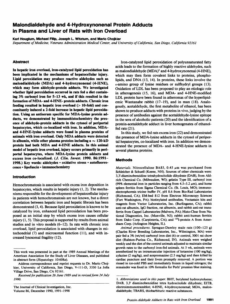
Hepatic iron concentration. Rats fed a carbonyl iron diet for 5wk attained a moderate degree of iron overload that was 13-fold greater than diet-restricted controls (Table I). The bodyweight was similar in the two groups. The iron concentrationin the iron-fed rats was lower than that reported by Park et al.(22), but this may be due to our use of older animals (100-125g). Fig. 1 shows stainable iron in the liver of a control rat and arat fed the carbonyl iron diet for 13 wk (3,900 jug iron/g liver).The iron was present in all periportal areas (zone 1), and vir-tually absent around the terminal hepatic venules (zone 3).Iron was found in hepatocytes and distributed in a pericana-licular pattern.
Thiobarbituric acid reactivity. (TBARS), a measure of lipidperoxidation (25, 26), was significantly increased in liver ho-mogenates of animals fed the 3%carbonyl iron diet (Table I).The addition of ferritin (final concentration 1,300 Mg iron/gliver) during sample preparation did not increase the produc-
1992 K Houglum, M. Filip, J. Witztum, and M. Chojkier
Table L Hepatic Iron Concentration, TBARS, and Body Weightin Iron-Overloaded Rats
Experimentalgroup* Iron TBARS Body weight
Zg/g wet wt nmol/g g
Control(n = 5) 104±6 35±2 263±9
Carbonyl iron(n = 5) 1350±130t 74±3t 272±14
* Animals (100-125 g) were fed either a Purina chow (Ralston-Pur-ina Co.) (control) or a 3%carbonyl iron diet for 5 wk. Hepatic ironand TBARSwere measured as described in Methods; all values aremean±SEM. t Carbonyl iron fed versus control; P < 0.05.
tion of TBARSby normal liver (data not shown), suggestingthat the increased concentration of TBARSin livers of iron-fed rats reflects in vivo formation. Similarly, Bacon et al. (6)have reported increased conjugated dienes in the mitochon-dria of animals with comparable iron overload to ours.
Immunohistochemistry of hepatic tissue. Wehave pre-viously generated an antiserum, MAL-2, that specifically rec-ognizes MDA-lysine residues present on a variety of different
Figure 1. Perls' Prussian blue staining of liver. Paraffin sections ofliver from a control (A) and an iron-overloaded rat (3,900 /g/g wetwt) (B) were treated with Perls' Prussian blue, which stains for iron(80X).
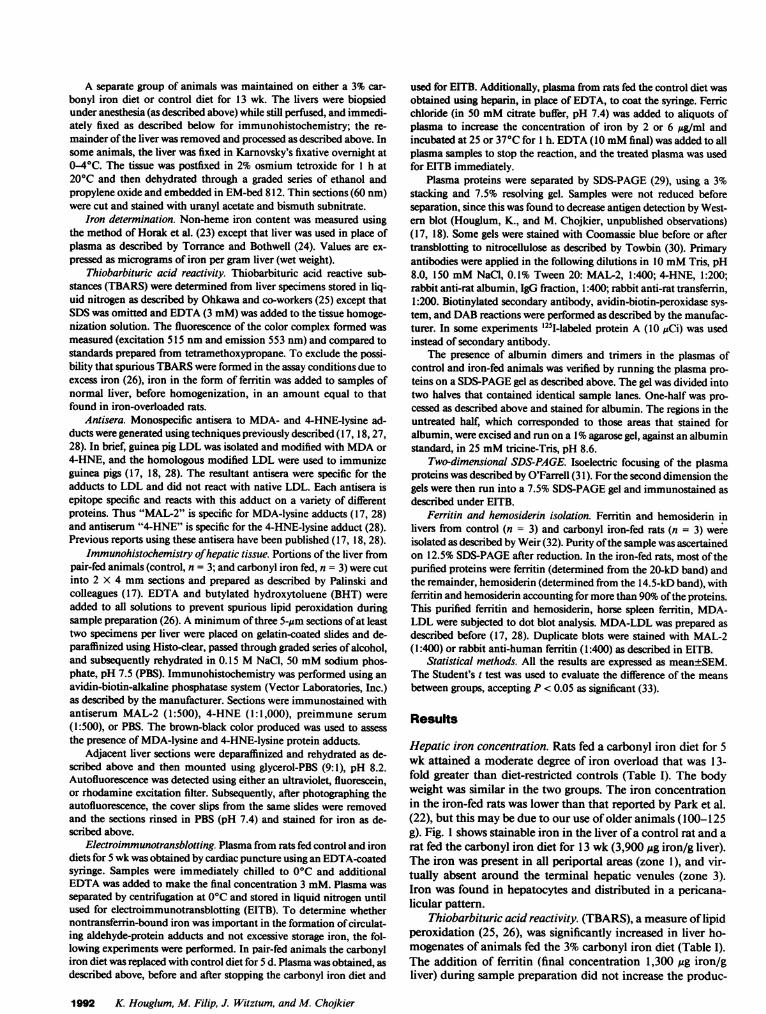
proteins (17, 18, 28). A fluorescent probe was chosen to en-hance the detection of specific MAL-2 staining in hepatocytesand siderotic nodules that contained a significant amount ofdense hemosiderin pigment, which we thought might mask thestaining by enzymatic methods. However, what was found wasan intense yellow-orange autofluorescence in untreated sec-tions (Fig. 2, B and C). The distribution of this autofluo-rescence was predominately in periportal hepatocytes (zone 1)and co-localized to the same hepatocytes with stainable iron inthe identical tissue sections (Fig. 2, B and C and Fig. 1 B).Autofluorescence was absent around the terminal hepaticvenule (zone 3) in iron-overloaded livers (Fig. 2, B and C) andalso in liver sections obtained from control animals (Fig. 2 A).To our knowledge, autofluorescence in experimental ironoverload has not previously been described, however Lillie andFullmer (34) suggested that a non-hemosiderin yellow pig-ment, that has been described in patients with hemochroma-tosis, is lipofuscin. Lipofuscin contains fluorophores, whichare products of lipid peroxidation (35). Indeed, lipofuscin asdetected by its characteristic electron microscopic pattern (36)was abundant in the periportal hepatocytes of iron-overloadedanimals (Fig. 2 D) but rare in controls (data not shown). Peri-portal (zone 1) hepatocytes showed a diffuse as well as vesicu-lar pattern of MAL-2 reactivity that diminished in zone 2 andwas absent around the terminal hepatic venules (zone 3) (Fig. 3B). The vesicular pattern was most prominent in the cyto-plasm of hepatocytes and no nuclear staining was identified(Fig. 3 B). Staining for the MDA-lysine epitope (Fig. 3 B) andiron (Fig. 1 B) co-localized to zone 1 hepatocytes, but neitherwas present in zone 3 hepatocytes. Hemosiderin, a dense yel-low pigment in immunostained sections, can be seen in Fig. 3B. The sections shown in Fig. 3 B were obtained from ananimal with a hepatic iron load of 3,900 ,g/g of liver and arerepresentative sections. Adjacent sections incubated withpreimmune serum or PBS showed no specific staining, how-ever sinusoidal staining was still apparent and not diminishedby the alkaline phosphatase inhibitor levamisole. Some liversections obtained from control animals showed a rare hepato-cyte with staining, among multiple portal triads viewed. Thismay represent either nonspecific staining or specific staining indegenerating hepatocytes. The same pattern of nonspecific si-nusoidal staining, as seen in section of iron-loaded liver, wasalso demonstrated in control liver sections (Fig. 3 A). No dif-ference was seen between control and iron-fed animals in sec-tions stained with 4-HNE (data not shown). The reason for thedifference between 4-HNE and MAL-2 staining is not clear. Inother experiments using CCl4-intoxicated animals, bothMDA- and 4-HNE-protein adducts in the liver are seen in thesame zonal distribution (37), however 4-HNE consistentlystained less intense (Houglum, K. and M. Chojkier; unpub-lished observations). This may reflect a difference in the sensi-tivity of the antisera or a less abundant antigen.
Ferritin and hemosiderin isolated from iron-overloadedanimals had no detectable MDA-protein adducts (data notshown). Therefore, it seems unlikely that the MDA-proteinadducts seen in Fig. 3 B represent either ferritin or hemosid-erin.
Plasma adducts. Because reactive aldehydes formed bylipid peroxidation in iron-overloaded hepatocytes were shownto modify hepatic proteins, we also investigated whetherMDA- or 4-HNE-lysine adducts were present in the plasma ofiron-overloaded rats. Plasma was collected in the presence of
Protein-Aldehyde Adducts in Rats with Iron Overload 1993
a~~~ ~ ~ ~~~~~A
'.4~~~~~~~~~~~~~~~~~~~~~:
;, .i t . .
:i: ~i , t t 6
,- ,+ z z Ww-
Figure 2. Co-localization of autofluorescence and iron. Liver sections (used in Fig. 1) from a control (A) and iron-overloaded rat (3,900 Ag/gwet wt) (B and C) were prepared as described in Methods. Autofluorescence was detected using a rhodamine excitation/emission cube. Ahigher magnification of B is shown in C (A and B, 120X; C, 240X). The electron micrograph (D) demonstrates typical lipofuscin particleswhich were abundant in the periportal hepatocytes of iron overloaded rats (bar is 1 ,um).
excess EDTA in order to prevent lipid peroxidation and theformation of aldehyde adducts during the processing of sam-ples (26, 28). Plasma containing an equal amount of proteinfrom both control and iron-fed rats was subjected to SDS-PAGEand then used for Western blotting with antiserumMAL-2. Western blots of plasma proteins from the control ratswere negative with MAL-2 when stained with DAB-peroxi-dase. In contrast, MAL-2 consistently reacted with several dif-ferent protein bands prepared from the iron-overloaded rats(Fig. 4). In subsequent experiments, even using the more sen-sitive 251I-labeled protein A method, the 120-kD band wasminimally detected in control animals (data not shown). Thelowest molecular weight protein detected had a 66 kD molwt, similar to albumin. Fig. 5 shows the results of a Westernblot of plasma from an iron-overloaded rat in which the nitro-cellulose was divided into two parts and half probed with theMAL-2 antiserum and the other half exposed to antiserumspecific for rat albumin. Several bands were positive for albu-min, at 66 kD and between 92 and 200 kD, suggesting thatalbumin monomers, dimers, and higher molecular weight ag-gregates were present. These bands also reacted with MAL-2.However, there were additional proteins that had the MDA-lysine epitope, between 66 and 92 kD. The existence of albu-min dimers and trimers was confirmed first by isolating theputative polymers of albumin by SDS-PAGEgel electrophore-
sis and then by running them on a 1% agarose gel. Identicalmobility, compared with authentic rat albumin, indicated thatsome of the high molecular weight proteins were indeed albu-min (data not shown). Two-dimensional gel electrophoresisconfirmed that albumin contained MDA-lysine adducts (seearrow, Fig. 6), but also identified the presence of several otherproteins containing MDAadducts. These were present be-tween 66 and 200 kD and had different isoelectric points thanthat of native albumin. Possibly, some of the higher molecularweight proteins were albumin aggregates whose isoelectricpoint was altered by the MDAmodification.
Western blots of plasma proteins were also done with theantiserum specific for 4-HNE. Proteins from normal ratsshowed minimal reactivity at 120 kD, compared with sev-eral different bands that were identified in the proteins ofiron-overloaded rats (Fig. 7). The presence of 4-HNE adductslike MDAadducts in normal plasma was not unexpected, andmost likely represents the presence of 4-HNE-lysine adductsunder normal conditions (17). The marked increase in4-HNE-lysine adducts present in iron-loaded animals proba-bly represents the enhanced formation of reactive 4-HNEunder experimental conditions. A direct comparison of theprotein adducts recognized by antisera MAL-2 and 4-HNE isshown in Fig. 8. Note that antiserum 4-HNE recognizes someof the same modified proteins as MAL-2 but does not recog-
1994 K. Houglum, M. Filip, J. Witztum, and M. Chojkier
.1. Bi, .-, .:
N..P,,A4L.,:''.
1' = - 200
-:..:.l116
-
._wr;_- 6646
2 3 4
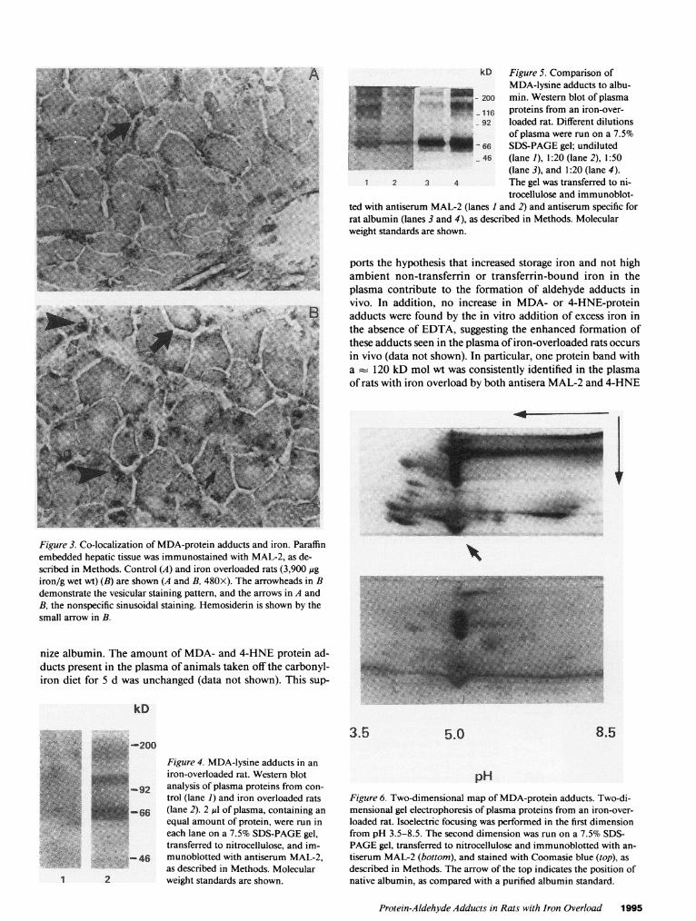
Figure 5. Comparison ofMDA-lysine adducts to albu-min. Western blot of plasmaproteins from an iron-over-loaded rat. Different dilutionsof plasma were run on a 7.5%SDS-PAGEgel; undiluted(lane 1), 1:20 (lane 2), 1:50(lane 3), and 1:20 (lane 4).The gel was transferred to ni-trocellulose and immunoblot-
ted with antiserum MAL-2 (lanes I and 2) and antiserum specific forrat albumin (lanes 3 and 4), as described in Methods. Molecularweight standards are shown.
ports the hypothesis that increased storage iron and not highambient non-transferrin or transferrin-bound iron in theplasma contribute to the formation of aldehyde adducts invivo. In addition, no increase in MDA- or 4-HNE-proteinadducts were found by the in vitro addition of excess iron inthe absence of EDTA, suggesting the enhanced formation ofthese adducts seen in the plasma of iron-overloaded rats occursin vivo (data not shown). In particular, one protein band witha 120 kD mol wt was consistently identified in the plasmaof rats with iron overload by both antisera MAL-2 and 4-HNE
40
Figure 3. Co-localization of MDA-protein adducts and iron. Paraffinembedded hepatic tissue was immunostained with MAL-2, as de-scribed in Methods. Control (A) and iron overloaded rats (3,900 jigiron/g wet wt) (B) are shown (A and B, 480X). The arrowheads in Bdemonstrate the vesicular staining pattern, and the arrows in A andB, the nonspecific sinusoidal staining. Hemosiderin is shown by thesmall arrow in B.
nize albumin. The amount of MDA- and 4-HNE protein ad-ducts present in the plasma of animals taken off the carbonyl-iron diet for 5 d was unchanged (data not shown). This sup-
kD
:- - 200Figure 4. MDA-lysine adducts in an
iron-overloaded rat. Western blot92 analysis of plasma proteins from con-
E. g Etrol (lane 1) and iron overloaded rats-qg~66(lane 2). 2 Wd of plasma, containing an
equal amount of protein, were run ineach lane on a 7.5% SDS-PAGEgel,transferred to nitrocellulose, and im-
-46 munoblotted with antiserum MAL-2,as described in Methods. Molecular
1 2 weight standards are shown.
3.5 5.0 8.5
pHFigure 6. Two-dimensional map of MDA-protein adducts. Two-di-mensional gel electrophoresis of plasma proteins from an iron-over-loaded rat. Isoelectric focusing was performed in the first dimensionfrom pH 3.5-8.5. The second dimension was run on a 7.5% SDS-PAGEgel, transferred to nitrocellulose and immunoblotted with an-tiserum MAL-2 (bottom), and stained with Coomasie blue (top), asdescribed in Methods. The arrow of the top indicates the position ofnative albumin, as compared with a purified albumin standard.
Protein-Aldehyde Adducts in Rats with Iron Overload 1995
kD
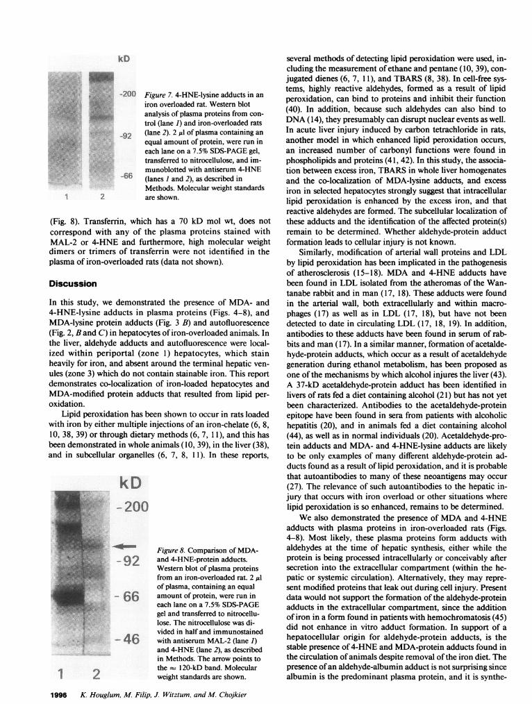
-200 Figure 7. 4-HNE-lysine adducts in aniron overloaded rat. Western blotanalysis of plasma proteins from con-trol (lane 1) and iron-overloaded rats
92 (lane 2). 2 Al of plasma containing an
t equal amount of protein, were run ineach lane on a 7.5% SDS-PAGEgel,transferred to nitrocellulose, and im-munoblotted with antiserum 4-HNE
-66 (lanes I and 2), as described inMethods. Molecular weight standards
2 are shown.
(Fig. 8). Transferrin, which has a 70 kD mol wt, does notcorrespond with any of the plasma proteins stained withMAL-2 or 4-HNE and furthermore, high molecular weightdimers or trimers of transferrin were not identified in theplasma of iron-overloaded rats (data not shown).
Discussion
In this study, we demonstrated the presence of MDA- and4-HNE-lysine adducts in plasma proteins (Figs. 4-8), andMDA-lysine protein adducts (Fig. 3 B) and autofluorescence(Fig. 2, Band C) in hepatocytes of iron-overloaded animals. Inthe liver, aldehyde adducts and autofluorescence were local-ized within periportal (zone 1) hepatocytes, which stainheavily for iron, and absent around the terminal hepatic ven-ules (zone 3) which do not contain stainable iron. This reportdemonstrates co-localization of iron-loaded hepatocytes andMDA-modified protein adducts that resulted from lipid per-oxidation.
Lipid peroxidation has been shown to occur in rats loadedwith iron by either multiple injections of an iron-chelate (6, 8,10, 38, 39) or through dietary methods (6, 7, 11), and this hasbeen demonstrated in whole animals (10, 39), in the liver (38),and in subcellular organelles (6, 7, 8, 11). In these reports,
kD-200
Figure 8. Comparison of MDA-
-92 and 4-HNE-protein adducts.Western blot of plasma proteinsfrom an iron-overloaded rat. 2 1lof plasma, containing an equal
- 66 amount of protein, were run ineach lane on a 7.5% SDS-PAGEgel and transferred to nitrocellu-lose. The nitrocellulose was di-vided in half and immunostained
-46 with antiserum MAL-2 (lane 1)and 4-HNE (lane 2), as describedin Methods. The arrow points tothe 120-kD band. Molecularweight standards are shown.
several methods of detecting lipid peroxidation were used, in-cluding the measurement of ethane and pentane (10, 39), con-jugated dienes (6, 7, 1 1), and TBARS(8, 38). In cell-free sys-tems, highly reactive aldehydes, formed as a result of lipidperoxidation, can bind to proteins and inhibit their function(40). In addition, because such aldehydes can also bind toDNA(14), they presumably can disrupt nuclear events as well.In acute liver injury induced by carbon tetrachloride in rats,another model in which enhanced lipid peroxidation occurs,an increased number of carbonyl functions were found inphospholipids and proteins (41, 42). In this study, the associa-tion between excess iron, TBARSin whole liver homogenatesand the co-localization of MDA-lysine adducts, and excessiron in selected hepatocytes strongly suggest that intracellularlipid peroxidation is enhanced by the excess iron, and thatreactive aldehydes are formed. The subcellular localization ofthese adducts and the identification of the affected protein(s)remain to be determined. Whether aldehyde-protein adductformation leads to cellular injury is not known.
Similarly, modification of arterial wall proteins and LDLby lipid peroxidation has been implicated in the pathogenesisof atherosclerosis (15-18). MDAand 4-HNE adducts havebeen found in LDL isolated from the atheromas of the Wan-tanabe rabbit and in man (17, 18). These adducts were foundin the arterial wall, both extracellularly and within macro-phages (17) as well as in LDL (17, 18), but have not beendetected to date in circulating LDL (17, 18, 19). In addition,antibodies to these adducts have been found in serum of rab-bits and man (17). In a similar manner, formation of acetalde-hyde-protein adducts, which occur as a result of acetaldehydegeneration during ethanol metabolism, has been proposed asone of the mechanisms by which alcohol injures the liver (43).A 37-kD acetaldehyde-protein adduct has been identified inlivers of rats fed a diet containing alcohol (21) but has not yetbeen characterized. Antibodies to the acetaldehyde-proteinepitope have been found in sera from patients with alcoholichepatitis (20), and in animals fed a diet containing alcohol(44), as well as in normal individuals (20). Acetaldehyde-pro-tein adducts and MDA- and 4-HNE-lysine adducts are likelyto be only examples of many different aldehyde-protein ad-ducts found as a result of lipid peroxidation, and it is probablethat autoantibodies to many of these neoantigens may occur(27). The relevance of such autoantibodies to the hepatic in-jury that occurs with iron overload or other situations wherelipid peroxidation is so enhanced, remains to be determined.
Wealso demonstrated the presence of MDAand 4-HNEadducts with plasma proteins in iron-overloaded rats (Figs.4-8). Most likely, these plasma proteins form adducts withaldehydes at the time of hepatic synthesis, either while theprotein is being processed intracellularly or conceivably aftersecretion into the extracellular compartment (within the he-patic or systemic circulation). Alternatively, they may repre-sent modified proteins that leak out during cell injury. Presentdata would not support the formation of the aldehyde-proteinadducts in the extracellular compartment, since the additionof iron in a form found in patients with hemochromatosis (45)did not enhance in vitro adduct formation. In support of ahepatocellular origin for aldehyde-protein adducts, is thestable presence of 4-HNE and MDA-protein adducts found inthe circulation of animals despite removal of the iron diet. Thepresence of an aldehyde-albumin adduct is not surprising sincealbumin is the predominant plasma protein, and it is synthe-
1996 K. Houglum, M. Filip, J. Witztum, and M. Chojkier
1 2
sized exclusively by the hepatocyte. It is of interest that onlyMDA-lysine and not the 4-HNE-lysine epitope was identifiedwith albumin. This difference could be methodological anddue to an enhanced ability to detect MDA-lysine because of amore sensitive antiserum. Alternatively, the difference couldreflect the high reactivity of 4-HNE (13), that might react withproteins within a cellular compartment near to its site of for-mation, which does not contain albumin. MDA, however, isfreely diffusible (46), and may be more widely distributed inboth the intra- and extracellular compartments. With the ex-ception of albumin, the pattern of proteins containing MDAand 4-HNE adducts is similar (Fig. 8). Of particular interest isa prominent 120-kD protein. Further work will be requiredto assess the biological and clinical relevance of the 120-kDprotein as well as the other plasma protein adducts. The de-tection of such adducts might be useful as a noninvasivemarker of the disease process and allow one to judge the ade-quacy of therapy in patients with iron overload.
The demonstration in this study that MDA-lysine adductsin the liver co-localize with iron provides strong evidence forthe occurrence of iron-catalyzed lipid peroxidation in vivo.Characterization of the plasma and hepatocyte proteins thatare modified may provide clues to clarify the mechanism bywhich iron overload leads to cellular injury. Our laboratoryhas previously shown that the highly reactive aldehydes thatform as products of lipid peroxidation as well as acetaldehydefrom the oxidation of ethanol, stimulate collagen gene expres-sion in cultured fibroblasts (47, 48). Wehave proposed thatlipid peroxidation could be a link between tissue injury andtissue fibrogenesis (48). Furthermore, aldehyde adducts withDNAoccur in vitro (14), and they have been implicated in thepathogenesis of some cancers (49). Whether the basis of carci-nogenesis in hemochromatosis is linked to this intriguing pro-cess remains to be elucidated. Lipid peroxidation and the re-sultant formation of aldehyde adducts, as those identified inthis study, may have a role in these processes which ultimatelyleads to hepatic injury, fibrosis, and hepatocellular carcinoma.
Acknowledgments
The authors wish to thank Susan Butler, Seppo Yla-Herttuala, MichaelRosenfeld, and SamParthasarathy for their help and advice in prepara-tion and use of the antisera, Abbyann Sisk and Martina Buck for theirtechnical assistance in performing the electron microscopy and im-munohistochemistry, and Kris Beaver for her skillful preparation ofthis manuscript.
This study was supported by United States Public Health Servicegrants DK-07202, DK-38652, and HL-14197 (Specialized Center ofResearch), and grants from the Veterans Administration. M. Chojkieris a recipient of a Research Career Development Award (VeteransAdministration).
References
1. Powell, L. W., M. L. Bassett, and J. W. Halliday. 1980. He-mochromatosis: 1980 update. Gastroenterology. 78:374-381.
2. Bomford, A., and R. Williams. 1976. Long term results of vene-section therapy in idiopathic haemochromatosis. Q. J. Med. (newseries). 45:611-623.
3. Isaacson, C., H. C. Seftel, K. J. Keeley, and T. H. Bothwell. 1961.Siderosis in the Bantu: the relationship between iron overload andcirrhosis. J. Lab. Clin. Med. 58:845-853.
4. Risdon, R. A., M. Barry, and D. M. Flynn. 1975. Transfusional
iron overload: the relationship between tissue iron concentration andhepatic fibrosis in thalassaemia. J. Path. 116:83-95.
5. Bonkovsky, H. L., J. F. Healey, P. R. Sinclair, J. F. Sinclair, andJ. S. Pomeroy. 1981. Iron and the liver. Acute and long-term effects ofiron-loading on hepatic haem metabolism. Biochem. J. 196:57-64.
6. Bacon, B. R., A. T. Tavill, G. M. Brittenham, C. H. Park, andR. 0. Recknagel. 1983. Hepatic lipid peroxidation in vivo in rats withchronic iron overload. J. Clin. Invest. 71:429-439.
7. Bacon, B. R., C. H. Park, G. M. Brittenham, R. O'Neil, and A. S.Tavill. 1985. Hepatic mitochondrial oxidative metabolism in rats withchronic dietary iron overload. Hepatology. 5:789-797.
8. Masini, A., T. Trenti, E. Ventura, D. Ceccarelli-Stanzani, and U.Muscatello. 1984. Functional efficiency of mitochondrial membraneof rats with hepatic chronic iron overload. Biochem. Biophys. Res.Commun. 124:462-469.
9. Shedlofsky, S. I., H. L. Bonkowsky, P. R. Sinclair, J. F. Sinclair,W. J. Bement, and J. S. Pomeroy. 1983. Iron loading of culturedhepatocytes. Biochem. J. 212:321-330.
10. Dougherty, J. J., W. A. Croft, and W. G. Hoekstra. 1981. Effectof ferrous chloride and iron-dextran on lipid peroxidation in vivo invitamin E and selenium adequate and deficient rats. J. Nutr.111:1784-1796.
11. Bacon, B. R., J. F. Healey, G. M. Brittenham, C. H. Park, J.Nunnari, A. S. Tavill, and H. L. Bonkovsky. 1986. Hepatic micro-somal function in rats with chronic dietary iron overload. Gastroenter-ology. 90:1844-53.
12. Peters, T. J., M. J. O'Connell, and R. J. Ward. 1985. Role offree-radical mediated lipid peroxidation in the pathogenesis of hepaticdamage by lysosomal disruption. In Free Radicals In Liver Injury. G.Poli, H. H. Cheesman, M. U. Dianzani, and T. F. Slater, editors.I. R. L. Press Ltd., Oxford, England. 107-115.
13. Schauenstein, E., H. Esterbauer, and H. Zollner. 1977. Alde-hydes in Biological Systems. Pion Limited, London. 102 pp.
14. Sodum, R. S., and F.-L. Chung. 1988. 1,N2-Ethenodeoxygua-nosine as a potential marker for DNAadduct formation by trans-4-hydroxy-2-nonenal. Cancer Res. 48:320-323.
15. Steinberg, D., S. Parthasarathy, T. F. Carew, J. C. Khoo, andJ. L. Witztum. 1989. Beyond Cholesterol. Modifications of low-den-sity lipoprotein that increase its atherogenicity. N. Engi. J. Med.320:915-924.
16. Haberland, M. E., and A. M. Fogelman. 1987. The role ofaltered lipoproteins in the pathogenesis of atherosclerosis. Am. Heart J.113:573-577.
17. Palinski, W., M. E. Rosenfeld, S. Yla-Herttuala, G. C. Gurtner,S. S. Socher, S. W. Butler, S. Parthasarathy, T. E. Carew, D. Steinberg,and J. L. Witztum. 1989. Low density lipoprotein undergoes oxidativemodification in vivo. Proc. Natl. Acad. Sci. USA. 86:1372-1376.
18. Yla-Herttuala, S., W. Palinski, M. E. Rosenfeld, S. Parthasa-rathy, T. Carew, S. Butler, J. L. Witztum, and D. Steinberg. 1989.Evidence for the presence of oxidatively modified low density lipopro-tein in atherosclerotic lesions of rabbit and man. J. Clin. Invest.84:1086-1095.
19. Haberland, M. E., D. Fong, and L. Cheng. 1988. Malondialde-hyde-altered protein occurs in atheroma of Wantanabe heritable hy-perlipidemic rabbits. Science (Wash. DC). 241:215-218.
20. Niemela, O., F. Klajner, H. Orrego, E. Vidins, L. Blendis, andY. Israel. 1987. Antibodies against acetaldehyde-modified protein epi-topes in human alcoholics. Hepatology. 7:1210-1214.
21. Lin, R. C., R. S. Smith, and L. Lumeng. 1988. Detection of aprotein-acetaldehyde adduct in the liver of rats fed alcohol chronically.J. Clin. Invest. 81:615-619.
22. Park, C. H., B. R. Bacon, G. M. Brittenham, and A. S. Tavill.1987. Pathology of dietary carbonyl iron overload in rats. Lab. Invest.57:555-63.
23. Horak, E., D. C. Hohnadel, and F. W. Sunderman, Jr. 1975.Modified method for analysis of serum iron. Ann. Clin. Lab. Sci.5:303-307.
24. Torrance, J. D., and T. H. Bothwell. 1980. Tissue iron stores. In
Protein-Aldehyde Adducts in Rats with Iron Overload 1997
Iron. Vol. 1. J. Cook, editor. Churchill-Livingstone, Inc. New York.90-115.
25. Ohkawa, H., N. Ohishi, and K. Yagi. 1979. Assay for lipidperoxidation in animal tissue by thiobarbituric assay. Anal. Biochem.95:351-358.
26. Gutteridge, J. M. C., and G. J. Quinlan. 1983. Malondialde-hyde formation from lipid peroxides in the thiobarbituric acid test: therole of lipid radicals, iron salts and metal chelators. J. Appl. Biochem.5:293-299.
27. Steinbrecher, U. P., M. Fisher, J. L. Witztum, and L. K. Curtiss.1984. Immunogenicity of homologous low density lipoprotein aftermethylation, ethylation, acetylation, or carbamylation: generation ofantibodies specific for derivatized lysine. J. Lipid Res. 25:1109-1116.
28. Palinski, W., S. Yla-Herttuala, M. E. Rosenfield, S. W. Butler,S. A. Socher, S. Parthasarathy, L. K. Curtiss, and J. L. Witztum. 1990.Antisera and monoclonal antibodies specific for epitopes generatedduring oxidative modification of low density lipoprotein. Arterioscle-rosis. 10:325-335.
29. Laemmli, U. K. 1970. Cleavage of structural proteins duringthe assembly of the head of bacteriophage T4. Nature (Lond.).227:680-685.
30. Towbin, H., T. Staehelin, and J. Gordon. 1979. Electrophoresistransfer of proteins from polyacrylamide gels to nitrocellulose sheets:procedures and some applications. Proc. Natl. Acad. Sci. USA.76:4350-4354.
31. O'Farrell, P. H. 1975. High resolution two-dimensional electro-phoresis of proteins. J. Biol. Chem. 250:4007-4021.
32. Weir, M. P., J. F. Gibson, and T. J. Peters. 1984. Biochemicalstudies on the isolation and characterization of human spleen haemo-siderin. Biochem. J. 223:31-38.
33. Colton, T. 1974. Statistics in Medicine. T. Colton, editor. Little,Brown and Company., Boston. 99-150.
34. Lillie, R. D., and H. M. Fullmer. 1976. Histopathologic Tech-nic and Practical Histochemistry. McGraw-Hill Inc., New York.485-528.
35. Dillard, C. I., and A. L. Tappel. 1984. Fluorescent damageproducts of lipid peroxidation. Methods Enzymol. 105:337-341.
36. Miyagishi, T., N. Takahata, and R. Iizuka. 1967. Electronmicroscopic studies on the lipo-pigments in the cerebral cortex nervecells of senile and and Vitamin E deficient rats. Acta Neuropathol.9:7-17.
37. Houglum, K., D. Kaufer, and M. Chojkier. 1989. Cellularlocalization of malondialdehyde and 4-hydroxynonenal protein ad-ducts in hepatic lipid peroxidation and their prevention by d-a-tocoph-erol. Hepatology. 10:610a. (Abstr.)
38. Goldberg, L., L. E. Martin, and A. Batchelor. 1962. Biochemi-cal changes in the tissue of animals injected with iron. Biochem. J.83:291-298.
39. Dillard, C. J., J. E. Downey, and A. L. Tappel. 1984. Effect ofantioxidants on lipid peroxidation in iron-loaded rats. Lipids. 19:127-133.
40. Esterbauer, H. 1985. Lipid peroxidation products: formation,chemical properties and biological activities. In Free Radicals in LiverInjury. G. Poli, H. H. Cheesman, M. U. Dianzani, and T. F. Slater,editors. I. R. L. Press Ltd., Oxford, England. 2-47.
41. Benedetti, A., R. Fulceri, M. Ferrali, L. Ciccoli, H. Esterbauer,and M. Comporti. 1982. Detection of carbonyl functions in phospho-lipids of microsomes in CC14- and BrCCl3-poisoned rats. Biochem.Biophys. Acta. 712:628-638.
42. Benedetti, A., H. Esterbauer, M. Ferrali, R. Fulceri, and M.Comporti. 1982. Evidence for aldehydes bound to liver microsomalprotein following CC14 or BrCCl3 poisoning. Biochem. Biophys. Acta.711:345-356.
43. Sorrell, M. F., and D. J. Tuma. 1985. Hypothesis: alcoholicliver injury and the covalent binding of acetaldehyde. Alcoholism:Clin. Exp. Res. 9:306-309.
44. Israel, Y., E. Hurwitz, 0. Niemela, and R. Amnon. 1986. Mono-clonal and polyclonal antibodies against acetaldehyde-containing epi-topes in acetaldehyde-protein adducts. Proc. Natl. Acad. Sci. USA.83:7923-7927.
45. Grootveld, M., J. D. Bell, B. Halliwell, 0. I. Aruoma, A.Bombford, and P. J. Sadler. 1989. Non-transferrin-bound iron inplasma or serum from patients with idiopathic hemochromatosis. J.Biol. Chem. 264:4417-4422.
46. Esterbauer, H., G. Jurgens, 0. Quehenberger, and E. Koller.1987. Autoxidation of human low density lipoproteins: loss of polyun-saturated fatty acids and vitamin E and generation of aldehydes. J.Lipid Res. 28:495-509.
47. Brenner, D. A., and M. Chojkier. 1987. Acetaldehyde increasescollagen gene transcription in cultured human fibroblasts. J. Biol.Chem. 262:17690-17695.
48. Chojkier, M., K. Houglum, J. A. Solis-Herruzo, and D. A.Brenner. 1989. Stimulation of collagen gene expression by ascorbicacid in cultured human fibroblasts. A role for lipid peroxidation? J.Biol. Chem. 264:16957-16962.
49. Gupta, R. C., K. Earley, and F. F. Becker. 1988. Analysis ofDNAadducts in putative premalignant hepatic nodules and nontargettissues of rats during 2-acetylaminoflurene carcinogenesis. Cancer Res.48:5270-5277.
1998 K Houglum, M. Filip, J. Witztum, and M. Chojkier