Embed Size (px)
Citation preview
Mammography
9/24/2014 online
Radiographic studies of the breast
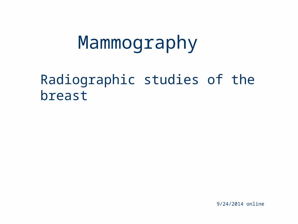
Breast Anatomy
Breast =mammary gland
Consist of glandular, fat, and fibrous tissue
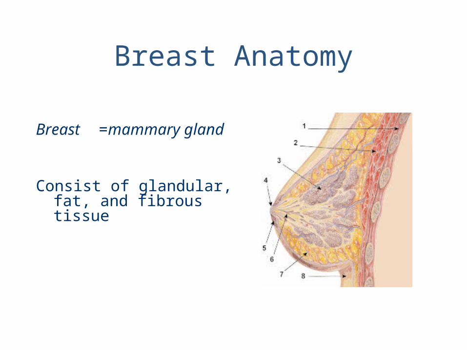
Anatomy cont’d
Female breasts are divided into 15 – 20 lobes
each made up of lobules
Supported by Cooper’s ligament which determines firmness

Lobule size
Affected by age and hormones (pregnancy)
Involution: process of decreasing lobule size with age and after pregnancy
-flatter, saggier breasts
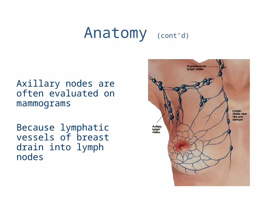
Anatomy (cont’d)
Axillary nodes are often evaluated on mammograms
Because lymphatic vessels of breast drain into lymph nodes
Tissue Variations
Breasts consists of both glandular and connective
Ability to visualize depends on amount of fat within and around breast lobules- provides contrast
Postpuberty breasts contain primarily dense connective tissue- harder to visualize

19 yr. old (never pregnant)
(dense breast)
24 yr. old (has children)
(fatty breast)
Mammograms comparing 2 different women
Cancer in tissues of breast –ducts (tubes that carry milk to nipple)
lobules (glands that make milk)
Can men get breast cancer?
Yes, but rare 1/100 compared to women
Definition of breast cancer
Breast Cancer
Nationally the 2nd leading cause of cancer-related deaths in women
What is first?lung cancer
Breast cancer in United States in 2009 (estimated):
New cases: 192,370 (female)
Deaths: 40,170 (female)

Breast Cancer Risk increases with:
Age
Hormonal history early menses late menopause pregnancy after age 30 Never had a child
Family historyIf daughter, mother, or sister has breast cancer
Pt.s in early stages respond well to treatment
Patients with advanced disease do poorly
Earlier diagnosis, better chance of survival
Mammography is best way for early detection!
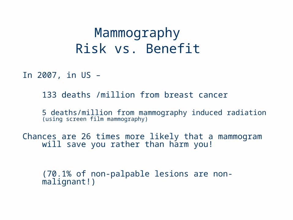
Mammography Risk vs. Benefit
In 2007, in US –
133 deaths /million from breast cancer
5 deaths/million from mammography induced radiation (using screen film mammography)
Chances are 26 times more likely that a mammogram will save you rather than harm you!
(70.1% of non-palpable lesions are non-malignant!)

What are your chances beating Breast Cancer ?
If cancer is confined to breast, there is a 97% survival rate for 5 years
Incidence of breast cancer stable since 1988-
-mortality rate decreased by 29%- mainly do to early detection

At what age should a woman have her first mammogram?
Used to be: once a year after age 40
In Nov. 2009, U.S. Preventive Services Task Force updated recommendations:
No routine screening mammography in women aged 40 to 49 years!
50-74 should have mammogram every other year
Optional every other year before age of 50 years:
individual choice based on family history and pt's weighing of specific benefits and harms

American College of Radiology and Society of Breast Imagingstrongly disagree!
Annual screening mammography should begin at age 40!
Mammography only every other year in women 50-74 would miss 19 to 33 percent of cancers that could be detected by annual screening!
History of breast cancer detection
When was the first radical mastectomy introduced?1898
What year was the radiographic appearance of breast cancer first reported?
1913
When did mammography became a reliable diagnostic tool? in 1950s when industrial grade x-ray film introduced
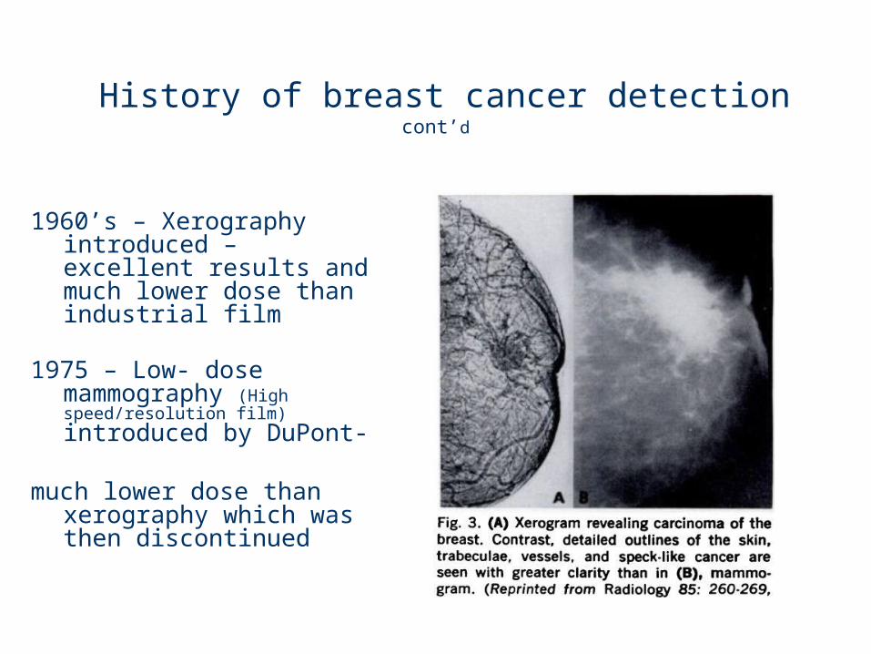
History of breast cancer detection cont’d
1960’s – Xerography introduced – excellent results and much lower dose than industrial film
1975 – Low- dose mammography (High speed/resolution film) introduced by DuPont-
much lower dose than xerography which was then discontinued
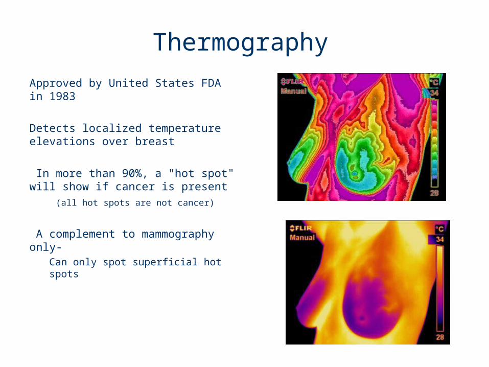
Thermography
Approved by United States FDA in 1983
Detects localized temperature elevations over breast
In more than 90%, a "hot spot" will show if cancer is present (all hot spots are not cancer)
A complement to mammography only-
Can only spot superficial hot spots
MQSA (Mammography Quality Standards Act)
1992 – MQSA passed by Congress, not enacted until 1994
Mammography became 1st and only federally regulated imaging exam, which mandated:
Formal training and continuing education
Required regular inspection of equipment
Documentation of quality assurance
Report results, follow-up, track pts, and monitor outcomes
Types of Mammograms
Baseline mammogram: very 1st mammogram (or 1st mammo. after surgery)
Screening mammogram: all mammos after baseline- if pt. asymptomatic (no known breast problems)
Diagnostic Mammogram: when woman presents with clinical evidence of:
Breast disease
Palpable mass or other symptom

Typical Mammography Unit
Equipment is C-arm
SID is fixed at 24 – 26”
Mammography Equipment
1st dedicated mammography unit -1969
Designed to produce high-contrast and high-resolution images
More precise control of kVp, mA, and exposure time
Low kVp : 25 – 28
AEC (automated exposure control)
Grid with ratio: 4:1, or 5:1 200 lines/inch
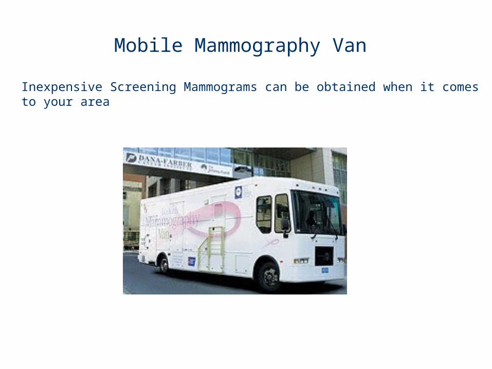
Mobile Mammography Van
Inexpensive Screening Mammograms can be obtained when it comes to your area
Screen-Film Systems
Now largely replaced by digital imaging
Mammography cassettes contain a single screen
Film is single emulsion
Extended time processing can be usedto reduce dose and increase contrast

Digital MammographyState of the art!
No film, no chemical processing
Much better definition
Less compression needed
Radiation dose about 22% less
Fewer repeats do to poor technique selection
Digital mammography cont’d
Images easily sent over internet
Can give pt. CD of images
Possible downside:1st digital images compared to
previous film images can give false positives due to increased sensitivity

Procedure
Complete, careful history and physical assessment!
Take notes on location of scars, palpable masses, skin abnormalities, and nipple alterations
Examine previous mammograms for positioning, compression, and exposure factors

Procedure (con’t)
Have Pt put on gown with opening in front
Breasts must be bared for imagingCloth will cause image artifact
Remove deodorant and powder from axilla and breast:
It can mimic calcifications on image!
Procedure (cont’d)
Explain procedure to pt., including possibility for additional projections
Consider natural mobility of breast before positioning
Support breast firmly so that nipple is directed forward in profile
Apply proper compression
Place ID markers
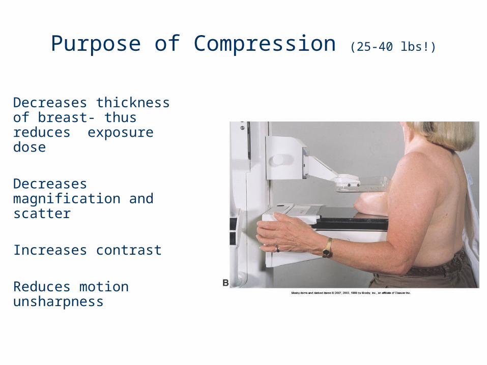
Purpose of Compression (25-40 lbs!)
Decreases thickness of breast- thus reduces exposure dose
Decreases magnification and scatter
Increases contrast
Reduces motion unsharpness

MagnificationDigital Mammography now makes “mag films” obsolete
Uses increase OID to magnify image
Increases visibility of small structures
Why does Radiation dose increase with magnification even though technique is not increased?-(breast is closer to source)

Routine Mammography Projections and Positions
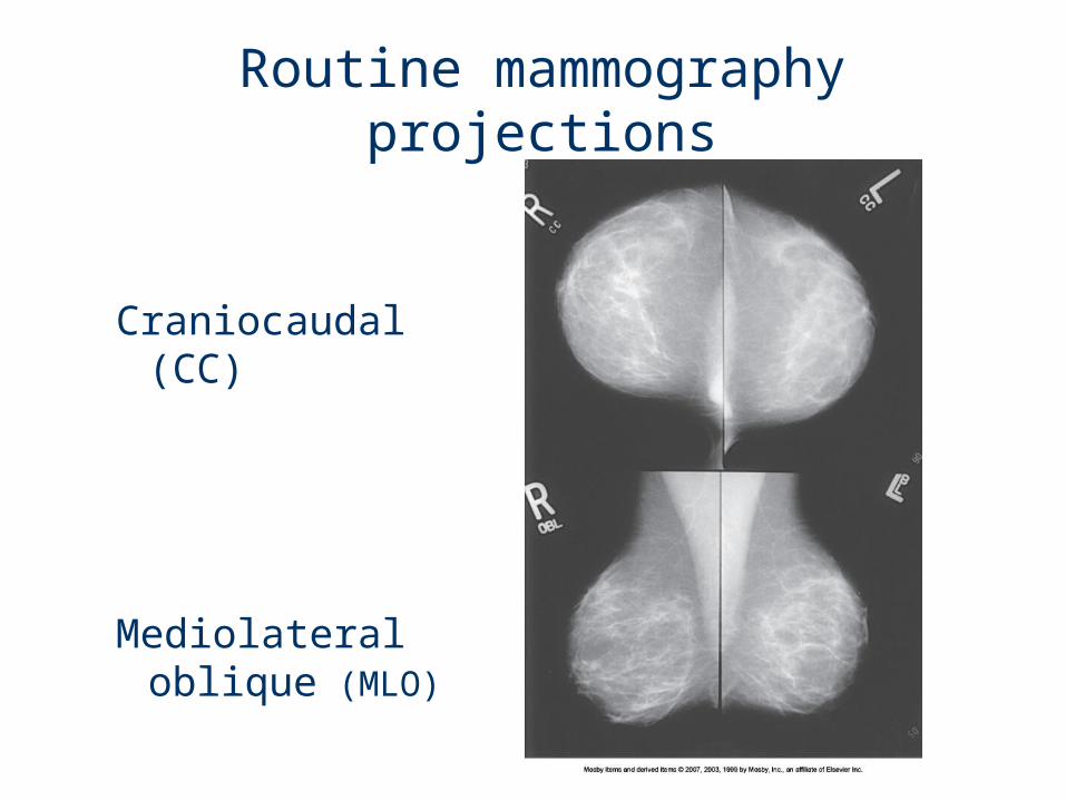
Routine mammography projections
Craniocaudal (CC)
Mediolateral oblique (MLO)
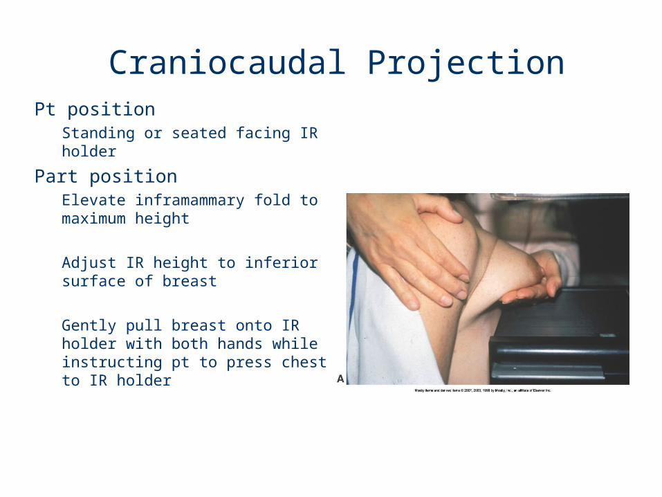
Craniocaudal ProjectionPt position
Standing or seated facing IR holder
Part positionElevate inframammary fold to maximum height
Adjust IR height to inferior surface of breast
Gently pull breast onto IR holder with both hands while instructing pt to press chest to IR holder
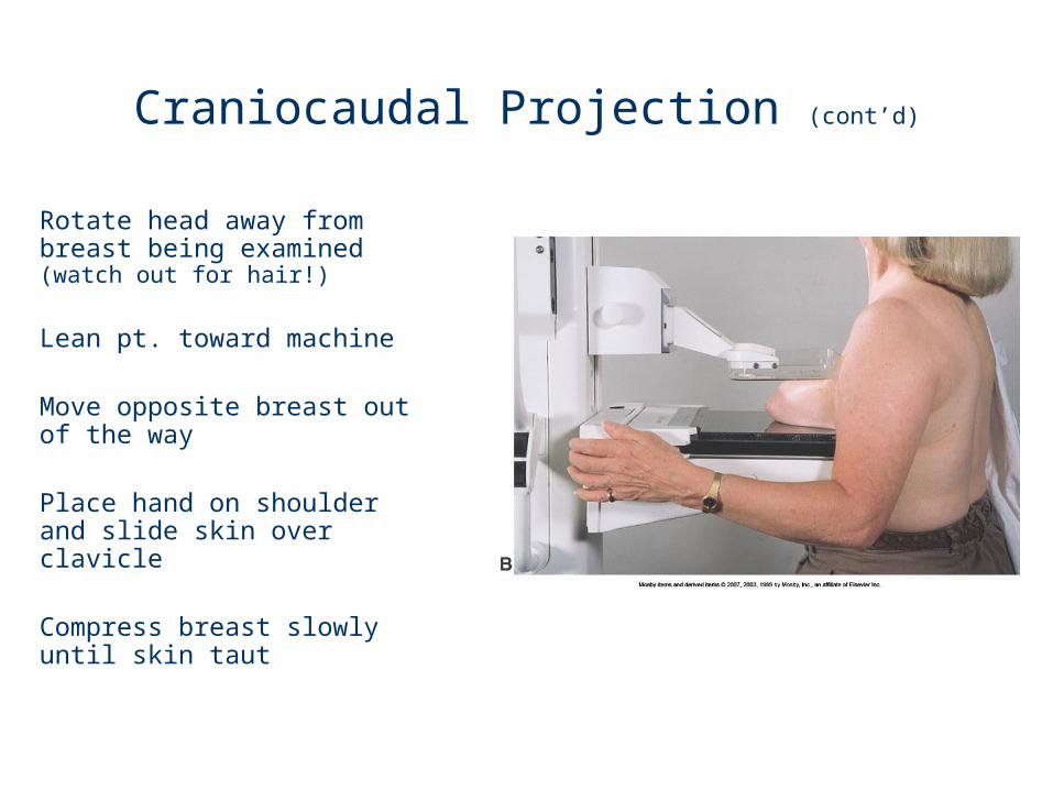
Craniocaudal Projection (cont’d)
Rotate head away from breast being examined (watch out for hair!)
Lean pt. toward machine
Move opposite breast out of the way
Place hand on shoulder and slide skin over clavicle
Compress breast slowly until skin taut

Mediolateral Oblique Projection
PositionCenter breast with nipple in profile
Hold breast up and out
Compress breast slowly until taut
Pull down on abdominal tissue to open inframammary fold
Instruct pt. to hold opposite breast laterally, out of anatomy of interest
Exposure on suspended respiration
Release compression immediately!

Breast Implants
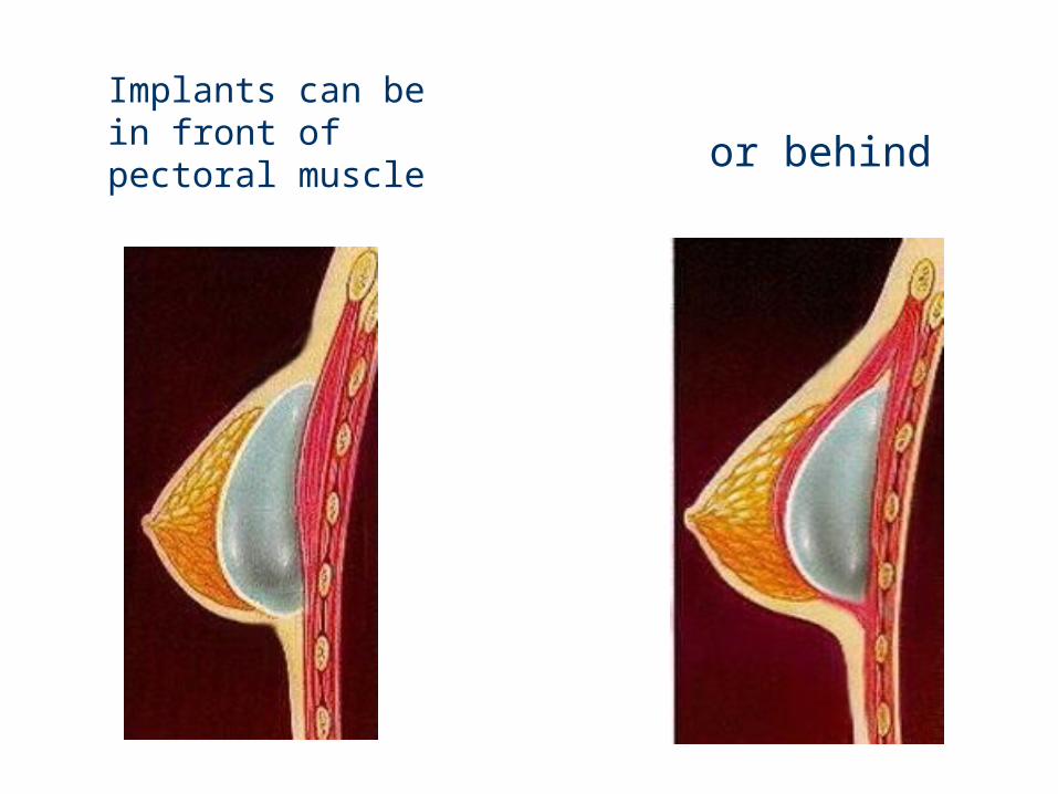
Implants can be in front of pectoral muscle
or behind
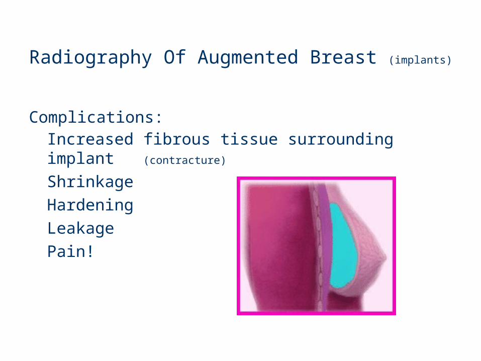
Radiography Of Augmented Breast (implants)
Complications:Increased fibrous tissue surrounding implant (contracture)
Shrinkage HardeningLeakagePain!
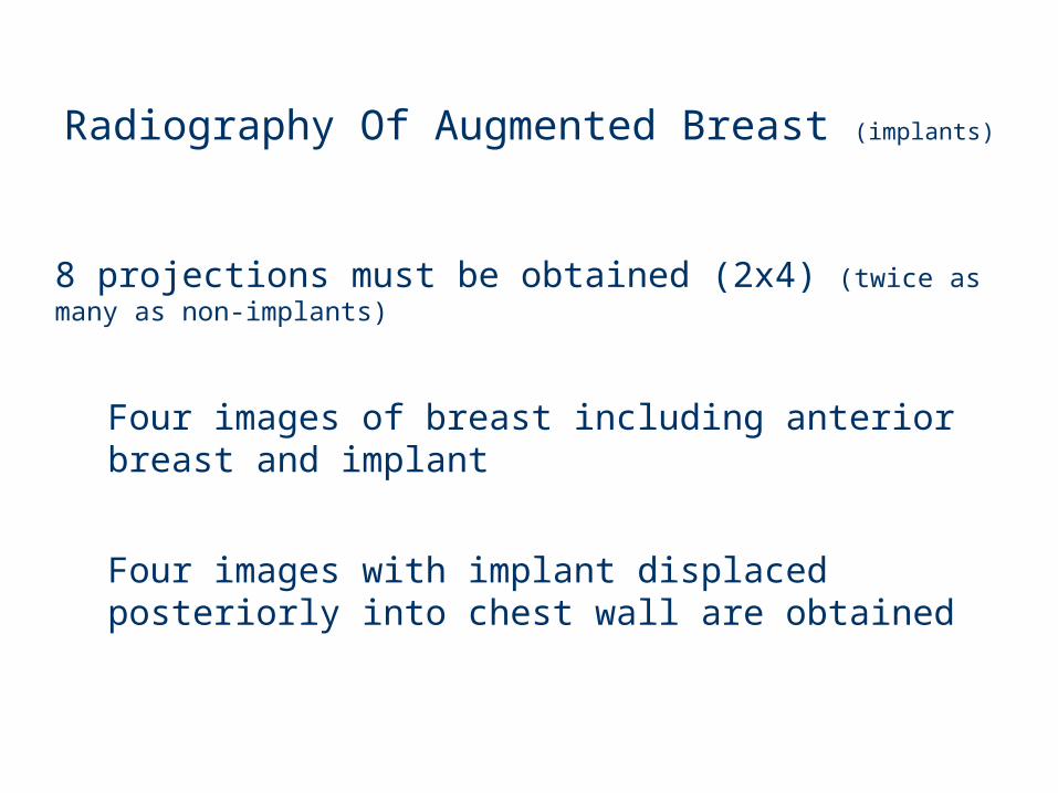
Radiography Of Augmented Breast (implants)
8 projections must be obtained (2x4) (twice as many as non-implants)
Four images of breast including anterior breast and implant
Four images with implant displaced posteriorly into chest wall are obtained
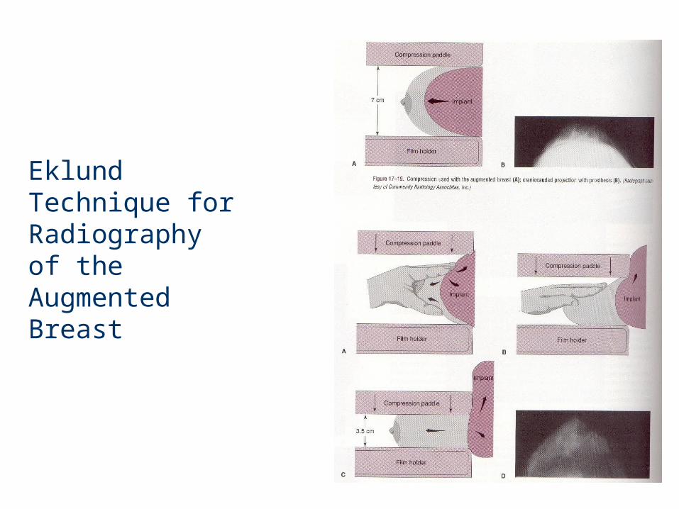
Eklund Technique for Radiographyof the Augmented Breast

Treatment For Breast Cancer
Lumpectomy
Partial or radical mastectomy
Radiation
Chemotherapy
(recent study shows that lumpectomy or mastectomy may be no more beneficial than radiation and chemotherapy)
Lesion
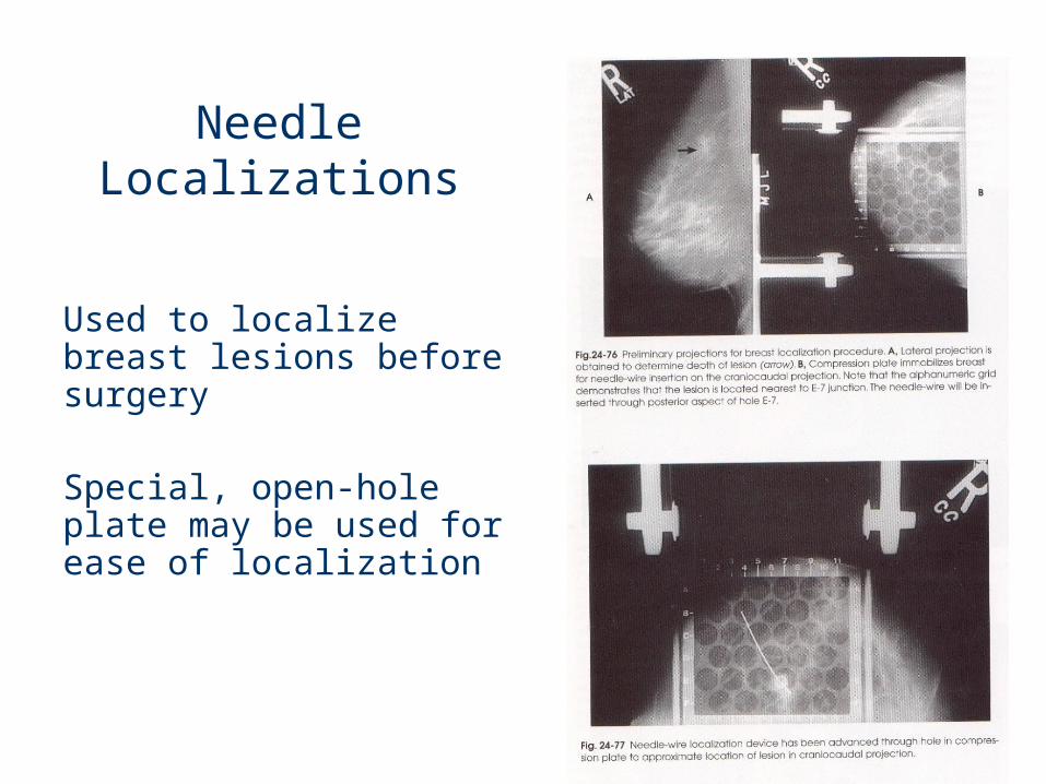
Needle Localizations
Used to localize breast lesions before surgery
Special, open-hole plate may be used for ease of localization
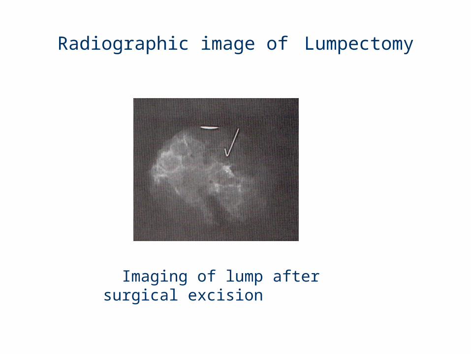
Radiographic image of Lumpectomy
Imaging of lump after surgical excision
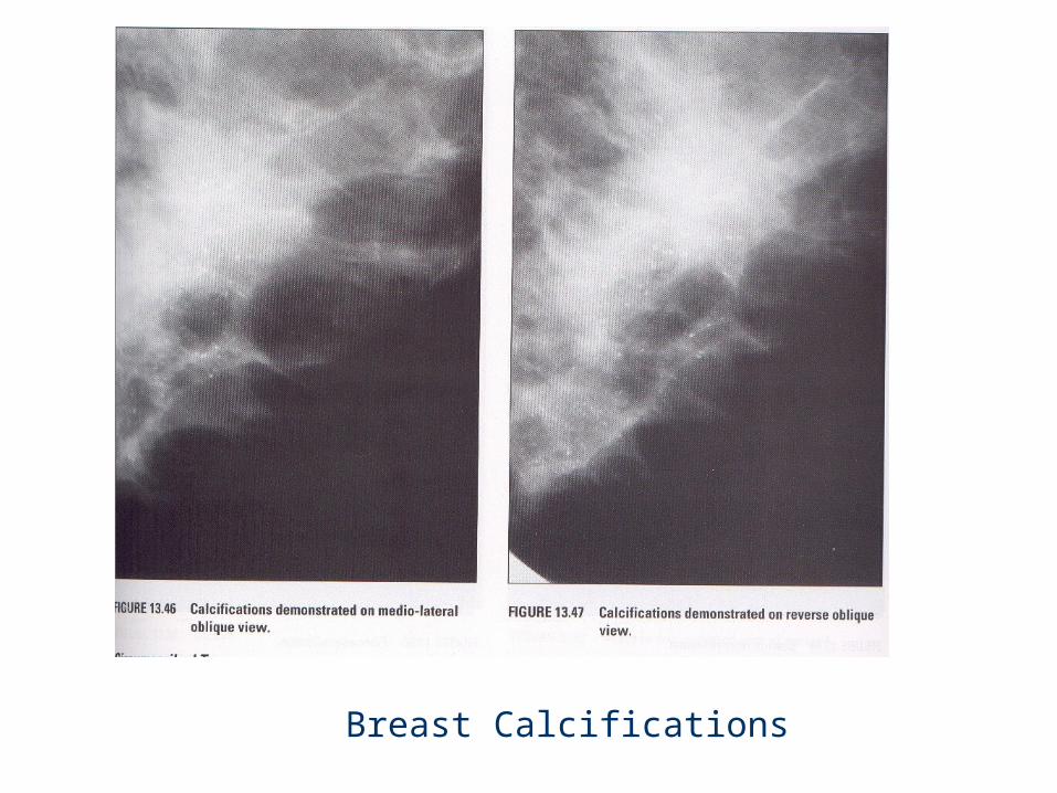
Breast Calcifications

Calcified Milk Ducts

Benign Cyst