Embed Size (px)
Citation preview

1
Management of challenging radioiodine treatment protocols: a case series and review of the
literature
Joseph Waller1,2, Courtney A. Lawhn-Heath1, Cathleen Edmonds3, Chloee Wendorf1, Brandon
Holmes3, Michael White, Miguel Pampaloni1, Chienying Liu4, Robert R. Flavell1.
1. University of California San Francisco, Department of Radiology and Biomedical Imaging, 505 Parnassus Ave, San Francisco, CA 94143
2. Drexel University College of Medicine, 2900 W Queen Ln, PA 19129 3. University of California San Francisco, Radiation Safety and UCOP, San Francisco, CA
94143 4. University of California San Francisco, Department of Medicine, San Francisco, CA
94143
Corresponding Author: Robert Flavell
Box 0946, UCSF, San Francisco, CA 94143
(415) 353-3638. [email protected]
First Author: Joseph Waller (student)
5214 Schuyler St, Philadelphia, PA 19129
(703) 297-0222. [email protected]
Word Count: 2960
Disclosures: N/A.
Financial Support: N/A
Short Running Title: Thyroid Cancer 131I Treatment Protocols
J of Nuclear Medicine Technology, first published online November 20, 2020 as doi:10.2967/jnmt.120.255307
2
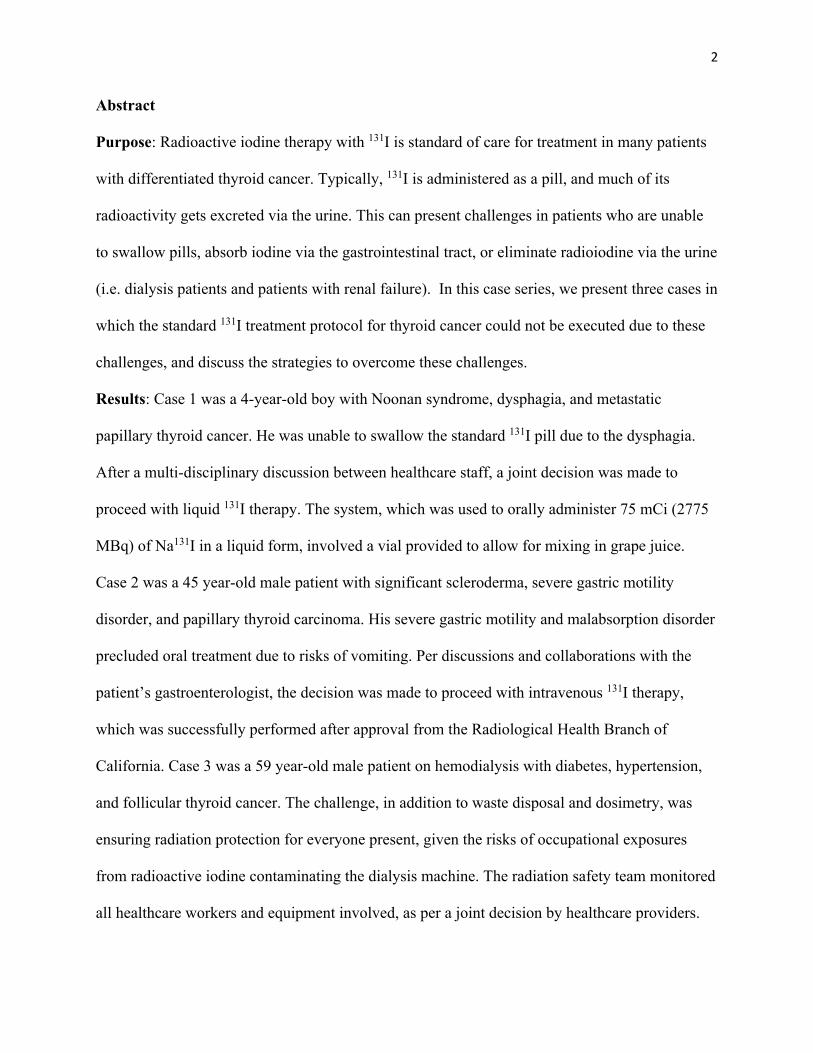
Abstract
Purpose: Radioactive iodine therapy with 131I is standard of care for treatment in many patients
with differentiated thyroid cancer. Typically, 131I is administered as a pill, and much of its
radioactivity gets excreted via the urine. This can present challenges in patients who are unable
to swallow pills, absorb iodine via the gastrointestinal tract, or eliminate radioiodine via the urine
(i.e. dialysis patients and patients with renal failure). In this case series, we present three cases in
which the standard 131I treatment protocol for thyroid cancer could not be executed due to these
challenges, and discuss the strategies to overcome these challenges.
Results: Case 1 was a 4-year-old boy with Noonan syndrome, dysphagia, and metastatic
papillary thyroid cancer. He was unable to swallow the standard 131I pill due to the dysphagia.
After a multi-disciplinary discussion between healthcare staff, a joint decision was made to
proceed with liquid 131I therapy. The system, which was used to orally administer 75 mCi (2775
MBq) of Na131I in a liquid form, involved a vial provided to allow for mixing in grape juice.
Case 2 was a 45 year-old male patient with significant scleroderma, severe gastric motility
disorder, and papillary thyroid carcinoma. His severe gastric motility and malabsorption disorder
precluded oral treatment due to risks of vomiting. Per discussions and collaborations with the
patient’s gastroenterologist, the decision was made to proceed with intravenous 131I therapy,
which was successfully performed after approval from the Radiological Health Branch of
California. Case 3 was a 59 year-old male patient on hemodialysis with diabetes, hypertension,
and follicular thyroid cancer. The challenge, in addition to waste disposal and dosimetry, was
ensuring radiation protection for everyone present, given the risks of occupational exposures
from radioactive iodine contaminating the dialysis machine. The radiation safety team monitored
all healthcare workers and equipment involved, as per a joint decision by healthcare providers.
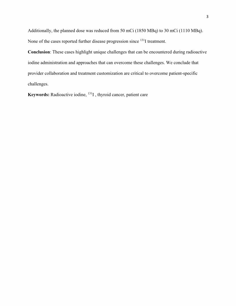
3
Additionally, the planned dose was reduced from 50 mCi (1850 MBq) to 30 mCi (1110 MBq).
None of the cases reported further disease progression since 131I treatment.
Conclusion: These cases highlight unique challenges that can be encountered during radioactive
iodine administration and approaches that can overcome these challenges. We conclude that
provider collaboration and treatment customization are critical to overcome patient-specific
challenges.
Keywords: Radioactive iodine, 131I , thyroid cancer, patient care
4
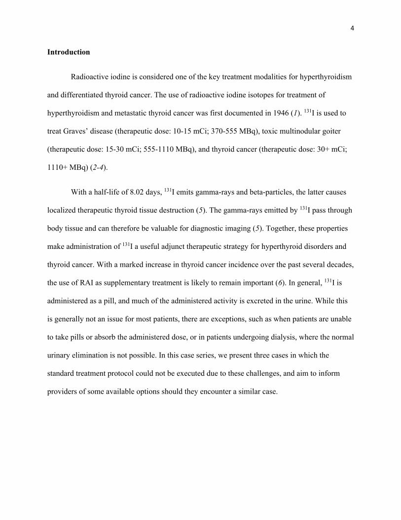
Introduction
Radioactive iodine is considered one of the key treatment modalities for hyperthyroidism
and differentiated thyroid cancer. The use of radioactive iodine isotopes for treatment of
hyperthyroidism and metastatic thyroid cancer was first documented in 1946 (1). 131I is used to
treat Graves’ disease (therapeutic dose: 10-15 mCi; 370-555 MBq), toxic multinodular goiter
(therapeutic dose: 15-30 mCi; 555-1110 MBq), and thyroid cancer (therapeutic dose: 30+ mCi;
1110+ MBq) (2-4).
With a half-life of 8.02 days, 131I emits gamma-rays and beta-particles, the latter causes
localized therapeutic thyroid tissue destruction (5). The gamma-rays emitted by 131I pass through
body tissue and can therefore be valuable for diagnostic imaging (5). Together, these properties
make administration of 131I a useful adjunct therapeutic strategy for hyperthyroid disorders and
thyroid cancer. With a marked increase in thyroid cancer incidence over the past several decades,
the use of RAI as supplementary treatment is likely to remain important (6). In general, 131I is
administered as a pill, and much of the administered activity is excreted in the urine. While this
is generally not an issue for most patients, there are exceptions, such as when patients are unable
to take pills or absorb the administered dose, or in patients undergoing dialysis, where the normal
urinary elimination is not possible. In this case series, we present three cases in which the
standard treatment protocol could not be executed due to these challenges, and aim to inform
providers of some available options should they encounter a similar case.
5
Case 1
Case 1 was a boy with a past medical history of Noonan syndrome with dysphagia
diagnosed at birth. At four years of age, the patient came to our institution with an enlarged right
thyroid lobe and was diagnosed with multiple thyroid nodules on a neck ultrasound. The
ultrasound demonstrated a diffusely abnormal thyroid gland with microcalcifications, and
bilateral enlarged cervical lymph nodes were abnormal with suspicious microcalcifications.
These findings were highly suspicious for thyroid malignancy, and a fine-needle aspiration of the
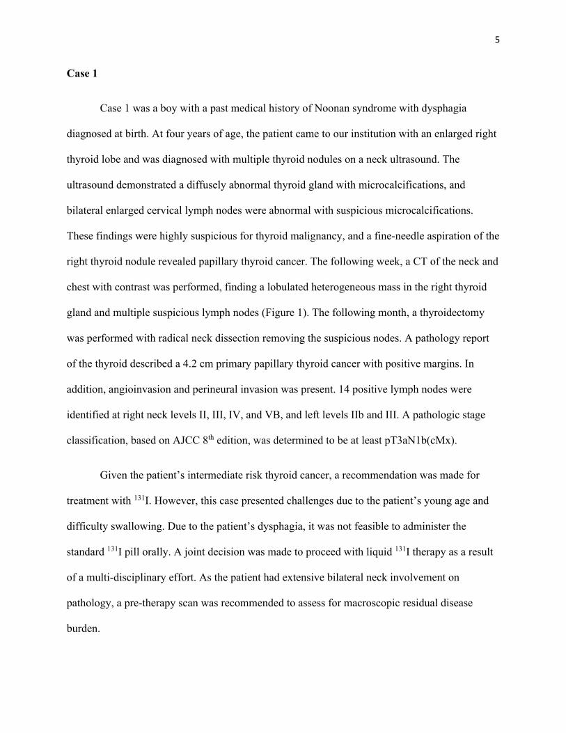
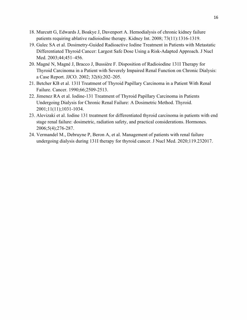
right thyroid nodule revealed papillary thyroid cancer. The following week, a CT of the neck and
chest with contrast was performed, finding a lobulated heterogeneous mass in the right thyroid
gland and multiple suspicious lymph nodes (Figure 1). The following month, a thyroidectomy
was performed with radical neck dissection removing the suspicious nodes. A pathology report
of the thyroid described a 4.2 cm primary papillary thyroid cancer with positive margins. In
addition, angioinvasion and perineural invasion was present. 14 positive lymph nodes were
identified at right neck levels II, III, IV, and VB, and left levels IIb and III. A pathologic stage
classification, based on AJCC 8th edition, was determined to be at least pT3aN1b(cMx).
Given the patient’s intermediate risk thyroid cancer, a recommendation was made for
treatment with 131I. However, this case presented challenges due to the patient’s young age and
difficulty swallowing. Due to the patient’s dysphagia, it was not feasible to administer the
standard 131I pill orally. A joint decision was made to proceed with liquid 131I therapy as a result
of a multi-disciplinary effort. As the patient had extensive bilateral neck involvement on
pathology, a pre-therapy scan was recommended to assess for macroscopic residual disease
burden.
6
Since 131I is volatile, liquid administration required special preparations. Specifically, to
avoid staff exposure to 131I, a pharmacist provided a 5-10 mL oral solution of 131I mixed with
grape juice in a French square glass vial with a screw cap. The patient drank through a straw
with a spinal needle that was attached to poke the Teflon septum within the cap. These
preparations were designed to minimize evaporation of liquid 131I. An anterior and posterior
whole-body scan with SPECT/CT imaging of the neck was taken 24 hours after oral
administration of 1.5 mCi (55.5 MBq) Na131I (123I was not available as a liquid for the diagnostic
scan). Focal uptake was noted in the thyroid bed region in the neck, consistent with remnant
thyroid tissue. There were no visible suspicious cervical or distant foci of radioiodine uptake to
suggest metastasis.
The following day, the patient was admitted (as per the patient’s legal guardian’s request
due to situations in the home) to our hospital for high dose RAI therapy. For the therapeutic dose
of 75 mCi (2775 MBq) of Na131I in a liquid form, the dose was again mixed with grape juice and
ingested via a similar method as in the pre-therapy scan.
The patient was discharged two days after being admitted, and returned the following
week for the post-therapy anterior and posterior whole-body scan supplemented by SPECT
imaging of the neck (Figure 2). A year after therapy, the patient received a follow-up neck
ultrasound to evaluate the surgical bed. Post-surgical changes related to total thyroidectomy were
observed, while no new suspicious soft tissue nodules or lymph nodes were found. There were a
few bilateral non-specific soft tissue nodules, but they have been unchanged from his last
ultrasound the previous year, and were felt to represent postsurgical and post-treatment changes.
The most recent lab results using an ultra-sensitive thyroglobulin assay showed an expected very
7
low level of thyroglobulin (0.6 ug/L; normal range is 1.4 – 29.2 ug/L) and undetectable
thyroglobulin antibodies of <2.00 IU/ml.
Case 2
Case 2 is a 45-year-old man with a medical history of scleroderma with severe gastric
motility disorder. An ultrasound followed by fine-needle aspiration a year prior to treatment led
to a diagnosis of papillary thyroid carcinoma. Imaging showed bilateral solid thyroid nodules
with microcalcifications. Later that year, the patient underwent thyroidectomy with radical neck
dissection. Features typical of papillary thyroid carcinoma, including papillary architecture,
nuclear crowding, nuclear clearing, and nuclear groove and intranuclear pseudoinclusion were
observed in sections of both the right and left thyroid lobes. In both lobes, there were two foci
and the cancer was confined to the thyroid with negative margins.
The patient was then referred to our service for RAI. A major challenge identified in this
patient’s treatment was his medical history of scleroderma resulting in gastric motility and
malabsorption disorders precluding oral intake and requiring total parenteral nutrition (TPN).
Due to these challenges, the risks of treatment with oral 131I in this patient included significant
difficulties in absorbing the pill and a very high risk of reflux and vomiting. As a result, and per
discussions with the patient’s treatment team, the decision was made to proceed with 131I therapy
via intravenous administration.
However, in contrast with case 1, this required special dispensation from the state for
intravenous administration due to the different route (IV vs. PO). Therefore, a one-time request
was placed to the Radiological Health Branch of California for dispensation to administer IV
8
131I, which was granted in April of 2018. This request was an amendment to the license for a
one-time IV usage. In addition, a vendor had to be located to provide a sterile form for IV. The
patient was pretreated with two daily intramuscular injections of 0.9 mg of thyrogen. Following
state board approval, the 50 mCi (1850 MBq) Na131I used was administered through an
intravenous catheter (IV). No immediate adverse events were observed.
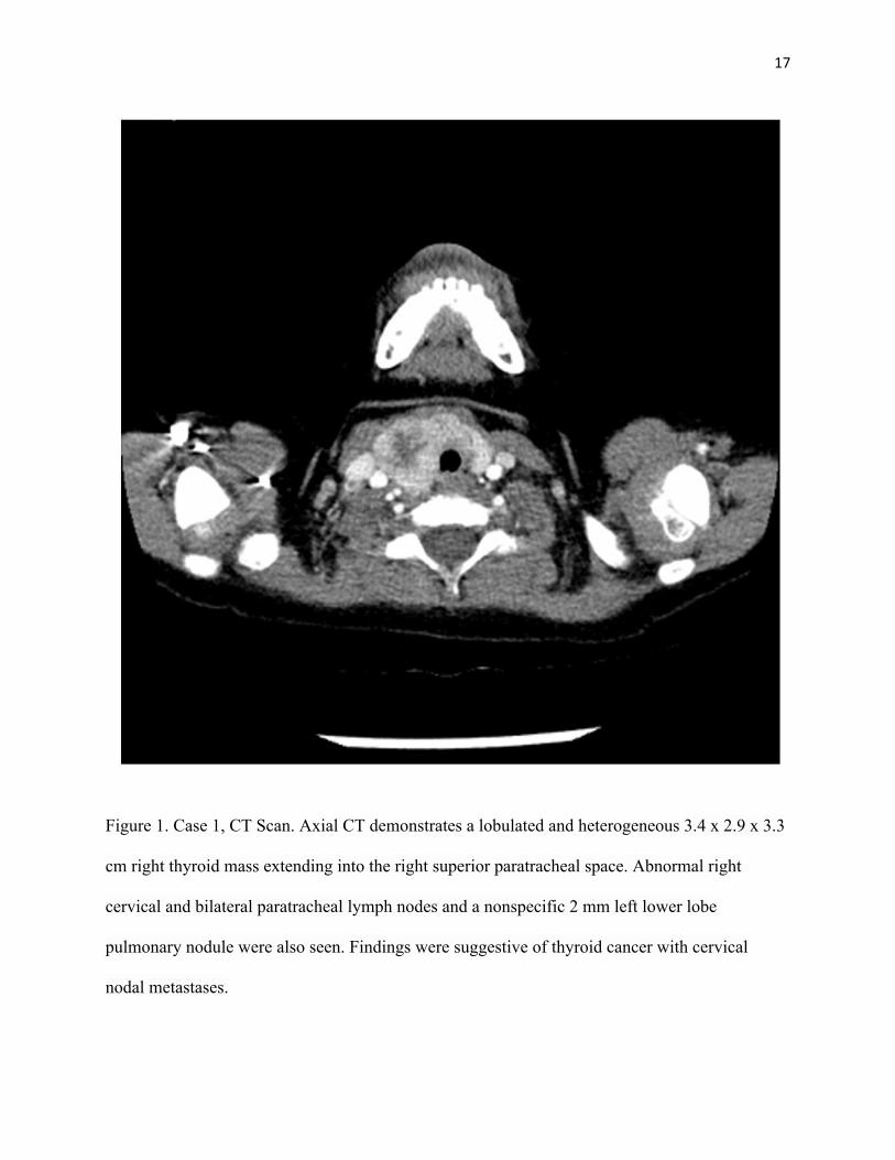
Post-therapy whole-body SPECT/CT imaging of the neck was taken the following week
did not find any distant metastatic disease (Figure 3). This year, a neck ultrasound found that the
patient was status post thyroidectomy without new or suspicious findings. The most recent
thyroglobulin lab-work taken (four months ago) revealed thyroglobulin levels of 0.1 ug/L (nearly
undetectable, reference range for intact thyroid: 2.8-40.9 ug/L and thyroglobulin antibodies of 1
IU/mL (within the normal range of < or = 1 IU/mL).
Case 3
Case 3 is a 59-year-old man with diabetes mellitus, hypertension, and end stage renal
disease on hemodialysis. One challenge with hemodialysis in the context of radioactive iodine
treatment is ensuring radiation protection for everyone present, as there is a risk of the
radioactive iodine contaminating the dialysis machine and increasing occupational exposures. In
addition to the contamination control and occupational exposure issues, there are concerns with
waste disposal, dosimetry, and patient release issues. Finally, since the elimination of the 131I is
largely through the dialysate, the administered dose may need to be modified from typical
protocol.

9
The patient was referred from outside of our medical center for treatment with
radioactive iodine after surgery for an 8 cm, pT3aNx, follicular thyroid cancer with capsular
vascular invasion. The patient underwent a whole-body scan with 123I. Anterior and posterior
images of the neck and entire body were taken 24 hours after oral administration of 2.59 mCi
(95.83 MBq) Na123I. No distant uptake was found to suggest metastasis, and the focal uptake
noted within the thyroid bed was consistent with residual thyroid tissue after recent
thyroidectomy.
The two major problems in patients with end stage renal disease on dialysis are
requirement of dose reduction due to negligible renal clearance of radioiodine and the radiation
protection during dialysis. The patient’s hemodialysis requirement presented challenges
(including radioiodine accumulation and radiation exposure) for radiation protection of the
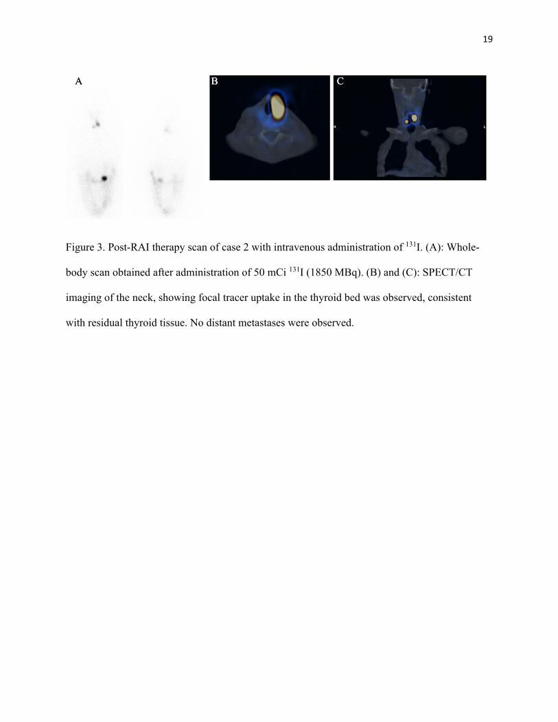
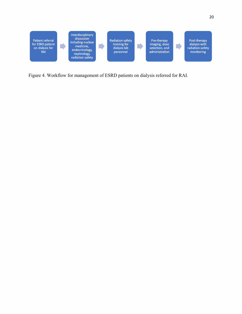
providers who would be caring for the patient during dialysis. Our workflow for managing this
case is illustrated on figure 4. Specifically, multidisciplinary meetings were held involving
radiation safety, nuclear medicine, endocrinology, and nephrology. The decision was made that
the radiation safety team would monitor all the personnel and equipment involved, check for
contamination of dialysis machines and dialysis ports, and hold the kits for decay as needed,
including the first 3-4 dialysis sessions after the 131I dose. Training materials for dialysis lab
personnel are included as a supplemental file. Additionally, all dialysis unit staff received
training from radiation safety personnel.
The following month, the patient received 29.8 mCi (1102.6 MBq) of Na131I as a pill for
the treatment of follicular thyroid cancer. The dose selection was lowered (following a
discussion among healthcare staff and consultation of the relevant literature) from an anticipated
10
dose of 50 mCi (1850 MBq) due to the use of dialysis, to reduce radiation dose to the marrow
secondary to the lowered clearance. The patient was admitted to our hospital for this procedure.
The patient was prepared for therapy by thyroid hormone withdrawal. The patient’s
outpatient dialysis record showed a normal session the day before the therapy without any
adverse events. Following collaboration between the nuclear medicine and radiation safety
teams, primary nephrologist, and outpatient nephrologist, the patient received dialysis the day
after the radioactive iodine therapy, with intermittent hemodialysis (IHD) for 3.5 hours planned
for 3 consecutive days following RAI. The patient tolerated the IHD well. Radiation safety
checked the dialysis machines and ports for contamination, and due to the effluent volume of 800
ml/minute, no contamination was detected. There were no complications nor radiation risks after
careful planning.
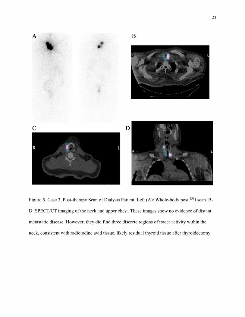
The post-therapy 131I scan supplemented by SPECT/CT imaging of the neck found no
evidence of distant metastatic disease (Figure 5). Most recently, a year and two weeks after the
radioactive iodine therapy, the patient underwent anterior and posterior imaging of the neck and
entire body without SPECT 24 hours after oral administration of 2.24 mCi (82.88 MBq) Na123I.
No abnormal radioiodine uptake was observed to suggest recurrent or metastatic disease.
Discussion
Case 1 described a patient unable to take pills due to developmental anomalies and young
age. At present, 131I pills are the standard medicinal preparation for RAI treatment of
hyperthyroidism and thyroid cancer. Bekier et al. concluded that administration of capsular 131I is
11
a safe formulation for treatment of thyroid disease, demonstrating that gastric radiation dose
from 131I pills was only high locally and was below the level that would cause tissue necrosis (7).
However, unique patient challenges, such as dysphagia, may limit or restrict safe administration
of oral 131I in pill form. The patient in case 1 and his family cited an inability to swallow pills,
and was consequently administered liquid oral 131I. No adverse events took place, and dose was
administered safely without exposure to technologist staff.
Previous studies concerning 131I therapy also describe administration difficulties and
alternative treatment protocols similar to those discussed here (Table 1). Aside from difficulty
swallowing pills, Aamri et al. reported pill-related instances of patient-caused radiation hazard,
pill adherence to the container, and technologist mishandling of the substance (8). Additionally,
Halpem et al. proposed the formation of iodine-gelatin complexes in the GI tract as a possible
mechanism for lower thyroidal uptake of capsular 131I relative to the liquid form (9). One
proposed solution involved endoscopically depositing the solid 131I pill into the stomach—
effectively minimizing risk of spillage, exposure, and incorrect administration (10). However,
endoscopy is invasive and carries risks for the patient.
Rini et al. commented on the more-than-intended irradiative impact of encapsulated 131I –
particularly when used as a diagnostic tracer – compared to liquid-form 131I (11). Using a pill-
form 131I tracer, this group observed a mean diagnostic 131I uptake 14% lower than the
corresponding therapeutic 131I uptake (44% versus 58%), reflecting the increased uptake when a
pill is delivered with a therapeutic dose than a diagnostic dose. They concluded that in
hyperthyroid patients treated with 131I liquid therapy compared to encapsulated 131I, uptake of
diagnostic doses of 131I liquid better predicts uptake of therapeutic doses of 131I liquid (11). It is
therefore recommended that health teams familiarize themselves with liquid and intravenous
12
(IV) 131I administrative safety and technique. Overall, while administration of 131I in a pill form
is used as the first option, it is important to remember that liquid administration remains a safe,
and effective alternative, provided appropriate radiation safety precautions are followed (3,12).
Unlike Case 1, Case 2 did not have dysphagia, and could swallow, but he had significant
gastrointestinal dysmotility which often manifested as episodes of pseudoobstruction with
gastroparesis. Therefore, the patient was largely dependent on total parental nutrition. Thus, the
decision to use IV treatment was due to severe gastric motility and malabsorption precluding oral
or gastrotomy tube treatment, and was made after months of ongoing discussion. Advantages of
IV treatment relative to oral 131I therapy include enhanced diagnostic accuracy, rapidity, and the
ability to treat patients with impaired gastro-intestinal absorption (13). The risks associated with
IV treatment include the liquid iodine’s volatility which makes it more dangerous to handle and
increases the risk of major spills and exposure to technologists. Meticulous care was undertaken
and no spill or other adverse event took place.
Patients are more prone to experience nausea as 131I dosage increases (14,15).
Additionally, patients demonstrating multiple disease processes including gastroesophageal
reflux disease, gastroparesis, gastric outlet obstruction, and other similar conditions are at
increased risk of vomiting (15). Overall, this case demonstrates that intravenous administration
of 131I is a safe and effective alternative in cases where great difficulty with oral administration
and gastrointestinal absorption is anticipated.
Case 3 evidences an instance of end stage renal disease complicating radioactive iodine
treatment for follicular thyroid cancer. In patients with normal renal function, the 131I would be
excreted through the urinary system; thus, impaired renal function complicates iodine clearance
and theoretically potentiates blood radioactivity risks. Although the dialysis machine would
13
likely compensate to some extent for this decreased clearance, hemodialysis patients are still
impacted by decreased clearance of 131I (16). In addition, there are risks of dialysis staff and
machinery contamination (16). In fact, the main challenge with this patient was the radioactive
dialysate waste, and managing staff exposure and training. Contingency plans are necessary for
addressing fluid spills during dialysis (17). Mercutt et al. supplied an illustrated schematic and
protocol for safe hemodialysis of patient in need of ablative 131I RAI therapy, much of which
mirrors the methods described here (18). Additionally, under dosimetry guidance, high-dose IV
treatment of differentiated thyroid cancer patients is safe and may also reduce radiation dose
(19). To limit risks of bone marrow toxicity and further renal insult, therapy doses should not
exceed the prescribed amount in any patient, including those on dialysis, as Magné et al. have
recommended. In their dialysis patient case report, a radioiodine dosage no more than 125% of
the currently prescribed concentration (20). Table 2 describes other successful oral 131I
administration cases for patients with end-stage renal disease on hemodialysis.
Prior studies have suggested that dialysis patients have increased systemic retention of
131I, and therefore have suggested reductions in the administered 131I dose, although no uniform
consensus exists. Citing rapid iodine clearance, Betcher et al. concluded that dialysis reduces
effective radiation dose and necessitates larger 131I treatment doses to achieve equivalent
outcomes of patients with normal renal function (21). Jimenez et al. administered the same doses
of 131I to hemodialysis patients that patients with normal renal function receive. In their small
cohort (n=3), this therapeutic protocol provided 131I dosages and avoided patient radiation
overexposure (22). Alevizaki et al. utilized a 40-50% activity reduction of 131I , finding that none
of their patients experienced short-term side effects or presented with detectable thyroglobulin
levels on their first post therapy evaluation (n=5, inclusion criteria = end stage renal disease)

14
(23). Vermandel et al. concluded that an approximately 30% reduction from nominal 131I dosage
struck the best balance between hematologic toxicity and treatment efficacy (n=6, inclusion
criteria = ESRD patients undergoing hemodialysis) (24). Following their lead, we chose to
reduce the dosage despite some of the literature recommending no change or even increased
doses; our case involved a 40% reduction from our typical 50 mCi (1850 MBq) to 30 mCi (1110
MBq) to minimize radiation exposure to marrow secondary to reduced clearance of 131I.
Conclusion
In summary, 131I is a common treatment for hyperthyroidism and thyroid cancer, and
most patients may be treated using standardized protocols as defined by The Society of Nuclear
Medicine and Molecular Imaging (12). We have presented three cases which presented technical
difficulties for nuclear medicine and radiation safety staff. All three cases included patients that
received alternatives protocols due to their specific clinical status/challenges (dysphagia in Case
1; severe gastric dysmotility in Case 2; End stage renal disease on hemodialysis in Case 3), and
all cases reported no further disease progression since 131I treatment. These shared cases should
be of particular use to nuclear medicine technologists and physicians presented with clinically
similar challenging cases. Through this case series of patient-specific applications of 131I RAI
therapy, we hope to inform providers of alternatives to standard RAI treatment protocols, and the
importance of provider collaboration and treatment customization to overcome patient-specific
challenges.
Financial disclosure: None.
Disclaimers: None.
Acknowledgements: None.

15
References
1. Hertz S and Roberts A. Radioactive Iodine In The Study Of Thyroid Physiology. JAMA. 1946;131:81.
2. Ross DS, Burch HB, Cooper DS, et al. 2016 American Thyroid Association Guidelines for Diagnosis and Management of Hyperthyroidism and Other Causes of Thyrotoxicosis. Thyroid. 2016;26:1343–1421.
3. Bahn R, Burch H, Cooper D, et al. Hyperthyroidism and other Causes of Thyrotoxicosis: Management Guidelines of the American Thyroid Association and American Association of Clinical Endocrinoloigists. Endocrine Practice. 2011;17:456–520.
4. Fred A Mettler and Milton J Guiberteau, Essentials of Nuclear Medicine Imaging, 6th edition, 2012.
5. Wyszomirska A. Iodine-131 for therapy of thyroid diseases. Physical and biological basis. Nucl Med Rev Cent East Eur. 2012;15(2):120‐123. Published 2012 Aug 28.
6. Lim H, Devesa SS, Sosa JA, Check D, Kitahara CM. Trends in Thyroid Cancer Incidence and Mortality in the United States, 1974-2013. JAMA. 2017;317:1338.
7. Bekier A et al. 131-Iodine capsules in thyroid therapy: An individually controlled study of their uptake kinetics as compared to liquid 131-Iodine. Eur J Nucl Med. 1985;10;25-28.
8. Al Aamri M, Ravichandran R, Binukumar JP, Al Balushi N. Therapeutic applications of radioactive 131-iodine: Procedures and incidents with capsules. Indian journal of nuclear medicine: IJNM. 2016;31(3):176-8.
9. Halpem S, Alazraki N, Littenberg R, Hurwitz S, Green J, Kunsa J, Ashburn W. 131I Thyroid Uptakes: Capsule Versus Liquid. Journal of Nuclear Medicine. 1973;14(7);507-510.
10. Shields A., Johnson J. Endoscopic administration of capsular therapeutic iodine-131 in patients unable to swallow: A novel technique. JMNT. 2011;1353.
11. Rini JN, Vallabhajosula S, Zanzonico P, Hurley JR, Becker DV, Goldsmith SJ. Thyroid uptake of liquid versus capsule 131I tracers in hyperthyroid patients treated with liquid 131I. Thyroid. 1999;9(4):347-52.
12. Silberstein EB, Alavi A, Balon HR, et al. The SNMMI practice guideline for therapy of thyroid disease with 131I 3.0. J Nucl Med. 2012;53(10):1633-1651.
13. Kriss JP. Uptake of Radioactive Iodine After Intravenous Administration of Tracer Doses. J Clin Endocrinol Metab. 1951;11(3);289–297.
14. Honour AJ, Myant NB, Rowlands EN. Secretion of 131I in digestive juices and milk in man. Clin Sci. 1952;11;449-62.
15. Wartofsky L, van Nostrand D. Thyroid Cancer: A Comprehensive Guide to Clinical Management. 2016;692-3.
16. Bhat M, Mozzor M, Chugh S, Buddharaju V, Schwarcz M, Valiquette G. Dosing of radioactive iodine in end-stage renal disease patient with thyroid cancer. Endocrinol Diabetes Metab Case Rep. 2017;2017:17-0111.
17. Homer L, Smith AH. Radiation protection issues of treating hyperthyroidism with 131I in patients on haemodialysis. Nucl Med Commun . 2002;23;261-264.
16
18. Murcutt G, Edwards J, Boakye J, Davenport A. Hemodialysis of chronic kidney failure patients requiring ablative radioiodine therapy. Kidney Int. 2008; 73(11):1316-1319.
19. Gulec SA et al. Dosimetry-Guided Radioactive Iodine Treatment in Patients with Metastatic Differentiated Thyroid Cancer: Largest Safe Dose Using a Risk-Adapted Approach. J Nucl Med. 2003;44;451–456.
20. Magné N, Magné J, Bracco J, Bussière F. Disposition of Radioiodine 131I Therapy for Thyroid Carcinoma in a Patient with Severely Impaired Renal Function on Chronic Dialysis: a Case Report. JJCO. 2002; 32(6):202–205.
21. Betcher KB et al. 131I Treatment of Thyroid Papillary Carcinoma in a Patient With Renal Failure. Cancer. 1990;66;2509-2513.
22. Jimenez RA et al. Iodine-131 Treatment of Thyroid Papillary Carcinoma in Patients Undergoing Dialysis for Chronic Renal Failure: A Dosimetric Method. Thyroid. 2001;11(11);1031-1034.
23. Alevizaki et al. Iodine 131 treatment for differentiated thyroid carcinoma in patients with end stage renal failure: dosimetric, radiation safety, and practical considerations. Hormones. 2006;5(4);276-287.
24. Vermandel M., Debruyne P, Beron A, et al. Management of patients with renal failure undergoing dialysis during 131I therapy for thyroid cancer. J Nucl Med. 2020;119.232017.
17
Figure 1. Case 1, CT Scan. Axial CT demonstrates a lobulated and heterogeneous 3.4 x 2.9 x 3.3
cm right thyroid mass extending into the right superior paratracheal space. Abnormal right
cervical and bilateral paratracheal lymph nodes and a nonspecific 2 mm left lower lobe
pulmonary nodule were also seen. Findings were suggestive of thyroid cancer with cervical
nodal metastases.

18
Figure 2. Case 1, Post-therapy Scan of patient 1. Left (A): Whole body scan obtained after
administration of 75 mCi 131I (2775 MBq). Right (B): SPECT imaging of the neck. These images
are consistent with residual thyroid tissue, as focal tracer uptake was noted within the thyroid
resection bed.
19
Figure 3. Post-RAI therapy scan of case 2 with intravenous administration of 131I. (A): Whole-
body scan obtained after administration of 50 mCi 131I (1850 MBq). (B) and (C): SPECT/CT
imaging of the neck, showing focal tracer uptake in the thyroid bed was observed, consistent
with residual thyroid tissue. No distant metastases were observed.
20
Figure 4. Workflow for management of ESRD patients on dialysis referred for RAI.
21
Figure 5. Case 3, Post-therapy Scan of Dialysis Patient. Left (A): Whole-body post 131I scan. B-
D: SPECT/CT imaging of the neck and upper chest. These images show no evidence of distant
metastatic disease. However, they did find three discrete regions of tracer activity within the
neck, consistent with radioiodine avid tissue, likely residual thyroid tissue after thyroidectomy.
22
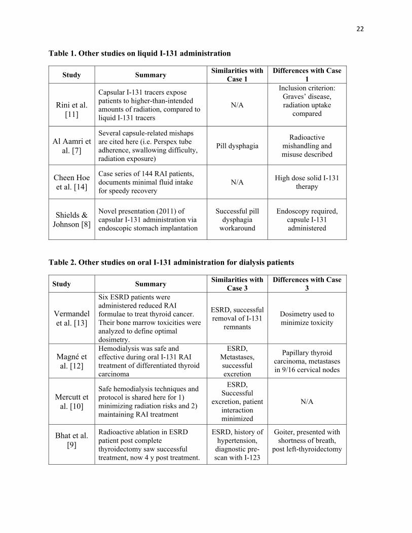
Table 1. Other studies on liquid I-131 administration
Study Summary Similarities with
Case 1 Differences with Case
1
Rini et al.
[11]
Capsular I-131 tracers expose patients to higher-than-intended amounts of radiation, compared to liquid I-131 tracers
N/A
Inclusion criterion: Graves’ disease, radiation uptake
compared
Al Aamri et al. [7]
Several capsule-related mishaps are cited here (i.e. Perspex tube adherence, swallowing difficulty, radiation exposure)
Pill dysphagia Radioactive
mishandling and misuse described
Cheen Hoe et al. [14]
Case series of 144 RAI patients, documents minimal fluid intake for speedy recovery
N/A High dose solid I-131
therapy
Shields & Johnson [8]
Novel presentation (2011) of capsular I-131 administration via endoscopic stomach implantation
Successful pill dysphagia
workaround
Endoscopy required, capsule I-131 administered
Table 2. Other studies on oral I-131 administration for dialysis patients
Study Summary Similarities with
Case 3 Differences with Case
3
Vermandel et al. [13]
Six ESRD patients were administered reduced RAI formulae to treat thyroid cancer. Their bone marrow toxicities were analyzed to define optimal dosimetry.
ESRD, successful removal of I-131
remnants
Dosimetry used to minimize toxicity
Magné et al. [12]
Hemodialysis was safe and effective during oral I-131 RAI treatment of differentiated thyroid carcinoma
ESRD, Metastases, successful excretion
Papillary thyroid carcinoma, metastases in 9/16 cervical nodes
Mercutt et al. [10]
Safe hemodialysis techniques and protocol is shared here for 1) minimizing radiation risks and 2) maintaining RAI treatment
ESRD, Successful
excretion, patient interaction minimized
N/A
Bhat et al. [9]
Radioactive ablation in ESRD patient post complete thyroidectomy saw successful treatment, now 4 y post treatment.
ESRD, history of hypertension,
diagnostic pre-scan with I-123
Goiter, presented with shortness of breath,
post left-thyroidectomy