Embed Size (px)
Citation preview
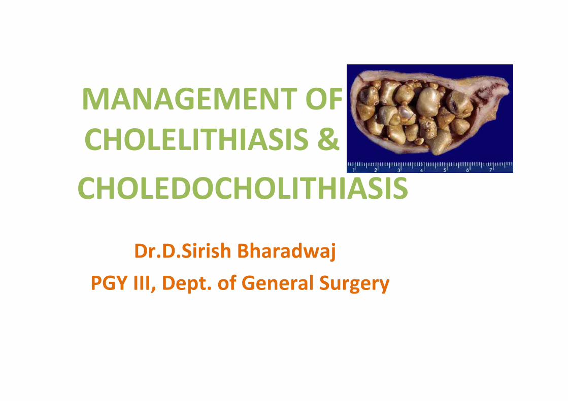
MANAGEMENT OF CHOLELITHIASIS &
CHOLEDOCHOLITHIASIS
Dr.D.Sirish Bharadwaj PGY III, Dept. of General Surgery
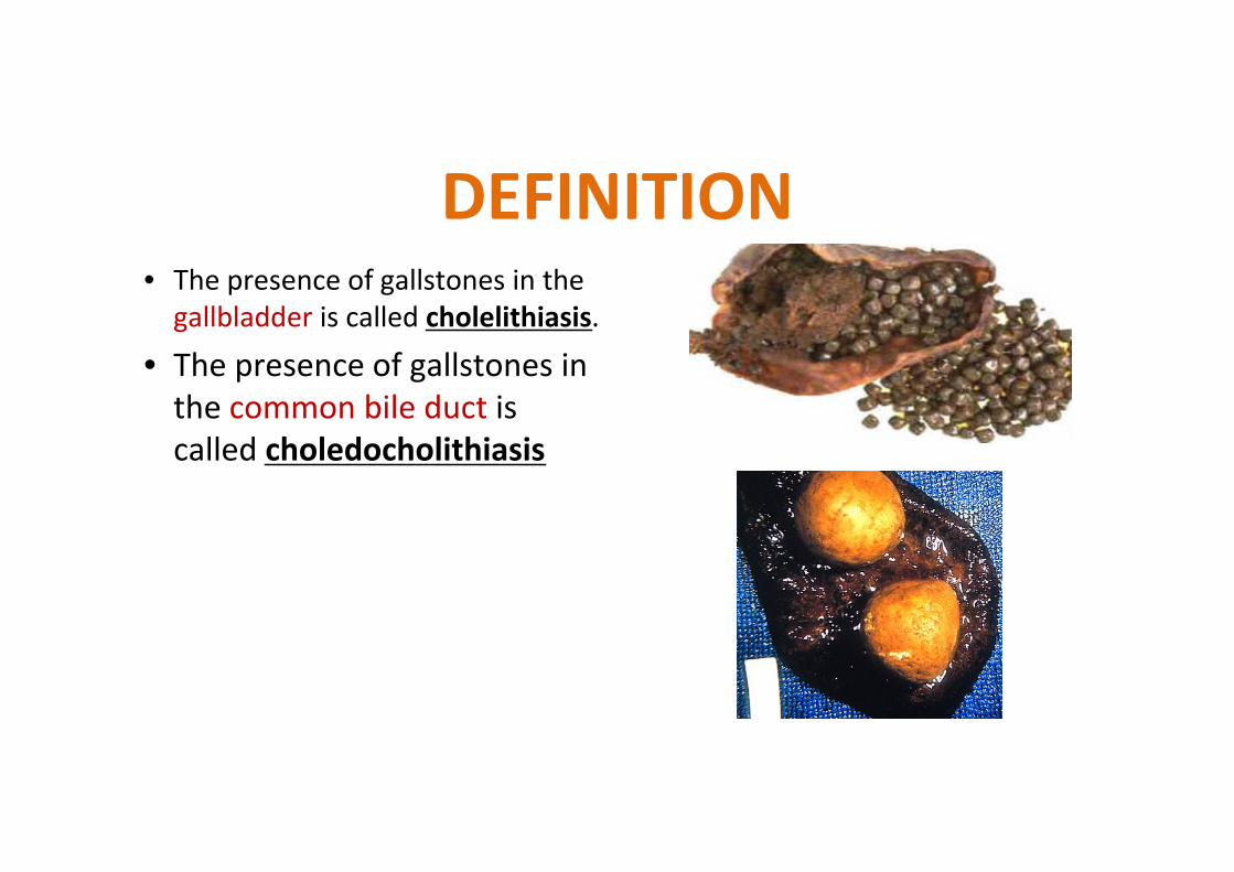
• The presence of gallstones in the gallbladder is called cholelithiasis.
• The presence of gallstones in the common bile duct is called choledocholithiasis
DEFINITION
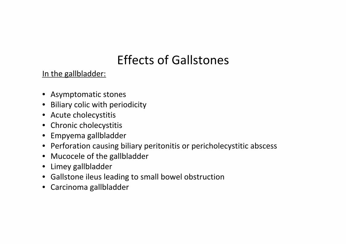
Effects of GallstonesIn the gallbladder:
• Asymptomatic stones• Biliary colic with periodicity • Acute cholecystitis• Chronic cholecystitis• Empyema gallbladder• Perforation causing biliary peritonitis or pericholecystitic abscess• Mucocele of the gallbladder• Limey gallbladder• Gallstone ileus leading to small bowel obstruction• Carcinoma gallbladder
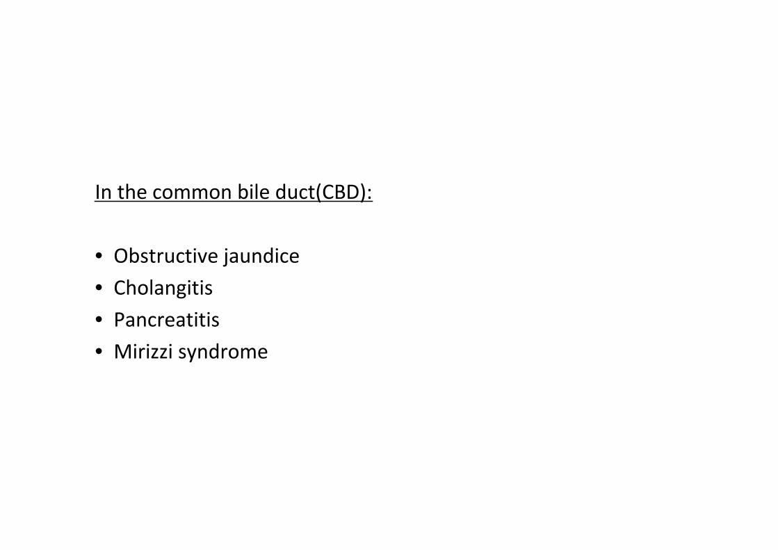
In the common bile duct(CBD):
• Obstructive jaundice• Cholangitis• Pancreatitis• Mirizzi syndrome
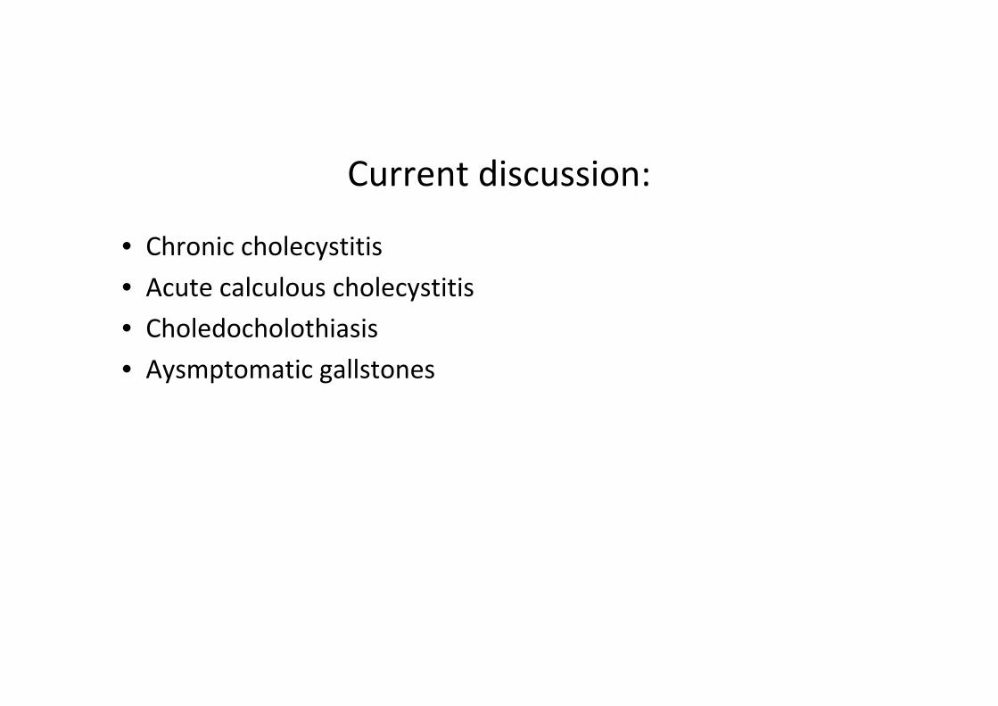
Current discussion:
• Chronic cholecystitis• Acute calculous cholecystitis• Choledocholothiasis• Aysmptomatic gallstones
CLINICAL PRESENTATION
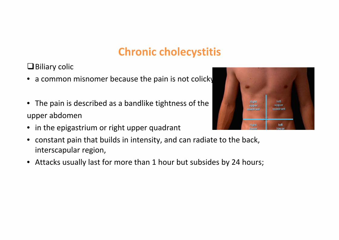
Chronic cholecystitisBiliary colic • a common misnomer because the pain is not colicky
• The pain is described as a bandlike tightness of the upper abdomen • in the epigastrium or right upper quadrant • constant pain that builds in intensity, and can radiate to the back, interscapular region,
• Attacks usually last for more than 1 hour but subsides by 24 hours;
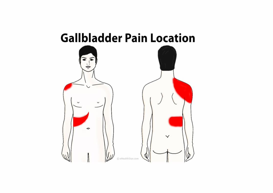
may be associated with nausea and vomiting. Bloating and belching – 50%Intolerance to fatty mealsFlatulent dyspepsiaThe physical examination usually normal if they are pain‐free.During an episode of biliary colic, mild right upper quadrant tenderness may be present.
Acute calculous cholecystitis• Right upper quadrant pain, similar in severity but much longer in duration than pain from previous episodes of biliary colic
• fever, nausea, and vomiting. On physical exam, • right upper quadrant tenderness and guarding are usually present inferior to the right costal margin, distinguishing the episode from simple biliary colic.
• When inflammation spreads to the peritoneum, patients develop more diffuse tenderness, guarding and rigidity.
• A mass, the gallbladder and adherent omentum, is occasionally palpable,
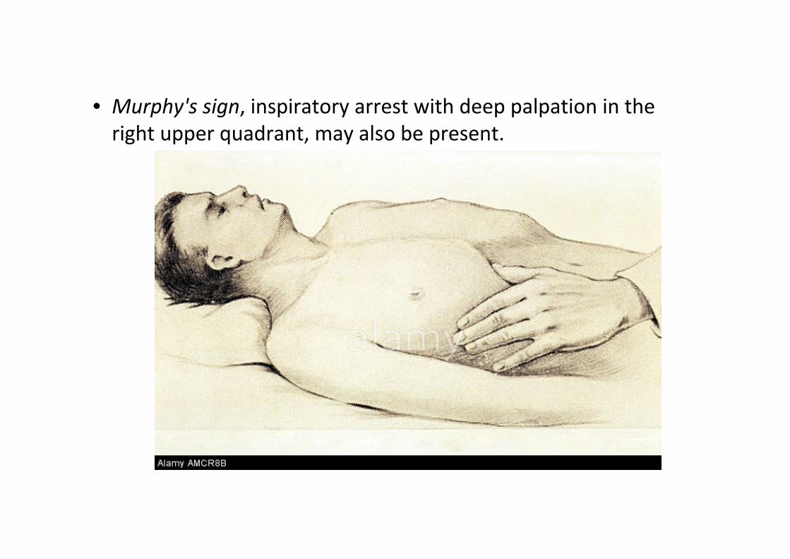
• Murphy's sign, inspiratory arrest with deep palpation in the right upper quadrant, may also be present.

• A mild leukocytosis is usually present (12,000‐14,000 cells/mm3).
• mild elevations in: serum bilirubin (>4 mg/dL), alkaline phosphatase, transaminases, amylase may be present.

CholedocholithiasisCommon bile duct stones may be silent and are often discovered incidentally.
• In these patients, biliary obstruction is transient, and laboratory tests may be normal.
Clinical features suspicious for biliary obstruction(obstructive/surgical jaundice) due to common bile duct stones:
• biliary colic, • jaundice, • pruritis• Pale/clay coloured stools, and • dark of the urine.
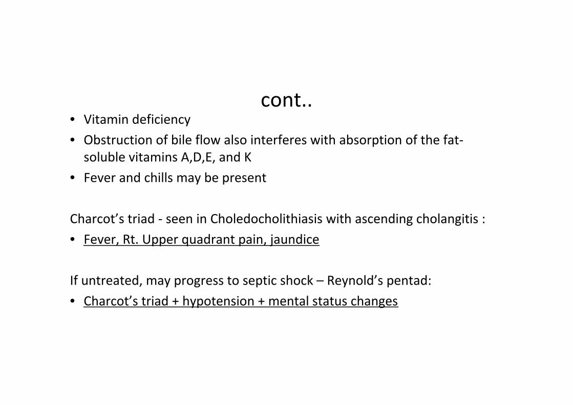
cont..• Vitamin deficiency• Obstruction of bile flow also interferes with absorption of the fat‐soluble vitamins A,D,E, and K
• Fever and chills may be present
Charcot’s triad ‐ seen in Choledocholithiasis with ascending cholangitis :• Fever, Rt. Upper quadrant pain, jaundice
If untreated, may progress to septic shock – Reynold’s pentad:• Charcot’s triad + hypotension + mental status changes
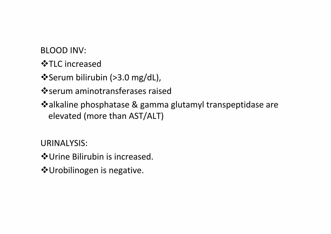
BLOOD INV:TLC increasedSerum bilirubin (>3.0 mg/dL), serum aminotransferases raised alkaline phosphatase & gamma glutamyl transpeptidase are elevated (more than AST/ALT)
URINALYSIS:Urine Bilirubin is increased.Urobilinogen is negative.

Aysmptomatic gallstones
• 10% of males and 20% of females• Gallstones detected incidentally while performing USG abdomen for some other reasons.
MANAGEMENT

INVESTIGATIONS
• Laboratory investigations– LFT’s– Hepatitis B&C viral serology– Urine
• Imaging– Non‐invasive/ minimally invasive– Invasive
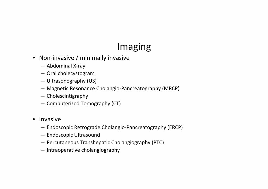
Imaging • Non‐invasive / minimally invasive
– Abdominal X‐ray– Oral cholecystogram– Ultrasonography (US)– Magnetic Resonance Cholangio‐Pancreatography (MRCP)– Cholescintigraphy– Computerized Tomography (CT)
• Invasive– Endoscopic Retrograde Cholangio‐Pancreatography (ERCP) – Endoscopic Ultrasound– Percutaneous Transhepatic Cholangiography (PTC)– Intraoperative cholangiography

X‐ray abdomen
• X‐rays: 15% stones are radiopaque,• May show Mercedes‐Benz sign
• porcelain GB may be seen. • Air in biliary tree(Pneumobilia)• emphysematous GB wall.
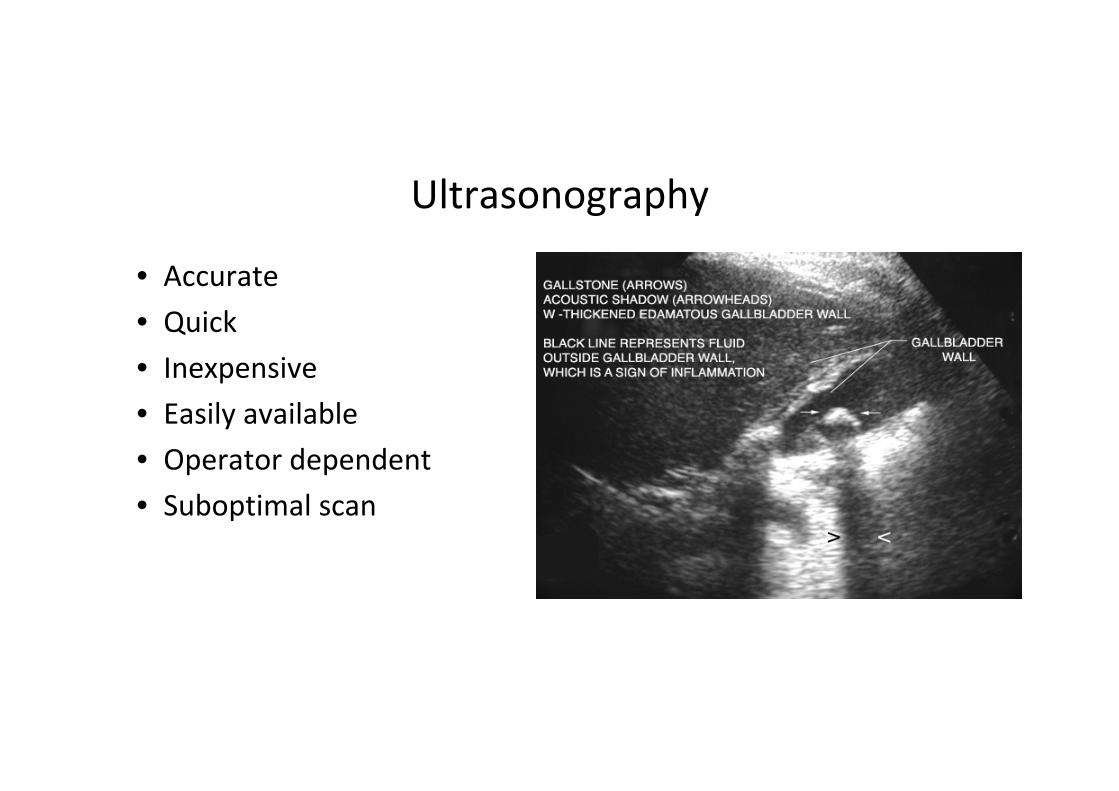
Ultrasonography
• Accurate• Quick• Inexpensive• Easily available• Operator dependent• Suboptimal scan
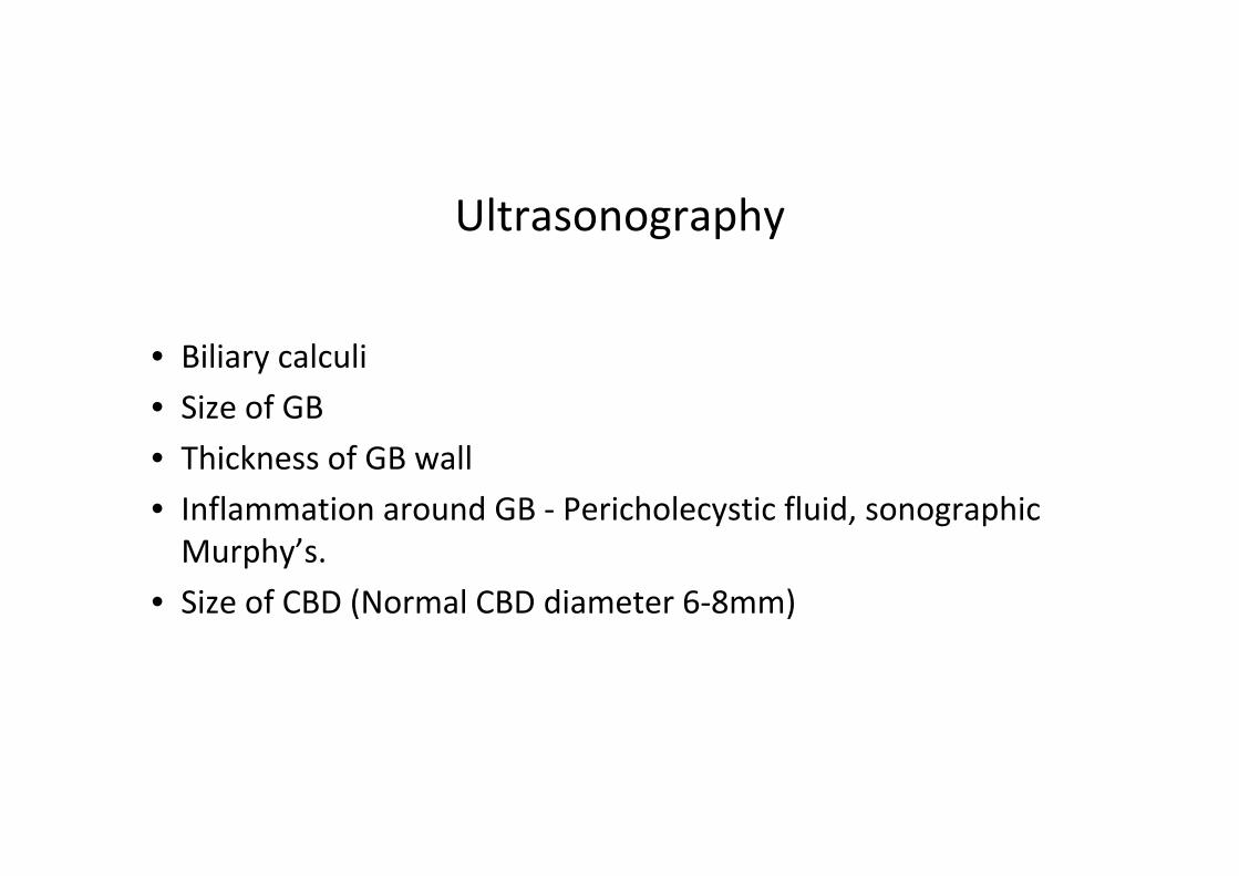
Ultrasonography
• Biliary calculi• Size of GB• Thickness of GB wall• Inflammation around GB ‐ Pericholecystic fluid, sonographic Murphy’s.
• Size of CBD (Normal CBD diameter 6‐8mm)

Ultrasonography
• Intra and extra‐hepatic biliary dilatation and level of obstruction.
• 70‐95 % sensitive.• 80‐100 % specific.

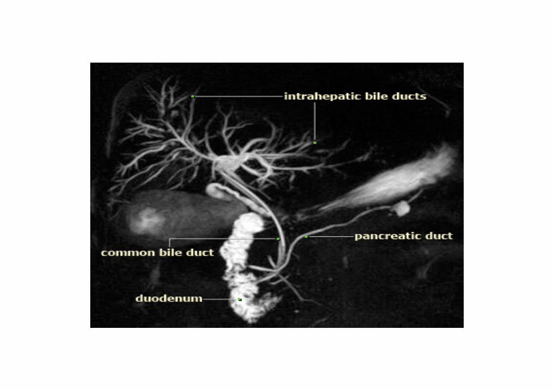
MRCP
• Non‐invasive• Contrast not required.• Demonstrates
– Ductal obstruction – Visualizes stones– Strictures– Other intra & extra ductal abnormalities

Cholescintigraphy• Technitium 99 (Tc99‐IDA chelate complex).• HIDA/ PIPIDA/ DISIDA scan.• Gallbladder visualized within 30min to 1 hour in absence of disease.
• Not visualized in Acute cholecystitis• 97 % sensitive and 94 % specific.• Diagnose obstruction.

HIDA•Bile leaks (detect and quantify).
•CBD obstruction appears as non visualization of small intestine.
•No external radiation exposure to the patient.
•Less helpful when the patient is fasting for more than 5 days, with a 40% false‐positive rate.
CT Scan
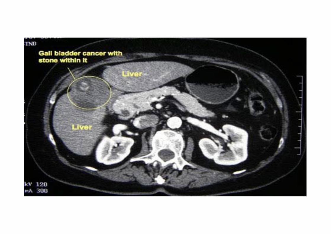
• Not useful in benign biliary disease.
• Useful when Carcinoma gallbladder is suspected
• Gall stones often not visualized.
• Cholecystitis is underdiagnosed.
• Higher dose of radiation.
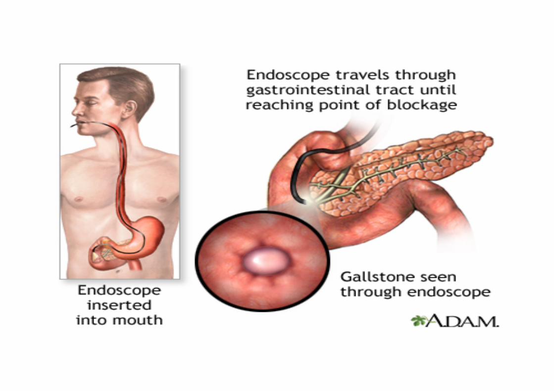
Endoscopic UltrasoundProcedure:• USG probe passed through an upper GI endoscope and kept in pylorus/duodenum area
• High frequency used ‐ 20‐40Mhz
Evaluates Pancreato‐biliary system.Detection of microlithiasisCholedocholithiasis Evaluation of benign and malignant strictures.Detects regional lymphnodesRelationship to vascular structures.
Endoscopic Ultrasound
Advantages:• High resolution imaging.• Less invasive.• No exposure to radiation.• Aspiration of a cyst or FNAC.
Disadvantages:• Higher operator dependency.• Cost and availability.• Visualization is limited to 8 cm.
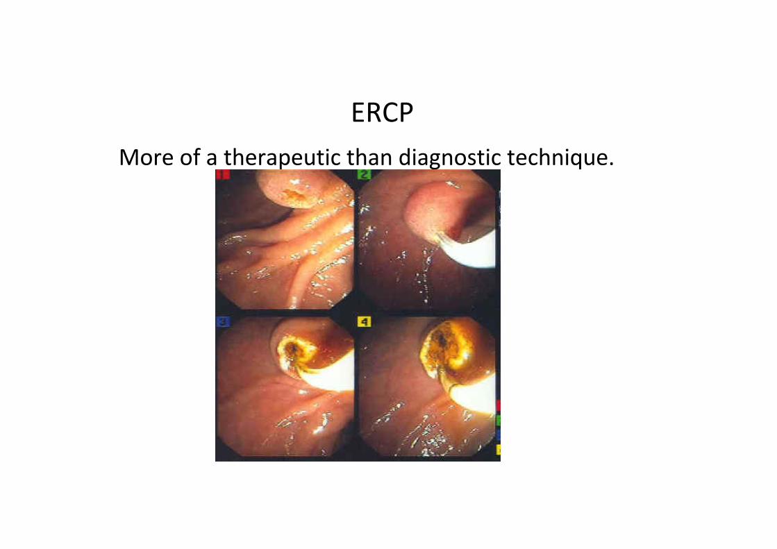
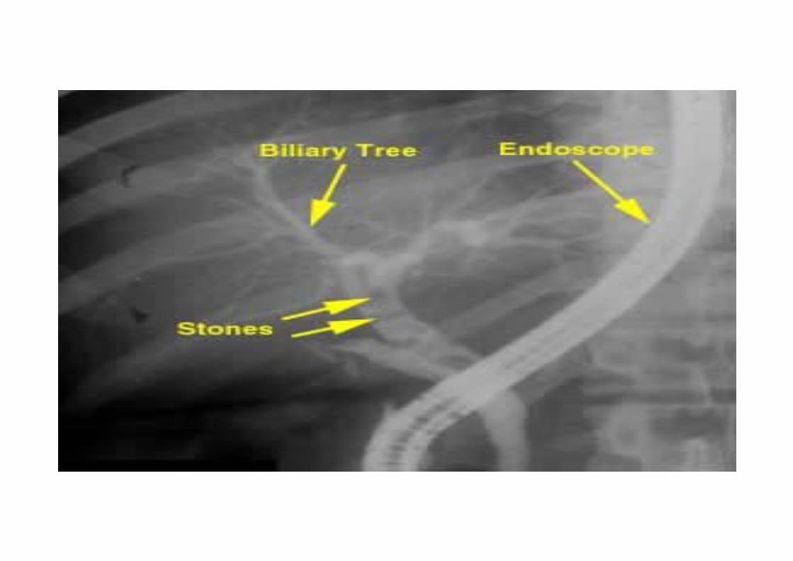
ERCPMore of a therapeutic than diagnostic technique.
ERCP – Diagnostic
• Gold standard of imaging for biliary tree.• Detects stones or malignant strictures• Identifies the cause and level of obstruction


Percutaneous Transhepatic Cholangiography (PTC)
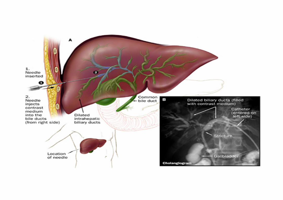
Percutaneous Transhepatic Cholangiography (PTC)• More of a palliative technique.• Bile ducts are cannulated directly.• Demonstrates areas of stricture/obstruction.• Effective in pts with a dilated biliary ductal system
Indications:• When ERCP fails or is not possible.• Stenting for biliary drainage.• Prior to biliary drainage procedure.
Contraindications:• bleeding tendency, • Unfit for surgery, • Hydatid Cysts, • Ascites, CLD (chronic liver disease)
TREATMENT
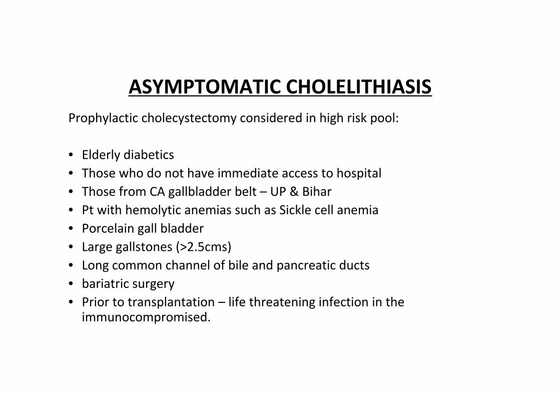
ASYMPTOMATIC CHOLELITHIASISProphylactic cholecystectomy considered in high risk pool:
• Elderly diabetics• Those who do not have immediate access to hospital• Those from CA gallbladder belt – UP & Bihar • Pt with hemolytic anemias such as Sickle cell anemia • Porcelain gall bladder• Large gallstones (>2.5cms)• Long common channel of bile and pancreatic ducts• bariatric surgery• Prior to transplantation – life threatening infection in the immunocompromised.
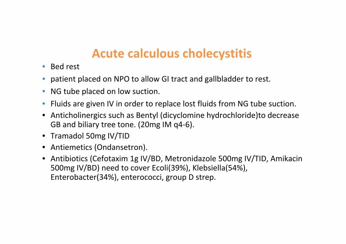
Acute calculous cholecystitis• Bed rest• patient placed on NPO to allow GI tract and gallbladder to rest.• NG tube placed on low suction.• Fluids are given IV in order to replace lost fluids from NG tube suction.• Anticholinergics such as Bentyl (dicyclomine hydrochloride)to decrease GB and biliary tree tone. (20mg IM q4‐6).
• Tramadol 50mg IV/TID• Antiemetics (Ondansetron).• Antibiotics (Cefotaxim 1g IV/BD, Metronidazole 500mg IV/TID, Amikacin 500mg IV/BD) need to cover Ecoli(39%), Klebsiella(54%), Enterobacter(34%), enterococci, group D strep.
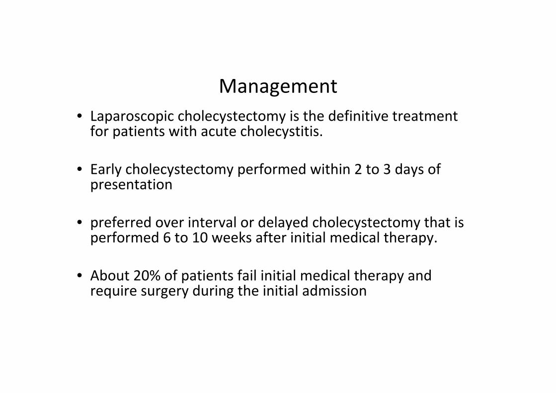
Management• Laparoscopic cholecystectomy is the definitive treatment for patients with acute cholecystitis.
• Early cholecystectomy performed within 2 to 3 days of presentation
• preferred over interval or delayed cholecystectomy that is performed 6 to 10 weeks after initial medical therapy.
• About 20% of patients fail initial medical therapy and require surgery during the initial admission

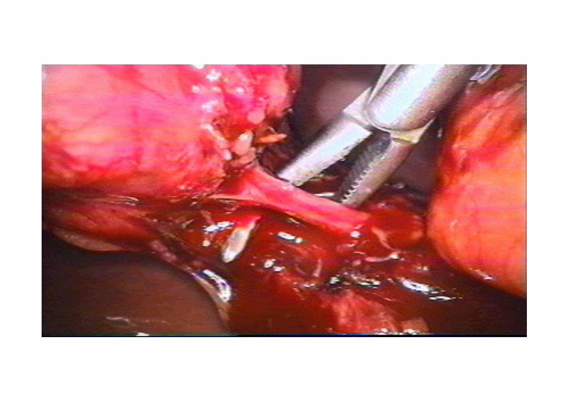
• Occasionally, the inflammatory process obscures the structures in the triangle of Calot, precluding safe dissection and ligation of the cystic duct.
• In these patients, partial cholecystectomy, cauterization of the remaining gallbladder mucosa &
drainage avoid injury to the common bile duct.
• In patients considered too unstable to tolerate a laparotomy, percutaneous cholecystostomy under local anesthesia can be performed to drain the gallbladder.
• This procedure leaves the gallbladder in place, which may be a source of ongoing sepsis.
• Drainage and IV antibiotics, followed by interval laparoscopic cholecystectomy, can then be performed after 3 to 6 months to allow the patient to recover and the acute inflammation to resolve.

Chronic Cholecystitis• Observation & dietary/lifestyle changes for pts with very mild symptoms
• Elective laparoscopic cholecystectomy with CBD exploration in pts with severe/recurrent symptoms
• Diabetic patients should have a cholecystectomy promptly because they are at higher risk for acute cholecystitis or even gangrenous cholecystitis.
• Pregnant women with symptomatic gallstones who fail expectant management with dietary modification can safely undergo surgery during the second trimester
SUPPORTIVE OR DIETARY MANAGEMENT
• Low fat diet • Powdered supplements high in protein and carbohydrates• Cooked fruits• Rice or tapioca• Lean meats• Smashed potatoes• Non gas forming vegetables
The following to be avoided
• Eggs • Cream • Pork • Fried foods, cheese and rich dressings• Gas forming vegetables ‐ Legumes• Alcohol
Non operative treatment: For pts where Lap chole contraindicated Generally unsuccessful and used rarely
• Dissolution with oral bile salt therapy (Ursodeoxycholic acid, Chenodeoxycholic acid)• Contact dissolution – cannulation of GB & infusion of organic solvent ‐ MTBE• ESWL ‐ generally combined with oral dissolution treatment to help dissolve the
fragmented pieces of the original gallstone.• Intracorporeal lithotripsy
•For solitary stones that are less than 2 centimeters in diameter.
•The patient sits in a tub of water.
• High‐energy, ultrasound shock waves are directed through the abdominal wall toward the stones.
•The shock waves travel through the soft tissues of the body and break up the stones.
•The stone fragments are then usually small enough to be passed through the bile duct and into the intestines.
Extracorporeal shock wave lithotripsy (ESWL)
CHOLEDOCHOLITHIASIS
Treatment:
ERCP sphincterotomy with a balloon sweep and extraction of the stone followed by Laparoscopic cholecystectomy in the same admission.
ERCP ‐ Therapeutic
Indications:
if expertise in laparoscopic common bile duct exploration is not available. worsening cholangitis, ampullary stone impaction, biliary pancreatitis, multiple comorbidities, and cirrhosis

Various applications:
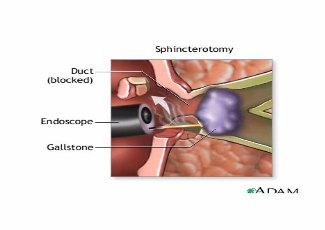
• Endoscopic sphincterotomy/papillotomy
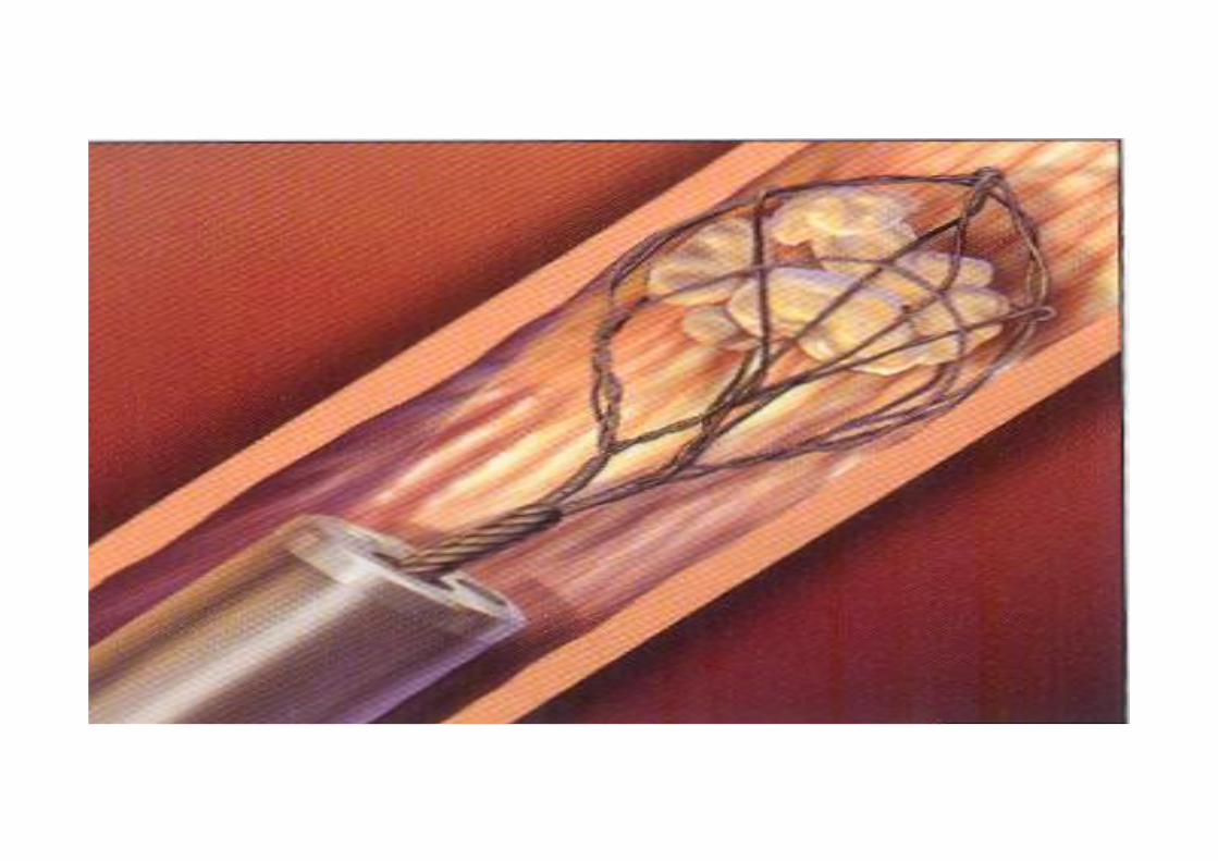
• Removal of stones
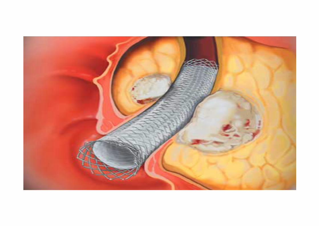
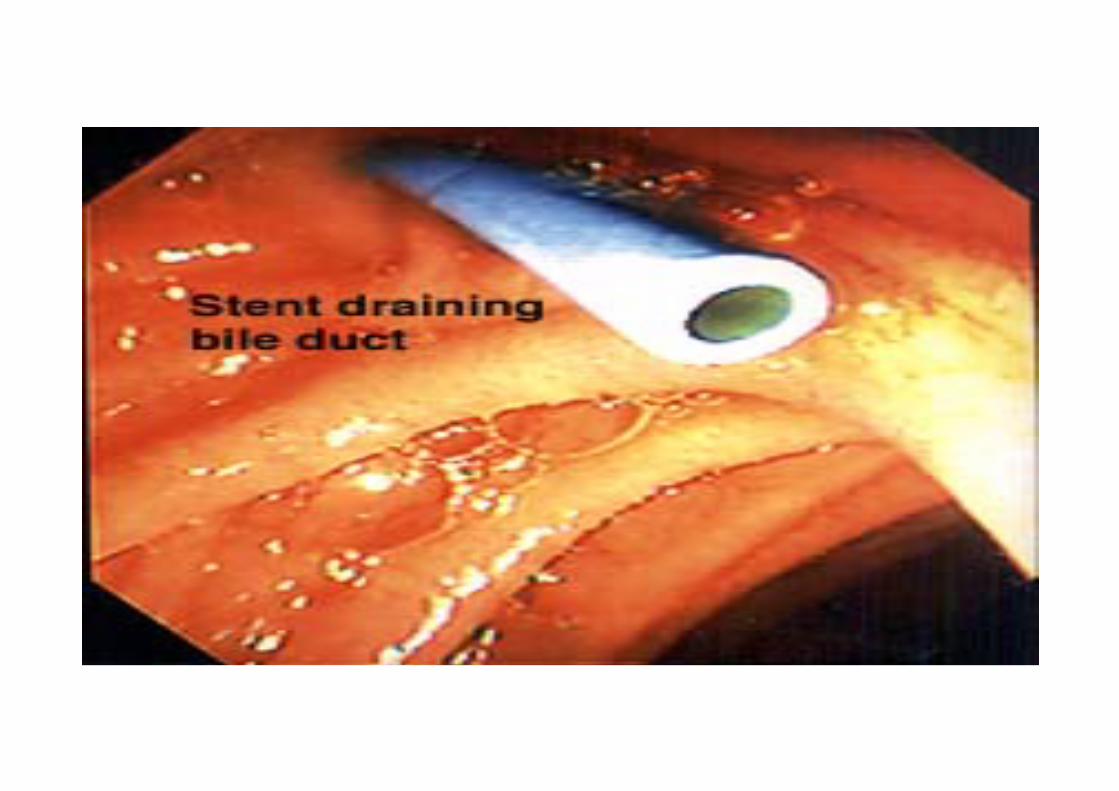
• Insertion of stents
• Dilation of strictures
• Extraction of worms
Contra‐indications of ERCP
• Acute Pancreatitis• Pancreatic Pseudocyst• Previous Pancreato‐duodenectomy• Coagulation disorders• Recent Myocardial Infarction• H/o contrast dye anaphylaxis• Not fit for surgery
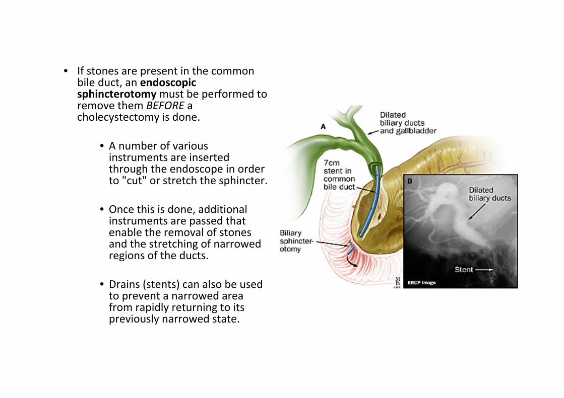
• If stones are present in the common bile duct, an endoscopic sphincterotomy must be performed to remove them BEFORE a cholecystectomy is done.
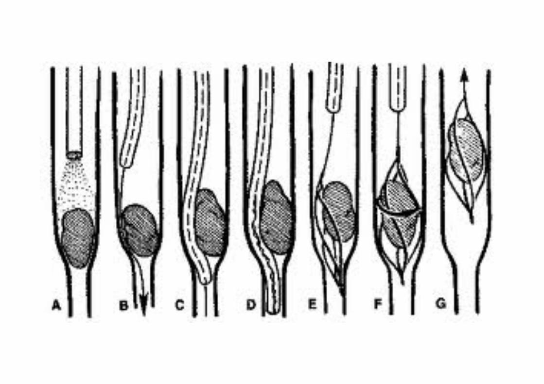
• A number of various instruments are inserted through the endoscope in order to "cut" or stretch the sphincter.
• Once this is done, additional instruments are passed that enable the removal of stones and the stretching of narrowed regions of the ducts.
• Drains (stents) can also be used to prevent a narrowed area from rapidly returning to its previously narrowed state.


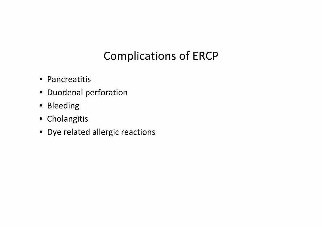
Complications of ERCP
• Pancreatitis• Duodenal perforation• Bleeding• Cholangitis• Dye related allergic reactions
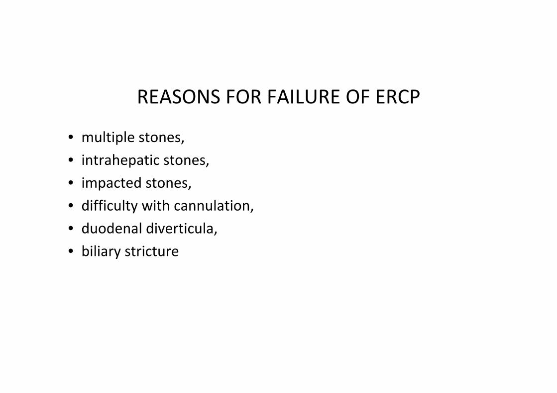
REASONS FOR FAILURE OF ERCP
• multiple stones, • intrahepatic stones, • impacted stones, • difficulty with cannulation, • duodenal diverticula, • biliary stricture

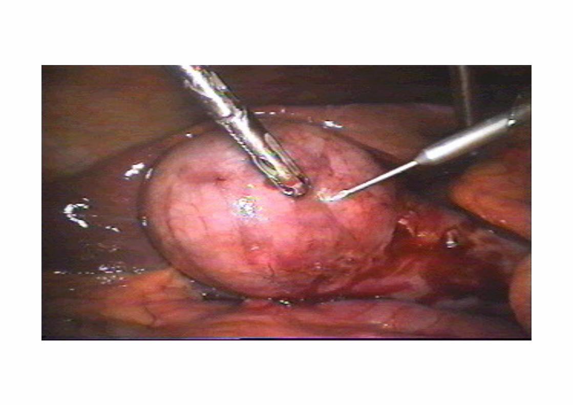
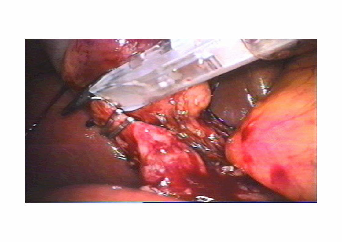
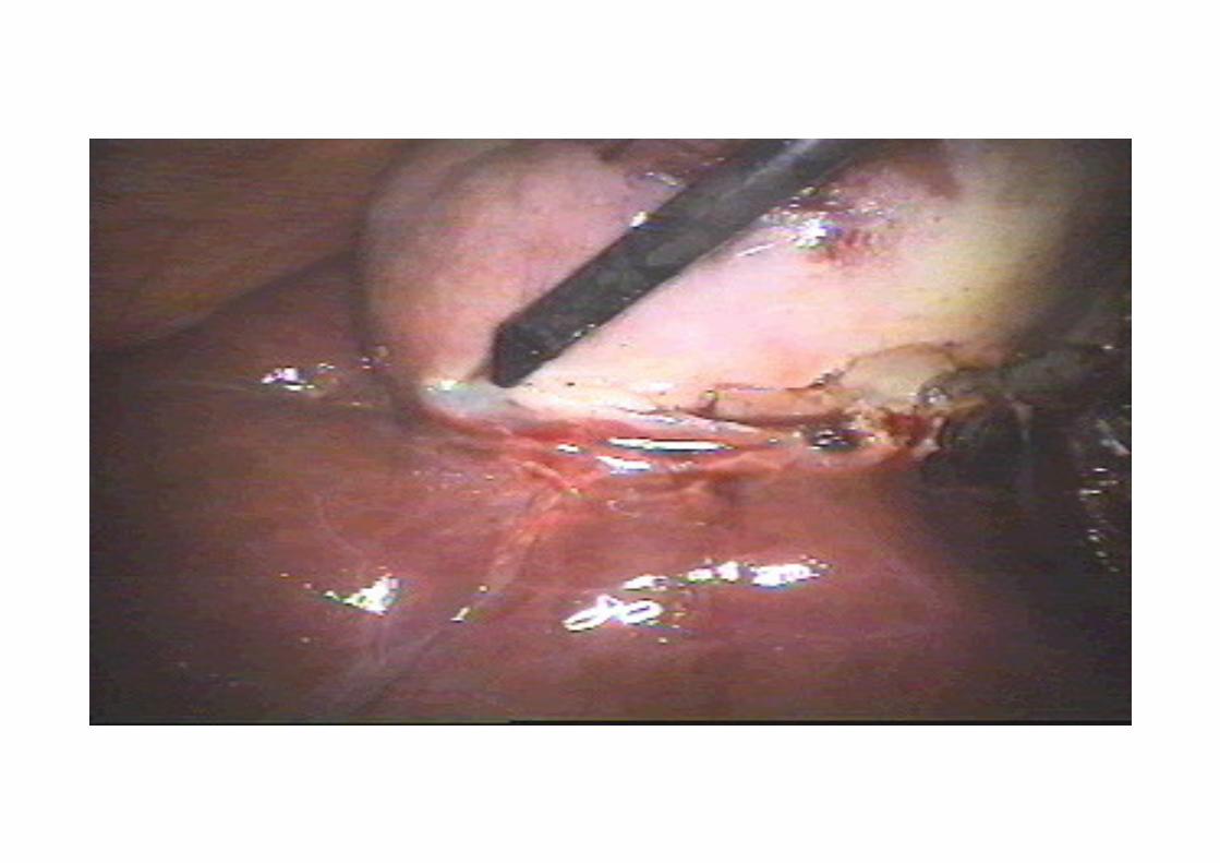
LAPAROSCOPIC CHOLECYSTECTOMY


• CBD stones identified but not removed during cholecystectomy – ERCP for stone extraction.
OPEN CHOLECYSTECTOMY• performed as a conversion from an attempted laparoscopic cholecystectomy (4‐35%) or when
• Laparoscopic facility is not available
Indications for Open Cholecystectomy:Poor pulmonary or cardiac reserve Suspected or known gallbladder cancer Cirrhosis and portal hypertension Third‐trimester pregnancy Combined procedure
Intraoperative Cholangiography
• An intraoperative cholangiogram at the time of cholecystectomy will document the presence of common bile duct stones.
Indications: • Elevated preoperative liver enzymes (AST, ALT, ALP, bilirubin) • Unclear anatomy during laparoscopic dissection • Suspicion of intraoperative injury to biliary tract • Dilated common bile duct on preoperative imaging • Gallstone pancreatitis without endoscopic clearance of common bile duct
• Jaundice • Large common bile duct and small stones • Unsuccessful preoperative endoscopic retrograde cholangiopancreatography for choledocholithiasis

CBD EXPLORATION
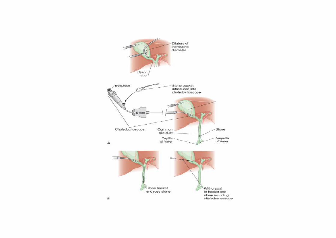
LAPAROSCOPIC CBD EXPLORATION
• Laparoscopic common bile duct exploration through: (choledochoscope)cystic duct or with formal choledochotomy allows the stones to be retrieved during the same procedure.
• If the expertise and instrumentation for laparoscopic common bile duct exploration are not available:
a drain should be placed and left adjacent next to the cystic duct &ERCP with stone extraction is performed the following day.
Open Common Bile Duct Exploration
• An open common bile duct exploration should be performed if endoscopic intervention is not available or not feasible because of anatomic restrictions or expertise.
• If a choledochotomy is performed, a T tube is left in place. • The purpose of the T tube is to provide access to the biliary system for postoperative radiologic stone extraction.
• Completion cholangiography via the T tube documents stone removal.

• Stones impacted in the ampulla may be difficult for both endoscopic ductal clearance and common bile duct exploration.
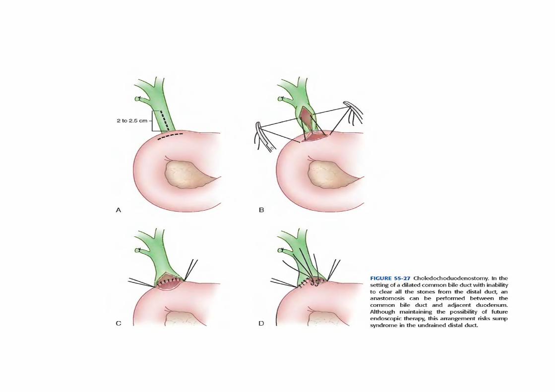
• In these cases, transduodenal sphincteroplasty and stone extraction should be performed; if this is not successful,
• a choledochoduodenostomy or a Roux‐en‐Y choledochojejunostomy should be performed.
• Sump syndrome associated with choledochoduodenostomy
Hepatico‐jejunostomy
• For intra hepatic stones
Complications of surgery:Can be divided into a)Intraoperative b)Post operative

Intraoperative complications
related to dangerous anatomy and pathology ‐ chronic or acute inflammation‐obscured anatomy & increased vasularity.
Insufficient preoperative assessment of a complicated situation is another reason
Insufficient experience, inadequate exposure/incision/assistance
Anatomical variations like narrow common bile duct can be mistaken for cystic duct
Hemmorhage represents potential danger because attempts at hemostasis by placing clamps with obstructed and insufficient view may result in inadvertent clamping of the rt or common hepatic artery
In such conditions hemmorhage to be controlled by digital compression or by clamping of hepatoduodenal ligament to localize its precise origin – Pringles maneuver
Post operative complications:
Bile duct injury Spilled/Lost stones Post cholecystectomy pain Retained bile stonesGall stone ileusAcute cholangitis Recurrent pyogenic cholangitis Bile leak Choledochoduodenal fistula Post cholecystectomy diarrheaWound pain
BILE DUCT INJURY:
During open or laparoscopic cholecystectomy injury to CBD is an unsual but devastating complication.
Risk factors are: ‐surgical inexperience ‐inappropriate exposure ‐variable biliary anatomy ‐aggressive attempts at hemostasis ‐inflammation in the porta

•LOST STONES:In the era of laparoscopic cholecystectomy,inadvertent opening of gall bladder with spillage of stones is seen in 20‐30% of casesRisk factors include:
‐cholecystitis‐presence of pigmented stones‐if stones are >15
Delayed consequences like chronic abscess,fistula,wound infection and bowel obstruction may occur
Most dropped stones settle in Morrison’s pouch/retro hepatic space‐chronic abscess
TREATMENT
Extensive irrigationSignificant attempt to retain lost stonesA course of broad spectrum antibioticsDocumentation of perforation in the operative notes
• RETAINED BILIARY STONES:
Retained CBD stones/secondary common duct stones can be identified upto 2yrs following cholecystectomy
Endoscopic removal of these stones via generous sphincterotomy is the treatment
THANK YOU


![CHOLELITHIASIS [Autosaved]](https://img.pdfslide.net/doc/110x75/577ce5051a28abf1038fa5b3/cholelithiasis-autosaved.jpg)