Embed Size (px)
Citation preview
Management of Fungating Tumors andPressure Ulcers in a Patient With Stage IVCutaneous Malignant Melanoma
Anne F. Walsh, MSN, ANP-BC, ACHPN, CWOCN ƒMarianne Bradley, MSN, CNS, CWOCN ƒ Kim Cavallito, FNP-BC, CWCN
People with advanced disease often face many distressingsymptoms. These symptoms can be further compoundedand complicated by the presence of a wound or wounds.This article presents a case involving a patient being caredfor at home with fungating tumors secondary to stage IVcutaneous malignant melanoma and pressure ulcers. Thegoal of care was to offer symptom management optionsfor the distressing symptoms that he and his family facedwith regard to these wounds: pain, odor, bleeding, andexcessive drainage.
KEY WORDSfungating tumors, malignant melanoma, pressure ulcers,skin cancer, wounds
Patients with advanced disease often have distressingsymptoms that can be confounded and further com-plicated by the presence of a wound or wounds. The
wounds can be a constant physical reminder of the patient’sillness. The wound etiology in a patient with advanced dis-ease can include pressure-related ulcers, vascular-relatedulcers, a primary skin cancer,metastasis from anothermalig-nancy, a Kennedy terminal ulcer, or a Marjolin ulcer (ma-lignant transformation of a chronic wound).1
The skin is the largest organ in the body, and whenmajor complications occur in the skin, this can be 1 of themost devastating concerns to both patients and caregivers.Patients may develop a Kennedy terminal ulcer, which isa complication of the dying process resulting frommultiple-system organ failure.2 It usually develops in the sacrococ-cygeal region and is pear or butterfly shaped (Photo 1).
Loved ones may feel a sense of failure or blame when apressure ulcer develops despite their best caregiving efforts.
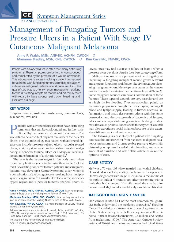
Malignant wounds may present as either fungating orulcerating. A fungating malignant wound grows outwardand appears fungus or cauliflower-like (Photo 2). An ulcer-ating malignant wound develops as a crater as the cancererodes through the skin into deeper tissue layers (Photo 3).Some malignant wounds can have a combination of thesefeatures. These types of wounds are very vascular and areat a high risk for bleeding. They are also often painful asthe tumor progresses through the tissue layers, cutting offblood and lymph supply, leading to further necrosis, in-flammation, and tissue destruction. Along with the tissuedestruction and the overgrowth of bacteria and fungus,odor can be a major distressing symptom. Leaking exudatemay also cause pruritus. Patientswith these types ofwoundsmay also experience social isolation because of the exten-sive disfigurement and embarrassment.
The following is a case study of a patient with fungatingtumors at his anterior chest from recurrent malignant cuta-neous melanoma and 2 unstageable pressure ulcers. Hisdistressing symptoms included pain, bleeding, and a largeamount of exudate and odor. This article reviews theoptions of care.
CASE STUDY
Mr J was a 70-year-old white, married man with 2 children.Heworked as a sailor spendingmuch time in the open sun.He was diagnosed with stage III cutaneous melanoma ofhis right shoulder 5 months ago after presenting with amole that had changed. His wife noted the size had in-creased, and Mr J noted some bloody exudate on his shirt.
BACKGROUND: SKIN CANCER
Skin cancer is cited as 1 of the most common malignan-cies in the elderly, and the incidence is growing.3 The SkinCancer Foundation estimates skin cancer diagnoses in theUnited States each year as follows: squamous cell carci-noma, 700 000; basal cell carcinoma, 2.8 million; and deathsfrom melanoma, 8790.4 The American Cancer Societyestimated 76 690 newmelanoma cases in the United States
Anne F. Walsh, MSN, ANP-BC, ACHPN, CWOCN, is an nurse practi-tioner in hospice at the Visiting Nurse Service of New York.
Marianne Bradley, MSN, CNS, CWOCN, is a nursing instructor instaff development at the Visiting Nurse Service of New York, Bronx.
Kim Cavallito, FNP-BC, CWCN, is a nurse manager at Calvary HospitalWound Center, Bronx, New York.
Address correspondence to Anne F. Walsh, MSN, ANP-BC, ACHPN,CWOCN, Visiting Nurse Service of New York, 1250 Broadway, 7thFloor, New York, NY 10001 ([email protected]).
The authors have no conflicts of interest to disclose.
DOI: 10.1097/NJH.0000000000000059
208 www.jhpn.com Volume 16 & Number 4 & June 2014
2.1 ANCC Contact Hours
Symptom Management Series
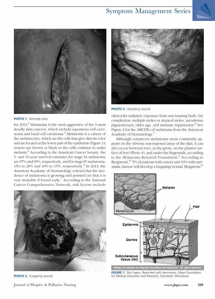
for 2013.5 Melanoma is the most aggressive of the 3 mostdeadly skin cancers, which include squamous cell carci-noma and basal cell carcinoma.3 Melanoma is a cancer ofthe melanocytes, which are the cells that give skin its colorand are located at the lower part of the epidermis (Figure 1);tumors are brown or black as the cells continue to makemelanin.6 According to the American Cancer Society, the5- and 10-year survival estimates for stage IA melanomaare 97% and 95%, respectively, and for stage IVmelanoma,15% to 20% and 10% to 15%, respectively.5 In 2013, theAmerican Academy of Dermatology echoed that the inci-dence of melanoma is growing and pointed out that it isvery treatable if found early.7 According to the NationalCancer Comprehensive Network, risk factors include
ultraviolet radiation exposure from sun/tanning beds, faircomplexion, multiple moles or atypical moles, xerodermapigmentosum, older age, and immune suppression.8 SeeFigure 2 for the ABCDEs of melanoma from the AmericanAcademy of Dermatology.7
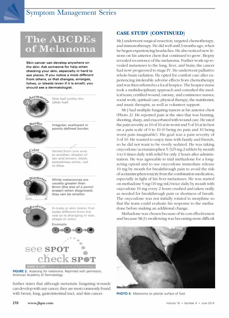
Although cutaneous melanoma most commonly ap-pears on the obvious sun-exposed areas of the skin, it canalso occur between toes, in the groin, on the plantar sur-face of feet (Photo 4), and under the fingernails, accordingto the Melanoma Research Foundation.9 According toBergstrom,10 5% of patients with cancer and 10%withmet-astatic disease will develop a fungatingwound. Bergstrom10
PHOTO 1. Kennedy ulcer.
PHOTO 2. Fungating wound.
PHOTO 3. Ulcerating wound.
FIGURE 1. Skin layers. Reprinted with permission, Mayo Foundationfor Medical Education and Research, Rochester, Minnesota.
Journal of Hospice & Palliative Nursing www.jhpn.com 209
Symptom Management Series
further states that although metastatic fungating woundscandevelopwith any cancer, they aremost commonly foundwith breast, lung, gastrointestinal tract, and skin cancer.
CASE STUDY (CONTINUED)
Mr J underwent surgical resection, targeted chemotherapy,and immunotherapy.He didwell until 3months ago,whenhe began experiencing headaches. He also noticed new le-sions on his anterior chest that continued to grow. Biopsyrevealed recurrence of the melanoma. Further work-up re-vealed metastases to the lung, liver, and brain; the cancerhad now progressed to stage IV. He underwent palliativewhole-brain radiation. He opted for comfort care after ex-periencing intolerable adverse effects from chemotherapyandwas then referred to a local hospice. The hospice nursetook a multidisciplinary approach and consulted the med-ical team; certified wound, ostomy, and continence nurses;social work; spiritual care; physical therapy; the nutritionist;and music therapist, as well as volunteer support.
Mr J had multiple fungating tumors at his anterior chest(Photo 2). He reported pain at the sites that was burning,shooting, sharp, and exacerbatedwithwound care.He ratedthe pain severity as 10 of 10 at its worst and 5 of 10 at its beston a pain scale of 0 to 10 (0 being no pain and 10 beingworst pain imaginable). His goal was a pain severity of3 of 10. He wanted to enjoy time with family and friends,so he did not want to be overly sedated. He was takingoxycodone/acetaminophen 5/325mg 2 tablets bymouth4 to 6 times daily with relief for only 2 hours after adminis-tration. He was agreeable to trial methadone for a long-acting opioid and to use oxycodone immediate release10 mg by mouth for breakthrough pain to avoid the riskof acetaminophen toxicity from the combinationmedication,especially in light of his liver metastases. He was startedonmethadone 5mg (10mg/mL) twice daily bymouthwithoxycodone 10 mg every 2 hours crushed and taken orallyas needed for breakthrough pain or shortness of breath.The oxycodone was not initially rotated to morphine sothat the team could evaluate his response to the metha-done before making an additional change.
Methadonewas chosen because of its cost effectivenessand because Mr J’s swallowing was becomingmore difficult
FIGURE 2. Assessing for melanoma. Reprinted with permission,American Academy of Dermatology.
PHOTO 4. Melanoma on plantar surface of foot.
210 www.jhpn.com Volume 16 & Number 4 & June 2014
Symptom Management Series
and the team had access to methadone intensol on for-mulary. Methadone was also considered because of thesuspected neuropathic component of the pain from Mr J’spain descriptors of burning and shooting pain. Althougha Cochrane review found it no more effective for neuro-pathic pain thanmorphine,11 in these authors’ experiences,methadone has been effective for neuropathic pain.12-14
Mr J and his family kept a record of his daily breakthroughmedication needs to allow for aggressive titration of theopioids as needed. He did well on this regimen requiring2 to 3 breakthrough pain medication doses daily, includ-ing a dose 30 to 60 minutes before the wound care. Hereported a pain level of 3 both overall and during woundcare,whichwas acceptable to him.As his swallowingwors-ened, he was rotated to morphine sulfate 15 mg (20mg/mL)by mouth with good results. Morphine is considered thestandard initial opioid of use.15 The oxycodone intensolis more costly, and because Mr J had no contraindicationstomorphine, such as advanced renal disease, the transitionwasmadewithout any problems. The team reevaluated hispain regularly during visits and over the telephone.
Mr J and his family were also upset by the odor, the bleed-ing, and the large amount of exudate from the fungatingwound at his chest and from the sacral pressure ulcer. Thisled to the need for frequent clothing and linen changes.He becamemore withdrawn and embarrassed by the odorand had a poor body image. His spouse was fatiguedandworried hewould become depressed. Theywere bothfrightened by the amount of bleeding occurring duringthe wound care.
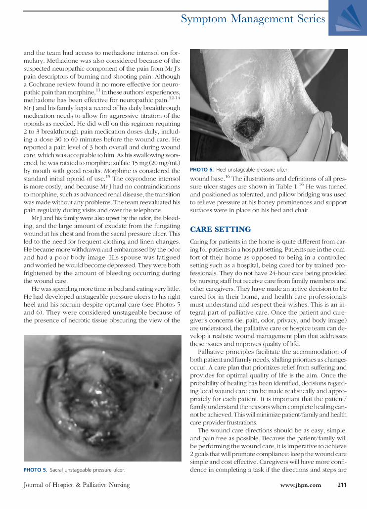
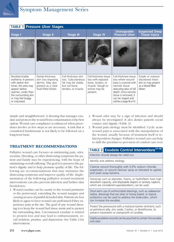
Hewas spendingmore time in bed and eating very little.He had developed unstageable pressure ulcers to his rightheel and his sacrum despite optimal care (see Photos 5and 6). They were considered unstageable because ofthe presence of necrotic tissue obscuring the view of the
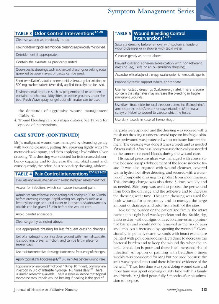
wound base.16 The illustrations and definitions of all pres-sure ulcer stages are shown in Table 1.16 He was turnedand positioned as tolerated, and pillow bridging was usedto relieve pressure at his boney prominences and supportsurfaces were in place on his bed and chair.
CARE SETTING
Caring for patients in the home is quite different from car-ing for patients in a hospital setting. Patients are in the com-fort of their home as opposed to being in a controlledsetting such as a hospital, being cared for by trained pro-fessionals. They do not have 24-hour care being providedby nursing staff but receive care from family members andother caregivers. They have made an active decision to becared for in their home, and health care professionalsmust understand and respect their wishes. This is an in-tegral part of palliative care. Once the patient and care-giver’s concerns (ie, pain, odor, privacy, and body image)are understood, the palliative care or hospice team can de-velop a realistic wound management plan that addressesthese issues and improves quality of life.
Palliative principles facilitate the accommodation ofboth patient and family needs, shifting priorities as changesoccur. A care plan that prioritizes relief from suffering andprovides for optimal quality of life is the aim. Once theprobability of healing has been identified, decisions regard-ing local wound care can be made realistically and appro-priately for each patient. It is important that the patient/family understand the reasonswhen complete healing can-not beachieved.Thiswillminimizepatient/family andhealthcare provider frustrations.
The wound care directions should be as easy, simple,and pain free as possible. Because the patient/family willbe performing the wound care, it is imperative to achieve2 goals thatwill promote compliance: keep thewound caresimple and cost effective. Caregivers will have more confi-dence in completing a task if the directions and steps are
PHOTO 6. Heel unstageable pressure ulcer.
PHOTO 5. Sacral unstageable pressure ulcer.
Journal of Hospice & Palliative Nursing www.jhpn.com 211
Symptom Management Series
simple and straightforward. A dressing that manages exu-date andprotects thewound fromcontamination is the bestoption. Wound care compliance is enhanced when proce-dures involve as few steps as are necessary. A task that isconsidered burdensome is not likely to be followed on along-term basis.
TREATMENT RECOMMENDATIONS
Palliative wound care focuses on minimizing pain, odor,exudate, bleeding, or other distressing symptoms the pa-tient and family may be experiencing, with the hope ofminimizing overall suffering. The goal is to preserve the pa-tient’s dignity and to help promote quality of life. The fol-lowing are recommendations that may minimize thedistressing symptoms and improve quality of life. Imple-mentation of the following palliative wound treatmentinterventions can also prevent infection and further skinbreakdown.1. Wound exudate can be caustic to the wound perimeter
or the periwound, extending the wound margins andcausing an area of painful denuded skin. Patients are lesslikely to agree to havewound care performed if they ex-perience pain at the site. The goal of any wound dress-ing is to keep the wound bed tissue moist and to protectthe surrounding skin. Uncontained exudate is relatedto protein loss and may lead to embarrassment, so-cial isolation, pruritus, and depression. See Table 2 forinterventions.
2. Wound odor may be a sign of infection and shouldalways be investigated. It also denies patients socialcontact and dignity (Table 3).
3. Wound pain etiology must be identified. Cyclic acutewound pain is associated with the manipulation ofthe wound, usually because of treatment itself or re-lated position changes. Palliative wound care can helpto shift the priorities to provision of comfort care over
TABLE 1 Pressure Ulcer Stages
Stage I Stage II Stage III Stage IVUnstageablePressure Ulcer
Suspected DeepTissue Injury
Nonblanchableerythema. In personswith darker skintones, the area mayappear darker,warmer, cooler thanthe surrounding skinor may feel boggyor indurated.
Partial-thicknessskin loss exposingdermis. May alsopresent as a clearfluid-filled blister.
Full-thickness skinloss. Subcutaneousfat may be visible,but not bone,tendon, or muscle.
Full-thickness tissueloss with exposedbone, tendon, ormuscle. Slough oreschar may bepresent.
Full-thickness tissueloss where woundbase is covered withnecrotic tissueobscuring view of fulldepth.Oncenecrotictissue is removed, itcan be staged andwill bea stage III or IV.
Purple or maroondiscolored intactskin or may presentas a blood-filledblister.
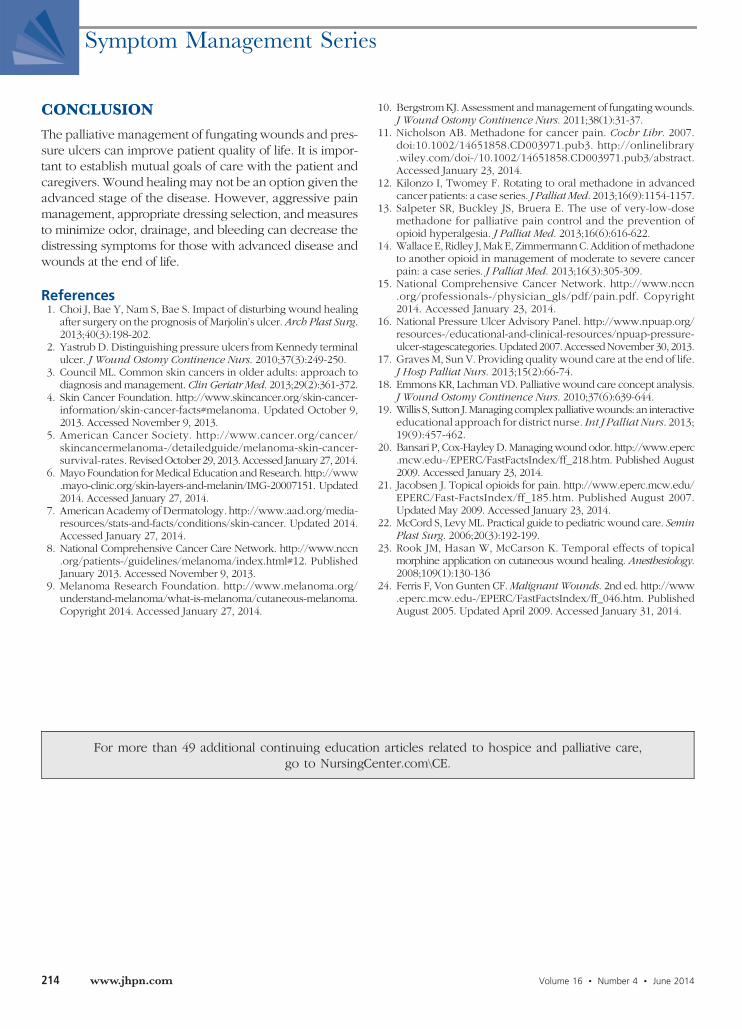
TABLE 2 Exudate Control Interventions17-19
Infection should always be ruled out.
Identify and address etiology.
Cleanse wound thoroughly with 0.9% sodium chloride,soap/water, or wound cleanser spray as tolerated to agitateand wash away bacteria.
Dressings such as alginates, foams, or hydrofibers have highabsorbent capacity, and disposable diapers or sanitary napkins,which are considered superabsorbent, can be used.
Short-term use of antimicrobial dressings, such as cadexemoriodine, dressings that are silver or honey based, or topicalantibiotics can be used to address the bioburden, whichcan increase the exudate.
Protect the periwound with a moisture barrier ointment, suchas petroleum jelly, zinc oxide, Cavilon, or dimethicone, toprevent maceration or overgrowth of candida.
Highly exudativewounds canbepouched to contain the exudateand odor.
212 www.jhpn.com Volume 16 & Number 4 & June 2014
Symptom Management Series
the demands of aggressive wound management(Table 4).
4. Wound bleeding can be a major distress. See Table 5 foroptions of interventions.
CASE STUDY (CONTINUED)
Mr J’s malignant wound was managed by cleansing gentlywith wound cleanser, patting dry, spraying lightly with 1%metronidazole spray, and then applying a hydrofiber silverdressing. This dressingwas selected for its increased absor-bency capacity and to decrease the microbial count and,consequently, the odor. As a secondary dressing, abdomi-
nal padswere applied, and the dressingwas securedwith amesh net dressing retainer to avoid tape on his fragile skin.The periwound was protected with a moisture barrier oint-ment. The dressing was done 3 times a week and as neededif itwas soiled.Afrin nasal spraywasused topically as neededto the tumor to control bleeds during the wound care.
His sacral pressure ulcer was managed with conserva-tive bedside sharps debridement of the loose necrotic tis-sue. It was also irrigated with wound cleanser, coveredwith a hydrofiber silver dressing, and securedwith a water-proof composite dressing to protect from incontinence.This dressing change was also done 3 times weekly andas needed. Skin prep was used to protect the periwoundfrom both the drainage and the adhesive and to increasethe dressing wear time. The same dressing was used forboth wounds for consistency and to manage the largeamount of drainage and odor from both of the sites.
To ease the burden on the patient and family, the intacteschar at his right heel was kept clean and dry. Stable, dry,intact eschar, without signs of infection, serves as a protec-tive barrier and should not be removed as the risk of painand limb loss is increased by opening the wound.16 Occa-sionally, in palliative care, wounds with intact eschar arepainted with povidone-iodine (Betadine) to decrease thebacterial burden and to keep the wound dry when the ar-terial circulation is poor and there is an increased risk ofinfection. An option of painting with Betadine 3 timesweekly was considered for Mr J but not used because thearea was dry and intact and there is limited evidence of thebenefit.24 Thus, less time was spent doing wound care andmore time was spent enjoying quality time with his familyand friends. Mr J died peacefully 5 months after his admis-sion to hospice.
TABLE 3 Odor Control Interventions17-20
Cleanse wound as previously noted.
Useshort-termtopicalantimicrobialdressingsaspreviouslymentioned.
Debridement if appropriate.
Contain the exudate as previously noted.
Odor-specific dressings such as charcoal dressings or baking sodasprinkled between layers of gauze can be used.
Short-termDakin’s solutionormetronidazole (asagelor solution,or500 mg crushed tablets twice daily applied topically) can be used.
Environmental products such as peppermint oil or an opencontainer of charcoal, kitty litter, or coffee grounds under thebed, Fresh Wave spray, or gel odor eliminator can be used.
TABLE 4 PainControl Interventions17-19,21-23
Evaluate and revaluatepainwitha validatedpain assessment tool.
Assess for infection, which can cause increased pain.
Administer an effective short-acting oral analgesic 30 to 60minbefore dressing change. Rapid-acting oral opioids such as afentanyl lozenge or buccal tablet or intravenous/subcutaneousopioids can be given 15 min before the wound care.
Avoid painful antiseptics.
Cleanse gently as noted above.
Use appropriate dressing for less frequent dressing changes.
Use of a hydrogel is best in a cleanwoundwithminimal exudate;it is soothing, prevents friction, and can be left in place forseveral days.
Usemoisture retentivedressings todecrease frequencyof changes.
Apply topical 2% lidocaine jelly22 3-5minutesbeforewound care.
Topicalmorphine-basedhydrogel: 10mg (10mg/mL)ofmorphineinjection in 8 g of Intrasite hydrogel 1-3 times daily.21 Thereis limited research available. There is some evidence that topicalmorphine may impair wound healing if healing is the goal.23
TABLE 5 Wound Bleeding ControlInterventions17-19
Saturate dressing before removal with sodium chloride orwound cleanser or in shower with tepid water.
Cleanse gently as noted above.
Prevent dressing adherence/desiccation with nonadherentdressing (eg, Telfa or an oil-emulsion dressing).
Assessbenefitsofadjunct therapy: localorsystemichemostaticagents.
Provide systemic support where appropriate.
Use hemostatic dressings (Calcium-alginate). There is someconcern that alginates may increase the bleeding in fragilemalignant wounds.
Use silver nitrate sticks for local bleeds or adrenaline (Epinephrine),aminocaproic acid (Amicar), or oxymetazoline (Afrin nasalspray) off-label to wound to vasoconstrict the tissue.
Use dark towels in case of hemorrhage.
Journal of Hospice & Palliative Nursing www.jhpn.com 213
Symptom Management Series
CONCLUSION
The palliative management of fungating wounds and pres-sure ulcers can improve patient quality of life. It is impor-tant to establish mutual goals of care with the patient andcaregivers. Wound healing may not be an option given theadvanced stage of the disease. However, aggressive painmanagement, appropriate dressing selection, and measuresto minimize odor, drainage, and bleeding can decrease thedistressing symptoms for those with advanced disease andwounds at the end of life.
References1. Choi J, Bae Y, Nam S, Bae S. Impact of disturbing wound healing
after surgery on the prognosis of Marjolin’s ulcer.Arch Plast Surg.2013;40(3):198-202.
2. Yastrub D. Distinguishing pressure ulcers fromKennedy terminalulcer. J Wound Ostomy Continence Nurs. 2010;37(3):249-250.
3. Council ML. Common skin cancers in older adults: approach todiagnosis and management. Clin Geriatr Med. 2013;29(2):361-372.
4. Skin Cancer Foundation. http://www.skincancer.org/skin-cancer-information/skin-cancer-facts#melanoma. Updated October 9,2013. Accessed November 9, 2013.
5. American Cancer Society. http://www.cancer.org/cancer/skincancermelanoma-/detailedguide/melanoma-skin-cancer-survival-rates. RevisedOctober 29, 2013.Accessed January27, 2014.
6. Mayo Foundation forMedical Education and Research. http://www.mayo-clinic.org/skin-layers-and-melanin/IMG-20007151. Updated2014. Accessed January 27, 2014.
7. AmericanAcademyofDermatology. http://www.aad.org/media-resources/stats-and-facts/conditions/skin-cancer. Updated 2014.Accessed January 27, 2014.
8. National Comprehensive Cancer Care Network. http://www.nccn.org/patients-/guidelines/melanoma/index.html#12. PublishedJanuary 2013. Accessed November 9, 2013.
9. Melanoma Research Foundation. http://www.melanoma.org/understand-melanoma/what-is-melanoma/cutaneous-melanoma.Copyright 2014. Accessed January 27, 2014.
10. BergstromKJ. Assessment andmanagement of fungatingwounds.J Wound Ostomy Continence Nurs. 2011;38(1):31-37.
11. Nicholson AB. Methadone for cancer pain. Cochr Libr. 2007.doi:10.1002/14651858.CD003971.pub3. http://onlinelibrary.wiley.com/doi-/10.1002/14651858.CD003971.pub3/abstract.Accessed January 23, 2014.
12. Kilonzo I, Twomey F. Rotating to oral methadone in advancedcancer patients: a case series. J PalliatMed. 2013;16(9):1154-1157.
13. Salpeter SR, Buckley JS, Bruera E. The use of very-low-dosemethadone for palliative pain control and the prevention ofopioid hyperalgesia. J Palliat Med. 2013;16(6):616-622.
14. WallaceE, Ridley J,MakE, ZimmermannC.Addition ofmethadoneto another opioid in management of moderate to severe cancerpain: a case series. J Palliat Med. 2013;16(3):305-309.
15. National Comprehensive Cancer Network. http://www.nccn.org/professionals-/physician_gls/pdf/pain.pdf. Copyright2014. Accessed January 23, 2014.
16. National Pressure Ulcer Advisory Panel. http://www.npuap.org/resources-/educational-and-clinical-resources/npuap-pressure-ulcer-stagescategories.Updated2007.AccessedNovember 30, 2013.
17. Graves M, Sun V. Providing quality wound care at the end of life.J Hosp Palliat Nurs. 2013;15(2):66-74.
18. Emmons KR, LachmanVD. Palliativewound care concept analysis.J Wound Ostomy Continence Nurs. 2010;37(6):639-644.
19. Willis S, Sutton J.Managingcomplexpalliativewounds: an interactiveeducational approach for district nurse. Int J Palliat Nurs. 2013;19(9):457-462.
20. Bansari P, Cox-HayleyD.Managingwound odor. http://www.eperc.mcw.edu-/EPERC/FastFactsIndex/ff_218.htm. Published August2009. Accessed January 23, 2014.
21. Jacobsen J. Topical opioids for pain. http://www.eperc.mcw.edu/EPERC/Fast-FactsIndex/ff_185.htm. Published August 2007.Updated May 2009. Accessed January 23, 2014.
22. McCord S, LevyML. Practical guide to pediatric wound care. SeminPlast Surg. 2006;20(3):192-199.
23. Rook JM, Hasan W, McCarson K. Temporal effects of topicalmorphine application on cutaneous wound healing. Anesthesiology.2008;109(1):130-136
24. Ferris F, Von Gunten CF.Malignant Wounds. 2nd ed. http://www.eperc.mcw.edu-/EPERC/FastFactsIndex/ff_046.htm. PublishedAugust 2005. Updated April 2009. Accessed January 31, 2014.
For more than 49 additional continuing education articles related to hospice and palliative care,go to NursingCenter.com\CE.
214 www.jhpn.com Volume 16 & Number 4 & June 2014
Symptom Management Series