Embed Size (px)
Citation preview

MANAGEMENT OF SPINE TUBERCULOSIS
DR.P.ABHILASH2ND YR ORTHO PG
POTT’S DISEASE
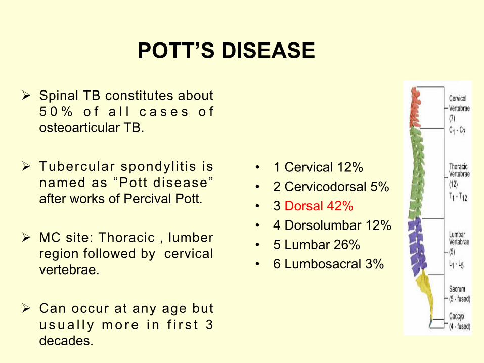
Ø Spinal TB constitutes about 5 0 % o f a l l c a s e s o f osteoarticular TB.
Ø Tubercular spondyl i t is is named as “Pott disease” after works of Percival Pott.
Ø MC site: Thoracic , lumber region followed by cervical vertebrae.
Ø Can occur at any age but u s u a l l y m o r e i n f i r s t 3 decades.
• 1 Cervical 12%• 2 Cervicodorsal 5%• 3 Dorsal 42%• 4 Dorsolumbar 12%• 5 Lumbar 26%• 6 Lumbosacral 3%

PATHOLOGY
Pott’s disease is usually secondary to an extraspinal source of infection.
Typically, more than one vertebra is involved.
Infection occurs through haematological spread ,generally the arteries or through Batson’s plexus of
veins in axial skeleton.

Pathogenesis of TB Spine

Patterns of Vertebral Involvement
Four patterns :• Paradiscal • Central• Anterior• Appendiceal (Posterior)
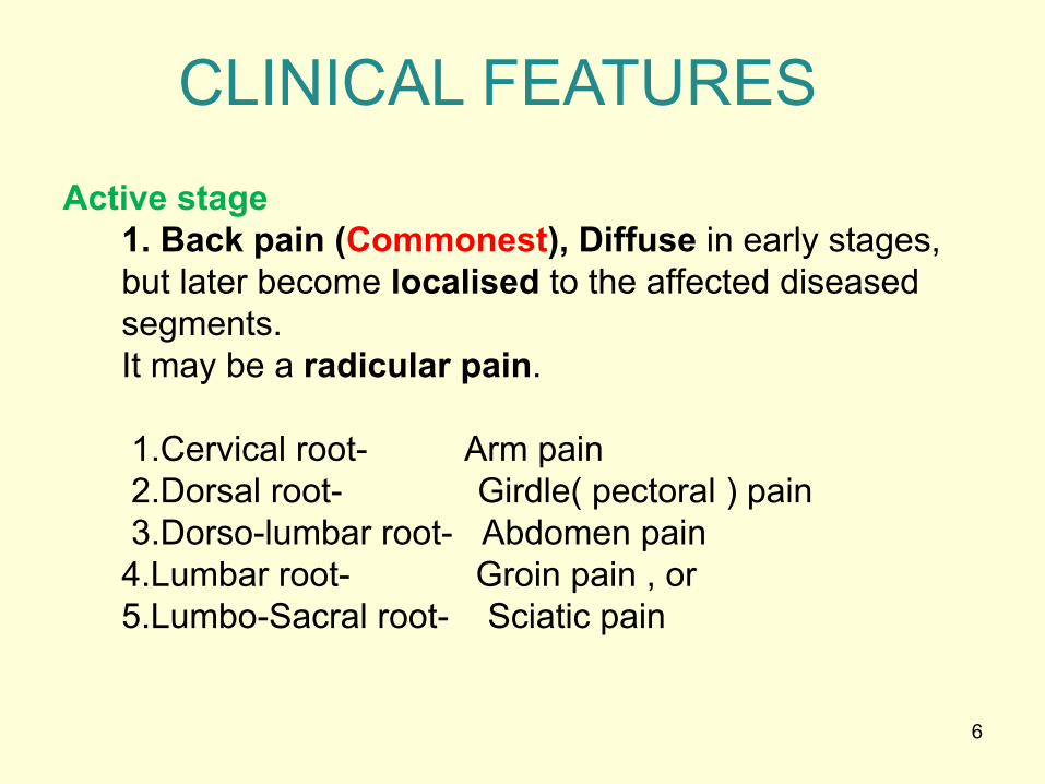
Active stage1. Back pain (Commonest), Diffuse in early stages, but later become localised to the affected diseased segments.It may be a radicular pain.
1.Cervical root- Arm pain 2.Dorsal root- Girdle( pectoral ) pain 3.Dorso-lumbar root- Abdomen pain4.Lumbar root- Groin pain , or 5.Lumbo-Sacral root- Sciatic pain
CLINICAL FEATURES
6

2.Spine Stiffness: spasm of para-vertebral muscle
3.Night cries4.Deformity: Knuckle
/Gibbus/Kyphus.5.Cold abscess: May be
present6.Paraplegia (if neglected in
early stages)
7

7.Constitutional Symptoms (Only in 20% cases): Malaise, weight loss, loss of appetite, night sweats, evening rise of temperature.
B. Healed stageNo systemic features but deformity persists.Radiological evidence of bone healing
8
Patient may present with cold abcess or due to its compression effects:-
Retropharyngeal abscess --Dysphagia,dyspnea, Hoarseness of voice Mediastinal abscess --Dysphagia Psoas abscess -- Flexion deformity of hip
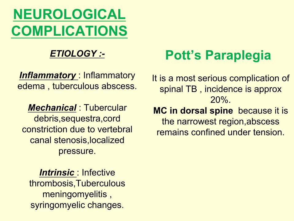
NEUROLOGICAL COMPLICATIONS
ETIOLOGY :-
Inflammatory : Inflammatory edema , tuberculous abscess.
Mechanical : Tubercular debris,sequestra,cord
constriction due to vertebral canal stenosis,localized
pressure.
Intrinsic : Infective thrombosis,Tuberculous
meningomyelitis , syringomyelic changes.
It is a most serious complication of spinal TB , incidence is approx
20%.MC in dorsal spine because it is
the narrowest region,abscess remains confined under tension.
Pott’s Paraplegia
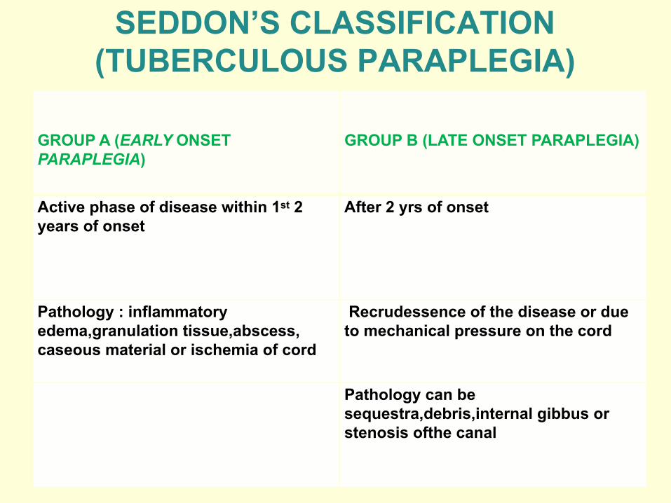
SEDDON’S CLASSIFICATION(TUBERCULOUS PARAPLEGIA)
GROUP A (EARLY ONSET PARAPLEGIA)
GROUP B (LATE ONSET PARAPLEGIA)
Active phase of disease within 1st 2 years of onset
After 2 yrs of onset
Pathology : inflammatory edema,granulation tissue,abscess, caseous material or ischemia of cord
Recrudessence of the disease or due to mechanical pressure on the cord
Pathology can be sequestra,debris,internal gibbus or stenosis ofthe canal

PARA/QUADRIPLEGIA (predominantly based on motor weakness)
STAGE
I Negligible Patient unaware of neuralogical
deficits,plantar extensor &/or ankle clonus
II MildPatient aware of deficit but manages to walk with support(spastic paresis)
III ModerateNon ambulatory because of paralysis ( in extension ), sensory deficits less than 50 %
IV SevereStage III + Flexor spasms/ paralysis in flexion / sensory deficit more than 50% / sphincters involved
CLINICAL FEATURES
DIFFERENTIAL DIAGNOSIS
q Congenital defects like block vertebra,Schmorl’s disease, Scheurermann’s disease.
q Infetious conditions like Acute pyogenic,Typhoid spine,Brucella spondylitis,Mycotic Spondylitis,Syphillis
q Tumours Conditions :-q Benign : Hemangioma,Giant cell tumour,Aneurysmal
bone cyst.q Malignant :Secondaries Ewing’s
sarcoma,Osteogenic sarcoma,Multiple myeloma.
q Traumatic conditions

INVESTIGATIONSqCLINICO RADIOLOGICAL &• LAB STUDIES• Mantoux / tuberculin skin test• Microbiological studies• Histopathological study• CT SCAN• MRI SCAN• USG• RADIONUCLIDE SCAN• MYELOGRAPHY
BASIC PRINCIPLES OFMANAGEMENT
• Early diagnosis
• Medical treatment
• Aggressive surgical approach
• Prevent deformity
• Best outcome
TREATMENT
• Aim of treatment is to achieve healing of disease & to prevent,detect early & promptly any complication like paraplegia.
• Rest: Bed rest for pain relief & to prevent further collapse & dislocation of diseased vertebrae.
• For cervical spineMinerva jacket & collar

• Building up of patient’s resistance : High protein diet
• ATT : This remains the cornstone of management, completed by rest,nutritional support & splinting, as necessary.
• There is difference of opinion regarding the duration of drug therapy.
• Short course chemotherapy for 9-10months has shown good results in patients.
• Antibiotics : For persistently draining sinuses which get secondary infection.
• Bed sore care & to treat other comorbid conditions.
• Mobilisation : Gradual as improvement begins sit & walk,the spine is supported with collar(cervical),brace (dorso-lumber spine)
• Cold abcesses may subside with ATT,if present superficially may need aspration(antigravity insertion of needle through a zig-zag tract) or evacuation.
• Sinuses: Mostly heal within 6-12 weeks. If no improvement Excision of tract

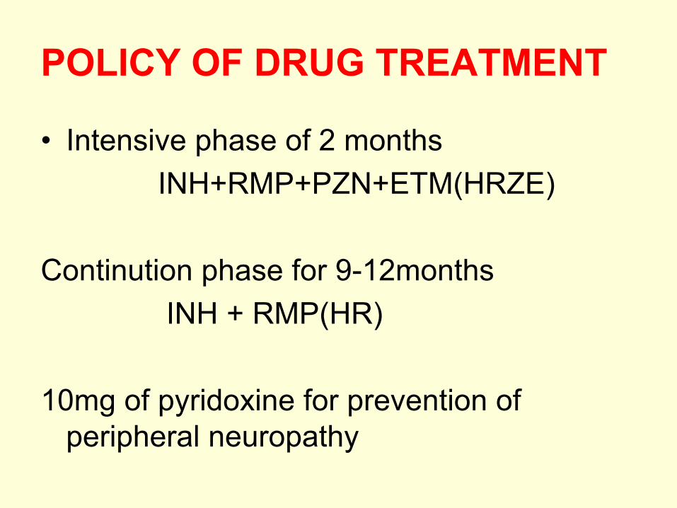
POLICY OF DRUG TREATMENT
• Intensive phase of 2 months INH+RMP+PZN+ETM(HRZE)
Continution phase for 9-12months INH + RMP(HR)
10mg of pyridoxine for prevention of peripheral neuropathy
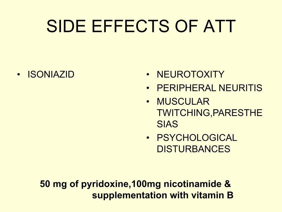
SIDE EFFECTS OF ATT
• ISONIAZID • NEUROTOXITY• PERIPHERAL NEURITIS• MUSCULAR
TWITCHING,PARESTHESIAS
• PSYCHOLOGICAL DISTURBANCES
50 mg of pyridoxine,100mg nicotinamide & supplementation with vitamin B

RIFAMPICIN • HEPATOTOXICITY• FLU-LIKE SYNDROME• ERYTHEMATOUS
REACTION• RED BROWN
DISCOLOURATION OF BODY FLUIDS
PYRAZINAMIDE •HEPATOTOXICITY•ARTHRALGIA•FLUSHING
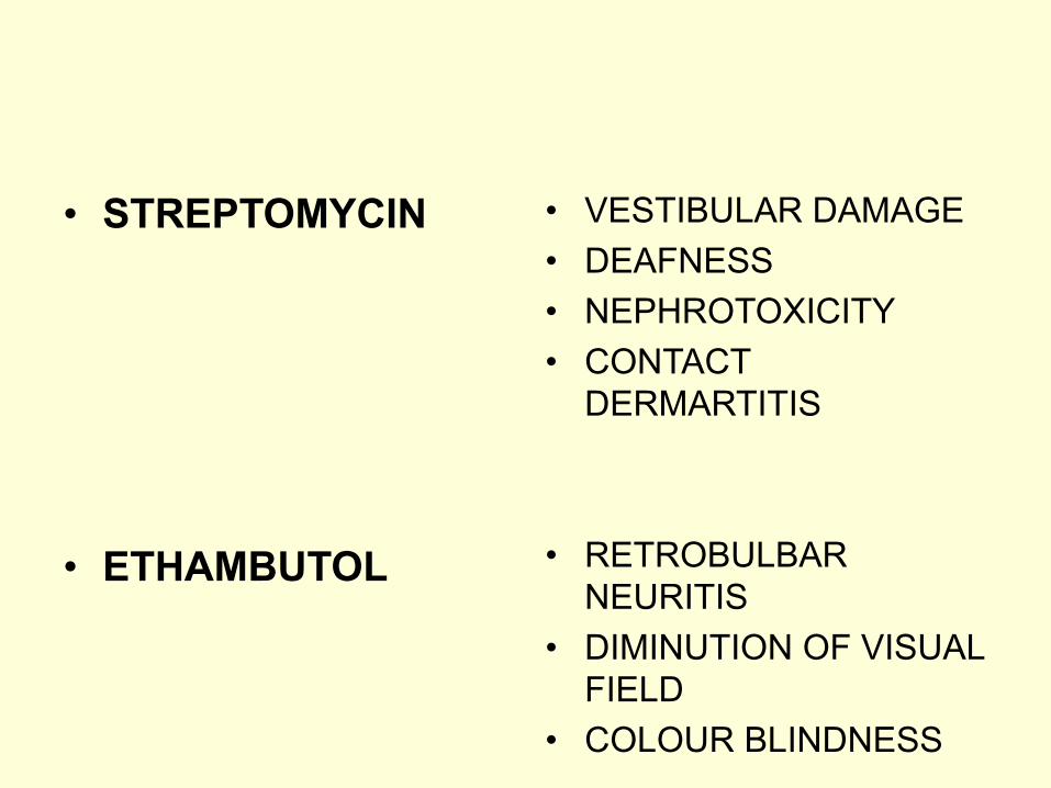
• STREPTOMYCIN
• ETHAMBUTOL
• VESTIBULAR DAMAGE• DEAFNESS• NEPHROTOXICITY• CONTACT
DERMARTITIS
• RETROBULBAR NEURITIS
• DIMINUTION OF VISUAL FIELD
• COLOUR BLINDNESS
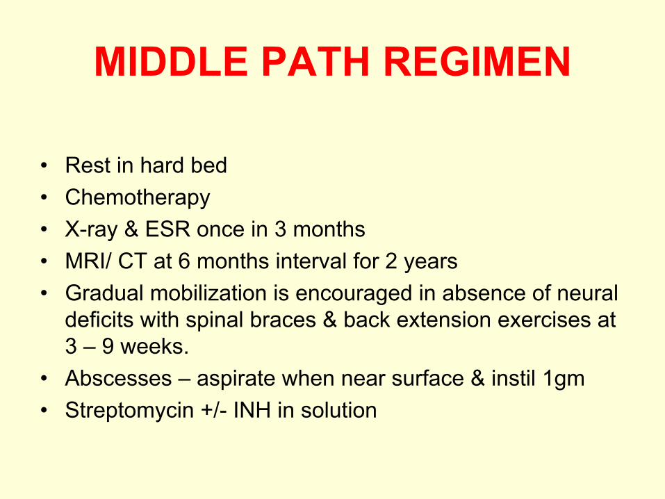
MIDDLE PATH REGIMEN
• Rest in hard bed• Chemotherapy• X-ray & ESR once in 3 months• MRI/ CT at 6 months interval for 2 years• Gradual mobilization is encouraged in absence of neural
deficits with spinal braces & back extension exercises at 3 – 9 weeks.
• Abscesses – aspirate when near surface & instil 1gm• Streptomycin +/- INH in solution
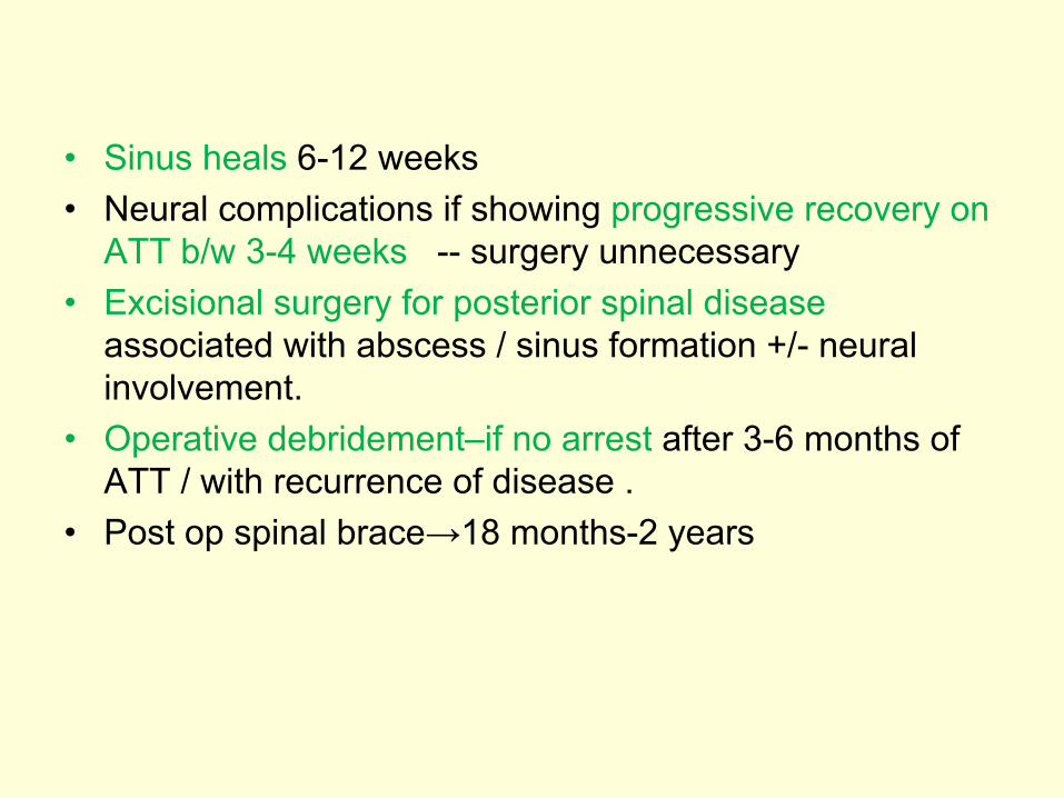
• Sinus heals 6-12 weeks • Neural complications if showing progressive recovery on
ATT b/w 3-4 weeks -- surgery unnecessary• Excisional surgery for posterior spinal disease
associated with abscess / sinus formation +/- neural involvement.
• Operative debridement–if no arrest after 3-6 months of ATT / with recurrence of disease .
• Post op spinal brace→18 months-2 years

DRUGS IN MIDDLE PATH REGIMEN
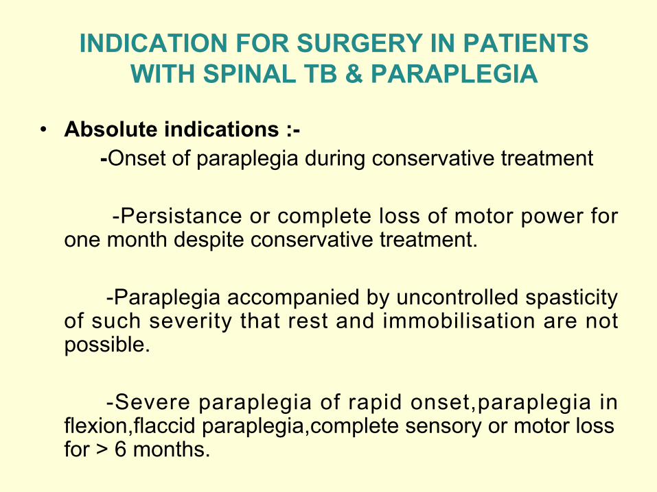
INDICATION FOR SURGERY IN PATIENTS WITH SPINAL TB & PARAPLEGIA
• Absolute indications :- -Onset of paraplegia during conservative treatment
-Persistance or complete loss of motor power for one month despite conservative treatment.
-Paraplegia accompanied by uncontrolled spasticity of such severity that rest and immobilisation are not possible.
-Severe paraplegia of rapid onset,paraplegia in flexion,flaccid paraplegia,complete sensory or motor loss for > 6 months.

• Relative Indication :- -Recurrent paraplegia even with paraplegia
that would cause no concern in first attack -Paraplegia with onset in old age -Painful paraplegia -complications such as UTI and stones• Rare Indications:- -Post. Spinal disease -spinal tumour syndrome -severe paralysis from cervical disease, -severe cauda equina paralysis


APPROACH1.Cervical spine – Anterior retropharyngeal
(smith-Robinson’s) Anterior approach – Anterior/Medial
border of sternocleidomastoid.2. Dorsal spine (D1 to L1) –
1 Transthoracic transpleural 2 Anterolateral decompression(D2 – L1)3. Lumbar spine – Anterolateral(Lumbovertebrotomy) Extraperitoneal Ant. Approach Posterior approach

Surgeries for Pott’s Paraplegia
1. Anterio-lateral decompression (MC)-Spine is opened up from its lateral side & access is made to the front and side of the cord.The cord is laid free from granulation tissue,caseous material,bony spur or sequestrum
2. Costo-transversectomy-Removal of 2 inches of rib&transverse processpus drained.
3. Radical debridement and arthrodesis(Hongkong operation)
4. Laminectomy & posterior stabilisation-Indicated in spinal tumour syndrome and paraplegia resulted from post. spinal disease.
Cervical spine: Anterior decompression is preffered.