Embed Size (px)
Citation preview
Visit the EuroSafe Imaging lounge at ECR 2018
© European Society of Radiology
Ask EuroSafe ImagingTips & Tricks
Paediatric Imaging Working Group
Managing Cone Beam CT Dose in Paediatric Dental ImagingRaija Seuri (HUS Medical Imaging Center, FI)
Cristina Almeida (Centro Hospitalar de Lisboa Central, PT)
Theocharis Berris (University of Crete, GR)
Visit the EuroSafe Imaging lounge at ECR 2018
© European Society of Radiology
Introduction
Cone beam CT (CBCT) has been widely used in dentistry for over 10 years (Li, 2013).
CBCT technology can provide multiple viewing angles and 3D reconstructions, which help in a more complete evaluation as compared to conventional dental imaging modalities (panoramic radiography, intraoral, etc.).
The doses associated with CBCT span a considerably wide range, depending a lot on the equipment (Li, 2013; Ludlow et al., 2008; Pauwels, 2012).
CBCT doses are generally lower than multidetector CT (MDCT) doses (Li, 2013).
CBCT doses are generally higher than the doses from conventional dental radiography (Li, 2013).
Visit the EuroSafe Imaging lounge at ECR 2018
© European Society of Radiology
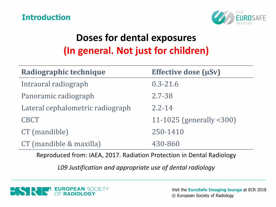
Introduction
Radiographic technique Effective dose (µSv)
Intraoral radiograph 0.3-21.6
Panoramic radiograph 2.7-38
Lateral cephalometric radiograph 2.2-14
CBCT 11-1025 (generally <300)
CT (mandible) 250-1410
CT (mandible & maxilla) 430-860
Reproduced from: IAEA, 2017. Radiation Protection in Dental Radiology
L09 Justification and appropriate use of dental radiology
Doses for dental exposures(In general. Not just for children)

Visit the EuroSafe Imaging lounge at ECR 2018
© European Society of Radiology
Introduction
Children are more sensitive to radiation than adults (Aps, 2013; IAEA, 2017).
Children also have longer life expectancy than adults. This means that the potential long term effects (cancer) due to past irradiations have more time to develop and manifest.
Dose to children is higher than dose to adults using the same exposure parameters. The same size of field-of-view (FOV) will cover a larger region in the case of a child.
ChildAdult
Adapted from: IAEA, 2017 Radiation Protection in Dental Radiology
L02 Special Considerations for Radiation Protection in Children
Visit the EuroSafe Imaging lounge at ECR 2018
© European Society of Radiology
Introduction
Taking into account that:
The possibility of radiation induced stochastic effects (cancer) cannot be ruled out
The probability of stochastic effects is cumulative
The frequency of dental radiographic examinations is high in children (IAEA, 2017)
Practitioners always need to keep radiation doses at the lowest reasonably achievable levels (ALARA Principle)
Visit the EuroSafe Imaging lounge at ECR 2018
© European Society of Radiology
Justification and Optimisation
Literature regarding the indications for CBCT use in dentomaxillofacial paediatric imaging remains limited (Oenning et al., 2017).
Thus, the appropriate use of CBCT in paediatric dental imaging needs to be based on proper justification and optimisation of examinations. Applying the basic principles of radiation protection should suffice for safe CBCT use in children (Aps, 2013).
Justification Optimisation

Visit the EuroSafe Imaging lounge at ECR 2018
© European Society of Radiology
Justification
Justification is related to the appropriate selection of an imaging technique in a given situation (IAEA, 2017).
“Any decision that alters the radiation exposure situation should do more good than harm.” (ICRP 103, 2007).
Justification of medical exposures is more stringent in children (IAEA, 2017).
Because of the complexity and uncertainty of the process; The assessment of risk vs benefit should be performed at an individual patient basis and the benefit should clearly outweigh the risk (Aps, 2013; IAEA, 2017). Good
Harm

Visit the EuroSafe Imaging lounge at ECR 2018
© European Society of Radiology
Justification
Justification should take dose into account.
Patients should not be subjected to screening or routine imaging examinations just because they are new to a dental practice.
Dentists should try to get as much relevant information as possible from previous examinations, patient history and clinical examination.
Care should be taken to AVOID REPEAT SCANS.
Adapted from: IAEA, 2017. Radiation Protection in Dental Radiology L09 Justification and appropriate use of dental radiology

Visit the EuroSafe Imaging lounge at ECR 2018
© European Society of Radiology 9
CBCT advised when:
High sharpness is needed when compared with MDCT (e.g. small anatomy/pathology)
Only a localized region needs to be scanned (e.g. single tooth region); large amount of dose can be saved through horizontal collimation
MDCT advised when:
Soft tissue discrimination is needed
Neurological symptoms
Contrast agent needed
MRI not available
Not unequivocally clear for certain applications whether CBCT or MDCT provides better diagnostic image quality at the same dose
Radiation Protection in Dental Radiology L09 Justification and appropriate use of dental radiology
JustificationCBCT or MDCT? (If no other modality can be used. General information. Not only for children)
Adapted from: IAEA, 2017. Radiation Protection in Dental Radiology L09 Justification and appropriate use of dental radiology

Visit the EuroSafe Imaging lounge at ECR 2018
© European Society of Radiology 10
Cone beam CT for dental and maxillofacial radiology (Evidence-based guidelines) (EC RP 172, 2012).
UK: Selection Criteria for Dental Radiography (Faculty of General Dental Practice).
Other national guidelines.
Radiation Protection in Dental Radiology L09 Justification and appropriate use of dental radiology
JustificationFurther Information - Referral Criteria
Adapted from: IAEA, 2017. Radiation Protection in Dental Radiology L09 Justification and appropriate use of dental radiology

Visit the EuroSafe Imaging lounge at ECR 2018
© European Society of Radiology
Optimisation
Use child sized protocols which should be reviewed and optimised periodically (IAEA, 2017).
A tube voltage of 90 kVp results in lowest doses for all sizes of patients (Pauwels et al, 2017).
Use lowest possible mAs keeping the image quality at clinically acceptable levels. Using lower mAs leads to larger dose reductions than lower kVp. Dose reduction up to 50% was can be achieved by reducing mAs for small head sizes (Pauwels et al., 2017; IAEA, 2017).
Use the lowest resolution needed to get the desired clinical information (prefer larger voxel sizes than smaller ones) (Librizzi et al., 2011).
Reduce field of view to the minimum possible.
Visit the EuroSafe Imaging lounge at ECR 2018
© European Society of Radiology
Optimisation
Shield the thyroid of patients. The thyroid gland seemed to receive four times more radiation in a 10-year-old than in an adolescent because of the anatomy of the patient (Theodorakou et al., 2012).
Qu et al. (2012) reported dose reduction of approximately 50% to the thyroid when collar was used.
Do not use thyroid shielding if region of interest is at the vertical level of the shielding (use scout image to verify) (IAEA, 2017; Hidalgo et al., 2015)
Consider using lead goggles in case of imaging the orbita. Up to 67% dose reduction has been observed (Prins et al., 2011).
Keep in mind that CBCT does not provide as good soft tissue differentiation as MDCT or MRI.
“Stitching” of multiple scans may lead to higher doses. Horizontal “stitching” is discouraged (IAEA, 2017).
Visit the EuroSafe Imaging lounge at ECR 2018
© European Society of Radiology
Optimisation
Consider the artefacts before deciding to expose paediatric patients to CBCT. If you expect them to be very bad, consider alternate imaging methods (Aps, 2013).
Use a quality assurance scheme for your equipment and processes.
Perform patient dosimetry.
Diagnostic reference levels (CBCT DRLs are not widely available yet); There is need for optimisation in CBCT (IAEA, 2017).
Visit the EuroSafe Imaging lounge at ECR 2018
© European Society of Radiology
Pediatric Dental CBCT dose can be reduced if appropriate, personalised justification and proper techniques are used.
Dentists should avoid routine CBCT scans and always consider conventional radiographic imaging methods if possible.
Conclusions

Visit the EuroSafe Imaging lounge at ECR 2018
© European Society of Radiology
References
Aps, J.K.M., 2013. Cone beam computed tomography in paediatric dentistry: overview of recent literature. European Archives of Paediatric Dentistry, 14(3), pp.131-140.
International Atomic Energy Agency (IAEA), 2107. Radiation Protection in Dental Radiology. Training material https://rpop.iaea.org/RPOP/RPoP/Content/AdditionalResources/Training/1_TrainingMaterial/Cardiology8520.htm.
Oenning, A.C., Jacobs, R., Pauwels, R., Stratis, A., Hedesiu, M., Salmon, B. and DIMITRA Research Group, 2017. Cone-beam CT in paediatric dentistry: DIMITRA project position statement. Pediatric Radiology, pp.1-9.
Li, G., 2013. Patient radiation dose and protection from cone-beam computed tomography. Imaging science in dentistry, 43(2), pp.63-69.
Ludlow, J.B. and Ivanovic, M., 2008. Comparative dosimetry of dental CBCT devices and 64-slice CT for oral and maxillofacial radiology. Oral Surgery, Oral Medicine, Oral Pathology, Oral Radiology, and Endodontology, 106(1), pp.106-114.
Pauwels, R., Beinsberger, J., Collaert, B., Theodorakou, C., Rogers, J., Walker, A., Cockmartin, L., Bosmans, H., Jacobs, R., Bogaerts, R. and Horner, K., 2012. Effective dose range for dental cone beam computed tomography scanners. European journal of radiology, 81(2), pp.267-271.
Pauwels et al. (2017) Determination of size-specific exposure settings in dental cone-beam CT. EurRadiol. 2017;27:279-285.

Visit the EuroSafe Imaging lounge at ECR 2018
© European Society of Radiology
References
International Commission on Radiological Protection, 2007. ICRP publication 103. Ann. ICRP, 37(2.4), p.2.
Hidalgo A et al. (2015) Effectiveness of thyroid gland shielding in dental CBCT using a paediatric anthropomorphic phantom. Dentomaxillofac Radiol. 44:20140285.
Prins, R., Dauer, L.T., Colosi, D.C., Quinn, B., Kleiman, N.J., Bohle, G.C., Holohan, B., Al-Najjar, A., Fernandez, T., Bonvento, M. and Faber, R.D., 2011. Significant reduction in dental cone beam computed tomography (CBCT) eye dose through the use of leaded glasses. Oral Surgery, Oral Medicine, Oral Pathology, Oral Radiology, and Endodontology, 112(4), pp.502-507.
Librizzi, Z.T., Tadinada, A.S., Valiyaparambil, J.V., Lurie, A.G. and Mallya, S.M., 2011. Cone-beam computed tomography to detect erosions of the temporomandibular joint: effect of field of view and voxel size on diagnostic efficacy and effective dose. American Journal of Orthodontics and Dentofacial Orthopedics, 140(1), pp.e25-e30.
Qu, X.M., Li, G., Sanderink, G.C.H., Zhang, Z.Y. and Ma, X.C., 2012. Dose reduction of cone beam CT scanning for the entire oral and maxillofacial regions with thyroid collars. Dentomaxillofacial Radiology, 41(5), pp.373-378.
Theodorakou, C., Walker, A., Horner, K., Pauwels, R., Bogaerts, R., Jacobs Dds, R. and SEDENTEXCT Project Consortium, 2012. Estimation of paediatric organ and effective doses from dental cone beam CT using anthropomorphic phantoms. The British Journal of Radiology, 85(1010), pp.153-160.
![Fundamentals of cone beam computed tomography for a ...Cone beam computed tomography (CBCT, also referred to as C-arm computed tomography [CT], cone beam volume CT, or flat panel CT)](https://img.pdfslide.net/doc/110x75/611ad245d6c77f53c63c9117/fundamentals-of-cone-beam-computed-tomography-for-a-cone-beam-computed-tomography.jpg)