Embed Size (px)
Citation preview

Critical-Thinking Questions: 1. How can brain research help explain how teens make decisions? (Brain imaging can show more brain activity for teens in the area that processes motivation and pleasure than that used for decision making. This indicates that teens may focus more on rewards and less on risks when making decisions.)
2. Describe how each brain-imaging tool highlighted in the article teaches something different about the relationship between the brain and drug use. (Structural MRI scans can show changes in a person’s brain structure as a result of using drugs. Functional MRIs [fMRI] show that teens may focus more on rewards and less on risks when making decisions—which can increase risks for using drugs. PET scans have shown how using drugs can cause changes in the way brain cells function.)
3. How might findings from brain research, such as the ABCD study, help doctors in their jobs? (Doctors can use brain research to better understand who might be at greater risk for disease, or how using drugs changes the brain. This information can give insight into prevention and possible treatments.)
Writing Prompts: • What are two ways using drugs may affect
the brain?
• Compare and contrast each of these three brain imaging technologies: structural MRI, fMRI, and PET.
• How might changes in the brain caused by using drugs make it more difficult for a person to stop using drugs?
Paired Reading, Writing Prompts: • “Wiring Your Brain,” headsup.scholastic
.com/students/wiring-your-brain
Writing Prompt: Evaluate the statement: “Using drugs can interfere with brain development.”
• “The Awesomely Evolved Human Brain,” headsup.scholastic.com/students /awesomely-evolved-human-brain
Writing Prompt: Explain the role of dopamine in the brain and how it might affect behavior.
Tiered Vocabulary Tools:Visit scholastic.com/headsup/brain -imaging-tools for vocabulary printables that support the student article and lesson.
Video Extension:“The Human Brain: Major Structures and Functions,” https://teens.drugabuse.gov /videos
After reading the article, watch this short video with your students and ask them what new information about the brain they learned. Discuss how brain imaging may have helped scientists to learn facts explained in the video. Have students write down at least one question they still have about the brain after reading the article and watching the video. Ask them to conduct additional research, and write a 3–4 paragraph report on their findings.
Student Work Sheet: “Can You Think Like a Neuroscientist?”The skills sheet on the reverse side has students imagine they are neuroscientists studying the brain.
Answer Key:
1) a. Structural MRI; structure. b. Starting from write-in box, upper right, clockwise: frontal lobe; prefrontal cortex; brain stem; cerebellum; occipital lobe; temporal lobe; parietal lobe
2) fMRI; function
3) a. PET scan; the cellular level b. You can conclude that using drugs contributes to a decrease in dopamine activity.
4) Structural MRI would be used to track anatomical changes in the prefrontal cortex because this technique produces a detailed map of brain structure.
5) fMRI imaging could help determine which areas of the brain are involved in making risky decisions. The technique shows which areas of the brain are most active during certain behaviors and functions.
[Continue to work sheet on next page.]
Additional Teaching Resources: • headsup.scholastic.com
/teachers
• teens.drugabuse.gov
Sup
ple
men
t to
Sch
olas
tic m
agaz
ines
. S
chol
astic
and
ass
ocia
ted
logo
s ar
e tr
adem
arks
and
/or
regi
ster
ed t
rad
emar
ks o
f S
chol
astic
Inc.
All
right
s re
serv
ed.
NID
A 1
6–17
; In
sert
1—
Up
f, S
co,
Cho
, JS
, S
W.
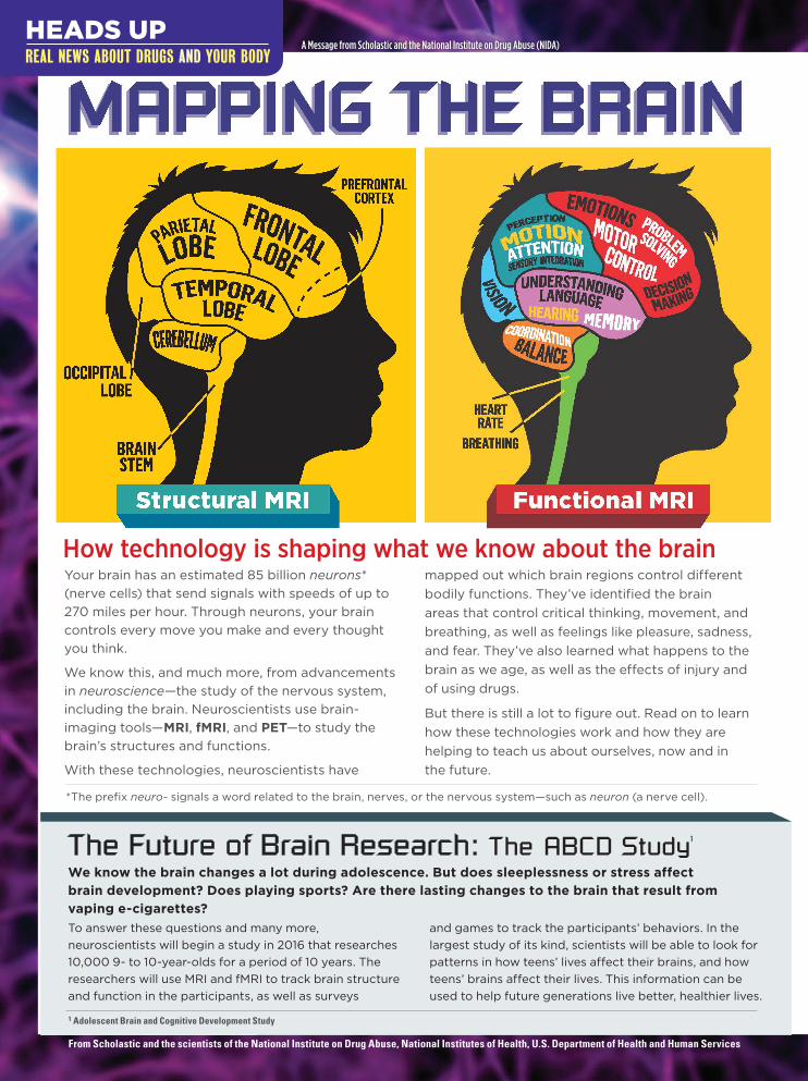
MAPPING THE BRAIN
HEADS UP REAL NEWS ABOUT DRUGS AND YOUR BODY TEACHER’S GUIDE
SUBJECT
• Science Literacy• English/Language Arts• Health/Life Skills
COMMON CORE STATE STANDARDS
• RST.9 Analyze structure of relationships among concepts in a text
• W.9 Draw evidence to support analysis and reflection
NEXT GENERATION SCIENCE STANDARDS
NATIONAL SCIENCE EDUCATION STANDARDS
• LS1.A Structure and Function
• LS1.D Information Processing
• Structure and Function in Living Things
• Personal and Community Health
The brain is the body’s most complex organ, controlling everything from our heartbeat to how we make important decisions. Through research and the use of brain-imaging tools, neuroscientists are learning just how critical the teen years are for brain development. This article explains for students how brain-imaging techniques work, how they apply to their lives, and also highlights some of the things neuroscientists have learned about drug use. It may also inspire them to want to learn more about neuroscience!

1. a. The image below shows regions of the brain. What type of scan is shown? What kind of information about the brain does it provide (structure, function, or cellular)?
b. Label the highlighted regions of the brain.
SCHOLASTIC.COM/HEADSUP
STUDENT WORK SHEET
Can You Think Like a Neuroscientist?Use the information from “Mapping the Brain” to answer the questions below and analyze real images of the brain. Record your responses on a separate sheet of paper as necessary.
FROM SCHOLASTIC AND THE SCIENTISTS OF THE NATIONAL INSTITUTE ON DRUG ABUSE, NATIONAL INSTITUTES OF HEALTH, U.S. DEPARTMENT OF HEALTH AND HUMAN SERVICES
2
NORMAL USING DRUGS
3
1
Imag
es: f
rom
top,
©de
radr
ian/
Flic
kr; ©
201
3, O
xfor
d U
nive
rsity
Pre
ss; B
rook
have
n N
atio
nal L
abor
ator
y.
2. The image at right shows brain activity levels while a person is laughing. What type of scan is shown? What kind of information does it provide?
3. a. These images (below right) were created using radiotracers that attached to dopamine receptors in the brain. What type of scan is shown? What kind of information does it provide?
b. Dopamine is the brain chemical that helps us feel pleasure. Dopamine levels are higher in the brain on the left. What can you conclude about how using drugs affects the brain?
4. What type of imaging technique would you use to learn about how the size of the prefrontal cortex changes as kids grow into adulthood? Explain your answer.
5. What type of imaging technique would you use to find out which areas of the brain are active when a person considers making a risky decision? Explain your answer.
HEADS UP REAL NEWS ABOUT DRUGS AND YOUR BODY
Your brain has an estimated 85 billion neurons* (nerve cells) that send signals with speeds of up to 270 miles per hour. Through neurons, your brain controls every move you make and every thought you think.
We know this, and much more, from advancements in neuroscience—the study of the nervous system, including the brain. Neuroscientists use brain- imaging tools—MRI, fMRI, and PET—to study the brain’s structures and functions.
With these technologies, neuroscientists have
mapped out which brain regions control different bodily functions. They’ve identified the brain areas that control critical thinking, movement, and breathing, as well as feelings like pleasure, sadness, and fear. They’ve also learned what happens to the brain as we age, as well as the effects of injury and of using drugs.
But there is still a lot to figure out. Read on to learn how these technologies work and how they are helping to teach us about ourselves, now and in the future.
*The prefix neuro- signals a word related to the brain, nerves, or the nervous system—such as neuron (a nerve cell).
How technology is shaping what we know about the brain
To answer these questions and many more, neuroscientists will begin a study in 2016 that researches 10,000 9- to 10-year-olds for a period of 10 years. The researchers will use MRI and fMRI to track brain structure and function in the participants, as well as surveys
and games to track the participants’ behaviors. In the largest study of its kind, scientists will be able to look for patterns in how teens’ lives affect their brains, and how teens’ brains affect their lives. This information can be used to help future generations live better, healthier lives.
The Future of Brain Research: The ABCD Study1
We know the brain changes a lot during adolescence. But does sleeplessness or stress affect brain development? Does playing sports? Are there lasting changes to the brain that result from vaping e-cigarettes?
Structural MRI
From Scholastic and the scientists of the National Institute on Drug Abuse, National Institutes of Health, U.S. Department of Health and Human Services
1 Adolescent Brain and Cognitive Development Study
Functional MRI
A Message from Scholastic and the National Institute on Drug Abuse (NIDA)

Imag
es: c
lock
wis
e fr
om to
p le
ft, ©
dera
dria
n/Fl
ickr
; © 2
013,
Oxf
ord
Uni
vers
ity
Pres
s; B
rook
have
n N
atio
nal L
abor
ator
y; b
ackg
roun
d: S
ebas
tian
Kaul
itzk
i/Sh
utte
rsto
ck.
SOMETHING WE’VE LEARNED
Dopamine is the brain chemical that helps us feel pleasure. By following radiotracers for dopamine receptors, PET scans have shown that using drugs heavily reduces the number of these receptors. Fewer receptors indicates less dopamine activity in the brain. This finding helps explain why people addicted to drugs experience less pleasure from everyday activities. They begin
to crave the drug to get their dopamine activity back up to normal.
WHAT IT SHOWS
Areas of the brain that are active during a task.
HOW IT WORKS
A person lies in an MRI machine while doing an activity such as looking at an image, hearing a sound, laughing at something funny, or completing a puzzle. The areas of the brain that are active during the behavior have increases in blood flow and blood oxygen levels. A computer analyzes these changes to map brain function.
SOMETHING WE’VE LEARNED
In studies where adolescents played a game to earn rewards, their brain scans showed higher activity in the area of the brain that processes motivation and pleasure (the nucleus accumbens2) compared with the area of the brain that guides thoughtful decision making (the prefrontal cortex). Scientists think this imbalance in activated brain regions may lead teens to focus more on the possible rewards of a decision than on any drawbacks. This could increase a person’s risk for using drugs.
WHAT IT SHOWS
A detailed image of the structure (size and shape) of tissues, organs, and bones. Also shows the presence of disease.
HOW ITWORKS A person lies still in an MRI machine, which surrounds the body with a magnetic field and emits radio waves. Hydrogen atoms in the water of tissues and bones absorb and then release the energy from the radio waves. A computer maps and measures these changes to create an image. Changes in the size of tissues (such as from diseases like cancer that cause tumors) can increase the amount of water in different parts of the body, which can be detected by MRI scans.
SOMETHING WE’VE LEARNED
MRI scans of the brain have shown that people who have been using drugs for a long time have a smaller prefrontal cortex than people who have not been using drugs. The prefrontal cortex is the area where decision making occurs.
Structural MRIStructural Magnetic Resonance Imaging
More Info: For additional facts about the brain, visit scholastic.com/headsup and teens.drugabuse.gov.
2 The nucleus accumbens is a brain structure located at the base of the frontal lobe deep inside the brain. It does not appear on the MRI scan shown on this page.
WHAT IT SHOWS
The brain and body at the cellular level.
HOW IT WORKS
PET scans use radioactive chemicals, called radiotracers, that are injected into the body. The radiotracers go to different areas depending on the chemical that is used. The PET machine detects the radiotracers and computer programs use colors to show their location.
Functional MRI (fMRI)Functional Magnetic Resonance Imaging
PET Positron Emission Tomography
The color areas in the fMRI above show brain regions active during laughter.
HIGH
LOW
A normal brain (left) has high levels of dopamine. Using drugs may make levels decrease (right).
NORMAL USING DRUGS
Temporal Lobe
Prefrontal Cortex
Occipital Lobe
Parietal Lobe
Cerebellum
Frontal Lobe
Brain Stem

Dear Teacher, The vocabulary list on the following pages is drawn from the “Mapping the Brain” student article and work sheet.
This vocabulary can be previewed with students prior to reading or reinforced with students afterward. Encourage students to incorporate these words into their writing and discussion of the “Mapping the Brain” student article and the “How to Think Like a Neuroscientist” work sheet.
The vocabulary list integrates two different tiers of vocabulary: words that would be used across several content areas, such as advancement, motivation, or process, and domain-specific words such as cellular, cerebral, and chemicals.
Some suggestions for students to help their understanding include:
• organizing concept maps that include word parts, synonyms, antonyms, and examples;
• composing memory aids that explain the words or use them in a meaningful context;
• employing the words to create newspaper articles, stories, or poems.
Sources: Unless otherwise noted, definitions below are sourced or adapted from:
• Grades 6–8: The American Education Publishing Children’s Dictionary
• Grades 9–12: Merriam-Webster Collegiate Dictionary
VOCABULARY LIST GRADES 6–12
[Continue to vocabulary sheet on next page.]
Supplement to “Mapping the Brain”
• Student Article: scholastic.com /headsup/mappingthebrain
• Teacher’s Guide (includes work sheet): scholastic.com/headsup/teachers /mappingthebrain

VOCABULARY LIST FROM “MAPPING THE BRAIN”
• absorb (verb): to take in or draw in
• activated (adjective): to be set into motion; to be made active
• addicted (adjective): to be dependent on something and not able to give it up, such as a drug
• adolescence (noun): the period of life when a child develops into an adult
• advancement (noun): the act, process, or result of moving forward; progress
• analyze (verb): to study closely and carefully
• atom (noun): the smallest component of an element that can exist by itself, consisting of protons, neutrons, and electrons
• brain stem (noun): the lower part of the brain that connects to the spinal cord and controls certain automatic functions, such as breathing
• cellular (adjective): of, related to, or made of cells from a living thing
• cerebellum (noun): the area of the brain located between the brain stem and the cerebrum that controls voluntary muscle coordination and balance
• cerebral (adjective): relating to or involving the cerebrum
• cerebrum (noun): the upper part of the brain that is split into two hemispheres. These two hemispheres are referred to as cerebral hemispheres.
• chemical (noun): a substance, such as an element or a mix of elements (compound), that is made by a chemical process
• complex (adjective): having a complicated structure; not simple
• crave (verb): to have a strong desire for something
• critical thinking (noun): thought process characterized by clear and rational thinking that relies on facts and evidence
• development (noun): the act or process of growing or causing something to become larger or more advanced
• dopamine (noun): a neurotransmitter chemical that helps transmit signals in the brain and is associated with feelings of pleasure
• drawback (noun): something that causes problems or creates a disadvantage
• emit (verb): to send out
• frontal lobe (noun): the front part of each cerebral hemisphere of the brain in which emotions, problem solving, motor control, and decision-making processes are controlled
• function (noun): the purpose or activity for which a thing exists or is used
• functional MRI (fMRI) (noun): a brain imaging tool that shows areas of the brain that are active during a behavior
• generation (noun): a group of people born and living during the same time
• hemisphere (noun): as in brain hemisphere; either of the two halves of the upper part of the brain in humans and other animals
• hydrogen (noun): a chemical element that has no color or smell and that is the simplest, lightest, and most common element. A single molecule of water contains two hydrogen atoms and one oxygen atom.
• imaging (noun): the act of creating a medical or scientific image that shows a picture of the inside of a body
• imbalance (noun): a state or condition when different things do not occur in equal or proper amounts
• inject (verb): to force a liquid into something, such as with a needle
• integration (noun): the act or process of combining different things
• lasting (adjective): existing or continuing for a long time
• lobe (noun): a somewhat rounded part of a body organ or division of a body organ
• magnetic (adjective): relating to a magnet or magnetism

VOCABULARY LIST FROM “MAPPING THE BRAIN”
• magnetic field (noun): a region of space near a magnetic body where magnetic forces can be detected
• motivation (noun): the condition of being eager to act or work
• nervous system (noun): the system of nerves in your body that sends messages for controlling movement and feelings, for example
• neuron (noun): a nerve cell that carries messages between the brain and other parts of the body and is a basic part of the nervous system
• neuroscience (noun): the area of science related to the nervous system, including the nerves, brain, and spinal cord
• neuroscientist (noun): a scientist who studies the nervous system, including the nerves, brain, and spinal cord
• nucleus accumbens (noun): the area of the brain that processes motivation and pleasure
• occipital lobe (noun): the lobe of each cerebral hemisphere located at the back of the brain that contains the visual-processing area
• organ (noun): a structure in a plant or animal that performs a specific function, such as the brain or the heart
• parietal lobe (noun): the rear part of each hemisphere in the upper part of the brain that contains an area concerned with sensory perception and integration, motion, and attention
• participant (noun): a person who is involved in an activity, event, or study
• perception (noun): the ability to notice or understand something using one of your senses
• positron emission tomography (PET) (noun): a tool that produces images of activity inside the body by detecting energy given off by radioactive substances that have been injected into the body
• prefrontal cortex (noun): the front part of the frontal lobe area of the brain that is involved in complex decision making and thinking
• process (noun): a series of actions that produce something to lead to a certain result
• radio wave (noun): an electromagnetic wave that is used for sending signals through the air without using wires
• radioactive (adjective): emitting energy or high-energy particles (radiation) as a result of the decay of unstable atoms
• radiotracer (noun): a radioactive substance injected into the body that is used to track and study processes in the body
• receptor (noun): a cell or group of cells that receives signals and has an attraction for specific chemicals
• reduce (verb): to make smaller in size, amount, or number
• region (noun): a particular area of something
• release (verb): to set free, or to allow a substance to enter the air, water, bloodstream, etc.
• scan (noun): a medical or scientific image that shows a picture of the inside of a body
• sensory (adjective): of or related to your physical senses (touch, taste, smell, sight, and hearing)
• structural MRI (noun): a brain imaging tool that shows a detailed image of the structure of tissues, organs, and bones in the body
• technology (noun): a machine or method that is created or invented by scientists to solve problems
• temporal lobe (noun): the large lobe of each cerebral hemisphere that is situated in front of the occipital lobe and contains a sensory area associated with hearing, understanding language, and memory
• tissue (noun): the group of cells that forms the parts and organs in a plant or animal
• tumor (noun): a mass of tissue that is made up of abnormal cells
• vaping (verb): inhaling and exhaling vapor from an electronic cigarette