Embed Size (px)
Citation preview
中華放射醫誌 Chin J Radiol 2004; 29: 87-91 87
We report neuroimaging studies of Marchiafava -Bignami disease (MBD) diagnosed on a 39-year-oldalcoholic man with acute clinical deterioration.Initially, cranial CT and MRI demostrated diffuselyedematous change from the genu to the splenium ofthe corpus callosum (CC) characteristic for acutestage of MBD. Decreased blood perfusion in bilat-eral parietal and occipital lobes was also noted inSPECT study. Prompt treatment with thiamine andvitamin B complex resulted in clinical improvement.8 months later, follow-up MR imaging showed typ-ical features of chronic MBD—atrophy with cysticchange in the CC.
To our knowledge, this is the first case of MBDreported in Taiwan literature.
Key words: Alcoholism; Brain, corpus callosum;Brain, CT; Brain, MRI; Brain, SPECT;Machiafava-Bignami disease
Marchiafava - Bignami disease (MBD), patholog-ically characterized by necrosis and demyelinationwithin the medial layer of the corpus callosum (CC), isa rare disorder observed in people with chronic alco-holism and/or malnutrition [1-3]. In the past, thediagnosis of MBD was only made in the postpartumexam. However, after the introduction of CT and espe-cially MRI, more cases were reported in its early stageand more chronic subtype were diagnosed in livingpeople. Nevertheless, efficient treatment has not estab-lished, the prognosis is still pessimistic [1, 4].
CASE REPORT
A 39-year-old right-handed locksmith had a 27-year history of alcohol dependency, varied in amountand kinds. He often drinks all day with irregular diet.About 2 weeks before, he experienced sudden onset ofslurred speech and mentality disturbance in themorning that never happened before. On admission, hewas drowsy with poor attention. Wide-based, ataxicgait with deviation to right was noted. He had normalmuscle power with mild hyperreflexia bilaterally. EEGfinding and the results of laboratory examinations wereunremarkable.
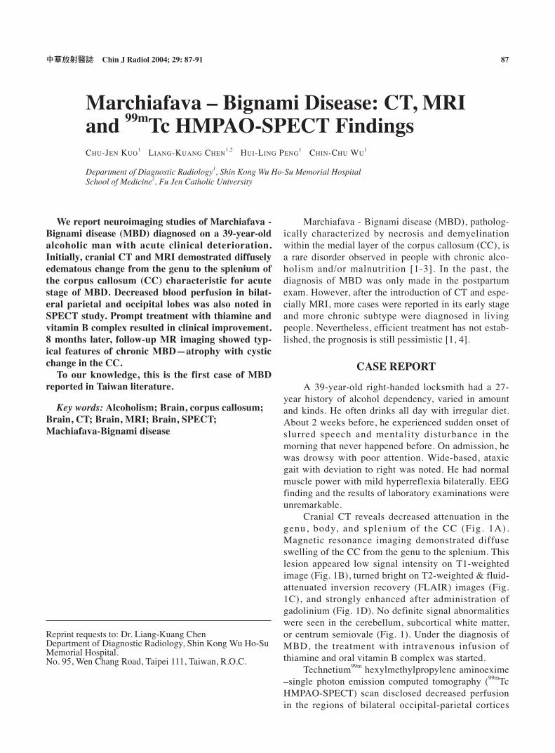
Cranial CT reveals decreased attenuation in thegenu, body, and splenium of the CC (Fig. 1A).Magnetic resonance imaging demonstrated diffuseswelling of the CC from the genu to the splenium. Thislesion appeared low signal intensity on T1-weightedimage (Fig. 1B), turned bright on T2-weighted & fluid-attenuated inversion recovery (FLAIR) images (Fig.1C), and strongly enhanced after administration ofgadolinium (Fig. 1D). No definite signal abnormalitieswere seen in the cerebellum, subcortical white matter,or centrum semiovale (Fig. 1). Under the diagnosis ofMBD, the treatment with intravenous infusion ofthiamine and oral vitamin B complex was started.
Technetium99m hexylmethylpropylene aminoexime–single photon emission computed tomography (99mTcHMPAO-SPECT) scan disclosed decreased perfusionin the regions of bilateral occipital-parietal cortices
Reprint requests to: Dr. Liang-Kuang ChenDepartment of Diagnostic Radiology, Shin Kong Wu Ho-SuMemorial Hospital.No. 95, Wen Chang Road, Taipei 111, Taiwan, R.O.C.
Marchiafava – Bignami Disease: CT, MRIand 99mTc HMPAO-SPECT FindingsCHU-JEN KUO
1 LIANG-KUANG CHEN1,2 HUI-LING PENG
1 CHIN-CHU WU1
Department of Diagnostic Radiology1, Shin Kong Wu Ho-Su Memorial HospitalSchool of Medicine2, Fu Jen Catholic University
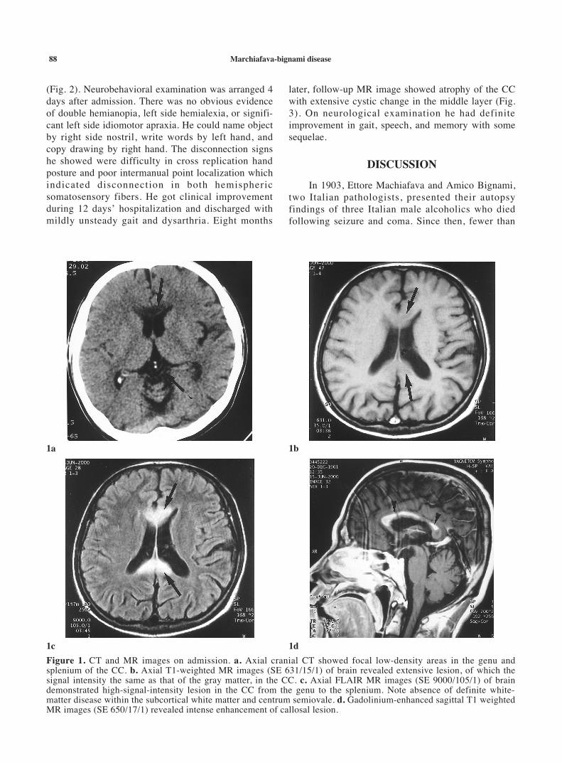
(Fig. 2). Neurobehavioral examination was arranged 4days after admission. There was no obvious evidenceof double hemianopia, left side hemialexia, or signifi-cant left side idiomotor apraxia. He could name objectby right side nostril, write words by left hand, andcopy drawing by right hand. The disconnection signshe showed were difficulty in cross replication handposture and poor intermanual point localization whichindicated disconnection in both hemisphericsomatosensory fibers. He got clinical improvementduring 12 days’ hospitalization and discharged withmildly unsteady gait and dysarthria. Eight months
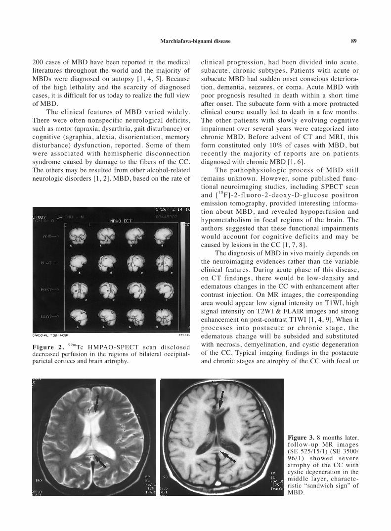
later, follow-up MR image showed atrophy of the CCwith extensive cystic change in the middle layer (Fig.3). On neurological examination he had definiteimprovement in gait, speech, and memory with somesequelae.
DISCUSSION
In 1903, Ettore Machiafava and Amico Bignami,two Italian pathologists, presented their autopsyfindings of three Italian male alcoholics who diedfollowing seizure and coma. Since then, fewer than
Marchiafava-bignami disease88
Figure 1. CT and MR images on admission. a. Axial cranial CT showed focal low-density areas in the genu andsplenium of the CC. b. Axial T1-weighted MR images (SE 631/15/1) of brain revealed extensive lesion, of which thesignal intensity the same as that of the gray matter, in the CC. c. Axial FLAIR MR images (SE 9000/105/1) of braindemonstrated high-signal-intensity lesion in the CC from the genu to the splenium. Note absence of definite white-matter disease within the subcortical white matter and centrum semiovale. d. Gadolinium-enhanced sagittal T1 weightedMR images (SE 650/17/1) revealed intense enhancement of callosal lesion.
1a 1b
1c 1d
200 cases of MBD have been reported in the medicalliteratures throughout the world and the majority ofMBDs were diagnosed on autopsy [1, 4, 5]. Becauseof the high lethality and the scarcity of diagnosedcases, it is difficult for us today to realize the full viewof MBD.
The clinical features of MBD varied widely.There were often nonspecific neurological deficits,such as motor (apraxia, dysarthria, gait disturbance) orcognitive (agraphia, alexia, disorientation, memorydisturbance) dysfunction, reported. Some of themwere associated with hemispheric disconnectionsyndrome caused by damage to the fibers of the CC.The others may be resulted from other alcohol-relatedneurologic disorders [1, 2]. MBD, based on the rate of
clinical progression, had been divided into acute,subacute, chronic subtypes. Patients with acute orsubacute MBD had sudden onset conscious deteriora-tion, dementia, seizures, or coma. Acute MBD withpoor prognosis resulted in death within a short timeafter onset. The subacute form with a more protractedclinical course usually led to death in a few months.The other patients with slowly evolving cognitiveimpairment over several years were categorized intochronic MBD. Before advent of CT and MRI, thisform constituted only 10% of cases with MBD, butrecently the majority of reports are on patientsdiagnosed with chronic MBD [1, 6].
The pathophysiologic process of MBD stillremains unknown. However, some published func-tional neuroimaging studies, including SPECT scanand [18F]-2-fluoro-2-deoxy-D-glucose positronemission tomography, provided interesting informa-tion about MBD, and revealed hypoperfusion andhypometabolism in focal regions of the brain. Theauthors suggested that these functional impairmentswould account for cognitive deficits and may becaused by lesions in the CC [1, 7, 8].
The diagnosis of MBD in vivo mainly depends onthe neuroimaging evidences rather than the variableclinical features. During acute phase of this disease,on CT findings, there would be low-density andedematous changes in the CC with enhancement aftercontrast injection. On MR images, the correspondingarea would appear low signal intensity on T1WI, highsignal intensity on T2WI & FLAIR images and strongenhancement on post-contrast T1WI [1, 4, 9]. When itprocesses into postacute or chronic stage, theedematous change will be subsided and substitutedwith necrosis, demyelination, and cystic degenerationof the CC. Typical imaging findings in the postacuteand chronic stages are atrophy of the CC with focal or
Marchiafava-bignami disease 89
Figure 2. 99mTc HMPAO-SPECT scan discloseddecreased perfusion in the regions of bilateral occipital-parietal cortices and brain artrophy.
Figure 3. 8 months later,follow-up MR images(SE 525/15/1) (SE 3500/96/1) showed severeatrophy of the CC withcystic degeneration in themiddle layer, characte-ristic “sandwich sign” ofMBD.
diffuse presence of hypointensity on T1WI and hyper-intensity on T2WI, particularly in the medial layer,that makes characteristic “sandwich sign” of MBD [1,4, 9]. Extracallosal lesions such as in subcorticalwhite matter, centrum semiovale, or anterior/posteriorcommissures have also been described [1, 2, 6].
Another specific process related to chronic alco-holism is Wernicke encephalopathy (WE) whichmainly involves the periventricular regions, the medialthalamic nuclei, third ventricular floor, and mammil-lary bodies. Although WE with similar clinical mani-festations as MBD, image study can make well differ-entiation between them. According to above-mentioned findings, we thought that our patient wassubjected to acute stage of MBD when initial studiesperformed with typical imaging pictures of this stage,and this disease progressed into chronic stage provedby characteristic findings on follow-up images.
In recent years several reported cases, as well asour own, developed acute or subacute MBD. Theywere admitted to a medical ward due to sudden onsetor rapid deterioration of neurological symptoms.Based on imaging studies, especially MRI, precisediagnosis was made immediately. Although there is nospecific therapy established for MBD, subsequenttreatment with thiamine or vitamin B complex resultedin good recovery [3-7]. Our findings, combined withother reports [3-7], suggest that acute or subacuteMBD may not have rapid course leading to death, andearly diagnosis with CT and MRI followed prompttreatment with thiamine or vitamin B complex appearsto improve the prognosis of MBD. ◆
REFERENCE
1. Christian GK, Beau MA, Rand CA, J. Daniel R, MarkL, Ruben CG. Marchiafava-Bignami disease: literaturereview and case report. NNBN 2000; 13: 67-76
2. Franco F, Fernando C, Manrico G, et al. Marchiafava-Bignami disease: computed tomograghic scan, 99mTcHMPAO-SPECT, and FLAIR MRI findings in a patientwith subcortical aphasia, alexia, bilateral agraphia, andleft-handed deficit of constructional ability. ArchNeurol 1999; 46: 107-110
3. Achim G, Christina B, Manfred O, Andreas S, MichaelGH. Marchiafava-Bignami disease: reversibility of neu-roimaging abnormality. J Comput Assist Tomogr 1998;22: 503-504
4. Muneshige T, Hiroshi M, Shoki T, Hiroshi O, Yasuto I,Yuzo I. A case of Marchiafava-Bignami disease withcomplete recovery: sequential imaging documentingimprovement of callosal lesions. Tohoku J Exp Med1997; 182: 175-179
5. Kazuya Y, Shotai K, Shuhei Y, Hiromi K, Katsuhisa N.Reversible corpus callosum lesions in a patient withMchiafava-Bignami disease: serial changes on MRI.Eur Neurol 1997; 37: 192-193
6. Ruiz MJ, Martínez PBA, Ruibal M, Urtasun M, VillanaJ, Martí MJF. Marchiafava-Bignami disease with wide-spread extracallosal lesion and favourable course.Neuroradiology 1999; 41: 40-43
7. Motoki S, Kenji I, Takeshi S, Masanobu U, Michio S.Reductions in the bilateral parietal and occipital cere-bral blood flow and metabolism in a patient withMarchifava-Bignami disease. J Neural 1999; 246: 607-608
8. Kazunari I, Yoshitaka I, Masahiro S, Hajime K, EtsuroM. Regional cerebral glucose metabolism and bloodflow in a patient with Marchifava-Bignami disease.AJNR 1999; 20: 1249-1251
9. Brian JF, Brian SK. Incidentally diagnosedMarchiafava-Bignami disease. AJR 1999; 173: 1713-1714
Marchiafava-bignami disease90
Marchiafava-bignami disease 91
Marchiafava-Bignami Disease:電腦斷層、磁振造影、單光子射出電腦斷層的影像表現
郭鑄仁1 陳良光1,2 彭惠玲1 吳金珠1
新光吳火獅紀念醫院 放射診斷科1
天主教輔仁大學 醫學院 醫學系2
我們提出Machiafava-Bignami Disease(MBD)神經影像上的發現,MBD在一位三十九
歲酗酒的男性被診斷出來,他呈現急性臨床變化。最初,頭部的電腦斷層和磁振造影影像指出
從胼胝體的膝部到喙部產生了瀰漫水腫樣的病變,此為急性期特殊表現。單光子射出電腦斷層
檢查指出在兩側大腦頂葉及枕葉有血流灌注減少的情形。即刻以Thiamine和維他命B群治療使
他在臨床上逐漸地好轉。8個月後,追蹤的磁振造影影像表現出慢性MBD典型特徵─胼胝體萎
縮合併胼胝體內層囊狀病變。依據我們所知,這是台灣文獻中有關MBD的第一篇個案報告。
關鍵詞:Machiafava-Bignami Disease;大腦,胼胝體;大腦,電腦斷層;大腦,磁振造
影;大腦,單光子射出電腦斷層掃描;酗酒