Embed Size (px)
Citation preview
Marginal Integrity of Glass Ionomer and All Ceramic Restorations
by
Christopher Mark Hanson Lieutenant, Dental Corps
United States Navy
A thesis submitted to the Faculty of the Comprehensive Dentistry Graduate Program
Naval Postgraduate Dental School Uniformed Services University of the Health Sciences
in paitial fulfillment of the requirements for the degree of Master of Science
in Oral Biology
June 2015

Naval Postgraduate Dental School Uniformed Services University of the Health Sciences
Bethesda, Maryland
CERTIFICATE OF APPROVAL
MASTER'S THESIS
This is to certify that the Master's thesis of
Christopher Mark Hanson
has been approved by the Examining Committee for the thesis requirement for the Master of Science degree in Oral Biology at the June 2015 graduation.
Thesis Committee:
ii
Ye, Ling, D.D.S., Ph.D. LCDR, DC, USN Thesi('\llP.ervisor, Dental Research Department
~~'JJ\fvAJ\,nM~ . ctfulinaro, Joseph, D.D.S, M.S.
CAPT, DC, USN Program Director, Comprehensive Dentistry Depa11ment
S:tX~ Kooistra, Scott, D.D.S CAPT, DC, USN
Operati~~:ellti !~~ Departm:L--
Huber, ayson, D.D.S., M.S. LCDR, DC, USN Comprehensive Dentistry Department
Arena, arc, D.M.D., M.S. CAPT,DC, SN
ad, Comprehensive Dentistry Depat1ment
Munro, Glenn, D.D.S., MBA CAPT, DC, USN Dean, Naval Postgraduate Dental School
The author hereby certifies that the use of any copyrighted material in the thesis manuscript titled:
Marginal Integrity of Glass Ionomer and All Ceramic Restorations
is appropriately acknowledged and, beyond brief excerpts, is with the permission of the copyright owner.
Clu·istopher Mark Hanson Lieutenant, Dental Corps Comprehensive Dentistty Graduate Program Naval Postgraduate Dental School JUN 2015
NAVAL POSTGRADUATE DENTAL SCHOOL CHRISTOPHER MARK HANSON
2015
This thesis may not be re-printed without the expressed written pe1mission of the author.
iii
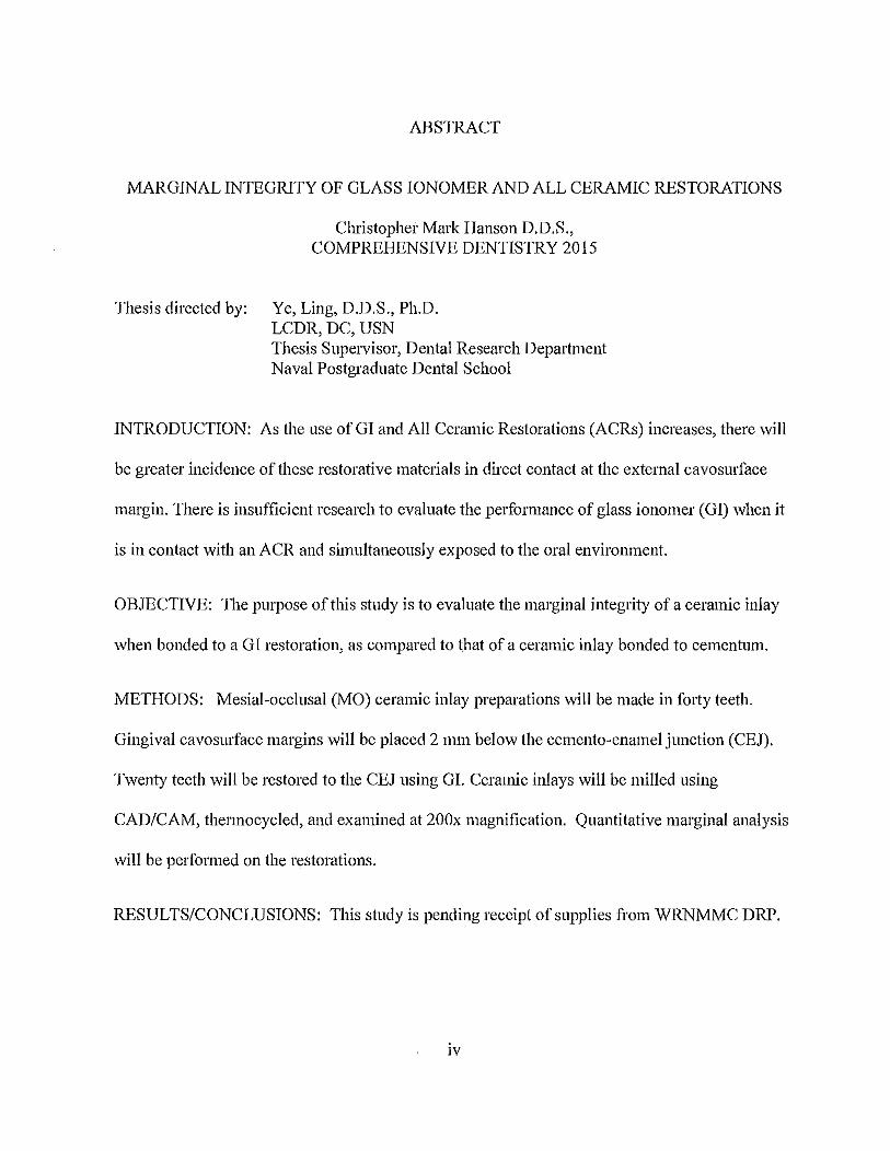
ABSTRACT
MARGINAL INTEGRITY OF GLASS ION OMER AND ALL CERAMIC RESTORATIONS
Clu·istopher Mark Hanson D.D.S., COMPREHENSIVE DENTISTRY 2015
Thesis directed by: Ye, Ling, D.D.S., Ph.D. LCDR, DC, USN Thesis Supervisor, Dental Research Department Na val Postgraduate Dental School
INTRODUCTION: As the use of GI and All Ceramic Restorations (ACRs) increases, there will
be greater incidence of these restorative materials in direct contact at the external cavosurface
margin. There is insufficient research to evaluate the performance of glass ionomer (GI) when it
is in contact with an ACR and simultaneously exposed to the oral environment.
OBJECTIVE: The purpose of this study is to evaluate the marginal integrity of a ceramic inlay
when bonded to a GI restoration, as compared to that of a ceramic inlay bonded to cementum.
METHODS: Mesial-occlusal (MO) ceramic inlay preparations will be made in forty teeth.
Gingival cavosurface margins will be placed 2 mm below the cemento-enamel junction (CEJ).
Twenty teeth will be restored to the CEJ using GI. Ceramic inlays will be milled using
CAD/CAM, thermocycled, and examined at 200x magnification. Quantitative marginal analysis
will be performed on the restorations.
RESULTS/CONCLUSIONS: This study is pending receipt of supplies from WRNMMC DRP.
IV
TABLE OF CONTENTS
Page
LIST OF TABLES..................................................................................................... vi
LIST OF ABBREVIATIONS.................................................................................... vii
CHAPTER
I. INTRODUCTION ..................................................................... .. 1
IL REVIEW OF THE LITERATURE ............................................ . 3
Indirect All Ceramic Restorations ............................................... 3
Resin Bonding.............................................................................. 4 The Use of Glass Iono111er ........................................................... 5 Marginal Integrity in Dental Restorations............................. 8 Challenging Restorative Conditions.................................. 9 Su111mary...... . . . . . . . . . . . . . . . . . . . . . . . . . . . . . . . . . . . . . . . . . . . . . . . . . . . . . . . . . . . . 11
III. MATERIALS AND METHODS ................................................ . 14
IV. RESULTS ................................................................................... . 17
v. DISCUSSION ............................................................................. . 17
VI. CONLUSIONS .......................................................................... .. 17
REFERENCES 18
v

LIST OF TABLES
Table Page
I. Treatment Groups ............................................................................... 15
vi

ACR AMR ART DME GI MCR MO NIDCR
LIST OF ABBREVIATIONS
All Ceramic Restoration All Metal Restoration Atraumatic Restorative Treatment Deep Margin Elevation Glass Ionomer Metal Ceramic Restoration Mesial-Occlusal National Institute for Dental and Craniofacial Research
vii
CHAPTER I: INTRODUCTION
Metal casting utilizing the lost wax teclmique probably dates back to ancient China or
Egypt. The first use of indirect cast metal dental restorations is generally attributed to Dr.
Swasney in 1890 (Asgar, 1998). With the introduction of improved casting methods by Taggert
and Jamieson in 1907, precision casting of full gold crowns became readily attainable (Schulein,
2005). Since that time, indirect extra-coronal cast metal dental restorations have demonstrated a
well-documented history of success (Small, 2008; Hagman, 1976). The first indirect cast
restorations were typically fabricated out of gold. They exhibited a coefficient of !henna!
expansion, wear characteristics and strength very similar to natural teeth (Hinman, 1907;
Macdonald, 1907). However, their disadvantages included poor esthetics and the high cost of
gold.
Ceramics are defined as non-organic, non-metallic materials created by heating minerals
at high temperatures (Shenoy & Shenoy, 2010). Dental ceramics are composite materials whose
structure can vary from amorphous to polycrystalline. A ceramic can be classified by the ratio of
glass to crystalline present in its composition. This ratio will determine the microstructure and
type of internal strncture exhibited by the ceramic. The microstructure of the ceramic will define
the characteristics and physical properties of the material (Giordano &McLaren, 2010).
As the desire for tooth-colored restorations increased, the metal ceramic restoration
(MCR) gained popularity (Cln·istensen, 2009). The first MCRs were developed by Weinstein in
the 1950's. The first MCRs were composed of porcelain powders using 11-15% percent K10
and subjected to temperatures of700-1200 C0• The MCR appeared more like a natural tooth,
while providing the strength necessary to function well under occlusal load (Christiansen, 2003).

However, the high opacity and occasionally visible metal margins left practitioners searching for
a better esthetic option (Helvey, 2010).
2

CHAPTER II: Review of the Literature
Indirect All -Ceramic Restorations
Indirect all-ceramic restorations (ACRs) were introduced independently by Horn,
Simonsen, and Calamia in the early 1980s (Spear & Holloway, 2008). They appealed to patients
who desired a metal-free restoration (Fasbinder, 2006). The ACR represented a significant
esthetic improvement over all-metal and metal-ceramic restorations (Kelly, 2004). Ceramic
materials with high aluminosilicate glass, such as feldspathic porcelain, contain fillers such as
leucite, nepheline, or albite added to improve their physical properties. These high glass content
ACRs appear very similar to natural teeth. However, due to the irregular microstructure of the
glass matrix infused with fillers, they do not possess fracture resistance comparable to natural
teeth. This limited ACRs composed primarily of glass to anterior areas (Kelly & Benetti, 2011).
To improve the physical properties of the ACR, filler particles such as lithium disilicate,
alumina, and spine! were added to the glass matrix. This gives greater strength to the restoration
(Kelly, 2008). With a high filler content and lower glass content, these ceramics have greater
fracture resistance. These improved ceramics can be used in areas with significant lateral and
protrusive forces applied to them, but where esthetics is still important (Spear & Holloway,
2008).
Most recently, ceramics with a polycrystalline structure have been introduced. Instead of
glass, these ceramics are composed of a strong matrix of alumina or zirconia. An additive,
known as a dopant, such as yttrium, cerium, and aluminum can be added to the zirconia matrix,
while magnesium is added to a matrix composed of alumina (Komine, Blatz & Matsumura,
2010). These dopants are added to improve the optical appearance of the well-ordered structure
of these ACRs (Luthard, Sandkuhl & Reitz, 1999). The polycrystalline structure has a much
3
higher resistance to fracture than the less dense and irregular composition of glass-containing
ACRs (Griggs, 2007).
The development of Computer Aided Design/Computer Aided Manufacturing
(CAD/CAM) of ceramic restorations have the increased ability of dentists to deliver high quality
ACRs (Miyazaki, Hotta, Kunii, Kuriyama, & Tamaki, 2009; Griggs, 2007). CAD/CAM ceramic
restorations can often be made in one appointment. This eliminates the need to fabricate a
physical master impression, stone casts, and provisional restoration (Mormann, 2006). By using
the CAD/CAM system, the dentist is able to keep the milling process in-office, thus bypassing
the need for the dental laboratory (Beuer, Schwieger & Edelhoff, 2008). The milling process is
faster, and most importantly, CAD/CAM restorations have become more accurate in terms of
anatomic appearance, and interproximal and occlusal contacts (Rocca, Bonnafous, Rizcalla &
Krejci, 2010). Research has shown that the Jong term acceptability and marginal integrity of
single crowns fabricated by CAD/CAM technology are similar to single crowns fabricated by
traditional laboratory methods over a 3 year time period (Wittneben, Wright, Weber, & Gallucci,
2009).
Resin Bonding
The ACR has benefited from advances in resin bonding (Hopp & Land, 2013). A
considerable advantage of the ACR is the dentist is able to adjust the shade of the final result by
using resin cements (Oztiirk & colleagues, 2013). There is an increasing range of shades in resin
cements that can be used to modify the shade and value of the restoration. The resin cement can
also block out any potential imperfections present in the prepared tooth from being externally
visible (Nie, Agustin & Douglas, 2014).
4

Modem bonding techniques involve treating the prepared tooth with phosphoric acid and
a resin bonding agent (Magne, Schlichting, Maia & Baraterie, 2010). The ACR is etched with
hydrofluoric acid, which exposes surface area for mechanical retention. The restoration is then
treated with silane to act as a coupling agent between the restoration and bonding resin cement
(Oba & colleagues, 2014). The silane agent forms a covalent hydrogen and chemical bond
between the organic matrix of the resin and the exposed hydroxyl group on the surface of the
treated ceramic (Anchieta, Rocha, Almeida, Freitas-Junior & Maitini, 2011).
The Use of Glass Ionomer in Restorative Dentistry
Glass ionomers (Gis) were developed to combine the favorable physical and esthetic
properties of resin composites with the fluoride release of silicate cements (Wilson & Kent,
1972). GI is created when a strontium or calcium alumino-flouro-silicate glass powder is mixed
with a polyalkenoic acid (Mitra, 1989). The first Gis set using only a chemical reaction initiated
by mixing the acid and base (Wilson and Prosser, 1984).During the setting process, the acid
groups are neutralized by the glass powder. GI performs well in the presence of moisture and
has low solubility when completely set (Croll, 2001). Water is the medium that facilitates ionic
exchange (Mitra, 1991). This reaction causes the release of fluoride ions. The amount of
fluoride released by GI cement into the prepared tooth was found to be statistically greater than
the fluoride output of other silicon-phosphate cements (Swartz, Phillips & Clark, 1984).
The addition of a photo-polymerizabale resin to traditional glass ionomer yields resin
modified glass ionomer (RMGl) (Croll & Helpin, 1994; Uno & Finger, 1996). The inclusion of
resin in the GI system decreases the setting time. RMGI enhances the cohesive strength of the
restorative material while maintain the tensile and compressive strength of traditional GI (Mitra,
1991; Mitra & Kedrowski, 1994). A recent improvement ofRMGl is known as a nano-ionomer
5

(Carvalho& colleagues, 2012). The decreased size of the filler particles results in a GI with high
polishability and better esthetic results (Croll, 2007).
Decreasing the base-to-acid ratio will yield a GI that can be used as luting cement. Glass
ionomer has been used successfully as luting cement for many years (Nicholson & Croll, 1997;
Hill & Lott, 2011 ), especially for pediatric patients in need for the delivery of stainless steel
crowns and orthodontic bands (Croll & Helpin, 1994).Another useful application of GI is as a
liner under direct restorations (Davidson, 1994). GI liners have positive clinical performance
when used to cover dentin prior to the acid etching step when placing direct composite
restorations (McLean &Wilson, 1977). Glass ionomer can also be an excellent choice as a liner
when a restoration is close to the pulp of the tooth (Prabhakar, Subhadra, Kurtlrnkoti & Shubha,
2008; Rusin, Agee, Suchko & Pashley, 2010). The low modulus of elasticity, and potential for
self-repair following the inevitable micro-fractures seen in the setting process, make GI an
excellent layer between tooth structure and a more brittle direct restorative materials (Ferrari,
1999).
Unlike resin composites, which rely on acid etching and micromechanical retention, GI
bonds chemically to tooth structure via chelation (Mount, 1991). In this process, calcium ions
chelate carboxyl groups on polyacrylic acid to form cross-linked chains of polymers. Carboxyl
groups present on the polymer chains bond with ions on the smface of tooth strncture and the
alumino-silicate powder (Mason & Ferrari, 1994). Over the first 2-3 days, calcium ions are
substituted for aluminum ions, forming an even tighter polymer chain. Over the next month,
available silicate ions react with water to create covalent bonds, increasing the strength of the
tooth-to-GI bond (Khoroushi & Keshani, 2013).
6
The ability of GI to provide extended fluoride release is of great advantage (Croll &
Nicholson, 2002). The slow and continuous release of fluoride by GI has an anti-cariogenic
effect adjacent to the margins of indirect restorations (Tantbirojn, 1997). Recurrent decay is a
major cause of failure of dental restorations (Kopperud, Tveit, Gaarden, Sandvik & Espelid,
2012; Mjor, 1996). This occurs when cariogenic bacteria colonize the area between restoration
and tooth surface. The acid released by these bacteria causes a drop in the local pH, which
demineralizes tooth structure (Torii & colleagues, 2001). Fluoride-releasing dental materials
inhibit the drop in pH at the cavosurface margin (Mayanagi, lgarashi, Washio, Domon-Tawaraya
& Takahashi, 2014).
The positive performance of GI makes it a frequently used restorative material (Frencken,
Leal, & Navarro, 2012). GI has a coefficient of thermal expansion similar to enamel and dentin
(Bullard, Leinfelder & Russell, 1988; Majety & Pujar, 2011). A similar coefficient ofthennal
expansion decreases microleakage and postoperative sensitivity (Weiner, 2011).
GI's ability to bond to tooth structure through chelation makes it very effective when
there is minimal enamel left for bonding (Croll & Nicholson, 2002). In areas ofbuccal or lingual
cervical recession, GI provides a tooth colored restoration with good marginal adaptation
(Siegward, Christiane & Valentin, 2010), when resin-based composite is not clinicaily ideal, but
an amalgam or gold restoration is not desired.
The fluoride releasing properties of GI make it an excellent choice for patients with high
caries risk. Patients with extensive treatment needs often require multi-phased treatment plans.
GI can provide anesthetic and functional restoration in the disease control phase of treatment,
prior to initiation of the corrective phase. Similarly, GI is the most commonly utilized
restorative material in atraumatic restorative treatment (ART) (Frencken, Songpaisan,
7

Phantumvanit & Pilot, 1994; Smales &Yip, 2002) This technique consists of removing gross
decay and placing a restoration to protect the tooth and inhibit further decay (Frenken &
colleagues, 2012). ART is often used in low-income areas, and in countries where modern
dental treatment may not be affordable or available (Frenken, 2010).
Research indicates that Class II resin-based composite and GI restorations have the
highest success rate when the cervical margins are placed in enamel due to the presence of more
enamel rods available for bonding (Demarco, Ramos, Mota, Fonnolo & Justino, 2001; Franco &
colleagues, 2006). However, the snccess rate of Class II restoration decreases when the cervical
margins are placed on dentin (Dietrich, Li:ische, Li:ische, & Ronlet, 1999).
To improve the longevity of restoration, the sandwich teclmique was developed to use GI
as a base under a resin composite restoration (Van Dijken, Kieri & Carlen, 1999). The technique
can improve the marginal adaptation of Class II resin composite restorations when gingival
margins are located in dentin (Dietrich, Li:ische, Li:ische, & Roule!, 1999). The "closed
sandwich" technique uses GI as the restorative material on the internal aspect of the preparation,
and maintains resin-based composite as the restorative material at the entire cavosurface margin.
The "open sandwich" variation maintains the glass ionomer as the external cavosurface margin
at the cervical area (Kirsten, Rached, Mazur, Vieira, & Souza, 2013; Suzuki & Jordan, 1990).
Marginal Integrity in Dental Restorations
The success of indirect dental restorations is directly related to the marginal adaptation.
Open margins collect plaque and have increased leakage and failure rates (Gardner, 1982).
Marginal integrity is expressed as the micromillimeter gap between dental restoration and fixed
margin of the tooth (Saltzberg, Cervalo, Holstein, Groom & Gottsegen, 1976). The margins of
dental restorations can be evaluated clinically using radiographs and tactile examination
8

(Dedmon, 1982). Marginal integrity is often measured in the laboratory using dye penetration
and electron microscopy (Glyn Jones, Grieve & Youngson, 1988) .
. The margins of an AMR made from gold have been found to reach 7 to 65 µmillimeters
(Lofstrom & Barakat, 1989). The marginal integrity of the MCR has been found to range from 6
to 34 µmillimeters (Donovan & Prince, 1985). Variance in marginal integrity in MCR can
depend on the type of metal used to fabricate the coping. At their inception, the ACR did not
have the ability to completely cover the margin. Instead of the crown fitting perfectly, the early
ACR relied upon the luting cement to seal the cavosurface margin. When the CAD/CAM ACR
entered into use, their marginal integrity left room for improvement, with have marginal integrity
ranging from 63- 161 µmillimeters (Sulaiman, Chai, Jameson &Wozniak, 1997). But, as the
scanning and milling technology has improved, current marginal integrity of ACR's has been
reported to range from 40 to 60 micromillemeters (Baig, Tan & Nicholls, 2010).
Challenging Restorative Conditions
Full coverage indirect restorations are often indicated after teeth have been subjected to a
series of direct restorations. Often these restorations have increased in size and depth at each
dental encounter (Hickel, Briishaver & Ilie, 2012). By the time an indirect restoration is
indicated, the margin is often sub-gingival. The ideal location of an ACR is one with ample
enamel present to bond with the resin cement (Della Bona & Kelly, 2008). As the margin of the
ACR extends towards the CEJ, there is decreasing enamel available. If ideal bonding cannot be
achieved on the external cavosurface margin, the longevity of the restoration may be
compromised (Demarco, Correa, Cenci, Moraes & Opdam, 2012). Dental restorations have the
highest success rate when they are placed above CEJ (Uischer, Lutz, Ochsenbein & Miihleman,
1978; Lefever, Gregor, Bartolotto & Krejci, 2012; Poggio, Chiesa, Scribante, Mekler, &
9

Colombo, 2013). However, there are many situations when this is not possible to place
restorations above CEJ.
When the margin of a restoration approaches the alveolar bone, the biologic width is
violated (Nethravathy, Vinoth & Thomas, 2013; Rosenberg, Cho & Garber, 1999). The
biological width is the area of the gingival tissue connected to the tooth above the height of the
alveolar bone (Garguilo, Wentz & Orban, 1961). Restorations should allow for at least 2-3mm of
biologic width (Vacek, Gher, Assad, Richardson & Giambarresi, 1994). If biologic width is
violated, periodontal bone loss and gingival inflammation can occur (Ingber, Rose & Coslet,
1977).Corrective Crown Lengthening (CCL) is a surgical procedure to remove hard and soft
periodontal tissue for a more accessible margin. This procedure involves gaining access and
surgically removing supporting bone to allow for a more supragingival margin and reestablishing
biologic width (Garguilo, Wentz & Orban, 1961). Although this is a proven technique, there are
possible disadvantages. Surgical complications such as post-operative bleeding and bacterial
infection can occur. Post-operative sensitivity and decreased ability to perform oral hygiene at
the surgical site immediately following the operation has been noted (Hoexter, 2006). Poor
gingival esthetic results and gingival recession can occur, resulting in a "black triangle"
surrounding the restored area (Nugala, Kumar, Sahitya & Krishna, 2012).
Margins that are placed with cavosurface above the gingiva are believed to have
higher success rates. One study found that subgingival margins with restorative overhangs lead
to changes in the types of bacteria found in the adjacent periodontium (Lang, Kiel &
Anderhalden, 1983). A supragingival margin makes the impression taking process easier and
more accurate. A restorative margin that is above the gingiva is easier to verify for proper fit.
Removing excess luting and cement and polishing the margins of the indirect restoration are also
10
better accomplished when the margin is in accessible location (Nugala, Kumar, Sahitya &
Krishna, 2012).
Recently, non-surgical techniques have been developed to place restorative margins in a
more ideal location. Deep margin elevation (DME), also termed proximal box elevation (PBE),
involves placing a resin-based composite to relocate the gingival margin in a more coronal
location. With the gingival margin more accessible, it can be isolated with a rubber dam. With
the margin more coronally, the dentist has better access and moisture control (Magne &
Spreafico, 2012).
The PBE technique has been used to raise proximal dentin margins prior to scanning for
indirect dental restorations. Using this composite placement teclmique, PBE could be used as an
alternative to other restorative techniques, such as placing the margin of the direct restoration in
a very subgingival location(Frankenberger & colleagues, 2013). PBE has been shown to provide
marginal integrity comparable to when ceramic restorations are placed in dentin (Zaruba,
Gohring, Wegehaupt, & Attin, 2013). However, by placing a layer of direct restorative material
at the cavosurface margin prior to an indirect restoration, the practitioner introduces another
restorative interface that could leak. When using techniques such as PBE, there are concerns that
there will be an increase in failure between the additional layer of restorative material
(Roggendorf & colleagues, 2012).
Summary
The ACR has many qualities that contribute to its increasing use in modern dentistry. It
has an appearance very similar to natural tooth structure (Pollington, 2011 ). Advances in the
science of dental materials are resulting in improved and stronger ACRs that can be placed in
areas with significant occlusal forces applied to them. CAD/CAM technology makes a strong
11

and esthetic ACR available in one visit (Walia, Thomas, Sandu & Santos, 2009). This makes the
CAD/CAM ACR a treatment option that appeals to both patients and providers.
GI has seen increased use since the 1970s (Torii & colleagues, 2001; Wilson & Kent,
1972). Its physical properties and bonding capabilities make GI a good choice for restoration
when extensive tooth structure has been lost (Croll & Nicholson, 2002). In particular, GI has
been used to successfully repair and extend the longevity of indirect restorations (Carlson,
Naguib, Cochran & Lund, 1990). The use of fluoride releasing GI helps to remineralize teeth
(Wiegand, Buchalla & Attin, 2007). Techniques such as the open sandwich restoration have
demonstrated the success of GI when used at the cervical margin (Kirsten, Rached, Mazur,
Vieira, & Souza, 2013).
Both direct and indirect restorations are most successful when placed in a supragingival
location with sufficient enamel at the cavosurface margin (Beznos, 2001 ). Recent techniques
such as DME and PBE have demonstrated the use of resin- based composites to place
cavosurface margins in a more accessible location. Repairing existing direct and indirect
restorations with direct restorative materials can be successful (Smales & Hawthorne 2004).
The ACR and GI are often used in conjunction. GI is frequently used for the delivery of
the ACR, as a liner and base to support all ceramic restorations (Snyder, Lang & Razzoog,
2003). . As the use of both glass ionomer and all ceramic restorations increases in dentistry,
there will be greater incidence of these restorative materials being used in direct contact. There
are documented situations when there is consistent marginal failure when two different direct
restorative materials are joined together (Brown, Swartz, Cochran & Phillips, 1993). However,
there is not sufficient research present to evaluate the performance of GI when it is in direct
contact with an ACR, and simultaneously exposed to the oral environment as an external
12

cavosurface margin. Therefore, purpose of this study is to evaluate the marginal leakage of a
ceramic inlay when bonded to a direct GI restoration, as compared to that of a ceramic inlay
bonded to cementum.
13
CHAPTER III: MATERIALS AND METHODS
This in vitro study quantifies and compares the marginal integrity of all-ceramic inlays
cemented to tooth structure; each mesial-occlusal (MO) cavity preparation includes a superficial ·
(enamel) and deep (cementum) gingival margin. The independent variables are: (1) margin
depth (two levels [enamel and cementum]); and (2) restorative material for margin elevation
(two levels [GI and RMGI]). The dependent, or outcome, variable is marginal integrity,
measured as the percentage of gingival cavosurface margin visibly closed when viewed at 200x
magnification.
Sample Size Determination. Using a sample size calculator developed by the University
of British Columbia Department of Statistics (http://wv.'W.stat.ubc.ca/-rollin/stats/ssize/n2.html)
with the following assumptions:
a (Type I error): 0.05
Sigma (common S.D.): 15% of the mean
Power: 0.80
Two-sided Test
The sample size needed to detect a 20% difference between mean values was calculated to be n =
9. For ease in statistical calculations, we elected to increase the sample size ton= 10.
Specimen Preparation. Forty caries-free, non-restored, extracted human third molars are
obtained from the National Institute for Dental and Craniofacial Research (NIDCR) (approved
Material Transfer Agreement). The teeth are cleaned of any contaminants or biologic debris and
stored in 0.5% chloramine Tat 4° C for up to six months until ready for use. Twenty-four hours
before beginning the study, all specimens are transferred to deionized water at 4° C. .
14
The buccal-lingual and mesial-distal dimensions (at the CEJ) of each specimen are
measured using a digital micrometer (Mitutoyo, Tokyo, Japan) and an area (mm2) calculated.
Specimens are assigned to four treatment groups (Table I) such that the mean dimensional areas
of all groups were equal.
Table 1. Treatment groups (n = 10 restorations).
Group Margin Placement Deep Margin Elevation DME Material
1 (Positive Control) Enamel -- None
2 (Negative Control) Cementum -- None
3 Cementum Yes Fuji JI LC
4 Cementum Yes Fuji IX
Standardized MO ceramic inlay preparations are placed (33% of overall width at buccal
lingual dimension of isthmus, 33% of overall occlusal depth, and 25% of overall depth at the
gingival cavosurface margin). Twenty teeth have 2 mm deep marginal elevation (DME) placed
using either a self-cured GI (Fuji IX, GC America, Alsip, Illinois) or light-cured RMGI (Fuji II
LC, GC America, Alsip, Illinois). Forty lithium disilicate porcelain ceramic inlays will be milled
from CEREC Block PC (Sirona, Charlotte, North Carolina), scanned by the CEREC Omnicam,
and milled by CEREC inLab MC XL system.
15

List of Procedures in Chronological Order
1. The intaglio surface of the inlays are treated using 5% hydrofluoric acid for 60
seconds, rinsed with water for 60 seconds, and treated with a silanating agent for 60
seconds.
2. The inlays are cemented with a dual-cured resin bonding system (Nexus NX3, Kerr,
Charlotte/North Carolina), following the manufacturer's instructions.
3. The margins of the restorations are polished according to the manufacturer's
recommendations.
Following 24 hours storage in distilled water at room temperature, the specimens are
subjected to thermal cycling (10,000 cycles; 5° CI 55° C). Following thermocycling, the
restorations were sectioned in half in the buccal-lingual dimension. The sections are examined
under a digital microscope (Hi-Rox) at 200x magnification.
Quantitate marginal analysis is performed on both the interface between the ACR and
either the GI or cementum composing the terminal margin of the tooth. Each interface is
classified as either possessing a closed or open margin, measured as the percentage of gingival
cavosurface margin visibly closed. A closed margin is defined as having complete continuity
between restorative materials, or between restorative materials and tooth structure. An open
margin indicates there is a gap between restorative materials.
Statistical Analysis. Mean(± standard deviation) marginal integrity (percentage of closed
margins) is calculated for each material (glass ionomer or resin-modified glass ionomer) and
substrate (enamel or cementum). Mean values are compared via a two-factor analysis of
variance (ANOV A) and, where indicated, Scheffe HSD post-hoc tests. Statistical analyses re
performed using Statistical Package forthe Social Sciences (SPSS) Version 18 computer
16
software (SPSS, Inc., Chicago, IL). All significance levels are set at a= 0.05. The null
hypothesis is that there is no difference in the marginal integrity of ceramic inlays cemented to
GI or tooth structure.
CHAPTERIV:RESULTS
The study proposal was approved by the Walter Reed National Military Medical Center
Department of Research Programs in September 2014 .. The funding for the project was
allocated in May 2015. Extracted teeth to be used as test specimen are being collected from
NIDCR. Data collection will begin as soon as the supplies are available.
CHAPTER V: DISCUSSION
Although we have yet to complete our data collection, it is highly likely that the
results of this test will resemble similar studies that see no difference in marginal integrity of
indirect restorations cemented to direct restorative material, when compared with dentin and
cementum (Frankenberger & colleagues, 2013). There is no doubt that the best margin of a
bonded ACR will have the best success when cemented to an enamel margin with perfect
isolation. However, in the clinical practice, there are many situations when it is not possible to
place cavosurface on enamel. The focus of this study is to examine what the best treatment
option will be when the dentist must decide to keep the cavosurface margin on cementum or an
existing GI restoration.
CHAPTER VI: CONCLUSION
The completion of this study will give guidance to dentists when dealing with this
common and challenging clinical situation in clinical practice.
17
REFERENCES
Anchieta R.B., Rocha E. P., Almeida E. 0., Freitas-Junior A.C., Martini AP. (2011 ). Bonding
all-ceramic restorations with two resins cement techniques: a clinical report of three-year
follow-up. European Joumal of Dentistry, 5, 478-485.
Asgar, K. (1998). Casting Metals in Dentistry: past-present-future. Advanced Dental
Restorations, 2(1), 33-43.
Baig M.R., Tan K.B., Nicholls J.I. (2010). Evaluation of the marginal fit ofa zirconia
ceramic computer-aided machined (CAM) crown system. Joumal of Prosthetic DentistTJ',
104( 4), 216-227.
Beuer F., Schweiger J., EdelhoffD. (2008). Digital dentistry: an overview ofrecent
developments for CAD/CAM generated restorations. British Dental Joumal, 204(9), 505-
511.
Beznos C. (2001 ). Microleakage at the cervical margin of composite Class II cavities with
different restorative teclmiques. Operative DentistTJ', 26(1), 60-69.
Brown K.B., Swartz M.L., Cochran M.A., Phillips R.W. (1993). The glass-ionomer-lined
cervical composite restoration: as in vitro investigation. Operative DentistTJ', 18(1), 17-27.
Bullard R.H., Leinfelder K.F., Russell C.M. (1988). Effect of coefficient of thermal
expansion on micro leakage. Journal of the American Dental Association, I I 6(7), 871-874.
Carlson T.J., Naguib E.A., Cocluan M.A., Lund M.R. (1990). Comparison of glass-ionomer
cements used to repair cast restorations. Operative DentistTJ', I 5(5), 162-166.
18

Carvalho F.G., Sampaia C.S., Fucio S.B., Carlo H.L., Correr-Sobrinho L., Puppin-Rontani R.M.
(2012). Effect of chemical and mechanical degradation on surface roughness of three glass
ionomers and a nanofilled resin composite. Operative Dentist1;1. 37(5), 509C517.
Cluistensen G.J. (2003). The confusing array of tooth-colored crowns. Journal of the American
Dental Association, 134(9), 1253-1255.
Christensen G. J. (2009). Porcelain-fused-to-metal versus zirconia-based restorations. 2009.
Joumal of the American Dental Association, 140(8), 136-139.
Croll T.P. (2001). Rapid setting, encapsulated glass-ionomer restorative cement. Co111pendiu111,
22(5), 442-448.
Croll T.P. (2007). Nanofilled resin-modified glass ionomer restorative cement. Contemporwy
Esthetics, 11, 14-17.
Croll T.P., Helpin M.L. (1994). Space maintainer cementation using light-hardened glass
ionomer/resin restorative cement. Journal of Dentist1yfor Children, 61, 246-248.
Croll T.P., Nicholson J.W., (2002). Glass ionomer cements in pediatric dentistry: review of the
literature. Pediatric Dentist1y, 24(5), 423-429.
Davidson C.L (1994). Glass ionomer bases under posterior composites. Joumal of Esthetic
Dentist1y, 6, 223-226.
Dedmon, H.W. (1982). Disparity in expert opinion on size of acceptable margin openings.
Operative Dentist1;1, 7, 97-101.
Della Bona A., Kelly J.R. (2008). The clinical success of all-ceramic restorations. Joumal of
the American Dental Association, 139(9 Suppl), 8S-13S.
Demarco F.F., Correa M.B., Cenci M.S., Moraes R.R., Opdam N.J. (2012). Longevity of
posterior composite restorations: not only a matter of materials. Dental Materials, 28(1 ),
19

87-101.
Demarco F.F., Ramos 0.L., Mota C.S., Formolo E., Justino L.M. (2001). Influence of different
restorative techniques on microleakage in Class II cavities with gingival wall in
cementum. Operative Dentist!)', 26(3), 253-259.
Dietrich T., Liische A.C., Liische G.M., Roulet J.F. (1999). Marginal adaptation of direct
composite and sandwich restorations in Class II cavities with cervical margins in dentine.
Journal of Dentistry, 27(2), 119-128.
Donovan T., Prince J. (1985). An analysis of margin configurations for metal-ceramic crowns.
Joumal of Prosthetic Dentistry, 53, 153-157.
Fasbinder D., (2006). Clinical performance ofchairside CAD/CAM restorations. Journal of the
American Dental Association, l 37(Suppl), 228-31 S.
Ferrari M. (1999). Use of glass-ionomers as bondings, linings, or bases. Cited In: Davidson CL,
Mjor IA, eds. Advances in Glass-Ionomer Cements. Berlin/Chicago: Quintessence
Publishing Company, 137-148.
Franco E.B., Benetti A.R., Ishikiriama S.K., Santiago S.L., Lauris J.R., Jorge M.F., Navarro
M.F. (2006). 5-year clinical performance of resin composite versus resin modified glass
ionomer restorative system in non-carious cervical lesions. Operative Dentist1y, 31(4), 403-
408.
Frankenberger R., Hehn J., Hajt6 J., Kramer N., Naumann M., Koch A., RoggendorfM. (2013).
Effect of proximal box elevation with resin composite on marginal quality of ceramic
inlays in vitro. Clinical Oral Investigations, 17(1 ), 177-183.
Frencken J.E. (2010). The ART approach using glass-ionomers in relation to global oral health
care. Dental Jo.faterials, 26(1 ), 1-6.
20

Frencken J.E., Leal S.C., Navarro M.F (2012). Twenty-five-year atraumatic restorative treatment
(ART) approach: a comprehensive overview. Clinical Oral Investigations, 16(5), 1337-1346.
Frencken J.E., Peters M.C., Manton D.J., Leal S.C., Gordan V.V., Eden E. (2012). Minimal
intervention dentistry for managing dental caries - a review: Report of a FDI task group.
International Dental Journal, 62(5), 223-2243.
Frencken J.E., Songpaisan Y., Phantumvanit P., Pilot T. (1994). An atraumatic restorative
treatment (ART) technique: evaluation after one year. International Dental Journal, 44(5),
460-464.
Gardner F.M. (1982). Margins of complete crowns--literature review. Journal of Prosthetic
Dentist/)'. 48(4), 396-400.
Gargiulo A., Wentz F., Orban B. (1961 ). Dimensions and relations of the dentogingival junction
in humans. Journal of Periodontology 32, 261-267.
Giordano R., McLaren E.A. (2010). Ceramics overview: classification by microstructure
and processing methods. Compendium ()/Continuing Education in Dentist1y, Nov-Dec,
31 (9), 682-684, 686, 688.
Glyn Jones J., Grieve AR., Y oungson C.C. (1988). Marginal leakage associated with three
posterior restorative materials. Journal ()f Dentist1y, 16(3), 130-134.
Griggs J.A. (2007). Recent advances in materials for all-ceramic restorations. Dental Clinics of
North America, 51(3), 713-727.
Hagman H. C. (1976). The evolution of metal castings for dentistry. Bulletin of Historical
Dentist1y, 24(2), 98-105.
Helvey, G. (2010). A history of dental ceramics. Co111pendiu111 of Continuing Education in
Dentist!)', 31 ( 4), 310-311.
Hickel R., Brtishaver K., Ilie N., (2013). Repair of restorations-criteria for decision
21
making and clinical recommendations. Dental Material!,; 29(1 ), 28-50.
Hill E, Lott J. (2011). A clinically focused discussion ofluting materials. Australian Dental
Journal, 56(1 Suppl), 67-76.
Hinman T. P. (1907). Methods of filling teeth with gold inlays. Items of Interest, 29 (1), 5861.
Hoexter D.L. (2006). Assuaging postoperative effects of periodontal surgery. Dentist1y Today,
25(4), 94-97.
Hopp C., Land M., (2013). Considerations for ceramic inlays in posterior teeth: a review.
Clinical, Cosmetic, and Jnvestigational Dentist1y, 18(5), 21-32.
Ingber J.S., Rose L.F., Coslet J.G. (1977). The "biologic width"--a concept in periodontics
and restorative dentistry. Alpha Omegan, 70(3), 62-65.
Kelly J.R. (2004). Dental ceramics: Current thinking and trends. Dental Clinics of North
America, 48(2), 513-530.
Kelly J.R. (2008). Dental ceramics: What is this stuff anyway? Journal of the
American Dental Association, 139(9 Suppl), 4S-7S.
Kelly J.R., Benetti P. (2011). Ceramic materials in dentistry: historical evolution and current
practice. Australian Dental Journal, 56(1 Suppl), 84-96.
Khoroushi M., Keshani F. (2013). A review of glass-ionomers: From conventional glass
ionomer to bioactive glass-ionomer. Journal of Dental Research, I 0( 4), 411-420.
Kirsten G.A., Rached R.N., Mazur R.F., Vieira S., Souza E.M. (2013). Effect of open-sandwich
vs. adhesive restorative techniques on enamel and dentine demineralization: an in
situ study. Journal of Dentistry, 41(10), 872-880.
Komine F., Blatz B., Matsumura H. (2010). Current status ofzirconia-based fixed restorations.
Journal o.fOral Science, 52(4), 531-539.
Kopperud S.E., Tveit A.B., Gaarden T., Sandvik L., Espelid I. (2012). Longevity of posterior
22
dental restorations and reasons for failure. European Journal of Oral Science, 120(6), 539-
548.
Lang N.P., Kiel R.A., Anderhalden K. (1983). Clinical and microbiological
effects of subgingival restorations with overhanging or clinically perfect margins. Journal of
Clinical Periodontology, 10(6), 563-578.
Lefever D., Gregor L., Bartolotto T., Krejci I. (2012). Supragingival relocation of subgingivally
located margins for adhesive inlays/onlays with different materials. Journal of Adhesive
Dentist1y 14(6), 561-567.
Lofstrom L.H., Barakat M.M., (1989). Scanning electron microscopic evaluation of
clinically cemented cast gold restorations. Journal of Prosthetic Dentistry, 61(6), 664-669.
Uischer B., Lutz F., Ochsenbein H., Miihleman H.R. (1978). Microleakage and marginal
adaptation of composite resin restorations, Journal o.f Prosthetic Dentisfly, 39,409-4 l 3.
Luthard R.G., Sandkuhl 0., Reitz B. (1999). Zirconia-TZP and alumina advanced technologies
for the manufacturing of the single crown. European Journal o.f Prosthodontic Restorative
Dentist1y, 7, 113-119.
Macdonald F.W. (1907).The evolution of the "inlay"' in dentistry. American Dental Journal, 6
(8), 50710.
Magne P., Schlichting L.H., Maia H.P., Baratieri L.N. (2010). In vitro fatigue resistance of
CAD/CAM composite resin and ceramic posterior occlusal veneers. Journal of Prosthetic
Dentistry, 104(3), 149-157.
Magne P., Spreafico R. C., (2012). Deep margin elevation: A paradigm shift. American
Journal of Esthetic Dentist1y 2(2), 86.
Majety K., Pujar M., (2011). In vitro evaluation ofmicroleakage of class II packable
23

composite resin restorations using flowable composite and resin modified glass ionomers as
intermediate layers. Journal of Conservative Dentistry, 14( 4), 414-417.
Mason P.N., Ferrari M. (1994). In vivo evaluation of glass ionomer cement adhesion to dentin.
Quintessence International, 25, 499-504.
Mayanagi G., Igarashi K., Washio J., Domon-Tawaraya H., Takahashi N. (2014). Effect of
fluoride-releasing restorative materials on bacteria-induced pH fall at the bacteria-material
interface: An in vitro model study. Journal of Dentistry, 42(1 ), 15-20.
McLean J.W., Wilson A.D. (1977). The clinical development of the glass ionomer cement. Some
clinical applications. Australian Dental Journal, 22(2), 120-127.
Mitra S.B. (1991 ). Adhesion to dentin and physical properties of a light cured glass-ionomer
liner/base. Journal of Dental Restorations, 70(1) 72-74.
Mitra S.B. (1989). Property comparisons of a light-cure and a self-cure glass ionomer liner.
Journal of Dental Restorations, 68, 274.
Mitra S.B., Creo A.L. (1989). Fluoride releasefi"om light-cure and se(f-cure glass ionomers.
Journal of Dental Restorations, 68, 274.
Mitra S.B., Kedrowski B.L. (1994). Long-term mechanical properties of glass ionomers. Dental
Materials, 10(2), 78-82.
Miyazaki T., Hotta Y., Kunii J., Kuriyama S., Tamaki Y. (2009). A review of dental CAD/CAM
status and future perspectives from 20 years of experience. Dental ·Materials Journal,
28(1 ), 44-56.
Mjor I.A. (1996). Glass-ionomer cement restorations and secondary caries: a preliminary
report. Quintessence International, 27(3), 171-174.
24
Mormann, W.H. (2006). The evolution of the CEREC system. Journal of the American Dental
Association, 137, 7s-13s.
Mount G.J. (1991). Adhesion of glass-ionomer cement in the clinical environment. Operative
Dentist1y, 16, 141-148.
Nethravathy R., Vinoth S.K., Thomas A.V., (2013). Three different surgical
techniques of crown lengthening: A comparative study. Journal of Pharmacy and Bioallied
Science, 5(Suppl 1 ), S 14-816.
Nicholson J.W., Croll T.P., (1997). Glass-ionomer cements in restorative dentistry.
Quintessence international, 28(11), 705-714.
Niu E., Agustin M., Douglas R.D., (2014). Color match of machinable lithium disilicate
ceramics: Effects of cement color and thickness. Journal qf Prosthetic DentisfJJ',
111 (1 ), 42-50.
Nugala B., Kumar B.S., Sahitya S., Krishna P.M. (2012). Biologic width and its importance
in periodontal and restorative dentistry. Journal qfConservative Dentist1J' 15(1), 12-17.
Oba Y., Koizumi H., Nakayama D., Ishii T., Akazawa N., Matsumura H. (2014). Effect of
silane and phosphate primers on the adhesive performance of atri-n-butylborane initiated
luting agent bonded to zirconia. Dental Materials Journal, 33(2), 226-232.
Oztiirk E., Chiang Y.C., Co~gun E., Bolay~., Hickel R, Ilie N. (2013). Effect ofresin shades on
opacity of ceramic veneers and polymerization efficiency through ceramics. Journal qf
DentisflJ', 4l(Suppl 5), 8-14.
Poggio C., Chiesa M., Scribante A., Mekler J., Colombo M. (2013). Microleakage in Class II
composite restorations with margins below the CEJ: in vitro evaluation of different
restorative techniques. Medicina Oral Patologfa Oral y Cirugfa Bucal, 18(5), 793-798.
25

Pollington S. (2011). Novel glass-ceramics for dental restorations. Journal of Conte111pora1y
Dental Practice, 12(1 ), 60-67.
Prabhakar A.R., Subhadra H.N., Kurthukoti A.J ., Shubha A.B., (2008). Sealing ability and
thermal diffusivity of cavity lining materials: an in vitro study. Journal of the Indian Society
of Pedodontics and Preventive Dentistry, 26(Suppl 2), S62-67.
Rocca G., Bonnafous F., Rizcalla N., Krejci I. (2010). A technique to improve the esthetics
aspects of CAD/CAM composite resin restorations. Journal of Prosthetic Dentist/)', 104( 4),
273-275.
Rosenberg E.S., Cho S.C., Garber D.A. (1999). Crown lengthening revisited. Compendium oft he
Continuing Education in Dentist!)', 20(6):527-32, 534, 536-538.
Rusin R.P., Agee K., Suchko M., Pashley D.H. (2010). Effect of a new desensitizing material
on human dentin permeability. Dental Materials, 26(6), 600-607.
Saltzberg, D.S., Ceravolo, F.J., Holstein, F., Groom, G., & Gottsegen, R. (1976). Scarming
electron microscope study of the junction between restorations and gingival cavosurface
margins. Journal of Prosthetic Dentist!)', 36,517.
Schulein T.M. (2005). Significant events in the history of operative dentistry. Joumal of the
HistOIJ' of Dentist!)', 53(2), 69.
Shenoy A., Shenoy N. (2010). Dental ceramics: An update. Joumal of Conservative Dentistry.
Oct; 13( 4), 195-203.
Siegward DH, Christiane R, Valentin R. (2010). Clinical perfonnance of cervical restorations-A
meta-analysis. Dental ~Materials, 26(12), 993-1000.
Smales R.J, Hawthorne W.S. (2004). Long-term survival of repaired amalgams, recemented
crowns and gold castings. Operative Dentist1J', 29(3), 249-253.
26

Smales R.J., Yip H.K. (2002). The atraumatic restorative treatment (ART) approach for the
management of dental caries. Quintessence International, 33(6), 427-432.
Small B.W. (2008). Cast gold: The standard of care for operative dentistry. Inside Dentist!)',
4(2).
Snyder M.D., Lang B.R., Razzoog M.E. (2003). The efficacy ofluting all-ceramic crowns with
resin-modified glass ionomer cement. Journal of the American Dental Association, 134(5),
609-612.
Spear F., Holloway J. (2008). Which all ceramic system is optimal for anterior esthetics? Journal
of the American Dental Association, 139 (Supplement), 19S-24S.
Sulaiman F., Chai J., Jameson L.M., Wozniak W.T. (1997). A comparison of the
marginal fit ofin-Ceram, IPS Empress, and Procera crowns. International Journal of
Prosthodontics, 10(5),478-484.
Suzuki M., Jordan R.E. (1990). Glass ionomer-composite sandwich technique. Journal of the
American Dental Association, 120(1), 55-57.
Swartz M.L., Phillips R.W., Clark H.E. (1984). Long-term :fluoride release from glass ionomer
cements. Journal of Dental Restorations, 63(2), 158-160.
Tantbirojn D., Douglas WH., Versluis A. (1997). Inhibitive effect ofa resin-modified glass
ionomer cement on remote enamel artificial caries. Caries Research, 31(4), 275-280.
Torii Y., Itota T., Okamoto M., Nakabo S., Nagamine M., Inoue K. (2001 ). Inhibition of
artificial secondary caries in root by fluoride-releasing restorative materials. Operative
Dentist1y, 26(1), 36-43.
Uno S., Finger W.J., Fritz U. (1996). Long-term mechanical characteristics ofresin-modified
glass ionomer restorative materials. Dental lvfateria/s, (1), 64-69.
27

Vacek J.S., Gher M.E., Assad D.A., Richardson A.C., Giambarresi L.I. (1994). The dimensions
of the human dentogingival junction. International Journal of Periodontics Restorative
Dentist1y, 14(2), 154-165.
Van Dijken J .W., Kieri C., Carlen M. (1999). Longevity of extensive class II open-sandwich
restorations with a resin-modified glass-ionomer cement. Journal of Dental Research, 78(7),
1319-1325.
Walia S., Thomas P.M., Sandu H., Santos G.C. (2009). Restoring esthetics with metal free
ceramics: A case report. Journal of the California Dental Association, 75(5), 353-355.
Weiner R., (2011). Liners and bases in general dentistry. Australian Dental Journal, 56(1),
11-22.
Wiegand A., Buchalla W., Attin T. (2007). Review on fluoride-releasing restorative materials
fluoride release and uptake characteristics, antibacterial activity and influence on caries
formation. Dental A1aterials, 23(3), 343-362.
Wilson A.D., Kent B.E. (1972). New translucent cement for dentistry. The glass ionomer
cement. British Dental Journal, 132(4), 133-135.
Wilson, A.D., Prosser H.K. (1984). A survey of inorganic and polyelectrolyte cements. Brittish
Dental Journal, 157, 449-454.
Wittneben J.G., Wright R.F.,Weber H.P., & Gallucci G.O. (2009). A systematic review of the
clinical performance of CAD/CAM single-tooth restorations. International Joumal of
Prosthodontic5~ 22(5), 466-471.
Zaruba M., Gohring T.N., Wegehaupt F.J., Attin T. (2013). Influence of a proximal margin
elevation technique on marginal adaptation of ceramic inlays. Acta Odontologica
Scandinavica, 71 (2), 317-324.
28