Embed Size (px)
Citation preview

Abstract— Random eye moment sleep is characterized
by random and rapid movement of eyes when a person sleeps. Rapid eye moment sleep occurs normally close to morning and usually for longest period of sleep. An infant spends most of his sleep under REM phase. There are some neurons in the brain stem called REM sleep-on cells. REM sleep on cells contributes 25% of total sleep. In fact REM sleep is an incorporation of procedural memory and spatial memory. During REM, the brain activities are similar to that of during waking hours. During REM sleep there is repetition of ideas and thoughts in the form of dreams. During REM, the sleep signals originated in the brain stem can be interpreted as information. So in this work we detected neuron components from .lsm image using Matlab. The main purpose of detecting these components is to show that one of neuron component called dendrite is responsible for storing, and receiving information. We have done preprocessing and extracted the features with the help using marker controlled watershed segmentation technique.
Index Terms— Dendrite, Memory, Encoding, Information, Segmentation, Neurons
I. INTRODUCTION
In human body, brain is the most complex organ. In brain structure the cerebrum is situated at the top. It is covered with a cortical layer. In human nervous system the human brain is situated in the centre and is surrounded by cranium. The surface of the forebrain is covered by a convoluted layer of neural tissues. It’s the function of the frontal lobe to govern restraint, forethought, logistic and abstract thought. Recollection of past events and information called recall or retrieval of memory are encoded and stored in the brain. In specification it is called remembering. At the time of recollection, the brain “recounts” a pattern of neural activity, in response to a particular event, that was originally generated and resounding the brain’s perception of the real
Manuscript received Dec 23, 2015; revised Jan 28, 2016. Marker controlled watershed segmentation for extracting features from neuron components
Madhulika is with the Amity University UP, Amity School of Engineering & Technology, Department of Computer Science & Engineering, Noida-201313( Phone: 9871907580;e-mail:mbhadauria@ amity.edu).
Dr. Abhay Bansal is the Amity University UP, India, Amity School of engineering & Technology, Department of Computer Science & Engineering, Noida-201313(e-mail: [email protected]).
Dr. Divakar Yadav is with the Jaypee University, Noida, India Department of Computer Science & Information Technology, (e-mail: [email protected]).
event(Mier et al 2010). From the outside world the information reaches to our senses and this information is ciphered and saved to memory from where it is retrieved later on. The first stage is the encoding. It plays an important role and the information reaches in the forms of synthetic and concrete stimuli. The second stage is the storage. It basically deals with storing events and because of this storing function of brain, we can store various events. Third process is the retrieval of information. For retrieval, information is located and is returned to our consciousness. Working memory keeps that information which is needed immediately, and it is in prefrontal lobe .This is the reason how human memorize phone numbers for long. Places, people visit and information that people gather is stored in the Hippocampus. It’s a curved structure below cerebral hemisphere.
There are transient patterns of neuronal communication in prefrontal lobe that is supported by short-term memory and they are dependent on regions of the frontal lobe and the prefrontal lobe. Behind the forehead there is prefrontal cortex which is located in front of brain. The prefrontal cortex is responsible for reasoning, behaviour regulation, making choices and envisions events or actions. This area of brain also supervises social control, such as emotional urges etc. In brain, centre prefrontal cortex is responsible for taking data in, with the help of body’s senses and initiate actions(Kapur et al 1995). Memory encoding begins with perception. It’s a biotic phenomenon, caused in the senses. For example, the memory of one’s first date when she met a person during dating, her visual system restores the physical features of person, such as his hair and eye colour, as well as her auditory system takes out the sound of his laughing style, aroma of his scent, touch of his hand etc(Frackowiak,1998). The hippocampus is the part of our brain where all of these sensations travel. Hippocampus along with the frontal cortex is responsible for the analysis of various auditory inputs and also is bound for remembering. These become part of our long-term memory. Different parts of brain store different bits of information and after some time these bits are recognized and retrieved to form an adhesive memory. The pattern of electricity and chemicals is used for encoding and storing information. As memory begins with consciousness, a nerve cell connects with other cell at a mark called synapse and all actions in our brain occur at these synapses.
Neurotransmitters are responsible for the release of chemical messengers. Neurotransmitters connect themselves to adjoining cells by diffusing spaces between cells. A typical brain has 100 trillion synapses and is formed by
Marker Controlled Watershed Segmentation for Extracting Features from Neuron Components
Madhulika, Member, IAENG, Abhay Bansal, Divakar Yadav
Proceedings of the World Congress on Engineering 2016 Vol I WCE 2016, June 29 - July 1, 2016, London, U.K.
ISBN: 978-988-19253-0-5 ISSN: 2078-0958 (Print); ISSN: 2078-0966 (Online)
WCE 2016

forming thousands of links. Feathery types of brain cells that receive electrical impulses are called dendrites (Hertz 1991). Connections among brain cells are not concrete, they keep on changing. Brain cells work together as a structure. They are specialized in handling different kinds of information and organizing themselves into various groups. When one brain unit cell sends signals to other brain cell the synapse between them get stronger(Shepherd & Gordon 1998).Brain rewires itself, its physical structure when there is new signal. The process of rewiring of the brain is called plasticity. The changes occur at the synapses and dendrites as a person learn and experience new things more and more. The brain organizes and reorganizes itself and forms memories triggered by an external input by experience(Hausser et al 2000).The more a person learn and practice, information are revised and reinforced and because of this knowledge and memory circuits are built in memory. For example, if someone plays a music note again and again, it causes repeated firing of some cells in some order and in the future due to this firing by the brain it becomes easier for him to repeat the music note. This is the reason people get better at doing of a task when people practice the task repeatedly [22]. Our brain begins to forget things if a person do not practice. Hippocampus is part of the brain which plays an important role in the dream formation. Dreams are the memories that are not being saved(Johnson et al 1984). Memories are diffused throughout the brain. Processes related to memory called Reveries and intellectual processing is done by hippocampus(Mier et al 2010).The brain functioning does not stop while sleeping and the proof are the dreams(Caldwell et al n.d).The process is shown in Figure 1.
Figure 1. Formation of dreams
II. DENDRITE
The neurons have one cellular extension called axon, the information input region of the neuron. A lot of information is received from branches in dendrites(Warren & Pitts 1943) as shown in Figure 2. The brain has many computational units for information. It is difficult to estimate number of cells in nervous system. Synaptic information is received by dendrites and it travels toward the cell body. A Large number of extensions are there in neurons called dendrites. They look like branches or spikes coming out of the cell body as shown in Figure 2.
Figure 2. Dendrites The chemical messages are received from the surfaces of
dendrites from other neurons(Shepherd & Gordon 1998).Neurotransmitters receive information about the brain, and send it to dendrites as shown in figure 3
Figure 3. Working of dendrites
A. Infrared video microscopy: Neuron slices can be seen in detail and neuronal processes can be patched-clamped under direct visual control. Infrared video microscopy helps us to visualize neurons and neuronal excitation in brain slices and this technique can be used as a tool for investigating neuronal functions. Infrared video microscopy (IVM) has the capability of showing 3-dimensional real-time images of neuron slices where the internal structure is shady to visible light(Shepherd & Gordon 1998)..Infrared illumination and differential contrast enhancement is used by infrared video microscopy. Infrared illumination can be obtained by placing infrared filters in front of the light source. Infrared sensitive camera is used to project the images. Neurons are considered as phased objects that change the phase of light wave passing through them. Nerve tissue has phase gradients that have to be converted into amplitude gradient to become visible. The process how visible neurons can be seen by amplitude gradient is shown in Figure 4.
.
Proceedings of the World Congress on Engineering 2016 Vol I WCE 2016, June 29 - July 1, 2016, London, U.K.
ISBN: 978-988-19253-0-5 ISSN: 2078-0958 (Print); ISSN: 2078-0966 (Online)
WCE 2016

Figure 4. Process of infrared video microscopy
The technology that was earlier used to precisely discover
or identify the presence or existence of the brain signals controlling arm movements can now translate brain signals into words. With the evolution in the field, there is a marked proleptical vestige that can help acutely paralyzed persons to speak with their thoughts. Voxels in brain are activated by stimulus. These responses from many voxels are detected by brain scanning techniques like fMRI and original stimulus can be decoded. The process of reading brain signals are shown in Figure 5.
Figure 5. Reading Brain Signals B. Non penetrating micro electroencephalography:
The lattice of minute micro electrodes was placed over the speech centers of the brain of a proffer with acute epileptic seizures for the study. The non penetrating micro electrodes were inculcated underneath the skull of the brain, but rest on the top without protruding into it. The doctors were able to
position humongous and accustomed electrodes on a proffer that already had an ephemeral fragmentary skull removal for locating the provenience of his seizures and cynically halt them. The micro electrodes were examined as impregnable to be set in speech areas of the brain as they do not trespass brain constituents which was used in laboratory devices resulted in a helping hand for the paralyzed persons to command cursor or an imitated arm. For decrypting speech signals from paralyzed persons, the electroencephalogram (EEG) electrodes are humongous and report many brain signals as shown in figure 6.
Figure 6. Process of Micro EEG
Two grids measuring 16 micro codes were spaced 1/25 inch to each other and were located over the facial motor cortex i.e. the muscles used in speaking (that commands movements of the mouth, lips, tongue and face) or Wernicke's area i.e. the part of the brain cinched to an understanding of language(Vogeley et al 2001). By using this methods electrical signals can be retrieved.
C. Translating Nerve Signals into Words: For detecting the electrical signals from the brain reproduced by nerve cells, microelectrodes were used for four days and approximately for one hour each day. Scientists recorded and studied brain signals when the patient ( i..e paralyzed person) reads and repeats the commonly used words like yes, no, hot, cold etc. Then a study was done by scientist at department of neurosurgery, University of Utah to analyze which type of brain signal is epitomized when each of the words are used for experimentation("The Brain",2015).The comparison between the two different signals representing two different words was done and they were able to differentiate brain signals with decent accuracy. But the experimentation has shown that the device was not best suitable for translating the thoughts of the paralyzed person into words voiced by the computer system. As noticed, the signals which were recorded from the facial motor cortex resulted to be more meticulous in discriminating brain signals depending on the words(Muzur et el 2002). In this work we implemented and shown that once dendrites get detected from brain images, the information stored in it can also be retrieved in the form of features.
Proceedings of the World Congress on Engineering 2016 Vol I WCE 2016, June 29 - July 1, 2016, London, U.K.
ISBN: 978-988-19253-0-5 ISSN: 2078-0958 (Print); ISSN: 2078-0966 (Online)
WCE 2016

III. METHODOLOGY
The steps used in this work are shown in figure 7, which are as follows:
a) Loading image b) Detection of neuron components i.e. soma, synapse and dendrite c) Preprocessing of dendrite image d) Features extraction e) Visualizations of results
A. Loading image: Image is loaded in Matlab as shown in figure 8. It shows that image of neurons named has been loaded in Matlab in RGB TIFF or .LSM file format("The Gould Lab",2013).
B. Tool used for detecting neuron components dendrites, soma, and synapse SynD is an image analysis routine to examine dendrite and synapse characteristics in immuno-fluorescence images and freely available on internet(Schmitz et al 2011).In this work SynD routine was extracted with the help of Matlab tool box and we have used this routine to load dendrite, synapse and soma. Below are the steps followed:
a) Load Image(LSM File format) b) Detecting Soma, Synapse and Dendrite c) Soma detection: Signals coming from dendrite are joined at soma and passed on neuron. It plays an active role to keep neuron cells functional as shown in figure 8. d) Dendrite detection: Dendrites are the cells which are responsible for storing information in brain. Its detection using mat lab is shown in figure 8. The stored information is interpreted in the form of neural signals. Contrast-Limited Adaptive Histogram Equalization was applied on these neural signals to generate histogram as shown in figure 9. e) Synapse detection: The passing of electrical and chemical signals from neurons or nerve cells are done by structure called synapse(Schmitz et al.,2011). Its detection from the brain image is shown in figure 8.
C. Preprocessing of dendrite image: After detecting the dendrites from the input image following preprocessing steps were performed before using the image for feature extraction in next step.
a) RGB to Gray: Before applying various intensity functions on the image it was converted to gray level for image enhancement(Kumar and Kaurn,2010) as shown in figure 8 (a). b) Intensity values adjustment: For a low contrast image, intensity values were remapped to increase the contrast of the image as shown in figure 8 (b). c) Adjusting limits as range: The range of the input values and output values were specified using imadjust Matlab function. Arguments to the function were passed as vectors in the form of range. These vectors depict intensity values (low- and high) and the scale on which mapping is done.
d) Contrast limited adaptive histogram equalization: It is used to increase the contrast of image globally. Intensity in
the images is fairly distributed. The lower contrast can gain higher contrast by applying this method. This method works on complete image and some functions applied on smaller region of image called tiles. The contrast of smaller region is enhanced, to match the histogram of target region. To remove artificial induced boundaries equalization is combined with bilinear interpolation(Histogram equalization,2015).The result after applying histogram equalization on the above mentioned image is shown in figure 8.
C. Features extraction:
In this work two methods were used for feature extraction viz. contrast limited adaptive histogram equalization and marker controlled watershed segmentation[23].
3.4a a) Marker controlled watershed segmentation: Under
this scheme of feature extraction following steps was followed.
Using gradient magnitude as the segmentation function
Mark the Foreground Objects Compute Background Markers Computing the Watershed transform of the
Segmentation Function
Gradients in images are used to extract features from it. It’s a directional change in intensity of an image(Clark & Andy 2009). Magnitude arithmetic with sobel edge masking is used for gradient computation. For an image high gradient is reflected at borders where as at surface it is low(Gauch & John 1999).Foreground markers are computed using morphological techniques to clean up the image called "opening-by-reconstruction" and "closing-by-reconstruction"(Bala 1999) as shown in figure 10. Flat maxima inside each image is created that can be located.The cleaned image obtained after above step is taken as input for computing back ground marker. Thresholding process is applied to obtain back ground pixels, represented in dark color. The background is made thin by computing the "skeleton by influence zones"(Salman et al 2010). This can be done by observing watershed ridge lines in result by figuring watershed transform as shown in figure 10.In matlab, functions can be used to modify gradient magnitude image so that regional maxima can be found on desired locations at foreground and background marker pixels(Parvati et al 2009).For input matrix of image, watershed regions are identified to compute label matrix. Watershed pixels are identified. Image is interpreted as topographic surface where gradient image grey level represents altitudes. High watershed represented by region edges and catchment basins represents low gradient region(Roerdink 2000).
D. Visualize the result: In results, brighter neighbor objects are merged with partially occluded darker objects. The label matrix can be used to display result as shown in Figure 10.
Proceedings of the World Congress on Engineering 2016 Vol I WCE 2016, June 29 - July 1, 2016, London, U.K.
ISBN: 978-988-19253-0-5 ISSN: 2078-0958 (Print); ISSN: 2078-0966 (Online)
WCE 2016

Figure 7. Flowchart showing feature extraction from Neuron components
IV. RESULTS AND CONCLUSIONS
The study shows that there are cells in which information regarding dreams is stored. Further from the work, it is clear that dendrites, soma and synapse can be detected from an image. Synapse is a structure that permits a neuron to pass an electrical or chemical signal to another cell. The Soma helps in joining and passing signals coming from dendrites. The soma and dendrite do not play an important role in the transmission of neural signal. Instead, these two structures maintain cell and keep the neuron functional. Dendrites component of
neurons act to propagate the electrochemical simulation received from one neural cell to the cell body. They integrate the synaptic inputs and determine the extent to which action potentials are produced by the neuron. So it can be concluded that dendrites can be detected and the information or data remain in dendrite for few minutes. This information can be retrieved in the form of signals. With the help of contrast adjustment in Matlab, intensity values can be adjusted to specified range. Histogram equalization and marker controlled watershed segmentation were applied on dendrite Images and features were extracted with the help of label matrix.
.
Figure 8 a)RGB to Gray b)Adjusting Intensity Values c)Image after Histogram Equalization d)Histogram Equalization e)Curve reflecting Histogram f)Adaptive Histogram Equalization
Proceedings of the World Congress on Engineering 2016 Vol I WCE 2016, June 29 - July 1, 2016, London, U.K.
ISBN: 978-988-19253-0-5 ISSN: 2078-0958 (Print); ISSN: 2078-0966 (Online)
WCE 2016
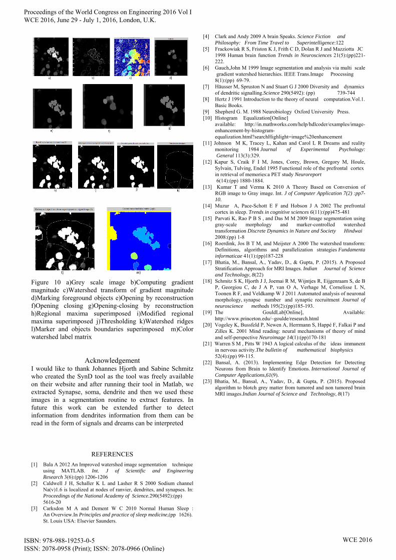
Figure 10 a)Grey scale image b)Computing gradient magnitude c)Watershed transform of gradient magnitude d)Marking foreground objects e)Opening by reconstruction f)Opening closing g)Opening-closing by reconstruction h)Regional maxima superimposed i)Modified regional maxima superimposed j)Thresholding k)Watershed ridges l)Marker and objects boundaries superimposed m)Color watershed label matrix
Acknowledgement
I would like to thank Johannes Hjorth and Sabine Schmitz who created the SynD tool as the tool was freely available on their website and after running their tool in Matlab, we extracted Synapse, soma, dendrite and then we used these images in a segmentation routine to extract features. In future this work can be extended further to detect information from dendrites information from them can be read in the form of signals and dreams can be interpreted
REFERENCES
[1] Bala A 2012 An Improved watershed image segmentation technique using MATLAB. Int. J of Scientific and Engineering Research 3(6):(pp) 1206-1206
[2] Caldwell J H, Schaller K L and Lasher R S 2000 Sodium channel Na(v)1.6 is localized at nodes of ranvier, dendrites, and synapses. In: Proceedings of the National Academy of Science.290(5492):(pp) 5616-20
[3] Carksdon M A and Dement W C 2010 Normal Human Sleep : An Overview.In Principles and practice of sleep medicine,(pp 1626). St. Louis USA: Elsevier Saunders.
[4] Clark and Andy 2009 A brain Speaks. Science Fiction and Philosophy: From Time Travel to Superintelligence:122
[5] Frackowiak R S, Friston K J, Frith C D, Dolan R J and Mazziotta JC 1998 Human brain function Trends in Neurosciences 21(5):(pp)221-222.
[6] Gauch,John M 1999 Image segmentation and analysis via multi scale gradient watershed hierarchies. IEEE Trans.Image Processing 8(1):(pp) 69-79.
[7] Häusser M, Spruston N and Stuart G J 2000 Diversity and dynamics of dendritic signalling.Science 290(5492): (pp) 739-744
[8] Hertz J 1991 Introduction to the theory of neural computation.Vol.1. Basic Books.
[9] Shepherd G. M. 1988 Neurobiology Oxford University Press. [10] Histogram Equalization[Online] available: http://in.mathworks.com/help/hdlcoder/examples/image-
enhancement-by-histogram-equalization.html?searchHighlight=image%20enhancement
[11] Johnson M K, Tracey L, Kahan and Carol L R Dreams and reality monitoring 1984 Journal of Experimental Psychology: General 113(3):329.
[12] Kapur S, Craik F I M, Jones, Corey, Brown, Gregory M, Houle, Sylvain, Tulving, Endel 1995 Functional role of the prefrontal cortex in retrieval of memories:a PET study Neuroreport 6(14):(pp) 1880-1884.
[13] Kumar T and Verma K 2010 A Theory Based on Conversion of RGB image to Gray image. Int. J of Computer Application 7(2) :pp7-10.
[14] Muzur A, Pace-Schott E F and Hobson J A 2002 The prefrontal cortex in sleep. Trends in cognitive sciences 6(11):(pp)475-481
[15] Parvati K, Rao P B S , and Das M M 2009 Image segmentation using gray-scale morphology and marker-controlled watershed transformation Discrete Dynamics in Nature and Society Hindwai 2008:(pp) 1-8
[16] Roerdink, Jos B T M, and Meijster A 2000 The watershed transform: Definitions, algorithms and parallelization strategies Fundamenta informaticae 41(1):(pp)187-228
[17] Bhatia, M., Bansal, A., Yadav, D., & Gupta, P. (2015). A Proposed Stratification Approach for MRI Images. Indian Journal of Science and Technology, 8(22)
[18] Schmitz S K, Hjorth J J, Joemai R M, Wijntjes R, Eijgenraam S, de B P, Georgiou C, de J A P, van O A, Verhage M, Cornelisse L N, Toonen R F, and Veldkamp W J 2011 Automated analysis of neuronal morphology, synapse number and synaptic recruitment Journal of neuroscience methods 195(2):(pp)185-193.
[19] The GouldLab[Online], Available: http://www.princeton.edu/~goulde/research.html
[20] Vogeley K, Bussfeld P, Newen A, Herrmann S, Happé F, Falkai P and Zilles K. 2001 Mind reading: neural mechanisms of theory of mind and self-perspective Neuroimage 14(1):(pp)170-181
[21] Warren S M , Pitts W 1943 A logical calculus of the ideas immanent in nervous activity.The bulletin of mathematical biophysics 52(4):(pp) 99-115.
[22] Bansal, A. (2013). Implementing Edge Detection for Detecting Neurons from Brain to Identify Emotions. International Journal of Computer Applications,61(9).
[23] Bhatia, M., Bansal, A., Yadav, D., & Gupta, P. (2015). Proposed algorithm to blotch grey matter from tumored and non tumored brain MRI images.Indian Journal of Science and Technology, 8(17)
Proceedings of the World Congress on Engineering 2016 Vol I WCE 2016, June 29 - July 1, 2016, London, U.K.
ISBN: 978-988-19253-0-5 ISSN: 2078-0958 (Print); ISSN: 2078-0966 (Online)
WCE 2016