Embed Size (px)
Citation preview

1
1
IntroductionMary K. Klassen-Fischer and
Ronald C. Neafie
Introduction
ProtozoaProtozoa are single-celled eukaryotic animals first dis-
covered by Antonie van Leeuwenhoek as he viewed Giar-dia in a personal enteric specimen through his own inven-tion (above photos). For a summary of the phylogeny of protozoa, see Table 1.1. A recent trend is to replace the term “protozoa” with “protista.” For these topics we retain “pro-tozoa” and reserve “protista” for a much larger group of single-celled organisms that include the algae, other single-celled photosynthetic organisms, and some of the water molds (slime molds).
Note that Pneumocystis jiroveci (previously called Pneu-mocystis carinii) is not included in this volume.1 Although this organism had been classified as a protozoon until the late 1980s, it is now considered a fungus.
Specimen Preparation The diagnosis of most human parasitic infections relies
upon the use of appropriate procedures for demonstrating the infecting organisms in feces, blood, urine, other body
fluids and tissues. The most commonly applied procedures are briefly reviewed below. For an in-depth presentation of particular procedures, appropriate laboratory guides and at-lases may be consulted. 2-4
Stool Fecal specimens may be preserved in 10% formalin, mer-
thiolate-iodine-formalin (MTF) solution, sodium acetate-acetic acid-formalin (SAF) solution, Schaudinn fluid, or polyvinyl alcohol (PVA) combined with Schaudinn fluid. Schaudinn fluid contains mercury, hence it is banned in many laboratories. The quality of the morphology of or-ganisms stained with trichrome or iron hematoxylin suf-fers when the modified fixative, in which zinc or copper is substituted for mercury, is used. Specimens should be first grossly examined to exclude macroscopic parasites such as roundworms or tapeworm proglottids. Concentration meth-ods may be applied to fresh or preserved fecal specimens to detect light infections. The two most common procedures are zinc sulfate flotation and formalin-ethyl acetate sedi-mentation.
Wet mount preparations for microscopic examination may be prepared in normal saline or stained with iodine, methylene blue, trichrome or iron hematoxylin. Special
Report Documentation Page Form ApprovedOMB No. 0704-0188
Public reporting burden for the collection of information is estimated to average 1 hour per response, including the time for reviewing instructions, searching existing data sources, gathering andmaintaining the data needed, and completing and reviewing the collection of information. Send comments regarding this burden estimate or any other aspect of this collection of information,including suggestions for reducing this burden, to Washington Headquarters Services, Directorate for Information Operations and Reports, 1215 Jefferson Davis Highway, Suite 1204, ArlingtonVA 22202-4302. Respondents should be aware that notwithstanding any other provision of law, no person shall be subject to a penalty for failing to comply with a collection of information if itdoes not display a currently valid OMB control number.
1. REPORT DATE JUN 2011 2. REPORT TYPE
3. DATES COVERED 00-00-2011 to 00-00-2011
4. TITLE AND SUBTITLE Introduction to Pathogenic Protozoa
5a. CONTRACT NUMBER
5b. GRANT NUMBER
5c. PROGRAM ELEMENT NUMBER
6. AUTHOR(S) 5d. PROJECT NUMBER
5e. TASK NUMBER
5f. WORK UNIT NUMBER
7. PERFORMING ORGANIZATION NAME(S) AND ADDRESS(ES) Inova Central Laboratory,2832 Juniper Street,Fairfax,VA,22031
8. PERFORMING ORGANIZATIONREPORT NUMBER
9. SPONSORING/MONITORING AGENCY NAME(S) AND ADDRESS(ES) 10. SPONSOR/MONITOR’S ACRONYM(S)
11. SPONSOR/MONITOR’S REPORT NUMBER(S)
12. DISTRIBUTION/AVAILABILITY STATEMENT Approved for public release; distribution unlimited
13. SUPPLEMENTARY NOTES See also ADA545141. Chapter 1 from e-book, Topics on the Pathology of Protozoan and InvasiveArthropod Diseases.
14. ABSTRACT
15. SUBJECT TERMS
16. SECURITY CLASSIFICATION OF: 17. LIMITATION OF ABSTRACT Same as
Report (SAR)
18. NUMBEROF PAGES
12
19a. NAME OFRESPONSIBLE PERSON
a. REPORT unclassified
b. ABSTRACT unclassified
c. THIS PAGE unclassified
Standard Form 298 (Rev. 8-98) Prescribed by ANSI Std Z39-18
2
1 • Topics on The paThology of proTozoan and invasive arThropod diseases
stains may be used to demonstrate specific organisms, such as a modified acid-fast stain or modified safranin stain for Cryptosporidium parvum and Cyclospora cayetanensis. Ultraviolet fluorescence microscopy demonstrates the auto-fluorescence of coccidian cysts such as C. cayetanensis and Isospora belli. Calcofluor white may be used to brighten the fluorescence of coccidian oocysts. Kits for detecting an-tigens in feces are commercially available for several com-mon parasites.
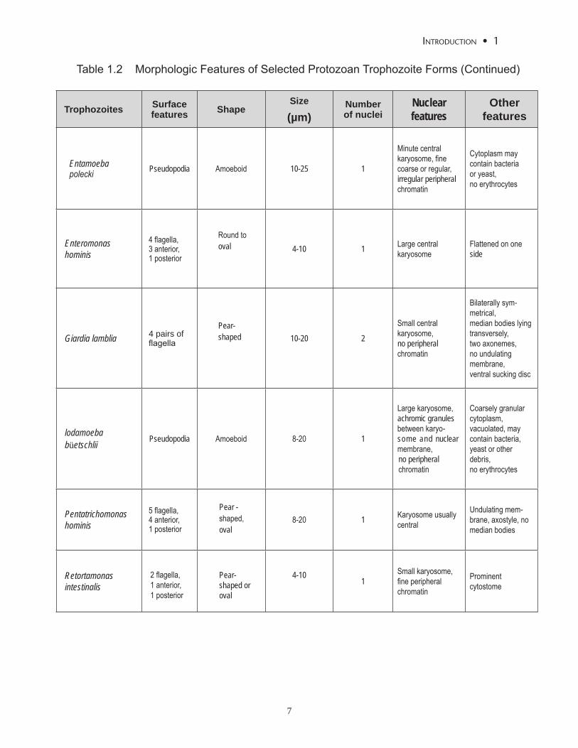
Important morphologic features of selected trophozoite forms are given in Table 1.2, and a summary of the com-parative morphologic features of selected cyst forms is pre-sented in Table 1.3.
Blood Identification of most parasites in blood requires the
preparation of stained thin or thick blood smears; however, specialized blood concentration techniques and commer-cial kits are available for the immunodetection of parasites, their antigens, and antibodies. Direct examination of drops of fresh blood or EDTA-preserved blood is useful for the detection of living motile trypomastigotes of Trypanosoma species. Preparation of thin or thick blood smears from fresh or EDTA-preserved blood followed by Giemsa, Wright or Dif-Quik® staining is useful for morphologic identifica-tion of species of Plasmodium, Babesia, Trypanosoma and Leishmania. For more details on blood smear preparation, see Topic 10. Specialized procedures exist for specific pro-tozoans, such as buffy coat concentration for Trypanosoma and Leishmania species and cytocentrifugation concentra-tion for Plasmodium and Leishmania species.5
Other fluids Cytology specimens that may be examined for protozoa
include urine, vaginal secretions, cerebrospinal fluid (CSF), aspirates of various tissues, and skin scrapings. Direct ex-amination of the sediment of the first portion of voided urine may detect the motile flagellates of Trichomonas vaginalis, especially in male patients. Demonstration of T. vaginalis trophozoites in female patients is usually done by prepar-ing wet mounts of vaginal swabs or scrapings. African trypanosomes and pathogenic free-living amebae (such as Naegleria fowleri) may be seen in CSF. Motile trypomas-tigotes of African trypanosomes can be found in aspirates from lymph nodes early in acute disease. Aspirates of bone marrow and spleen can demonstrate Leishmania donovani. Duodenal aspirates may be useful in demonstrating Giar-dia lamblia. Sigmoidoscopic material and aspirates of liver or lung abscesses may reveal trophozoites of Entamoeba histolytica. Cutaneous leishmaniasis can be diagnosed by finding amastigotes in stained smears prepared from scrap-ings of an ulcer.6
Tissue The examination of tissue specimens for protozoan infec-
tions may be accomplished in several ways. Fresh, unfixed biopsy material can be used to make touch preparations, to inoculate culture media, or infect experimental animals. Fixed tissue can be examined as stained histologic speci-mens.
Arthopods & Pentastomes Arthropods (phylum Arthropoda) are invertebrates with
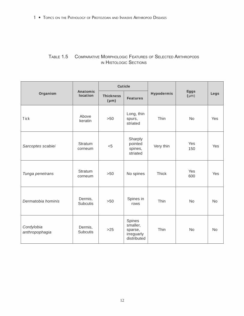
a chitinous exoskeleton, segmented body and jointed ap-pendages. A summary of the phylogeny of arthropods is given in Table 1.4. This e-book covers diseases caused by arthropods that invade human tissue, including tungiasis (Topic 17), myiasis (Topic 18), and infestation with the non-toxic mites, Sarcoptes and Demodex species (Topic 19). Arthropods have many other significant relationships to human disease as vectors, or toxin-producers; however, those diseases are not addressed in this volume. The com-parative morphologic characteristics of common parasitic arthropods is given in Table 1.5.
Pentastomes, or tongue worms, (Topic 16) are a class of parasitic animals that are similar to arthropods but that lack certain anatomic structures, such as circulatory and respi-ratory systems. The adults have oral hooks and the larvae have rudimentary legs; otherwise, they lack appendages.
Pseudoparasites and Artifacts It is important to be able to distinguish artifacts, con-
taminants, non-pathogenic protozoa and other microorgan-isms from pathogenic protozoa. Structures that may mimic pathogenic protozoa vary with the specimen type.
Stool specimens contain a variety of objects that can be mistaken for protozoa, including plant material, especially pollen. Stool may also contain nonpathogenic protozoa, such as Blastocystis hominis, Retortamonas intestinalis, Chilomastix mesnili, Enteromonas hominis, Endolimax nana, Iodamoeba buetschli, Entamoeba polecki, Entamoe-ba coli, Entamoeba hartmani and Entamoeba gingivalis. Spores of Myxobolus plectoplites, a fish pathogen, have been reported in fecal samples from patients who had re-cently eaten fish.7
In blood films, artifacts, such as platelets superimposed on erythrocytes, staining artifacts, and an array of contami-nants, may be mistaken for malarial parasites. Knowledge of the appearance of the normal constituents of blood pre-vents such errors.
Fluids from non-sterile body sites, such as sputum, are prone to contain foreign objects. Fluids from sterile body sites generally do not contain foreign objects, except in

3
inTroducTion • 1
cases where the fluid has become contaminated during col-lection or processing.
In tissue sections, one challenge is to distinguish minute protozoa from host cell structures. A common difficulty is discerning nuclear debris from amastigotes of Leishmania. Another problem may lie in deciding whether or not pig-ment deposition is due to Plasmodium falciparum infection. Yeast forms of some fungi, such as Basidiobolus ranarum, can bear a striking resemblance to amoebae. The intracy-toplasmic inclusions of cytomegalovirus may be mistaken for intracellular microorganisms if one does not observe the diagnostic cytoplasmic inclusions of this virus.
Delusional parasitosis (Morgellons Disease)
Delusional parasitosis is a condition characterized by an isolated delusion by individuals that they are infested by parasites, especially ectoparasites or intestinal parasites. The patient, who usually has no other psychiatric condi-tions, often describes various symptoms of the skin or near body orifices. It is not uncommon that more than one fam-ily member has the same symptoms in a shared delusion (folie à deux). The typical patient collects specimens from skin, feces, clothing or environment. Parasitologists and pa-thologists are consulted to rule out the presence of actual parasites. The samples often consist of fibers, keratin, scabs, hairs, food particles or flies. Most patients see numerous health care providers and fiercely reject negative findings, and are reluctant to consult a psychiatrist. A dermatologist may be more successful at convincing the patient to begin appropriate psychotropic drugs or other therapy.8,9
References1. Stringer JR, Beard CB, Miller RF, Wakefiled AE. A new name (Pneumocystis
jiroveci) for Pneumocystis from humans. Emerg Infect Dis. 2002;8:891-8962. Centers for Disease Control and Prevention, Center for Global Health, Laboratory
Diagnosis of Parasites of Public Concern. http://www.dpd.cdc.gov/dpdx/HTML/DiagnosticProcedures.htm Last modified 07/20/09.
3. Orihel TC, Ash LR. Parasites: A Guide to Laboratory Procedures and Identification. Chicago: ASCP Press; 1991.
4. Orihel TC, Ash, LR. Atlas of Human Parasitology. 4th ed. Chicago: ASCP Press; 1997.
5. Pethitory JC, Ardoin F, Ash LR, et al. Microscopic diagnosis of blood parasites following a cytoconcentration technique. Am J Trop Med Hyg. 1997;57:637-642.
6. Lesho EP, Neafie R, Wortmann G, Aronson N. Nonhealing skin lesinons in a sailor and journalist returning from Iraq. Cleveland Clinic J Med. 2005;72:93-94, 96, 98-99, 103-106.
7. Boreham RE, Hendrick S, O’Donoghue PJ, Stenzel DJ. Incidental finding of Myxobolus spores (protozoa: myxozoa) in stoll smaples from patients with gastronintestinal symptoms. J Clin Microbiol. 1998;36:3728-3730.
8. Koblenzer CS. The current management of delusional parasitosis and dermatitis artefacta. Skin Therapy Lett. 2010;15:1-3.
9. Morgellons disease: managing a mysterious skin condition. http://www.mayoclinic.com/health/morgellons-disease/sn00043. Last modified
02/13/2010.

4
1 • Topics on The paThology of proTozoan and invasive arThropod diseases
KINGDOM PROTOZOA
Subkingdom 1 Archezoa
Phylum MetamonadaClass Trepomonadea (intestinal flagellates)
Order Diplomonadida Giardia lamblia b (Topic 6)Order Enteromonadida Enteromonas hominis
Class RetortamonadeaOrder Retortamonadida Chilomastix mesnili, Retortamonas intestinalis
Phylum Parabasala (flagellates)
Class Trichomonadea (intestinal and related flagellates)Order Trichomonadida Trichomonas vaginalis
T. tenax, Pentatrichomonas hominis (Topic 7) Diantamoeba fragilis
Subkingdom 2 NeozoaInfrakingdom 1 Discicristata
Phylum Percolozoa (flagellates)Class Heterolobosea (flagellated amoebae)
Order Schizoprenida Naegleria fowleri (Topic 9)
Phylum Euglenozoa (flagellates)Class Kinetoplastidea (kinetoplastid flagellates)
Order Trypanosomatida Leishmania donovani, L. tropica (Topic 4 & Topic 5);L. infantum (Topic 5); L. major, L. braziliensis, L. mexicana, L. aethiopica, L. amazonensis, L. garnhami, L. guyanensis (Topic 4); L. colombien-sis, L. lainsoni, L. naiffi, L. panamensis, L. peruviana, L. pifanoi, L. shawi; Trypanosoma cruzi, T. rangeli (Topic 2); T. brucei gambiense, T. brucei rhodesiense (Topic 3)
Table 1.1 Classification of Parasitic Protozoa. a

5
inTroducTion • 1
Infrakingdom 2 Sarcomastigota
Phylum Amoebozoa (amoebae)
Subphylum LobosaClass Amoebaea (amoeba)
Order Acanthopodida Acanthamoeba castellanii, A. culbertsoni, A. hatchetti, A. polyphaga, Balamuthia mandrillaris (Topic 9)
Subphylum ConosaClass Entamoebidea (intestinal amoebae)
Order Euamoebida Entamoeba histolitica, E. coli, E. dispar,E. hartmanni, E. gingivalis, E. moshkoviskii, E. polecki, Endolimax nana, Iodoamoeba bütschlii (Topic 8); E. chattoni
Infrakingdom 3 Alveolata
Phylum Sporozoa (sporozoans)Class Coccidea
Order Eimeriida Cryptosporidium parvum, C. hominis, C. sp, Cyclospora cayetanensis, Isospora belli, Sarcocystis hominis, S. suihominis (Topic 13); S. lindemanni; Toxoplasma gondii (Topic 12)
Order Piroplasmida Babesia microti, B. divergens, B. gibsoni, Babesia sp (Topic 11)
Order Haemosporida Plasmodium falciparum, P. malariae, P. ovale,P. vivax (Topic 10)
Phylum Ciliophora (ciliates)Class Litostomatea
Order Vestibulifera Balantidium coli (Topic 15)
Table 1.1 Classification of Parasitic Protozoa. a (Continued)
a Adapted from: Cox, FEG. Taxonomy and classification of human parasites. Manual of Clinical Microbiology, 9th edition, Volume 2, section X Parasitology, chapter 132, editor in chief PR Murray, editors EJ Barron, JH Jorgensen, ML Landry, MA Pfaller, volume editor MA Pfaller, section editor LS Garcia. Washington DC, ASM Press, 2007. p 1991.

6
1 • Topics on The paThology of proTozoan and invasive arThropod diseases
Table 1.2 Morphologic Features of Selected Protozoan Trophozoite Forms
Trophozoites Surfacefeatures Shape
Size
(µm)Numberof nuclei
Nuclear features
Other features
Balantidium coli Cilia Ovoid 50-200 1 Large macronucleus Well-defined cyto-stome
Chilomastix mesnili
4 flagella, 3anterior, 1posterior
Round topear-
shaped6-24 1
Large or small karyosome,evenly or irregularlydistributed peripheralchromatin
Cytostome bordered by fibrils
Dientamoeba fragilis Pseudopodia Amoeboid 5-15 1 or 2
Karyosome frag- mented into 4-8 pieces, no peripheral chromatin
Finely granular vacuolated cyto-plasm, may contain bacteria,no erythrocytes
Endolimax nana Pseudopodia Amoeboid 6-12 1Large karyosome,no peripheral chromatin
Coarsely granu-lar cytoplasm, vacuolated, may contain bacteria, no erythrocytes
Entamoeba coli Pseudopodia Amoeboid 15-50 1
Large non-compacteccentric karyo-some,coarse irregular pe-ripheral chromatin
Coarsely granular cytoplasm, vacu-olated, may contain bacteria,yeast or other debris,no erythrocytes
Entamoeba dispar Pseudopodia Amoeboid 20-60 1
Small compact usually central karyosome, fine evenly distributed peripheral chromatin
Finely granular cytoplasm, rarely contain erythrocytes
Entamoeba gingivalis Pseudopodia Amoeboid 20 1
Small central karyo-some, fine granular regular peripheral chromatin
Finely granular cytoplasm, may contain bacteria, no erythrocytes, not found in feces
Entamoeba hartmanni Pseudopodia Amoeboid 5-12 1
Small compact usually central karyosome, fine evenly distributed peripheral chromatin
Finely granular cyto-plasm, may contain bacteria,no erythrocytes
Entamoeba histolytica Pseudopodia Amoeboid 20-60 1
Small compact, usually central karyosome, fine evenly distributed peripheral chro-matin
Finely granular cyto-plasm, may contain erythrocytes
7
inTroducTion • 1
Trophozoites Surfacefeatures Shape
Size
(µm)Numberof nuclei
Nuclear features
Other features
Entamoeba polecki Pseudopodia Amoeboid 10-25 1
Minute central karyosome, fine coarse or regular, irregular peripheral chromatin
Cytoplasm may contain bacteria or yeast, no erythrocytes
Enteromonas hominis
4 flagella, 3 anterior,1 posterior
Round tooval 4-10 1 Large central
karyosomeFlattened on one side
Giardia lamblia 4 pairs offlagella
Pear-shaped 10-20 2
Small central karyosome, no peripheral chromatin
Bilaterally sym-metrical,median bodies lying transversely, two axonemes,no undulating membrane,ventral sucking disc
lodamoeba büetschlii
Pseudopodia Amoeboid 8-20 1
Large karyosome, achromic granules between karyo-some and nuclear membrane, no peripheral chromatin
Coarsely granular cytoplasm, vacuolated, may contain bacteria, yeast or other debris, no erythrocytes
Pentatrichomonas hominis
5 flagella, 4 anterior, 1 posterior
Pear -shaped,oval
8-20 1 Karyosome usually central
Undulating mem-brane, axostyle, no median bodies
Retortamonas intestinalis
2 flagella, 1 anterior, 1 posterior
Pear-shaped or oval
4-101
Small karyosome, fine peripheral chromatin
Prominent cytostome
Table 1.2 Morphologic Features of Selected Protozoan Trophozoite Forms (Continued)

8
1 • Topics on The paThology of proTozoan and invasive arThropod diseases
Table 1.3 Comparative Morphologic Features of Selected Protozoan Cyst Forms
Cysts Type of cyst
Surface features Shape Size
(µm)Number of
nucleiNuclearfeatures Other features
Balantidium coli Not oocyst
Ciliawithin cyst wall
Round to oval 50-70 2
Large macro-nucleus,small micronucleus
Contractile vacuoles
Chilomastixmesnili Not oocyst No cilia
Round tolemon-shaped
6-10 1 Large, central karyosome
Hyaline knob ornipple-likeprotuberance,cytostome withfibrils
Cryptospo-ridiumspecies
Sporulated oocyst
Modifiedacid-fast in stool only
Round 4-6 Not visible – 4 nakedsporozoites
Cyclosporacayetanensis
Unsporu-latedoocyst
Modified acid-fast, doublecyst wall
Round 8-10 Not visible –
2 sporocystseach with 2sporozoites,greenish centralmass, refractileglobules
Endolimax nana Not oocyst No cilia
Round,oval orellipsoidal
5-10 4
Large centralkaryosome, noperipheralchromatin
Small granules
Entamoeba coli Not oocyst No cilia
Round,oval,triangular
15-30 8 (mature),1-4 (immature)
Large non-compact usuallyeccentrickaryosome,coarse irregularperipheralchromatin moreuniform than introphozoites
Chromatoidbodies withsplintered ends,central glycogenmass

9
inTroducTion • 1
Cysts Type of cyst
Surface features Shape Size
(µm)Number of
nucleiNuclearfeatures Other features
Entamoebadispar Not oocyst No cilia Round 10-20 4 (mature),
1-2 (immature)
Compact usually central small karyo-some, fine evenly distributed periph-eral chromatin,
Chromatoidbodies withrounded ends,central glycogenmass
Entamoebahartmanni Not oocyst No cilia Round 5-10 4 (mature),
1-2 (immature)
Small compactusually centralkaryosome, fineevenly distributedperipheralchromatin
Grape-likechromatoidbodies with bluntlyrounded ends
Entamoeba histolytica
Not oocyst No cilia Round 10-20 4 (mature),1-2 (immature)
Small compactusually centralkaryosome, fine evenly distrib-uted peripheral chromatin
Chromatoidbodies withrounded ends,central glycogenmass
Entamoebapolecki
Not oocyst No cilia Round, oval 10-15 1
Small central or ec-centric karyosome, fine regular periph-eral chromatin
Many grape-likechromatoidbodies ordark-staininginclusion mass
Enteromonashominis Not oocyst No cilia Oval or
ellipsoidal 4-8 2-4
1 or 2 nuclei ateach end, small central karyosome, no peripheralchromatin
Intracytoplasmicflagella
Giardia lamblia Not oocyst No cilia Ovoid to ellipsoidal 8-19 4 (mature)
2 (immature)
4 nuclei at one endcentral karyosome, no peripheral chromatin
Axonemes,intracytoplasmicfibrils lyingtransversely
Table 1.3 Comparative Morphologic Features of Selected Protozoan Cyst Forms (Continued)

10
1 • Topics on The paThology of proTozoan and invasive arThropod diseases
Cysts Type of cyst
Surface features Shape Size
(µm)Number of
nucleiNuclearfeatures
Other features
Iodamoebabuetschlii Not oocyst No cilia
Oval,ellipsoidal,triangular
5-20 1
Large eccen-tric karysome, no peripheral chromatin
Large glycogenvacuole
Isospora belli
Unsporulatedoocyst contains one immature sporont
Modified acid-fast Elongated 20-33 Not visible –
Mature oo-cyst contains 2 sporocystseach with 4sporozoites
E. bieneusiMicrosporidia Spore
Modified Trichrome stain
Oval 1-5 Not visible -
Retortamonasintestinalis Not oocyst No cilia
Pear-shaped oroval
4-9 1
Slightly ec-centric, small compactkaryosome,variableperipheralchromatin
Fibril andcytostome nearnucleus
Sarcocystisspecies
Sporulated oocyst
Thin-walled Elongated 15-19 Not visible –
2 sporocystseach with 4sporozoites,singlesporozoites fromruptured oocysts
Table 1.3 Comparative Morphologic Features of Selected Protozoan Cyst Forms (Continued)

11
inTroducTion • 1
1. Phylum Arthropoda
a. Subphylum mandibulata Class Insecta (fleas, flies, mosquitoes, bees, lice, bugs, beetles, caterpillars, fire ants)
b. Subphylum Chelicerata Class Arachnida (spiders, scorpions, mites, ticks)
c. Subphylum Onychophora (centipedes, millipedes)
2. Phylum Pentastomida
Class Pentastomata
Table 1.4 ProTozoa Seen in Human TiSSue SecTionS by
Size
Table 1.4 ProTozoa Seen in Human TiSSue SecTionS by
Size
Prepared by Mary Klassen 6/12/2007 Page 1
Table 1.4 claSSificaTion of PaTHogenic arTHroPodS and PenTaSTomeS
12
1 • Topics on The paThology of proTozoan and invasive arThropod diseases
Organism Anatomiclocation
Cuticle
Hypodermis Eggs (µm) Legs
Thickness(µm)
Features
Tick Abovekeratin >50
Long, thin spurs,striated
Thin No Yes
Sarcoptes scabiei Stratumcorneum <5
Sharply pointed spines, striated
Very thin Yes150 Yes
Tunga penetrans Stratumcorneum >50 No spines Thick Yes
600 Yes
Dermatobia hominis Dermis, Subcutis >50 Spines in
rows Thin No No
Cordylobia anthropophagia
Dermis, Subcutis >25
Spines smaller,sparse, irreguarly distributed
Thin No No
Table 1.5 comParaTive morPHologic feaTureS of SelecTed arTHroPodS
in HiSTologic SecTionS