Embed Size (px)
Citation preview
1
This accepted author manuscript is copyrighted and published by Elsevier. It is posted here by
agreement between Elsevier and MTA. The definitive version of the text was subsequently
published in 2015 Mol. Immunol. 65, 398–405. doi:10.1016/j.molimm.2015.02.014. Available
under license CC-BY-NC-ND.
MASP-1 of the complement system promotes clotting
via prothrombin activation
Lorenz Jenny a,b, József Dobó c, Péter Gál c, Verena Schroeder a,b
a University Clinic of Haematology, Haemostasis Research Laboratory, University Hospital Bern,
3010 Bern, Switzerland
b Department of Clinical Research, University of Bern, Bern, Switzerland;
c Institute of Enzymology, Research Centre for Natural Sciences, Hungarian Academy of Sciences,
Magyar tudósok krt 2, H-1113 Budapest, Hungary
Corresponding author:
Verena Schroeder, PhD
University Clinic of Haematology
Haemostasis Research Laboratory
University Hospital Inselspital
3010 Bern, Switzerland
Tel. +41 31 632 9618, Fax +41 31 632 1882
E-mail [email protected]
2
Abbreviations
CAPS, N-cyclohexyl-3-aminopropanesulfonic acid; CFT, clot formation time; CT, clotting time; FXa,
activated factor X; FXIII, factor XIII; MAp, MBL-associated protein; MASP, mannan-binding lectin-
associated serine protease; MBL, mannose binding lectin; MCF, maximum clot firmness; MES, 2-
(N-Morpholino)- ethanesulfonic acid; PAR4, protease activated receptor 4; PPP, platelet-poor
plasma; PT-DP, Prothrombin-depleted plasma; PVDF, polyvinylidene difluoride; rMASP-1cf,
MASP-1 catalytic fragment; SGMI, Schistocerca gregaria protease inhibitor (SGPI)-based MASP-
inhibitor; WB, whole blood.
Abstract
Mannan-binding lectin-associated serine protease-1 (MASP-1), a protein of the complement lectin
pathway, resembles thrombin in terms of structural features and substrate specificity, and it has
been shown to activate coagulation factors. Here we studied the effects of MASP-1 on clot
formation in whole blood (WB) and platelet-poor plasma (PPP) by thrombelastography and further
elucidated the underlying mechanism. Cleavage of prothrombin by MASP-1 was investigated by
SDS-PAGE and N-terminal sequencing of cleavage products. Addition of MASP-1 or thrombin to
WB and PPP shortened the clotting time and clot formation time significantly compared to
recalcified-only samples. The combination of MASP-1 and thrombin had additive effects. In a
purified system, MASP-1 was able to induce clotting only in presence of prothrombin. Analysis of
MASP-1-digested prothrombin confirmed that MASP-1 cleaves prothrombin at three cleavage
sites. In conclusion, we have shown that MASP-1 is able to induce and promote clot formation
measured in a global setting using the technique of thrombelastography. We further confirmed that
MASP-1-induced clotting is dependent on prothrombin. Finally, we have demonstrated that MASP-
1 cleaves prothrombin and identified its cleavage sites, suggesting that MASP-1 gives rise to an
alternative active form of thrombin by cleaving at the cleavage site R393.
Keywords
Coagulation; complement system; mannan-binding lectin-associated serine protease; MASP-1;
prothrombin; thrombelastography
3
1. Introduction
The complement system is an integral part of the innate immune system and serves to target,
opsonize and eliminate pathogens and foreign bodies from the circulation. More than 30 plasma
and membrane-bound proteins are involved in this process which can be subdivided into three
different pathways: the lectin, the alternative and the classical pathway. Upon recognition of a
target, all three pathways start a proteolytic cascade that leads to the formation of a C3 convertase
complex and ultimately ends in clearance of the target (Mayilyan et al., 2008; Ricklin et al., 2010).
Many intermediates of these pathways have additional properties, e.g. recruitment of inflammatory
cells or crosstalk with the coagulation system. Both the complement and the coagulation cascade
originate from the same ancestral pathways, they comprise several serine proteases with common
structural characteristics, and they show a high degree of interaction between specific
intermediates (Amara et al., 2010; Krem and Di Cera, 2002). As both systems serve as defense
mechanism in an environment with compromised barrier setting, they often activate each other
(Oikonomopoulou et al., 2012).
In the lectin pathway, mannose binding lectin (MBL) and ficolins recognize target molecules on
foreign and/or altered surfaces and thereby induce activation of the MBL-associated serine
proteases (MASPs) MASP-1, MASP-2, and MASP-3 (Krarup et al., 2007). MASPs 1-3 and the
enzymatically inactive MBL-associated proteins MAp19 and MAp44 are alternative splicing
products from two genes: MASP-1/MASP-3/Map44 and MASP2/MAp19 (Stover et al., 1999).
Among MASPs, MASP-2 is the only protease capable to form C3 convertase efficiently by cleaving
both C2 and C4. MASP-2 was also shown to cleave prothrombin in a similar way to activated factor
X (FXa) and hence to promote clot formation (Krarup et al., 2007; Yongqing et al., 2012). In
contrast, the function of MASP-3 remains unclear so far. It is speculated that MAp19 competes
with the MASPs for the MBL binding site inhibiting activation of the lectin pathway (Dahl et al.,
2001; Yongqing et al., 2012). MAp44 has also been shown to regulate the activity of the lectin
pathway (Pavlov et al., 2012).
4
The exact role of MASP-1 in the complement system has only recently been revised (reviewed in
Dobó et al., 2014). MASP-1 was known to cleave C2 but not C4, making it unable to form C3
convertase efficiently by itself. MASP-1 was therefore considered to have a merely supporting role
in MASP-2-mediated activation of the lectin pathway. Latest research, however, indicates a central
role for MASP-1 in the early lectin pathway by autoactivation and subsequent activation of MASP-2
and MASP-3, attributing MASP-1 the function of the major or even only initiator of the lectin
pathway (Héja et al., 2012a; Megyeri et al., 2013).
Even though MASP-1 belongs to the C1r/C1s/MASP family of serine proteases, its serine protease
domain is structurally more closely related to thrombin and trypsin. This is supported by the fact
that MASP-1 is more efficiently inhibited by antithrombin (in the presence of heparin) than by C1-
inhibitor (Gál et al., 2009; Parej et al., 2013). Due to its unusually wide substrate binding groove
and the resulting broad substrate specificity – which is atypical among complement proteases –
MASP-1 is able to interact with various targets outside the complement system. Due to its
thrombin-like activity, it is able to cleave fibrinogen and factor XIII (FXIII) (Krarup et al., 2008) and it
activates protease activated receptor 4 (PAR4) on endothelial cells (Megyeri et al., 2009);
however, its efficacy is significantly lower compared to thrombin. The A-subunit of FXIII and the
fibrinogen β-chain are cleaved in the same manner by MASP-1 as by thrombin, while the
fibrinogen α-chain is cleaved differently (Krarup et al., 2008). We have shown that clots produced
in plasma in presence of MASP-1 have an altered fibrin structure and presented first evidence that
MASP-1 may cleave prothrombin (Hess et al., 2012). First in vivo evidence for MASP-1
involvement in coagulation was obtained from MASP-1 and MBL knockout mice which showed
prolonged bleeding time on tail tip excision (Takahashi et al., 2012) and a significant decrease in
FeCl3-induced thrombogenesis (La Bonte et al., 2012). Taken together, MASP-1 seems to be a
major link between the complement system and coagulation but further work is needed to clarify
the exact underlying mechanisms and (patho-) physiological relevance.
The aim of this study was to investigate for the first time whether MASP-1 is able to influence or
even induce clot formation in human whole blood and how its effects compare with those of
thrombin. We went on to elucidate the underlying mechanisms in plasma and purified systems. In
5
particular, the role of prothrombin in MASP-1-mediated clotting and their specific interaction were
examined.
2. Materials and methods
2.1. MASP-1 protein
Large amounts of MASP-1 were required for the thrombelastographic experiments. However,
neither recombinant production nor plasma purification of pure full-length MASP-1 in sufficient
amounts have been achieved so far. We therefore used the recombinant MASP-1 catalytic
fragment (rMASP-1cf) for our experiments. It is a truncated form of MASP-1 consisting of the
CCP1-CCP2-SP domains while the N-terminal CUB1-EGF-CUB2 domains are deleted (Dobó et
al., 2009). In order to confirm that rMASP-1cf and full-length MASP-1 show the same effects on
coagulation, we performed a control experiment with active full-length MASP-1 (a kind gift from
Mikkel-Ole Skjoedt, Copenhagen, Denmark).
2.2. Specific protease inhibitors
SGMI-2 [Schistocerca gregaria protease inhibitor (SGPI)-based MASP-inhibitor] is a specific
inhibitor of MASP-2. It is a small protein inhibitor (MW 4009.5 Da) that was expressed in E.coli as
described earlier (Héja et al., 2012b). SGMI-2 was dissolved in water at a concentration of 2.83
mg/ml (706 µM) and used in the thrombelastographic experiments at a final concentration of 5
µmol/l. Hirudin (Novartis, Basel, Switzerland) specifically binds to the active site and exosite 1 of
thrombin species and does not interfere with MASP-1 activity (Parry et al., 1993; Presanis et al.,
2003). Hirudin was used in prothrombin cleavage experiments at a final concentration of 600
units/ml.
2.3. Blood collection and sample preparation
Whole blood (WB) was taken from healthy volunteers who had not taken any medication for 10
days. All volunteers gave written informed consent. Blood was drawn into Sarstedt Monovette®
6
tubes containing 0.106 mol/l sodium citrate. Platelet-poor plasma (PPP) was produced by
centrifugation at 1513 g for 20 min. Experiments were carried out within 4 h after blood sampling.
2.4. Thrombelastography experiments
Thrombelastographic measurements were performed in fresh WB and PPP following recalcification
(final concentration 12.5 mmol/l CaCl2) and addition of rMASP-1cf, thrombin, or a combination of
rMASP-1cf and thrombin. The mean MASP-1 plasma concentration is 11 µg/ml (equal to 143
nmol/l) (24). We used rMASP-1cf at final concentrations of 5, 10, and 20 µg/ml, corresponding to
110, 220, and 440 nmol/l. Thrombin (Calbiochem, Merck Chemicals, Nottingham, UK) was used at
final concentrations of 10, 20, and 40 nmol/l, as 26 nmol/l thrombin is the concentration generated
at approximately clot time (Brummel et al., 2002). Experiments were performed as follows: Into a
thrombelastography cup, we pipetted 16.9 µl of CaCl2 and 5.1 µl containing rMASP-1cf and/or
thrombin in Tris-buffered saline (TBS, 50 mmol/l Tris, 100 mmol/l NaCl, pH 7.4). Upon addition of
248 µl WB or PPP, the measurement was immediately started on a rotation-thrombelastometry
system (ROTEM®, Tem International, Munich, Germany) and ran for 1 h. Prothrombin-depleted
plasma (PT-DP, Hyphen BioMed, Neuville-sur-Oise, France) was used to test if MASP-1-mediated
clotting depended on the presence of prothrombin. A purified system containing 2 mg/ml (5.8
µmol/l) fibrinogen in presence or absence of 100 µg/ml (1.4 µmol/l) prothrombin (both from Hyphen
BioMed) in TBS was also used. Measurements using PT-DP or purified proteins were carried out
for 3 h to detect any delayed clot formation. We evaluated the standard ROTEM® parameters
clotting time (CT; representing the lag time until the onset of clotting), clot formation time (CFT;
representing the duration from onset of clotting until 20 mm of amplitude are reached), and
maximum clot firmness (MCF; a measure of visco-elastic properties of the formed clot). Each
measurement was performed in triplicate in WB and PPP samples from two individuals. For each
individual a recalcified-only sample without MASP-1 or thrombin served as baseline and the results
obtained with MASP-1 and/or thrombin were expressed in percentage relative to the baseline
values in order to correct for interindividual variation.
2.5. Prothrombin cleavage studies
7
Prothrombin (1.7 µmol/l / 120 µg/ml, Hyphen BioMed) and rMASP-1cf (1.75 µmol/l / 80 µg/ml) were
incubated for up to 180 min at 37°C in a final volume of 50 µl TBS-Buffer. After incubation, the
samples were mixed with 4x NUPAGE® sample buffer (4:1; Life Technologies, Carlsbad, USA),
boiled for 10 min at 70°C, and loaded onto NUPAGE® precast 4-12% (w/v) gradient gels. Precision
Plus Protein™ Dual Color Standards (10-250 kDa, Biorad, Hercules, USA) was used as protein
marker. MES (2-(N-Morpholino)-ethansulfonsäure, Life Technologies) SDS was used as running
buffer. For Western blotting we used a 0.2 µm polyvinylidene difluoride (PVDF) membrane and
CAPS-buffer (N-cyclohexyl-3-aminopropanesulfonic acid, 0.1 M, pH 11.0) (both from Life
Technologies). After the transfer the membrane was washed and stained with sulforhodamin
(sulforhodamin B 89.5 µM, 30% methanol (v/v), 0.1% acetic acid (v/v)). Bands were excised and
subjected to five cycles of Edman degradation and N-terminal sequencing on a ABI Procise cLC
494-device (Blue Lion Biotech, Carnation, USA).
3. Results
3.1. Effects of MASP-1 on clot formation studied by thrombelastography
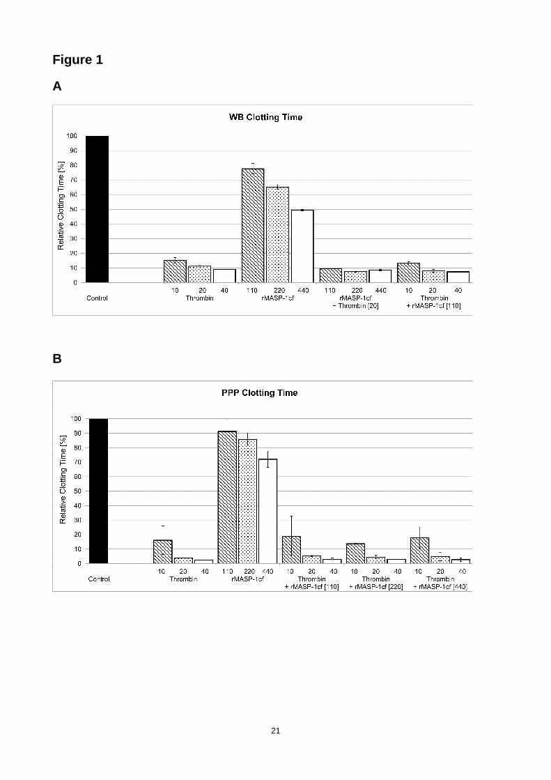
In WB, MASP-1 showed a dose-dependent reduction in CT as shown in Fig. 1a. Addition of 10
µg/ml rMASP-1cf shortened the CT by 34% compared to the recalcified-only sample, while
thrombin led to a reduction in CT by approximately 90%. Combination of both enzymes did not
lead to additive effects. As shown in Fig. 1b, addition of 10 µg/ml rMASP-1cf to PPP decreased the
CT by 15%. Again, thrombin shortened CT significantly more and the combination with rMASP-1cf
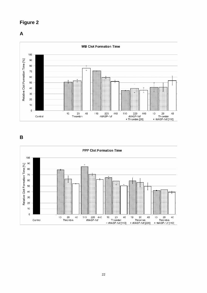
did not show any additional effect. However, a different picture was observed for CFT. MASP-1
reduced the CFT values of the recalcified sample by 41% in WB (Fig. 2a) and by 29% in PPP (Fig.
2b), while thrombin led to a decrease by 47% in WB and by 38% in PPP, respectively. The
combination of both enzymes clearly showed additive effects on CFT: reduction by 67% in WB and
by 47% in PPP. MASP-1 showed no consistent and significant effects on MCF (data not shown).
The results of the control experiment with full-length MASP-1 in WB and PPP showed reduced CT
8
and CFT and were consistent with the findings from the rMASP-1cf experiments (Fig. S1 and Table
S1 in the supplementary file).
3.2. Effects of MASP-1 are prothrombin-dependent
Based on earlier preliminary results (Hess et al., 2012) we hypothesized that MASP-1 may trigger
and/or promote clot formation via activation of prothrombin. We therefore performed
thrombelastographic measurements in PT-DP and a purified system in the absence and presence
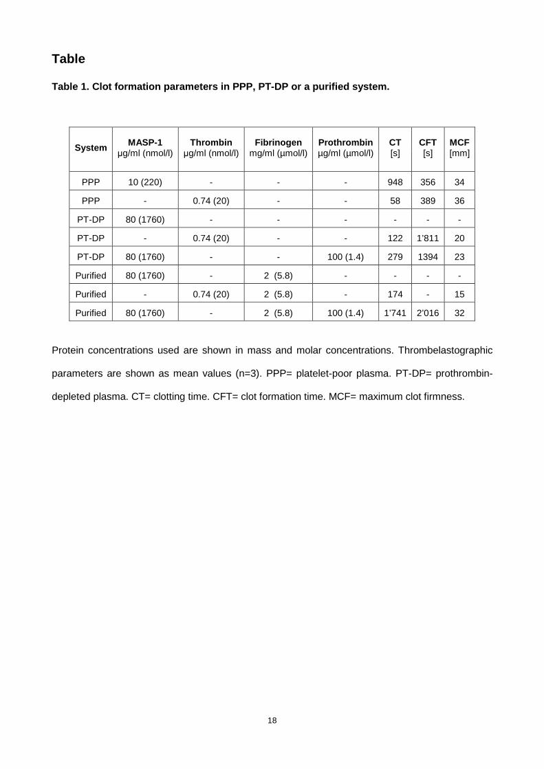
of prothrombin. The results are summarized in Table 1.
In PT-DP, even high concentrations of rMASP-1cf failed to induce clotting, whereas thrombin led
to clotting that was delayed compared with normal plasma. When PT-DP was reconstituted with
prothrombin, rMASP-1cf was able induce clotting. In a purified system containing fibrinogen and
thrombin, fibrin formation occurred, whereas MASP-1 alone failed to trigger fibrin formation.
However, when prothrombin was added to the purified system containing fibrinogen and rMASP-
1cf, we did observe fibrin formation. These results confirm that MASP-1-induced clot formation
relies on the presence of prothrombin.
It has been shown that MASP-1 is the main activator of MASP-2 Heja et al., 2012; Megyeri et al.,
2013) and that MASP-2 is able to cleave prothrombin (Krarup et al., 2007). Therefore, we wanted
to test whether the effects of MASP-1 on clotting observed in WB and PPP could be due to MASP-
1-mediated activation of MASP-2 and not due to direct action of MASP-1 itself.
Thrombelastographic measurements with rMASP-1cf in WB and PPP were repeated in the
presence of the specific MASP-2 inhibitor SGMI-2. Inhibition of MASP-2 did not lead to any
changes in CT and CFT (data not shown). These results suggest that MASP-2 is not involved in
MASP-1-mediated clotting of WB and PPP.
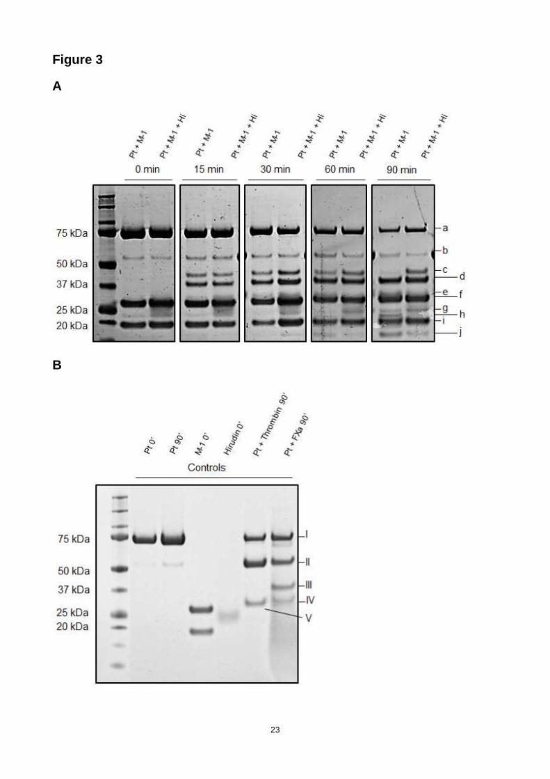
3.3. MASP-1 cleaves prothrombin
Our next aim was to confirm that MASP-1 is able to cleave prothrombin and to identify its cleavage
site(s). We incubated prothrombin with rMASP-1cf, stopped the reactions at different time points
and separated the resulting fragments by SDS-PAGE (as shown in Fig. 3a). Bands were identified
by N-terminal sequencing. Band a) corresponds to uncleaved prothrombin, f) to the MASP-1 heavy
9
chain, g) to a degradation product of the MASP-1 heavy chain, and i) to the MASP-1 light chain
(also shown in the control gel in Fig. 3b). Digestion by prothrombin in the presence of MASP-1
gave rise to several prothrombin fragments (shown in the first lane per time point): b) corresponds
to prethrombin-1 (=F2+LC+HC, arises through cleavage at R155), c) to a “meizothrombin
analogue” cleaved at R393 (instead of R320 as in FXa-mediated prothrombin cleavage), d)
corresponds to prethrombin-2 (=LC+HC, arises through cleavage at R271), e) to the fragment
F1.F2 (arises through cleavage at R271), h) to a truncated version of the α-thrombin heavy chain
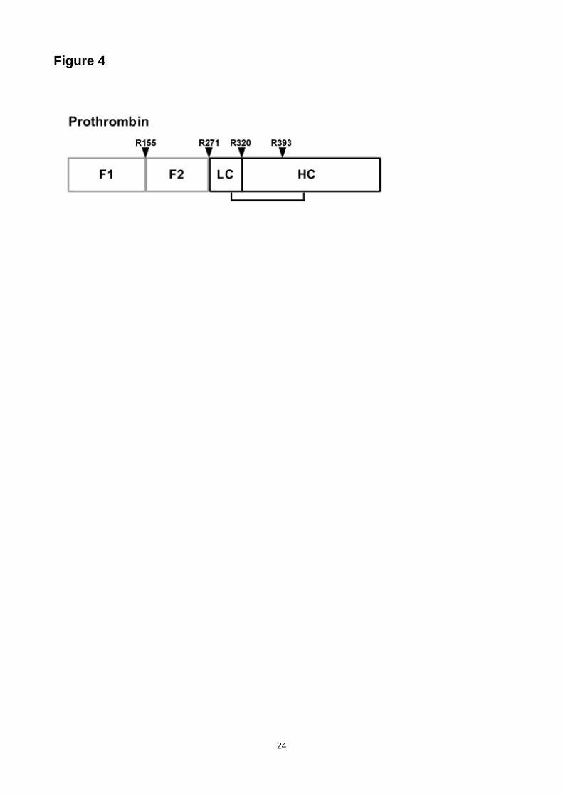
(from I321 to R393), and j) to the fragment F1. N-terminal sequencing has confirmed the following
cleavage sites and corresponding N-termini of the resulting fragments: R155 (SEGGS), R271
(TATSE) and R393 (NIEKI) (Fig. 4). Some cleavage at R155 to yield prethrombin-1 (band b) was
due to autodegradation of prothrombin (Fig. 3b). Over time, in the presence of MASP-1 most of the
prothrombin has been processed, after 90 min the “meizothrombin analogue” has almost
completely undergone further digestion, whereas prethrombin-2 seemed to accumulate. Band h)
corresponding to the truncated thrombin heavy chain started to visibly accumulate after
approximately 60 min. A time course digestion with 10 min steps and SDS-PAGE optimized for
band h) was able to detect this specific band already after 40 min (data not shown).
FXa cleaves prothrombin at R271 and R320, and the resulting products including α-thrombin and
meizothrombin are involved in its own back-cleavage (Haynes et al., 2012; Krishnaswamy, 2013)
Therefore we tested if similar mechanisms would apply to MASP-1-mediated prothrombin
cleavage, i.e. if thrombin species and/or intermediate cleavage products would significantly
contribute to prothrombin cleavage in presence of MASP-1. We incubated MASP-1 and
prothrombin in the presence of hirudin, a specific thrombin inhibitor which also inhibits active
thrombin-like intermediates such as meizothrombin. As shown in Fig. 3a (in the second lane per
time point), there were differences compared with the digestion lacking hirudin: prothrombin (band
a) decreased more slowly, prethrombin-1 (band b) decreased faster, the “meizothrombin analogue”
(band c) accumulated, and hardly any truncated thrombin (band h) and less fragment F1 (band j)
could be observed. These results suggest that MASP-1 may trigger initial prothrombin degradation
and give rise to active thrombin species and intermediates which then contribute to further
processing of prothrombin and intermediate cleavage products.
10
We also digested prothrombin with thrombin (Fig. 3b). After 90 min prethrombin-1 (band II)
accumulated, showing the preference of thrombin to cleave at R155.
4. Discussion
Various interactions between the complement system and blood coagulation have been discovered
(Amara et al., 2010). In this context the complement protease MASP-1 has moved into the focus of
interest in recent years, however, the relevance of its role in coagulation and the underlying
mechanisms are not yet fully understood. Most experiments on MASP-1 and coagulation were
conducted in isolated systems. In the present study, we investigated for the first time the effects of
MASP-1 on clot formation in whole blood by using the technique of thrombelastograhy which
simulates a more global setting.
We show that MASP-1 accelerates clotting times and promotes clot formation in WB and PPP. In
addition, MASP-1 and thrombin show an additive effect on CFT which is rather remarkable given
that thrombin is much more efficient in cleaving FXIII and fibrinogen than MASP-1 (Krarup et al.,
2008). This may suggest that MASP-1 exerts different functions than thrombin and not only mirrors
thrombin function in a less efficient way.
The influence of MASP-1 on CFT is more pronounced than its influence on CT. As CT reflects the
early onset of clotting and CFT measures the kinetics of later clot formation, our data suggest that
MASP-1 affects the later phase of clotting rather than the early phase. This delay in its impact on
clotting can be due to its lower efficiency towards substrates (Krarup et al., 2008), due to an
indirect effect on clotting or a combination of both. We have previously presented first evidence
that MASP-1 may activate prothrombin and thus drive clot formation in an indirect way (Hess et al.,
2012). This may explain why the effect of MASP-1 is more distinct in the later phase of clot
formation: prothrombin needs to be processed to thrombin by MASP-1 before it can support clot
formation, while the direct effects of MASP-1 on FXIII and fibrinogen is most likely too small to lead
11
to a visibly faster clot formation. This may also explain why there are additive effects of thrombin
and MASP-1 on CFT but not CT: with MASP-1 displaying only a negligible direct effect on clotting it
is initially not able to contribute to the immediate strong effect of thrombin. However, in the later
phase of clot formation, more and more additional thrombin is generated by MASP-1-mediated
prothrombin activation and this leads to a significant acceleration of CFT.
Although MASP-1 activates MASP-2 which, in turn, is able to promote clotting by prothrombin
cleavage similar to FXa-mediated cleavage (Krarup et al., 2007), inhibition of MASP-2 in WB and
PPP samples showed that MASP-2 does not further enhance the capability of MASP-1 to promote
clotting.
We further demonstrated that MASP-1-mediated clot formation is depending on prothrombin. In
PT-DP and a purified system, MASP-1 alone failed to induce clotting in the absence of
prothrombin, and supplementation with prothrombin was able to reconstitute clotting. These
experiments confirmed that MASP-1 induces clotting indirectly by activating prothrombin.
In the coagulation cascade, prothrombin is processed to thrombin by FXa, giving rise to several
intermediates exhibiting different functions (Krishnaswamy, 2013). Intriguingly, MASP-2 was shown
to cleave prothrombin in a similar manner, whereas prothrombin cleavage by MASP-1 was not
detected (Krarup et al., 2007). Here we show by SDS-PAGE analysis that MASP-1 is indeed
capable of cleaving prothrombin. N-terminal sequencing of cleavage products revealed three
cleavage sites on prothrombin, among which R393 is not cleaved by FXa, but it is a site of
autodegradation by α-thrombin during prolonged incubation (Bovill et al., 1995). R393, as R320, is
located between the interchain disulfide-bridge (C292-C438), therefore cleavage at R393 does not
lead to dissociation of the heavy and light chains but presumably maintains the two-chain
conformation and may give rise to an alternative active form of thrombin.
Similar to FXa-mediated prothrombin activation, several intermediate forms appear in the course of
prothrombin digestion by MASP-1. In FXa-mediated prothrombin activation, the intermediate
meizothrombin is enzymatically active and responsible for feedback-cleavage at position R155 of
prothrombin which is also a cleavage site for α-thrombin (Krishnaswamy, 2013). Also in MASP-1-
12
mediated prothrombin cleavage, intermediate forms and/or thrombin species contribute to further
processing of prothrombin and its cleavage products. Upon inhibition of thrombin species by
hirudin, the heavy chain of the putative alternative thrombin species (band h) does hardly appear.
Thus, it is likely that a thrombin species cleaved at R320 and therefore inhibitable by hirudin must
be involved. However, we were not able to isolate a fragment carrying the N-terminal sequence of
R320. An explanation for these findings could be that MASP-1 cleaves at both R320 and R393,
yielding a thrombin form resembling β-thrombin or even β-thrombin itself (Bovill et al., 1995). This
thrombin species could be responsible for the procoagulant action of MASP-1, as the appearance
of the R393 heavy chain (band h) at approximately 40 min roughly coincides with clotting at 32 min
in the purified system in thrombelastography.
Further work is ongoing to prove the existence and function of an alternative form of thrombin, and
to clarify the chronological order of prothrombin degradation by MASP-1 with the help of
prothrombin variants with mutated cleavage sites.
Our study may have (patho-) physiological implications. We have recently reported elevated
MASP-1 levels in patients with myocardial infarction (Frauenknecht et al., 2013) and in patients
with type-1 diabetes (Jenny et al., 2015). MASP-1 levels have also been shown to double during
acute-phase reactions (Thiel et al., 2012). We therefore believe that MASP-1 levels are elevated
and MASP-1 is activated in the proinflammatory and procoagulant environment of atherosclerosis
and contributes to clot formation by activation of coagulation factors and endothelial cells (Dobó et
al., 2014).
5. Conclusion
In summary, we have shown that MASP-1 is able to induce and promote clot formation in whole
blood and plasma, measured in a global setting using the technique of thrombelastography.
Experiments in PT-DP and a purified system confirmed that MASP-1-induced clotting is dependent
on prothrombin, as MASP-1 lacks any clotting activity in the absence of prothrombin. Finally, we
13
have demonstrated that MASP-1 cleaves prothrombin and identified its cleavage sites, suggesting
that MASP-1 gives rise to an alternative active form of thrombin.
Acknowledgements
The authors would like to thank Prof. Johann Schaller and Urs Kämpfer (Department of Chemistry
and Biochemistry, University of Bern) for their technical support and expertise in N-terminal protein
sequencing, Dr. Andrea Kocsis (Institute of Enzymology, Budapest, Hungary) for the preparation of
SGMI-2, and Dr. Mikkel-Ole Skjoedt (Copenhagen, Denmark) for the donation of full-length MASP-
1.
This work was supported by grants from the Swiss National Science Foundation (grant 140925),
OPO Foundation (Zurich, Switzerland), Hungarian Scientific Research Fund (OTKA grant
NK100834), and the János Bolyai Research Fellowship of the Hungarian Academy of Sciences.
Author contributions
L. Jenny designed and performed the experiments, analysed the data, and wrote the manuscript.
J. Dobó and P. Gál produced the rMASP-1 fragment and revised the manuscript. V. Schroeder
designed the study, analysed the data, and revised the manuscript.
Conflict of interest
The authors have no competing interests.
14
References
Amara, U., Flierl, M.A., Rittirsch, D., Klos, A., Chen, H., Acker, B., Brückner, U.B., Nilsson, B.,
Lambris, J.D., Huber-Lang, M., 2010. Molecular intercommunication between the
complement and coagulation systems. J. Immunol. 185, 5628-5636.
Bovill, E.G., Tracy, R.P., Hayes, T.E., Jenny, R.J., Bhushan, F.H., Mann, K.G., 1995. Evidence that
meizothrombin is an intermediated product in the clotting of whole blood. Arterioscler.
Thromb. Vasc. Biol. 15, 754-758.
Brummel, K.E., Paradis, S.G., Butenas, S., Mann, K.G., 2002. Thrombin functions during tissue
factor-induced blood coagulation. Blood 100, 148-152.
Dahl, M.R., Thiel, S., Matsushita, M., Fujita, T., Willis, A.C., Christensen, T., Vorup-Jensen, T.,
Jensenius, J.C., 2001. MASP-3 and its association with distinct complexes of the mannan-
binding lectin complement activation pathway. Immunity 15, 127-135.
Dobó, J., Harmat, V., Beinrohr, L., Sebestyén, E., Závodszky, P., Gál, P., 2009. MASP-1, a
promiscuous complement protease: structure of its catalytic region reveals the basis of its
broad specificity. J. Immunol. 183, 1207-1214.
Dobó, J., Schroeder, V., Jenny, L., Cervenak, L., Závodszky, P., Gál, P., 2014. Multiple roles of
complement MASP-1 at the interface of innate immune response and coagulation. Mol.
Immunol. 61, 69-78.
Frauenknecht, V., Thiel, S., Storm, L., Meier, N., Arnold, M., Schmid, J.P., Saner, H. & Schroeder,
V., 2013. Plasma levels of mannan-binding lectin (MBL)-associated serine proteases
(MASPs) and MBL-associated protein in cardio- and cerebrovascular diseases. Clin. Exp.
Immunol. 173, 112-120.
Gál, P., Dobó, J., Závodszky, P., Sim, R.B., 2009. Early complement proteases: C1r, C1s and
MASPs. A structural insight into activation and functions. Mol. Immunol. 46, 2745-2752.
Haynes, L.M., Bouchard, B.A., Tracy, P.B., Mann, K.G., 2012. Prothrombin activation by platelet-
associated prothrombinase proceeds through the prethrombin-2 pathway via a concerted
mechanism. J. Biol. Chem. 287, 38647-38655.
Héja, D., Kocsis, A., Dobó, J., Szilágyi, K., Szász, R., Závodszky, P., Pál, G., Gál, P., 2012a.
Revised mechanism of complement lectin-pathway activation revealing the role of serine
15
protease MASP-1 as the exclusive activator of MASP-2. Proc. Natl. Acad. Sci. USA., 109,
10498-10503.
Héja, D., Harmat, V., Fodor, K., Wilmanns, M., Dobó, J., Kékesi, K.A., Závodszky, P., Gál, P. &
Pál, G., 2012b. Monospecific inhibitors show that both mannan-binding lectin-associated
serine protease-1 (MASP-1) and -2 are essential for lectin pathway activation and reveal
structural plasticity of MASP-2. J. Biol. Chem. 287, 20290-20300.
Hess, K., Ajjan, R., Phoenix, F., Dobó, J., Gál, P., Schroeder, V., 2012. Effects of MASP-1 of the
complement system on activation of coagulation factors and plasma clot formation. PLoS
One 7, e35690.
Jenny, L., Ajjan, R., King, R., Thiel, S. & Schroeder, V., 2015. Plasma levels of mannan-binding
lectin-associated serine proteases MASP-1 and MASP-2 are elevated in type 1 diabetes
and correlate with glycaemic control. Clin. Exp. Immunol., in press.
Krarup, A., Wallis, R., Presanis, J.S., Gál, P., Sim, R.B., 2007. Simultaneous activation of
complement and coagulation by MBL-associated serine protease 2. PLoS One 2, e623.
Krarup, A., Gulla, K.C., Gál, P., Hajela, K., Sim, R.B., 2008. The action of MBL-associated serine
protease 1 (MASP1) on factor XIII and fibrinogen. Biochim. Biophys. Acta 1784, 1294-1300.
Krem, M.M., Di Cera, E., 2002. Evolution of enzyme cascades from embryonic development to
blood coagulation. Trends Biochem. Sci. 27, 67-74.
Krishnaswamy, S., 2013. The transition of prothrombin to thrombin. J. Thromb. Haemost. 11
(Suppl 1), 265-276.
La Bonte, L.R., Pavlov, V.I., Tan, Y.S., Takahashi, K., Takahashi, M., Banda, N.K., Zou, C., Fujita,
T., Stahl G.L., 2012. Mannose-binding lectin-associated serine protease-1 is a significant
contributor to coagulation in a murine model of occlusive thrombosis. J. Immunol. 188, 885-
891.
Mayilyan, K.R., Kang, Y.H., Dodds, A.W., Sim, R.B., 2008. The complement system in innate
immunity, in: Heine, H., Innate Immunity of Plants, Animals and Humans. Springer, Berlin
Heidelberg, pp. 219-236.
Megyeri, M., Makó, V., Beinrohr, L., Doleschall, Z., Prohászka, Z., Cervenak, L., Závodszky, P.,
Gál, P., 2009. Complement protease MASP-1 activates human endothelial cells: PAR4
16
activation is a link between complement and endothelial function. J. Immunol. 183, 3409-
3416.
Megyeri, M., Harmat, V., Major, B., Végh, Á., Balczer, J., Héja, D., Szilágyi, K., Datz, D., Pál, G.,
Závodszky, P., Gál, P., Dobó, J., 2013. Quantitative characterization of the activation steps
of mannan-binding lectin (MBL)-associated serine proteases (MASPs) points to the central
role of MASP-1 in the initiation of the complement lectin pathway. J. Biol. Chem. 288, 8922-
8934.
Oikonomopoulou, K., Ricklin, D., Ward, P.A., Lambris, J.D., 2012. Interactions between
coagulation and complement - their role in inflammation. Semin. Immunopathol. 34, 151-
165.
Parej, K., Dobó, J., Závodszky, P., Gál, P., 2013. The control of the complement lectin pathway
activation revisited: both C1-inhibitor and antithrombin are likely physiological inhibitors,
while α2-macroglobulin is not. Mol. Immunol. 54, 415-422.
Parry, M.A., Stone, S.R., Hofsteenge, J., Jackman, M.P., 1993. Evidence for common structural
changes in thrombin induced by active-site or exosite binding. Biochem. J. 290, 665-670.
Pavlov, V.I., Skjoedt, M.O., Siow Tan, Y., Rosbjerg, A., Garred, P., Stahl, G.L., 2012. Endogenous
and natural complement inhibitor attenuates myocardial injury and arterial thrombogenesis.
Circulation. 126, 2227-2235.
Presanis, J.S., Hajela, K., Ambrus, G., Gál, P., Sim, R.B., 2003. Differential substrate and inhibitor
profiles for human MASP-1 and MASP-2. Mol. Immunol. 40, 921-929.
Ricklin, D., Hajishengallis, G., Yang, K., Lambris, J.D., 2010. Complement: a key system for
immune surveillance and homeostasis. Nat. Immunol. 11, 785-97.
Stover, C.M., Thiel, S., Thelen, M., Lynch, N.J., Vorup-Jensen, T., Jensenius, J.C., Schwaeble,
W.J., 1999. Two constituents of the initiation complex of the mannan-binding lectin
activation pathway of complement are encoded by a single structural gene. J. Immunol.
162, 3481-3490.
Takahashi, K., Chang, W.C., Takahashi, M., Pavlov, V., Ishida, Y., La Bonte, L., Shi, L., Fujita, T.,
Stahl, G.L., Van Cott, E.M., 2011. Mannose-binding lectin and its associated proteases
(MASPs) mediate coagulation and its deficiency is a risk factor in developing complications
17
from infection, including disseminated intravascular coagulation. Immunobiology 216, 96-
102.
Thiel, S., Jensen, L., Degn, S.E., Nielsen, H.J., Gál, P., Dobó, J., Jensenius, J.C., 2012. Mannan-
binding lectin (MBL)-associated serine protease-1 (MASP-1), a serine protease associated
with humoral pattern-recognition molecules: normal and acute-phase levels in serum and
stoichiometry of lectin pathway components. Clin. Exp. Immunol. 169, 38-48.
Yongqing, T., Drentin, N., Duncan, R.C., Wijeyewickrema, L.C., Pike, R.N., 2012. Mannose-binding
lectin serine proteases and associated proteins of the lectin pathway of complement: two
genes, five proteins and many functions? Biochim. Biophys. Acta 1824, 253-262.
18
Table
Table 1. Clot formation parameters in PPP, PT-DP or a purified system.
System MASP-1 µg/ml (nmol/l)
Thrombin µg/ml (nmol/l)
Fibrinogen mg/ml (µmol/l)
Prothrombin µg/ml (µmol/l)
CT [s]
CFT [s]
MCF [mm]
PPP 10 (220) - - - 948 356 34
PPP - 0.74 (20) - - 58 389 36
PT-DP 80 (1760) - - - - - -
PT-DP - 0.74 (20) - - 122 1’811 20
PT-DP 80 (1760) - - 100 (1.4) 279 1394 23
Purified 80 (1760) - 2 (5.8) - - - -
Purified - 0.74 (20) 2 (5.8) - 174 - 15
Purified 80 (1760) - 2 (5.8) 100 (1.4) 1’741 2’016 32
Protein concentrations used are shown in mass and molar concentrations. Thrombelastographic
parameters are shown as mean values (n=3). PPP= platelet-poor plasma. PT-DP= prothrombin-
depleted plasma. CT= clotting time. CFT= clot formation time. MCF= maximum clot firmness.
19
Figure legends
Figure 1. Effects of rMASP-1cf on clotting time (CT ) measured by thrombelastometry. Dose-
dependent effects of rMASP-1cf, thrombin or combinations of both enzymes on the clotting time
(CT) measured by ROTEM® in (A) whole blood (WB) and (B) platelet-poor plasma (PPP). Results
are expressed relative to recalcified-only samples labelled as control. Enzyme concentrations are
in nanomolar. Error bars represent the standard deviation of six measurements.
Figure 2. Effects of rMASP-1cf on clot formation ti me (CFT) measured by
thrombelastometry. Dose-dependent effects of rMASP-1cf, thrombin or combinations of both
enzymes on the clot formation time (CFT) measured by ROTEM® in (A) whole blood (WB) and (B)
platelet-poor plasma (PPP). Results are expressed relative to recalcified-only samples labelled as
control. Enzyme concentrations are in nanomolar. Error bars represent the standard deviation of
six measurements.
Figure 3. Time-course of the digestion of prothromb in by MASP-1. (A) rMASP-1cf and
prothrombin were incubated for up to 90 min (shown in the first lane per time point). Hirudin was
added to abolish effects of thrombin species (shown in the second lane per time point). Bands
were identified as a) uncleaved prothrombin, b) prethrombin-1, c) “meizothrombin analogue”
cleaved at R393, d) prethrombin-2, e) fragment F1.F2, f) MASP-1 heavy chain, g) degradation
product of the MASP-1 heavy chain, h) shortened version of the α-thrombin heavy chain (cleaved
at R393), i) MASP-1 light chain, j) fragment F1. (B) The control gel shows prothrombin,
prothrombin (90 min incubation), rMASP-1cf (heavy and light chain), hirudin, prothrombin/thrombin
incubation (for 90 min) and prothrombin/FXa incubation (90 min) . The bands correspond to: I)
prothrombin, II) prethrombin-1, III) prethrombin-2, IV) fragment F1.F2, V) thrombin heavy chain.
Figure 4. Cleavage sites in prothrombin. Prothrombin domains are fragment 1 (F1), fragment 2
(F2), thrombin light chain (LC), and thrombin heavy chain (HC). LC and HC are connected by a
disulfide bond. All cleavage sites occur at arginine residues (R). Numbering of amino acids starts

20
at the first amino acid of F1. Identified cleavage sites for rMASP-1cf: R155, also cleaved by
thrombin/meizothrombin. R271, also cleaved by FXa. R393, cleavage site of MASP-1. Cleavage by
FXa at R320 between the light and heavy chain of thrombin leads to its activation.
21
Figure 1
A
B
22
Figure 2
A
B
23
Figure 3
A
B
24
Figure 4
25
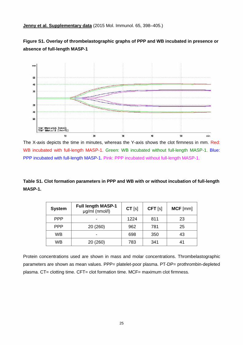
Jenny et al. Supplementary data (2015 Mol. Immunol. 65, 398–405.)
Figure S1. Overlay of thrombelastographic graphs of PPP and WB incubated in presence or
absence of full-length MASP-1
The X-axis depicts the time in minutes, whereas the Y-axis shows the clot firmness in mm. Red:
WB incubated with full-length MASP-1. Green: WB incubated without full-length MASP-1. Blue:
PPP incubated with full-length MASP-1. Pink: PPP incubated without full-length MASP-1.
Table S1. Clot formation parameters in PPP and WB w ith or without incubation of full-length
MASP-1.
System Full length MASP-1 µg/ml (nmol/l) CT [s] CFT [s] MCF [mm]
PPP - 1224 811 23
PPP 20 (260) 962 781 25
WB - 698 350 43
WB 20 (260) 783 341 41
Protein concentrations used are shown in mass and molar concentrations. Thrombelastographic
parameters are shown as mean values. PPP= platelet-poor plasma. PT-DP= prothrombin-depleted
plasma. CT= clotting time. CFT= clot formation time. MCF= maximum clot firmness.