Embed Size (px)
Citation preview
955
Special Articles
MASS RADIOGRAPHY IN THE EARLY
DIAGNOSIS OF CHEST DISEASE
E. G. W. HOFFSTAEDTM.D. Berlin
ASSISTANT PHYSICIAN, HOLYWOOD HALL SANATORIUM,WALSINGHAM, DURHAM
THE early diagnosis of pulmonary tuberculosis andother important chest diseases still leaves much to bedesired. Routine mass radiography of certain groupsof the population was’ recommended by the MedicalResearch Council (1942) to detect unsuspeoted cases inthe early asymptomatic stage. An ambitious programmewas initiated during the war by the then Minister ofHealth, and the results of three surveys made by theMedical Research Council (1945) gave impetus to thescheme, which has since gained the enthusiasticsupport of many able and experienced workers inthe field, particularly those who have witnessed thespectacular success of mass radiography in the ArmedForces.
Mass-radiography surveys of the civilian population,however, are a vastly different proposition from thescreening or sifting of military personnel under Serviceconditions. Brailsford (1943) was one of the first to castdoubt on the wisdom and practicability of the officialscheme ; and Toussaint and Pritchard (1945) developedinstead, with outstanding success, what has since beenknown as the Bermondsey scheme, in which practitionersare encouraged to send all their suspicious cases for
X-ray examination.It is perfectly true that " one type of case will be
detected only by mass radiography : the person with
unsuspected, possibly infective, chronic disease whose.symptoms are insufficient to take him to a doctor "
(Medical Research Council 1945). Active tuberculosis,however, has been found in only 0-4% of all the cases sofar examined by mass radiography, and not nearly allof these have been symptom-free. Though Kahan andClose (1944) report that in none of their 138 cases ofpulmonary tuberculosis picked up in a mass survey ofnaval ratings had the men ever reported sick or hadsymptoms such as to cause them to seek medical advice,in the surveys of the Medical Research Council (1945)of 79 " treffatment cases " (out of 21,280 persons radio-graphed) only 29 were symptomless, and of the 50 patientswith symptoms, 17 had already consulted a doctor. Inother words, in rather more than a fifth of the " unsus-pected " cases the patients had had symptoms for whichthey had sought medical advice, but in all of them thediagnosis had been missed because radiography was notused in time.No information is given about the incidence of symptoms
of non-tuberculous chest diseases found by mass radiography.The incidence of chest abnormalities other than tuberculosisdiscovered by mass radiography is 1—1-3%, but only a smallproportion of these conditions, which include cervical ribs,pleural thickening, and calcified glands, need treatment.There will hardly be many asymptomatic cases of bronchialcarcinoma, bronchiectasis, pneumoconiosis, or cardiovasculardiseases which show definite radiological abnormalities.
The facts (1) that only by mass radiography canpulmonary tuberculosis and certain other chest abnor-malities be detected in the presymptomatic stage (which isnot necessarily an early stage) ; (2) that even the presenceof certain symptoms, such as cough or lassitude, oftendoes not lead the patient to the doctor ; and (3) that inspite of definite radiological appearances physical signs
are sometimes absent and tubercle bacilli not found inthe sputum, have wrongly led to the conclusion thatmass radiography is the method of choice in diagnosing
early cases for treatment and thus in preventing thespread of pulmonary tuberculosis. It was hoped thatnation-wide periodical mass radiography would soon
become a reality, and the method was regarded as theanswer to the question of diagnosis.
THE CLINICAL ASPECT
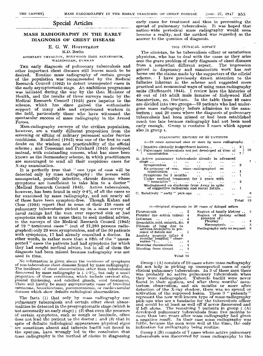
The clinician, be he tuberculosis officer or sanatoriumphysician, who has to deal with the cases as they arisesees the grave problem of early diagnosis of chest diseasesfrom a somewhat different aspect. The impressiongained in dispensary and sanatorium work has notborne out the claims made by the supporters of the officialscheme. - I have previously drawn attention to thefallacies inherent in the scheme and suggested morepractical and economical ways of using mass-radiographyunits (Hoffstaedt 1944). I review here the histories of40 out of 145 adult male inmates of Holywood HallSanatorium, co. Durham. In the table these 40 casesare divided into two groups-23 patients who had under-gone mass radiography before admission to the sana-torium ; and 20 cases where the diagnosis of pulmonarytuberculosis had been missed or had been establishedmuch too late because radiography had not been usedearly enough. Group 11 contains 3 cases which appearalso in group I.
DIAGNOSTIC HISTORY OF 40 PATIENTS
1-23 cases examined once or 11Wre by mass radiography
f Inactive clinically insignificant lesions.. 3 1A. Pulmonary tuberculosis not yet developed at time of r 10
L examination ............ 7 e
B. Active pulmonary tuberculosis already in advancedstage ................ 7 ,
Symptomless ............ 2
Ex-prisoner - of - war, mass - radiographed on
repatriation ............ 1
Symptoms for 3 months ........ 1Under medical treatment for 2 years with
" bronchitis " ... : ....... : 2Misdiagnosed on discharge from Army in spite .
of suggestive radiogram and rectal fistula.... 1
C. Relatively " early " pick-ups ........ 6
Total 23
II-Original diagnosis in 20 cases of delayed, action
Pleurodynia.... 2 Neglect of family history 5
Pleurisy (no action taken) 4 Neglect of history or/andInfluenza...... 4 presence of:
Bronchitis, cold, catarrh, &c. 6 Pleurisy .... 3
Fibrosis of lung due to Hsemoptysis .... 3asthma-bronchitis in pre- Radiography only on request 3sence of fistula ani .. 1
Anxiety neurosis (after 6months’ hospital obser-vation ") .. ’.... 1
Muscular rheumatism.. 1
Bleeding from throat.. 1
Total 20
Group I (A) consists of 10 cases where mass radiographydid not help in picking up unsuspected cases of earlyclinical pulmonary tuberculosis. In 3 of these men therewas probably no active pulmonary tuberculosis whenthey were radiographed. Tubercle bacilli were neverfound in their sputum, and after three months’ sana-torium observation, and six months or more afterdetection of the X-ray shadow, there was no spread oractivation of the supposed lesion. These 3 " patients "represent the now well-known type of mass-radiographypick-ups who are a headache for the tuberculosis officerand would be at least as well off if never discovered andif left alone. The remaining 7 patients in group I (A)developed pulmonary tuberculosis from five months tomore than two years after mass radiography had givena negative result. In their case mass radiography wasfutile, because the men were well at the time, the onlyindication for radiography being routine.Group I (B) consists of 7 cases where active pulmonary
tuberculosis was discovered by mass radiography to the
956
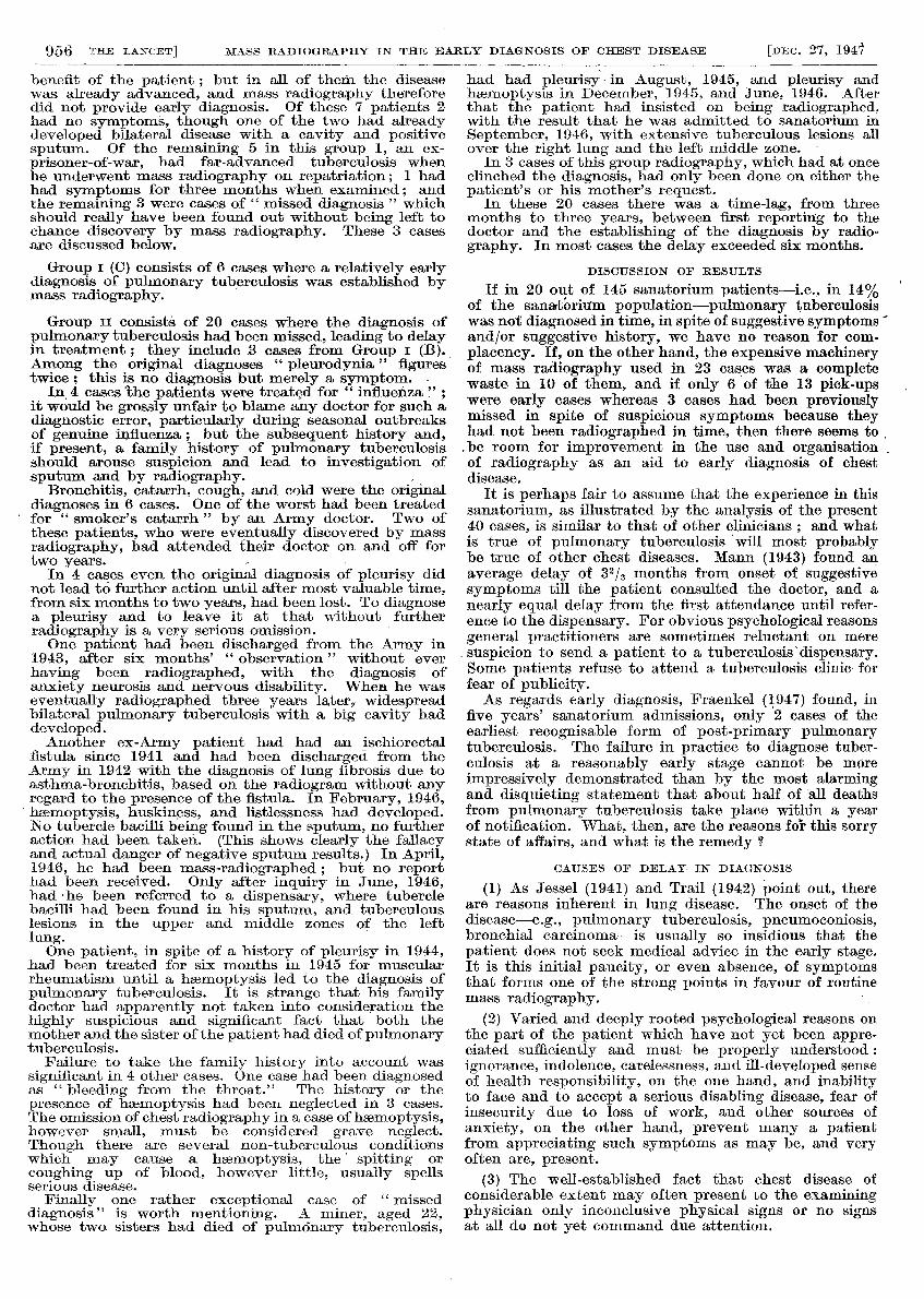
benefit of the patient ; but in all of them the diseasewas already advanced, and mass radiography thereforedid not provide early diagnosis. Of these 7 patients 2had no symptoms, though one of the two had alreadydeveloped bilateral disease with a cavity and positivesputum. Of the remaining 5 in this group 1, an ex-prisoner-of-war, had far-advanced tuberculosis whenhe underwent mass radiography on repatriation; 1 hadhad symptoms for three months when examined; andthe remaining 3 were cases of " missed diagnosis " whichshould really have been found out without being left tochance discovery by mass radiography. These 3 casesare discussed below.
Group I (C) consists of 6 cases where a relatively earlydiagnosis of pulmonary tuberculosis was established bymass radiography.
Group 11 consists of 20 cases where the diagnosis ofpulmonary tuberculosis had been missed, leading to delayin treatment ; they include 3 cases from Group I (B).,Among the original diagnoses " pleurodynia" figurestwice ; this is no diagnosis but merely a symptom.
In 4 casesthe patients were treated for " influenzait would be grossly unfair to blame any doctor for such adiagnostic error, particularly during seasonal outbreaksof genuine influenza ; but the subsequent history and,if present, a family history of pulmonary tuberculosisshould arouse suspicion and lead to investigation ofsputum and by radiography.
Bronchitis, catarrh, cough, and cold were the originaldiagnoses in 6 cases. One of the worst had been treated
for " smoker’s catarrh " by an Army doctor. Two ofthese patients, who were eventually discovered by massradiography, had attended their doctor on and off fortwo years. ’
In 4 cases even the original diagnosis of pleurisy didnot lead to further action until after most valuable time,from six months to two years, had been lost. To diagnosea pleurisy and to leave it at that without furtherradiography is a very serious omission.
’
One patient had been discharged from the Army in1943, after six months’ " observation " without everhaving been radiographed, with the diagnosis ofanxiety neurosis and nervous disability. When he waseventually radiographed three years later, widespreadbilateral pulmonary tuberculosis with a big cavity haddeveloped.Another ex-Army patient had had an ischiorectal
fistula since 1941 and had been discharged from theArmy in 1942 with the diagnosis of lung fibrosis due toasthma-bronchitis, based on the radiogram without anyregard to the presence of the fistula. In February, 1946,haemoptysis, huskiness, and listlessness had developed.No tubercle bacilli being found in the sputum, no furtheraction had been taken. (This shows clearly the fallacyand actual danger of negative sputum results.) In April,1946, he had been mass-radiographed ; but no reporthad been received. Only after inquiry in June, 1946,had he been referred to a dispensary, where tuberclebacilli had been found in his sputum, and tuberculouslesions in the upper and middle zones of the leftlung.
’
One patient, in spite of a history of pleurisy in 1944,had been treated for six months in 1945 for muscularrheumatism until a haemoptysis led to the diagnosis ofpulmonary tuberculosis. It is strange that his familydoctor had apparently not taken into consideration thehighly suspicious and significant fact that both themother and the sister of the patient had died of pulmonarytuberculosis.
Failure to take the family history into account wassignificant in 4 other cases. One case had been diagnosedas "bleeding from the throat." The history or thepresence of haemoptysis had been neglected in 3 cases.The omission of chest radiography in a case of haemoptysis,however small, must be considered grave neglect.Though there are several non-tuberculous conditionswhich may cause a haemoptysis, the’ spitting or
coughing up of blood, however little, usually spellsserious disease.
Finally one rather exceptional case of " misseddiagnosis " is worth mentioning. A miner, aged 22,whose two sisters had died of pulmonary tuberculosis,
had had pleurisy - in August, 1945, and pleurisy andhaemoptysis in December, 1945, and June, 1946. Afterthat the patient had insisted on being radiographed,with the result that he was admitted to sanatorium inSeptember, 1946, with extensive tuberculous lesions allover the right lung and the left middle zone.
In 3 cases of this group radiography, which had at onceclinched the diagnosis, had only been done on either thepatient’s or his mother’s request.
In these 20 cases there was a time-lag, from threemonths to three years, between first reporting to thedoctor and the establishing of the diagnosis by radio-graphy. In most cases the delay exceeded six months.
DISCUSSION OF RESULTS
If in 20 out of 145 sanatorium patients-i.e., in 14%of the sanatorium population-pulmonary tuberculosiswas not diagnosed in time, in spite of suggestive symptoms and/or suggestive history, we have no reason for com-placency. If, on the other hand, the expensive machineryof mass radiography used in 23 cases was a completewaste in 10 of them, and if only 6 of the 13 pick-upswere early cases whereas 3 cases had been previouslymissed in spite of suspicious symptoms because theyhad not been radiographed in time, then there seems to.be room for improvement in the use and organisationof radiography as an aid to early diagnosis of chestdisease.
It is perhaps fair to assume that the experience in thissanatorium, as illustrated by the analysis of the present40 cases, is similar to that of other clinicians ; and whatis true of pulmonary tuberculosis ’will most probablybe true - of other chest diseases. Mann (1943) found anaverage delay of 32/3 months from onset of suggestivesymptoms till the patient consulted the doctor, and anearly equal delay from the first attendance until refer-ence to the dispensary. For obvious psychological reasonsgeneral practitioners are sometimes reluctant on mere. suspicion to send a patient to a tuberculosis dispensary.Some patients refuse to attend a tuberculosis clinic forfear of publicity.As regards early diagnosis, Fraenkel (1947) found, in
five years’ sanatorium admissions, only 2 cases of theearliest recognisable form of post-primary pulmonarytuberculosis. The failure in practice to diagnose tuber-culosis at a reasonably early stage cannot be moreimpressively demonstrated than by the most alarmingand disquieting statement that about half of ’all deathsfrom pulmonary tuberculosis take place within- a yearof notification. What, then, are the reasons for this sorrystate of affairs, and what is the remedy ?
CAUSES OF DELAY IN DIAGNOSIS
(1) As Jessel (1941) and Trail (1942) point out, thereare reasons inherent in lung disease. The onset of thedisease-e.g., pulmonary tuberculosis, pneumoconiosis,bronchial carcinoma-is usually so insidious that thepatient does not seek medical advice in the early stage.It is this initial paucity, or even absence, of symptomsthat forms one of the strong points in favour of routinemass radiography. ’
(2) Varied and deeply rooted psychological reasons onthe part of the patient which have not yet been appre-ciated sufficiently and must be properly understood :ignorance, indolence, carelessness, and ill-developed senseof health responsibility, on the one hand, and inabilityto face and to accept a serious disabling disease, fear ofinsecurity due to loss of work, and other sources of
anxiety, on the other hand, prevent many a patientfrom appreciating such symptoms as may be, and veryoften are, present.
(3) The well-established fact that chest disease ofconsiderable extent may often present to the examiningphysician only inconclusive physical signs or no signsat all do not yet command due attention.
957
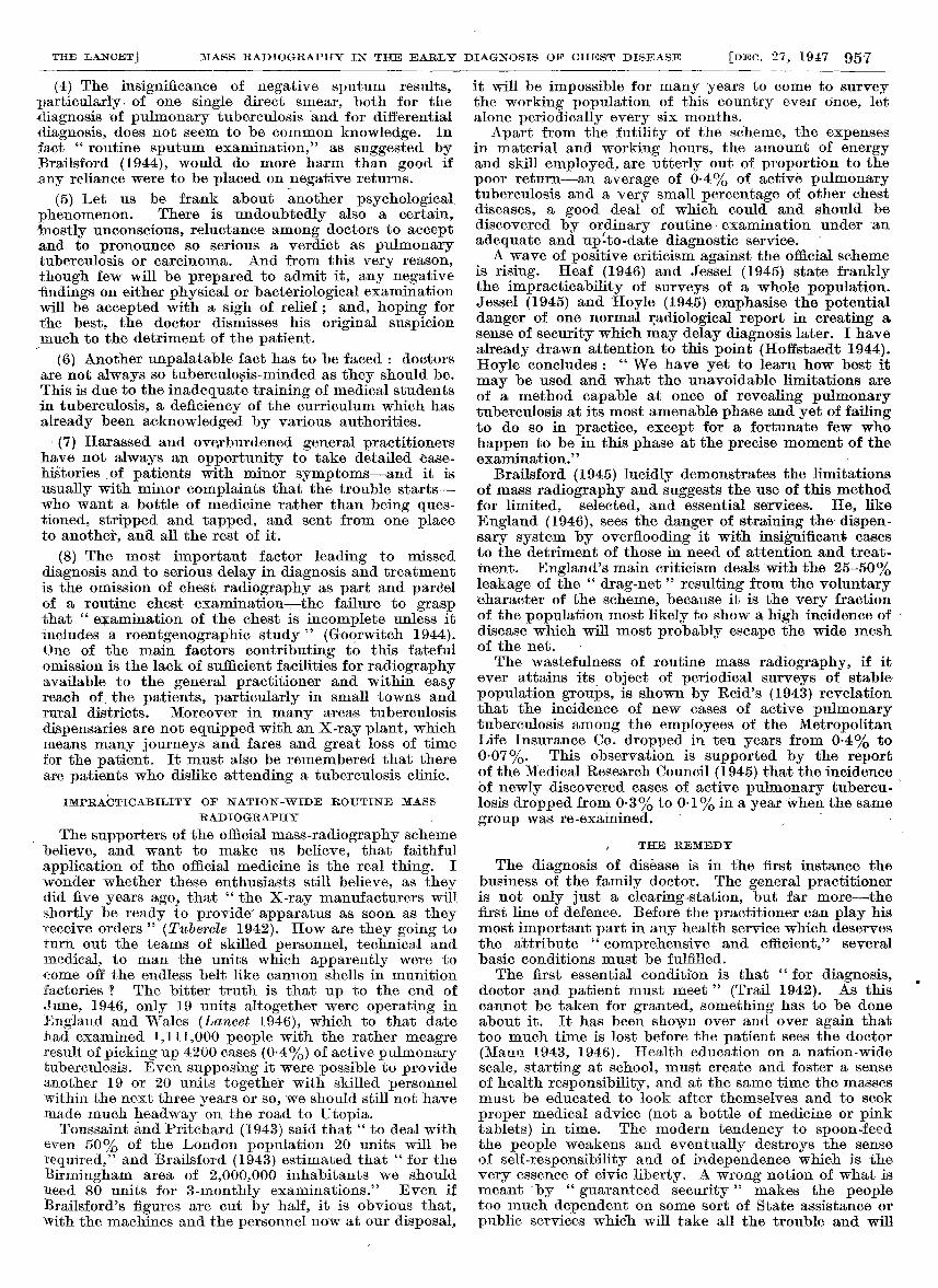
(4) The insignificance of negative sputum results,particularly of one single direct smear, both for the
diagnosis of pulmonary tuberculosis and for differentialdiagnosis, does not seem to be common knowledge. Infact " routine sputum examination," as suggested byBrailsford (1944), would do more harm than good if
any reliance were to be placed on negative returns.
(5) Let us be frank about another psychologicalphenomenon. There is undoubtedly also a certain,mostly unconscious, reluctance among doctors to acceptand to pronounce so serious a verdict as pulmonarytuberculosis or carcinoma. And from this very reason,though few will be prepared to admit it, any negativefindings on either physical or bacteriological examinationwill be accepted with a sigh of relief ; and, hoping forthe best, the doctor dismisses his original suspicionmuch to the detriment of the patient.
(6) Another unpalatable fact has to be faced : doctorsare not always so tuberculosis-minded as they should be.This is due to the inadequate training of medical studentsin tuberculosis, a deficiency of the curriculum which hasalready been acknowledged by various authorities.
(7) Harassed and overburdened general practitionershave not always an opportunity to take detailed case-histories of patients with minor symptoms-and it isusually with minor complaints that the trouble starts-who want a bottle of medicine rather than being ques-tioned, stripped and tapped, and sent from one placeto another, and all the rest of it.
(8) The most important factor leading to missed
diagnosis and to serious delay in diagnosis and treatmentis the omission of chest radiography as part and parcelof a routine chest examination -the failure to graspthat " examination of the chest is incomplete unless itincludes a roentgenographic study " (Goorwitch 1944).One of the main factors contributing to this fatefulomission is the lack of sufficient facilities for radiographyavailable to the general practitioner and within easyreach of the patients, particularly in small towns andrural districts. Moreover in many areas tuberculosis
dispensaries are not equipped with an X-ray plant, whichmeans many journeys and fares and great loss of timefor the patient. It must also be remembered that thereare patients who dislike attending a tuberculosis clinic.
IMPRACTICABILITY OF NATION-WIDE ROUTINE MASSRADIOGRAPHY ,
The supporters of the official mass-radiography schemebelieve, and want to make us believe, that faithful
application of the official medicine is the real thing. Iwonder whether these enthusiasts still believe, as theydid five years agos that " the X-ray manufacturers -willshortly be ready to provide- apparatus as soon as theyreceive orders " (Tubercle 1942). How are they going toturn out the teams of skilled personnel, technical andmedical, to man the units which apparently were toc-ome off the endless belt like cannon shells in munitionfactories ? The bitter truth is that up to the end ofJune, 1946, only 19 units altogether were operating inEngland and Wales (Lancet 1946), which to that datehad examined 1,111,000 people with the rather meagreresult of picking up 4200 cases (0-4%) of active pulmonarytuberculosis. Even supposing it were possible to provideanother 19 or 20 units together with skilled personnelwithin the next three years or so, we should still not havemade much headway on the road to Utopia.
Toussaint and Pritchard (1943) said that " to deal witheven 50% of the London population 20 units will berequired," and Brailsford (1943) estimated that " for theBirmingham area of 2,000,000 inhabitants we shouldlieed 80 units for 3-monthly examinations." Even ifBrailsford’s figures are cut by half, it is obvious that,with the machines and the personnel now at our disposal,
it will be impossible for many years to come to surveythe working population of this country even once, letalone periodically every six months.
Apart from the futility of the scheme, the expensesin material and working hours, the amount of energyand skill employed, are utterly out of proportion to thepoor return-an average of 0-4% of active pulmonarytuberculosis and a very small percentage of other chestdiseases, a good deal of which could and should bediscovered by ordinary routine examination under -anadequate and up’-to-date diagnostic service. ’
A wave of positive criticism against the official schemeis rising. Heaf (1946) and Jessel (1945) state franklythe impracticability of surveys of a whole population.Jessel (1945) and Hoyle (1945) emphasise the potentialdanger of one normal radiological report in creating asense of security which may delay diagnosis later. I havealready drawn attention to this point (Hoffstaedt 1944).Hoyle concludes :
" We have yet to learn how best itmay be used and what the unavoidable limitations areof a method capable at once of revealing pulmonarytuberculosis at its most amenable phase and yet of failingto do so in practice, except for a fortunate few whohappen to be in this phase at the precise moment of theexamination." ,
Brailsford (1945) lucidly demonstrates the limitationsof mass radiography and suggests the use of this methodfor limited, selected, and essential services. He, likeEngland (1946), sees the danger of straining the dispen-sary system by overflooding it with insignificant casesto the detriment of those in need of attention and treat-ment. England’s main criticism deals with the 25-50%leakage of the " drag-net " resulting from the voluntarycharacter of the scheme, because it is the very fractionof the population most likely to show a high incidence of disease which will most probably escape the wide meshof the net. ’
The wastefulness of routine mass radiography, if itever attains its object of periodical surveys of stable-population groups, is shown by Reid’s (1943) revelationthat the incidence of new cases of active pulmonarytuberculosis among the employees of the MetropolitanLife Insurance Co. dropped in ten years from 0-4% to0-07%. This observation is supported by the reportof the Medical Research Council (1945) that the incidenceof newly discovered cases of active pulmonary tubercu-losis dropped from 0-3% to 0-1% in a year when the samegroup was re-examined.
’
,
.
, THE REMEDY
The diagnosis of disease is in the first instance thebusiness of the family doctor. The general practitioneris not only just a clearing-station, but far more-thefirst line of defence. Before the practitioner can play hismost important part in any health service which deservesthe attribute "
comprehensive and efficient," severalbasic conditions must be fulfilled.The first essential condition is that " for diagnosis,
doctor and patient must meet " (Trail 1942). As thiscannot be taken for granted, something has to be doneabout it. It has been shown over and over again thattoo much time is lost before the patient sees the doctor(Mann 1943, 1946). Health education on a nation-widescale, starting at school, must create and foster a senseof health responsibility, and at the same time the massesmust be educated to look after themselves and to seek
proper medical advice (not a bottle of medicine or pinktablets) in time. The modern tendency to spoon-feedthe people weakens and eventually destroys the senseof self-responsibility and of independence which is thevery essence of civic liberty. A wrong notion of what ismeant by " guaranteed security" makes the peopletoo much dependent on some sort of State assistance orpublic services which will take all the trouble and will
958
readily come in whenever required-though then oftentoo late.
It therefore seems that, except for certain hazardousindustries where routine mass-radiography surveys areessential, it is a psychologically wrong approach, and notwithout danger, to bring the X-ray machine to the factoryand to the office. The better plan would be to see that thereally deserving people, the suspected cases, are sent tothe X-ray department through the agency of their owndoctor.The widespread indolence and complacency of so
many irresponsible and ignorant people must graduallybut systematically be overcome. The raising of the
school-leaving age is perhaps a unique opportunity forhealth education. The cinema should produce an equiva-lent to the B.B.C. talks of the Radio Doctor. There is
plenty of scope for effective health propaganda. Tomaintain the proper doctor-patient relationship, whichis the foundation of any satisfactory health service,people must be encouraged to see first of all their owndoctor.
It is certainly commendable that mass-radiographyunits direct their activities towards people in whom thepresence of a chest disease may be suspected ; but it is
definitely wrong for them to organise so-called opensessions and invite the public, by advertisements in localpapers and transport, to attend these sessions. This is
contrary to professional usage and does not fit in withthe generally accepted practice of outpatient clinics andpublic.rhealth clinics.
It is the general practitioner that must be encouraged,nay urged, to send patients for radiography. In theNational Health Service the health centres, as theygradually come into being, will provide facilities for
radiography. It is here that mobile mass-radiographyunits could be employed with the greatest benefit tothe public (Hoffstaedt 1947). Adequately equippedhealth centres for group practice, taking the place ofthe often poorly equipped one-man shop called the
surgery, will eventually give the practitioner the chanceof examining patients properly. Here also basic labora-
tory facilities must be provided. Last but not least, thehelp of a trained nurse our of a receptionist will enablethe practitioner to take proper case-histories, preferablyon standard case-sheets with relevant anamnestic and
symptomatic items, including family and occupationalhistory. After all, history-taking is one of the most impor-tant and revealing parts of an adequate examination.A much better training of the medical student in
tuberculosis and chest diseases and in the appraisal ofX-ray photographs is an urgent necessity. It must beunderstood by doctors that arguments about stethoscopeversus X rays are pointless, because physical chest exami-nation and radiography are not alternative but com-
plementary methods, each of them indispensable andessential, supplementing, never substituting, each other.Maxwell (1946) rightly postulates : " A practitioner whodid not insist on an X-ray examination of a suspiciouschest case, especially after haemoptysis, might be suedfor negligence." And Toussaint and Pritchard (1944)logically visualise the same - legal consequences foromission of a chest radiogram as those already existingunder the present law for neglect to radiograph a boneinjury in time. Even " the man in the street is coming toknow that a chest examination without a film is of
comparatively little value " (Tattersall 1946).THE PLACE OF ROUTINE MASS RADIOGRAPHY
In existing circumstances the pursuance of the officialscheme of indiscriminate routine mass radiography is
utopian. The Medical Research Council (1942) rightlysaid that " owing to the limitation of apparatus likelyto be available in this country concentration at thepresent time should be upon those groups where through
the nature of employment (e.g., silicosis producingindustries) or the composition of the population at riska relatively high incidence of unsuspected -pulmonarytuberculosis is likely to be found." Dick (1946), also
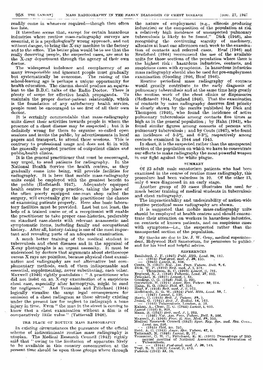
recognising the continuing scarcity of machinery,allocates at least one afternoon each week to the examina-tion of contacts and referred cases. Heaf (1946) andBrailsford (1944) recommend the use of the availableunits for those sections of the population where there isthe highest risk : hazardous industries, contacts, andsuspicious cases with symptoms. In hazardous industriesmass radiography should also be used for pre-employmentexamination (Gooding 1946, Heaf 1946).
Routine periodical mass radiography of contactswould greatly contribute to the early diagnosis of
pulmonary tuberculosis and at the same time help greatlythe routine, work of the chest clinics or dispensaries(Hoffstaedt 1944, England 1946). That the examinationof contacts by mass radiography deserves first priorityis clearly shown by the results published by Dick andThompson (1946), who found the incidence of activepulmonary tuberculosis among contacts five times as
high as in the general population ; by Hahn (1943), whofound similar figures among consorts of persons with
pulmonary tuberculosis ; and by Couts (1947), who foundan incidence of 5-5% and 6-3% respectively amongcontacts examined in 1944 and 1945.
In short, it is the suspected rather than the unsuspectedsection of the population on which we have to concentrateour efforts to make radiography the most powerful weaponin our fight against the white plague.
SUMMARY
Of 23 adult male sanatorium patients who had beenexamined in the course of routine mass radiography, thisprocedure had been valueless in 10. Of the other 13,only 6 were diagnosed in an early stage.Another group of 20 cases illustrates the need for -
much better training of medical students in tuberculosisand chest radiography.The impracticability and undesirability of nation-wide
routine periodical mass radiography are shown. ,
It is suggested that mobile mass-radiography unitsshould be employed at health centres and should concen-trate their attention on workers in hazardous industries,on contacts of known patients, and on suspicious caseswith symptoms-i.e., the suspected rather than the
unsuspected section of the population.My thanks are due to Dr. J. W. Gray, medical superinten-
dent, Holywood Hall Sanatorium, for permission to publish and for his kind and helpful advice.
REFERENCES
Brailsford, J. F. (1943) Publ. Hlth, Lond. 56, 117.— (1944) Post-grad. med. J. 20, 135.— (1945) Lancet, ii, 808.
Couts, B. (1947) Nat. Ass. Prev. Tuberc. Bull. 9, 6.Dick, W. P. (1946) Brit. med. J. i, 571.
— Thompson, B. C. (1946) Lancet, ii, 791.England, N. J. (1946) Tubercle, Lond. 27, 162.Fraenkel, E. (1947) Lancet, i, 84.Gooding, C. G. (1946) Ibid, ii, 891.Goorwitch, H. (1944) Amer. Rev. Tuberc. 50, 214.Hahn, R. G. (1943) Ibid, 47, 316.Heaf, F. (1946) Brit. med. J. ii, 975.Hoffstaedt, E. G. W. (1944) Publ. Hlth, Lond. 58, 7.
— (1947) Lancet, i, 120.Hoyle, C. (1945) Brit. J. Tuberc. 39, 1.Jessel, G. (1941) Brit. J. Radiol. 14, 162.
— (1945) Tuberculosis, London, p. 58.Kahan, A., Close, H. G. (1944) Lancet, i, 653.Lancet (1946) ii, 928.Mann, B. (1943) Brit. med. J. i, 283.
— (1946) Nat. Ass. Prev. Tuberc. Bull. 8, 166.Maxwell, J. (1946) Proc. R. Soc. Med. 39, 355.Medical Research Council (1942) Spec. Rep. Ser. med. Res. Coun.,
Lond. no. 246.— (1945) Ibid, no. 251.
Reid, A. C. (1943) Amer. Rev. Tuberc. 47, 9.Tattersall, N. (1946) Lancet, ii, 37.Toussaint, C. H. C., Pritchard, E. K. (1943) Proceedings of 26th
annual meeting of National Association for Prevention ofTuberculosis.
— — (1944) Post-grad. med. J. 20, 143.Trail, R. R. (1942) Lancet, ii, 413.Tubercle (1942) 23, 56.