Embed Size (px)
Citation preview
Int.J.Curr.Microbiol.App.Sci (2014) 3(11) 82-95
82
Original Research Article
Probiotic and functional characteristics of an indigenous Lactobacillus species isolated from traditional fermented product (Dahi-Chenna) of rural Odisha
Prangya Paramita Tripathy*, Mrutyunjay Suar, Jugal Kishore Das and Manish Ranjan Saini
School of Biotechnology, KIIT University, Bhubaneswar-751024, Odisha, India *Corresponding author
A B S T R A C T
Introduction
Probiotics are defined as live microorganisms which when administered in adequate amounts confer a health benefit on the host (FAO/WHO, 2002).
Lactobacillus and Bifidobacterium are the primarily studied and commonly used probiotics (Kleerebezem and Vaughan, 2009) and are GRAS (Generally regarded as
ISSN: 2319-7706 Volume 3 Number 11 (2014) pp. 82-95 http://www.ijcmas.com
K e y w o r d s
Probiotic, Autoaggregation, Acid and bile tolerance, Immunomodulation,
Probiotic microorganisms are increasingly used in preparation of neutraceuticals or in the treatment of infections. But the efficacy of a probiotic strain lies with its specific origin due to variation in gut microflora, different food habits and specific host-microbial interactions. As of now, Indian market is dominated with probiotics of Western origin and therefore an urgent need for exploring new indigenous probiotic strain is felt. In the present study an indigenous Lactobacillus species KSBT56 was isolated from a traditional dairy product of rural Odisha ( dahi chenna ) and identified as Lactobacillus plantarum both by biochemical and molecular typing methods. Further, KSBT56 was systematically studied for functional and probiotic characteristics e.g. acid tolerance, bile salt tolerance, adherence to colon epithelial cell line, cell surface hydrophobicity, auto aggregation, antimicrobial activity and in vitro immunomodulatory potential. The strain KSBT56 was able to survive in gastric acid conditions at pH 2.5 for 3 h and 2% bile salt for 2 h. Cellular autoaggregation of the strain was 39.3% and cell surface hydrophobicity was 63.3% with n-hexadecane. Occurrence of putative probiotic marker genes like fibronectin binding protein (fbp), mucus binding protein (mbp) and bile salt hydrolase (bsh) in KSBT56 were confirmed by PCR. The strain also showed adherence to colon epithelial HCT-116 cell line (7.43%). Increased expression of IL-10 in KSBT56 treated HCT-116 cell line shows the immunomodulatory potential of the strain. The strong antagonistic activity against seven different pathogens of KSBT56 was recently reported by us. Results suggested that Lactobacillus plantarum KSBT56 possesses in vitro probiotic potential and thus can be exploited for the development of indigenous functional foods.
Int.J.Curr.Microbiol.App.Sci (2014) 3(11) 82-95
83
safe) for consumption (Salminen et al., 1998). There has been an increase in the number of food supplements and pharmaceutical products that are being promoted on the basis of health claims based on several characteristics of strains of lactic acid bacteria, mainly from the genera Lactobacillus and Bacillus (McFarland and Elmer, 1997; Parvez et al., 2005; Hong et al., 2008). Probiotics are also known to alter the intestinal microflora balance favourably for the host, and thus must be able to survive in the gastrointestinal tract in order to exert their beneficial effects (De Vries et al., 2006). The organisms must be tolerant to low pH and bile toxicity prevalent in the upper digestive tract (Tuomola et al., 2001). The FAO/WHO guidelines also require the strain to have good adhesion to the intestinal epithelial cell lines and immunomodulatory potential to maintain gut homeostasis. The use of probiotics in food has become increasingly popular because of the multiple health benefits on the host.
Globally traditional products are gaining importance as a rich source of probiotic and functional lactobacilli (Reddy et al., 2007; Klayraung et al., 2008; Tinrat et al., 2011; Xiong et al., 2013). In India, no indigenous probiotic strain is available for commercialization and hence, Indian market is having probiotic products with western strains. Thus, the challenge before Indian researchers is to develop indigenous probiotic strains with specific health benefits. Traditional Indian fermented food products (dahi chenna, dahi, kanjii) are known for their unique style of fermentation and may harbour a rich flora of native Lactobacilli with potential probiotic properties. Many of the fermentative microflora in these traditional food products are not well studied and thus necessitated research on the isolation, identification, and characterization of such novel probiotic
strains from traditional food products. Dahi chenna is one such previously unexplored food product (which is mainly processed and consumed by rural people of Odisha, India) and which has been used in the present study to characterize the microflora present in it. The main objective of the present study was to characterize an indigenous isolate KSBT56 from dahi chenna by phenotypic and genotypic methods and to evaluate its in vitro probiotic potential.
Materials and Methods
Bacterial strains and culture conditions
The Lactobacillus isolate KSBT56 was grown in de Mann Rogosa Sharpe (MRS) broth (Hi Media Laboratories Pvt. Ltd, Mumbai, India) at 37oC for 4h and subcultured in the same media at 37oC for 24 h. Lactobacillus plantarum MTCC 1407 strain was cultured under similar conditions and used as a reference control in the study. All the strains used in the study are listed in Table 1.
Identification of the potential isolate
The biochemical and 16s rDNA sequencing of KSBT56 was already reported by Tripathy and Saini (2012) which confirmed the isolate as genus Lactobacillus. The molecular identification of KSBT56 was performed by PCR amplification. Colony PCR was performed with overnight (O/N) grown single colony of strain KSBT56 and reference cultures Lactobacillus casei (ATCC 9595) and Lactobacillus plantarum (MTCC 1407). Colonies were suspended in 150µL of sterile water and 2µL was taken as template for PCR. Cells were lysed at 95oC for 15 min. Sets of forward and reverse primers against the mub (Mucus binding protein), bsh (Bile salt hydrolase) and fbp (Fibronectin binding protein) gene of
Int.J.Curr.Microbiol.App.Sci (2014) 3(11) 82-95
84
Lactobacillus plantarum, are listed in Table 2 (Kaushik et al., 2009). Standard cycling conditions were: denaturation at 95oC for 30s, annealing at 51oC to 54oC for 1 min, extension at 72oC for 2 min with final extension of 10 min.
Screening for probiotic characteristics
Probiotic properties of the isolates were screened as per FAO/WHO (2002) guidelines that include acid and bile tolerance, antimicrobial activity, adhesion to colon epithelial cell line, cellular autoaggregation, cell surface hydrophobicity, antibiotic sensitivity and in vitro immunomodulatory activity.
Acid and bile salt tolerance
For acid tolerance MRS broth was adjusted to different pH values (1.5, 2.0, 2.5, and 3.0) with 1N HCl and the same (MRS broth) was supplemented with 1.5% and 2.0% (w/v) bile salts (MP Biomedicals, India Pvt. Ltd.) for bile salt tolerance. MRS broth with neutral pH 7.0 served as a control. All the broth tubes with different pH values and bile salt concentration were inoculated with 109
CFU/mL of O/N grown cultures of Lactobacilli and incubated at 37oC. Each tube containing 1 mL of culture was taken at 0, 1, 2 and 3 h interval; serially diluted in 0.1% peptone water, plated on MRS agar followed by incubation at 37oC for 48 h. The viable bacterial cell counts in terms of the colony forming units (CFU/mL) were recorded after 48 h. All the experiments were repeated thrice.
Cell aggregation
The freshly grown bacterial cells in MRS broth were harvested by centrifugation at 5000 × g for 10 min. The cell pellet was washed twice and resuspended in PBS to an
absorbance of 0.7 at 600 nm (AbsInitial). The suspension was centrifuged and the pellet was resuspended in equal volume of MRS broth, allowed to stand at 37oC for 2 h and the absorbance (AbsFinal) of upper suspension layer was measured using MRS broth as a reference. The percentage difference between the initial and final absorbance gives an index of cellular autoaggregation (Del Re et al., 2000; Tomas et al., 2005) that is expressed as follows: Aggregation (%) = 100 × (AbsInitial
AbsFinal)/Abs Initial
Cell surface hydrophobicity
Bacterial adhesion to hydrocarbons was determined according to the method described by Rosenberg et al. (1980). Bacterial cells grown in MRS broth at 37oC for 18 h were centrifuged at 8000 rpm for 10 min. The cell pellets were washed twice with phosphate urea magnesium (PUM) buffer, pH 7.0, resuspended in PUM buffer and the initial absorbance was adjusted to
0.7 OD at 600 nm (AbsInitial). Lactobacilli cell suspension was mixed with n-hexadecane or xylene (3:1), vortexed and incubated at 37oC for 10 min. The mixture was vortexed again and kept at 37oC for 1 h for phase separations. The aqueous phase was removed gently to measure its absorbance (AbsFinal) at 600 nm. The surface hydrophobicity (%) was calculated as per the following formula: Surface Hydrophobicity = 100 × (AbsInitial
AbsFinal)/ AbsInitial.
Adhesion assay
Adhesion of the isolated probiotic strain to HCT-116 colon epithelial cell line was carried out by using the method of Jacobsen et al. (1999). Briefly, DMEM without serum and antibiotics was added to each well of a 24-well tissue culture plate (Nest Biotech,
Int.J.Curr.Microbiol.App.Sci (2014) 3(11) 82-95
85
China) and incubated at 37°C for 30 min. Approximately 108 CFU/mL bacterial culture was suspended in 1.0 mL DMEM medium and added to the wells and incubated for 1 h at 37°C. Appropriate dilutions of the cell suspension were plated on MRS agar and incubated for 48 h at 37°C. The results were expressed as adhesion percentage, the ratio between adherent bacteria and added bacteria per well.
Immunomodulatory activity
Concentration of released cytokine IL-10, in response to bacterial infection was assessed with HCT-116 cell line (Wang et al., 2008) using commercially available ELISA kits (BD OptEIATM ELISA Kits, BD Biosciences, Pharmingen). HCT-116 cells were grown at 37°C in 5% CO2 in DMEM supplemented with 10% (v/v) fetal bovine serum, 100 U/mL penicillin, and 100 mg/mL streptomycin. The cells were seeded onto 24-well tissue culture plate and grown to 80% confluence. Cells of O/N grown Lactobacillus cultures were harvested by centrifugation at 8000 × g for 3 min, washed twice with PBS (pH 7.4) and resuspended in DMEM. To examine the influence of probiotics on cytokine production, both live and heat-inactivated Lactobacillus cultures were added to the HCT-116 cell culture medium at a concentration of 108 cells/mL. The limit of detection as described by the manufacturer was 1 pg/mL for all the assays. Results were statistically analysed by student s t-test.
Antibiotic susceptibility test
The antibiotic susceptibility was determined by using a standard disc diffusion technique according with the recommendations of National Commitee for Clinical Laboratory Standards (2008) (Charteris et al., 1998).
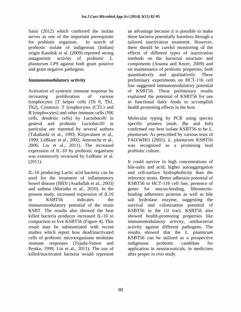
The tested antimicrobial drugs were ampicillin, streptomycin, kanamycin, tetracycline, carbenicillin, ofloxacin, cotrimoxazole and ciprofloxacin with concentrations of 1, 5, 10, 7, 7, 6, 11, and 8 (µg/mL), respectively and Mueller-Hinton agar (Merck, Darmstadt, Germany) was used for this purpose.
Results and Discussion
Molecular identification of the potential isolate
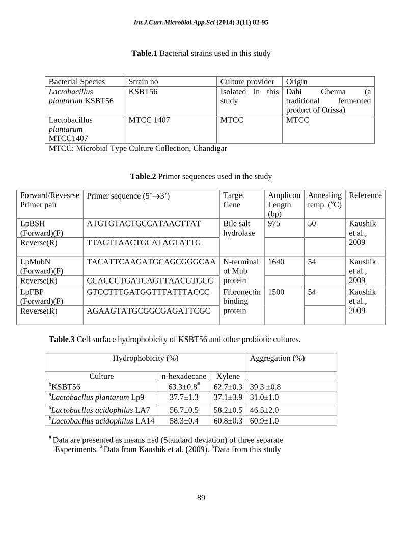
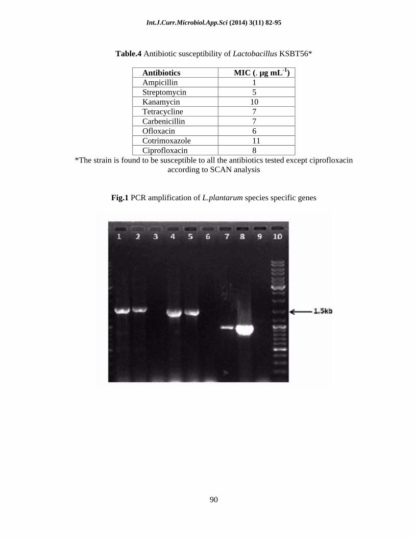
PCR amplification of unique fragments of 1.64 kbp, 1.5 kbp and 975 bp of the mub, fbp and bsh genes, respectively were detected by employing the L. plantarum species-specific primers (Fig. 1). The amplification indicates the presence of putative probiotic marker genes similar to that of the reference strain of L. plantarum. In silico analysis based on the homology search programme BLASTn with default parameters further revealed that these genes were absent from even closely related species like Lactobacillus pentosus, thereby confirming the strain KSBT56 to be a sub-species of L. plantarum.
Biochemical identification and 16s rDNA sequence of isolate KSBT56 showed the isolate belong to the genus Lactobacillus and was recently reported by Tripathy and Saini (2012). Further, molecular identification in this study confirms the bsh, fbp and mub gene amplification in both KSBT56 and Lactobacillus plantarum MTCC 1407 which substantiates KSBT56 as Lactobacillus plantarum. The amplification of above mentioned genes was also observed in probiotic L. plantarum 9 by Kaushik et al. (2009). From the above result KSBT56 was identified as L. plantarum and hence designated as L. plantarum KSBT56.
Int.J.Curr.Microbiol.App.Sci (2014) 3(11) 82-95
86
Screening for probiotic attributes of L. plantarum KSBT56
Acid and bile salt tolerance
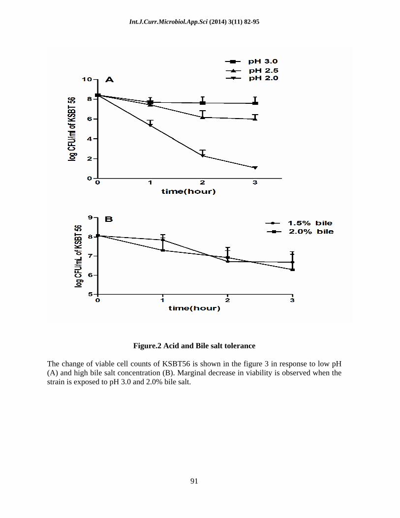
The viability of KSBT56 decreased by 1 log CFU mL-1 when exposed to pH 3.0 for 3 h. Similarly, the viability decreased by 1 log CFU mL-1 in 1 h and 3 log CFU mL-1 in 3 h at pH 2.5 (Fig. 2A). The marginal reduction in the viability of the isolate at low pH indicated good tolerance to acidic conditions prevalent in the stomach. KSBT56 also survived well in the presence of bile salt. The viability of KSBT56 in MRS broth containing 2.0% bile decreased by 2 log CFU mL-1 in 3 h (Fig. 2B). This indicates excellent bile tolerance of the isolated strain.
Cellular autoaggregation
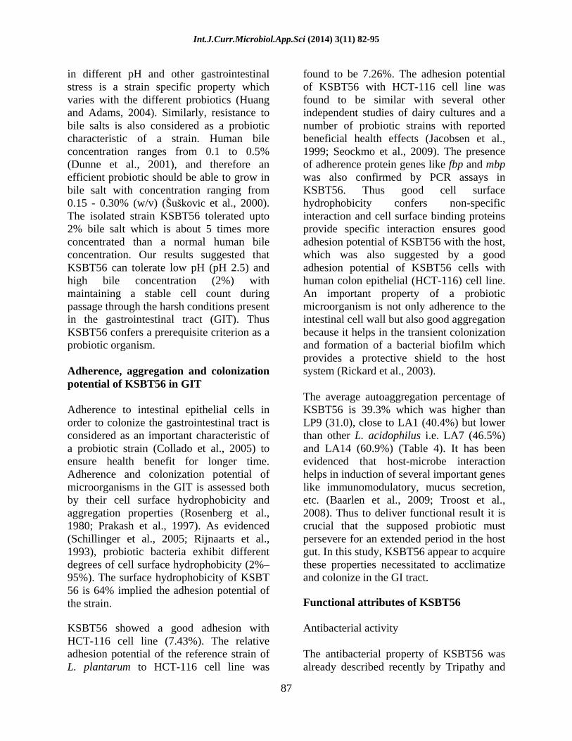
The cellular autoaggregation of KSBT56 was evaluated and compared with other standard probiotic cultures (Table 3). The average autoaggregation percentage of KSBT56 is 39.3% which was higher than LP9 (31.0), close to LA1 (40.4%) but lower than other L. acidophilus i.e. LA7 (46.5%) and LA14 (60.9%). The results indicate self-aggregation potential of KSBT56, a prerequisite characteristic of a probiotic strain.
Cell surface hydrophobicity and adhesion assay
The cell surface hydrophobicity and adherence values of the isolated probiotic strain indicate good probiotic potential of the isolate. Cell surface hydrophobicity of KSBT56 was found to be 63.3 ± 0.8 in n-hexadecane and 62.7 ± 0.3 in xylene which was more than the cell surface hydrophobicity of other standard probiotic strains (37.7% to 58.3% in n-hexadecane and 37.1% to 60.8% in xylene for the standard strains) (Table 3).
Immunomodulatory activity
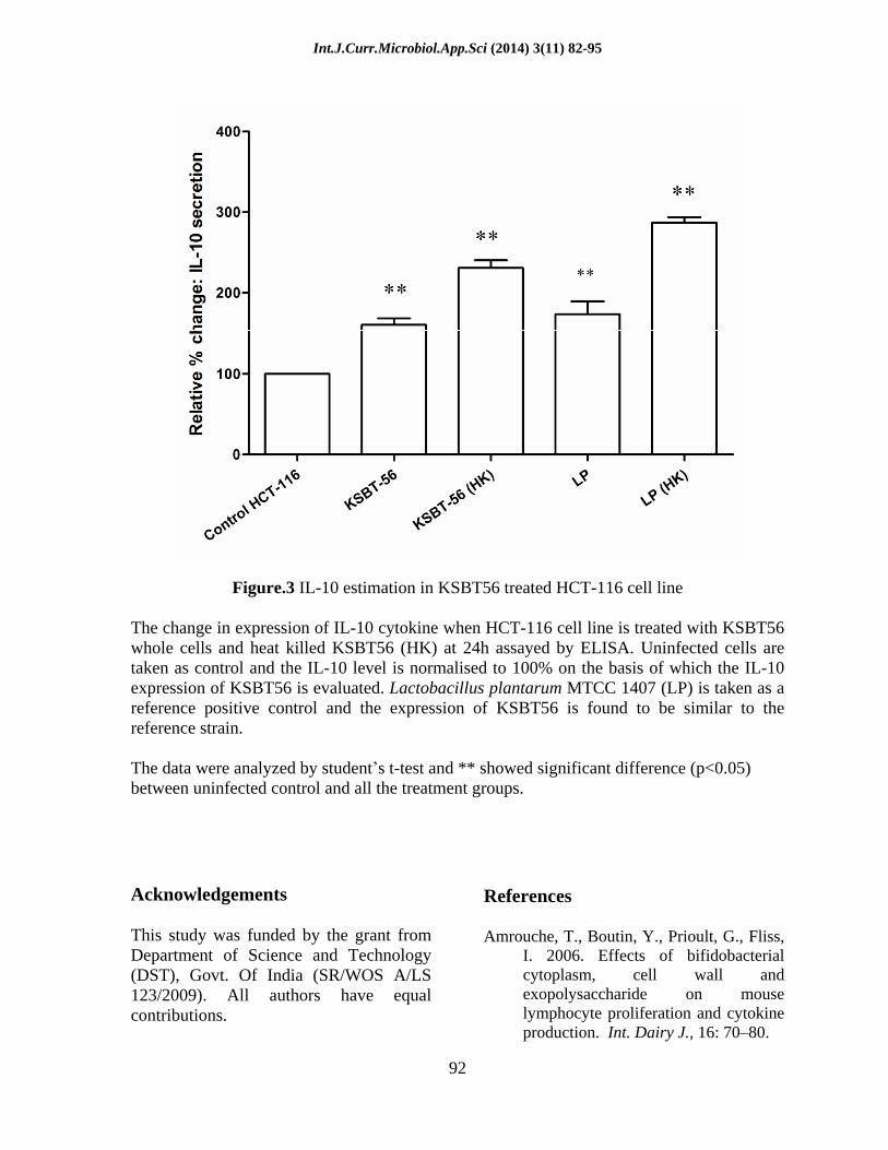
Live KSBT56 and heat killed KSBT56 demonstrated an increased expression of 1.6 and 2.31 times of IL-10 relative to control untreated cell line whereas Lactobacillus plantarum MTCC 1407 showed 1.73 and 2.86 times increase in IL-10 production over the untreated cell line respectively. The cells were also found to be viable after 24 h of infection which was confirmed by trypan blue dye exclusion method (Data not shown). The increased IL-10 expression by KSBT56 in comparison to control indicates the immunomodulatory potential of the KSBT56. The IL-10 expression of KSBT56 relative to the uninfected cell line is shown in Fig. 3.
Antibiotic susceptibility testing
The strain KSBT56 was found to be susceptible to all the antibiotics tested but resistant to ciprofloxacin (Table 4). Resistance to ciprofloxacin is an inherent resistance found in some Lactobacillus strains (Herreros et al., 2005). The antibiotic resistance in a probiotic strain could be transferred to the residential gut flora and is thus not a desirable characteristic of a probiotic strain. The susceptibility of KSBT56 to most of the antibiotics tested partially proves the safety of the strain.
Feasibility of KSBT56 isolate to survive under simulated gastrointestinal stress conditions
In the present study, the isolated strain KSBT56 tolerated acidic conditions better at pH 3.0 and pH 2.5 than at pH 2.0. Previous studies have also shown similar findings with L. plantarum strains, which could tolerate pH 2.5 and pH 3.0 (Sirilun et al., 2010). It has also been reported that survival
Int.J.Curr.Microbiol.App.Sci (2014) 3(11) 82-95
87
in different pH and other gastrointestinal stress is a strain specific property which varies with the different probiotics (Huang and Adams, 2004). Similarly, resistance to bile salts is also considered as a probiotic characteristic of a strain. Human bile concentration ranges from 0.1 to 0.5% (Dunne et al., 2001), and therefore an efficient probiotic should be able to grow in bile salt with concentration ranging from 0.15 - 0.30% (w/v) ( u kovic et al., 2000). The isolated strain KSBT56 tolerated upto 2% bile salt which is about 5 times more concentrated than a normal human bile concentration. Our results suggested that KSBT56 can tolerate low pH (pH 2.5) and high bile concentration (2%) with maintaining a stable cell count during passage through the harsh conditions present in the gastrointestinal tract (GIT). Thus KSBT56 confers a prerequisite criterion as a probiotic organism.
Adherence, aggregation and colonization potential of KSBT56 in GIT
Adherence to intestinal epithelial cells in order to colonize the gastrointestinal tract is considered as an important characteristic of a probiotic strain (Collado et al., 2005) to ensure health benefit for longer time. Adherence and colonization potential of microorganisms in the GIT is assessed both by their cell surface hydrophobicity and aggregation properties (Rosenberg et al., 1980; Prakash et al., 1997). As evidenced (Schillinger et al., 2005; Rijnaarts et al., 1993), probiotic bacteria exhibit different degrees of cell surface hydrophobicity (2%
95%). The surface hydrophobicity of KSBT 56 is 64% implied the adhesion potential of the strain.
KSBT56 showed a good adhesion with HCT-116 cell line (7.43%). The relative adhesion potential of the reference strain of L. plantarum to HCT-116 cell line was
found to be 7.26%. The adhesion potential of KSBT56 with HCT-116 cell line was found to be similar with several other independent studies of dairy cultures and a number of probiotic strains with reported beneficial health effects (Jacobsen et al., 1999; Seockmo et al., 2009). The presence of adherence protein genes like fbp and mbp was also confirmed by PCR assays in KSBT56. Thus good cell surface hydrophobicity confers non-specific interaction and cell surface binding proteins provide specific interaction ensures good adhesion potential of KSBT56 with the host, which was also suggested by a good adhesion potential of KSBT56 cells with human colon epithelial (HCT-116) cell line. An important property of a probiotic microorganism is not only adherence to the intestinal cell wall but also good aggregation because it helps in the transient colonization and formation of a bacterial biofilm which provides a protective shield to the host system (Rickard et al., 2003).
The average autoaggregation percentage of KSBT56 is 39.3% which was higher than LP9 (31.0), close to LA1 (40.4%) but lower than other L. acidophilus i.e. LA7 (46.5%) and LA14 (60.9%) (Table 4). It has been evidenced that host-microbe interaction helps in induction of several important genes like immunomodulatory, mucus secretion, etc. (Baarlen et al., 2009; Troost et al., 2008). Thus to deliver functional result it is crucial that the supposed probiotic must persevere for an extended period in the host gut. In this study, KSBT56 appear to acquire these properties necessitated to acclimatize and colonize in the GI tract.
Functional attributes of KSBT56
Antibacterial activity
The antibacterial property of KSBT56 was already described recently by Tripathy and
Int.J.Curr.Microbiol.App.Sci (2014) 3(11) 82-95
88
Saini (2012) which conferred the isolate serves as one of the important prerequisite for probiotic organism. In search of probiotic isolate of indigenous (Indian) origin Kaushik et al. (2009) reported strong antagonistic activity of probiotic L. plantarum LP9 against both gram positive and gram negative pathogens.
Immunomodulatory activity
Activation of systemic immune response by increasing proliferation of various lymphocytes [T helper cells (Th 0, Th1, Th2), Cytotoxic T lymphocytes (CTL) and B lymphocytes] and other immune cells (NK cells, dendritic cells) by Lactobacilli in general and probiotic Lactobacilli in particular are reported by several authors (Takahashi et al., 1993; Kirjavainen et al., 1999; LeBlanc et al., 2002; Amrouche et al., 2006; Liu et al., 2011). The increased expression of IL-10 by probiotic organisms was extensively reviewed by LeBlanc et al. (2011).
IL-10 producing Lactic acid bacteria can be used for the treatment of inflammatory bowel disease (IBD) (Asadullah et al., 2003) and asthma (Marinho et al., 2010). In the present study, increased expression of IL10 in KSBT56 indicates the immunomodulatory potential of the strain KSBT. The results also showed the heat killed bacteria produces increased IL-10 in comparison to live KSBT56 (Figure 4). This result may be substantiated with recent studies which report how dead/inactivated cells of probiotic microorganisms modulate immune responses (Tejada-Simon and Pestka, 1999; Lin et al., 2011). The use of killed/inactivated bacteria would represent
an advantage because it is possible to make these bacteria potentially harmless through a tailored inactivation treatment. However, there should be careful monitoring of the effects of different types of inactivation methods on the bacterial structure and components (Ananta and Knorr, 2009) and on maintenance of probiotic properties, both quantitatively and qualitatively. These preliminary experiments on HCT-116 cell line suggested immunomodulatory potential of KSBT56. These preliminary results explained the potential of KSBT56 for use in functional dairy foods to accomplish health promoting effects in the host.
Molecular typing by PCR using species specific primers (mub, fbp and bsh) confirmed our best isolate KSBT56 to be L. plantarum. As prescribed by various tests of FAO/WHO (2002), L. plantarum KSBT56 was recognized as a promising lead probiotic culture.
It could survive in high concentrations of bile-salts and acid; higher autoaggregation and cell-surface hydrophobicity than the reference strain. Better adhesion potential of KSBT56 to HCT-116 cell line, presence of genes for mucus-binding, fibronectin-binding adherence proteins as well as bile salt hydrolase enzyme, suggesting the survival and colonization potential of KSBT56 in the GI tract. KSBT56 also showed health-promoting properties like immunomodulatory activity, antibacterial activity against different pathogens. The results showed that the L. plantarum KSBT56 can be utilized as a prospective indigenous probiotic candidate for application in neutraceuticals, in medicines after proper in vivo study.
Int.J.Curr.Microbiol.App.Sci (2014) 3(11) 82-95
89
Table.1 Bacterial strains used in this study
Bacterial Species Strain no Culture provider Origin Lactobacillus plantarum KSBT56
KSBT56 Isolated in this study
Dahi Chenna (a traditional fermented product of Orissa)
Lactobacillus plantarum MTCC1407
MTCC 1407 MTCC MTCC
MTCC: Microbial Type Culture Collection, Chandigar
Table.2 Primer sequences used in the study
Forward/Revesrse Primer pair
Primer sequence (5 3 ) Target Gene
Amplicon Length (bp)
Annealing temp. (oC)
Reference
LpBSH (Forward)(F)
ATGTGTACTGCCATAACTTAT 975 50
Reverse(R) TTAGTTAACTGCATAGTATTG
Bile salt hydrolase
Kaushik et al., 2009
LpMubN (Forward)(F)
TACATTCAAGATGCAGCGGGCAA
1640 54
Reverse(R) CCACCCTGATCAGTTAACGTGCC
N-terminal of Mub protein
Kaushik et al., 2009
LpFBP (Forward)(F)
GTCCTTTGATGGTTTATTTACCC 54
Reverse(R) AGAAGTATGCGGCGAGATTCGC
Fibronectin binding protein
1500 Kaushik et al., 2009
Table.3 Cell surface hydrophobicity of KSBT56 and other probiotic cultures.
Hydrophobicity (%) Aggregation (%)
Culture n-hexadecane
Xylene bKSBT56 63.3±0.8# 62.7±0.3
39.3 ±0.8 aLactobacllus plantarum Lp9 37.7±1.3 37.1±3.9
31.0±1.0 aLactobacllus acidophilus LA7 56.7±0.5 58.2±0.5
46.5±2.0 bLactobacllus acidophilus LA14
58.3±0.4 60.8±0.3
60.9±1.0
# Data are presented as means ±sd (Standard deviation) of three separate Experiments. a Data from Kaushik et al. (2009). bData from this study
Int.J.Curr.Microbiol.App.Sci (2014) 3(11) 82-95
90
Table.4 Antibiotic susceptibility of Lactobacillus KSBT56*
Antibiotics MIC (. µg mL-1) Ampicillin 1 Streptomycin 5 Kanamycin 10 Tetracycline 7 Carbenicillin 7 Ofloxacin 6 Cotrimoxazole 11 Ciprofloxacin 8
*The strain is found to be susceptible to all the antibiotics tested except ciprofloxacin according to SCAN analysis
Fig.1 PCR amplification of L.plantarum species specific genes
Int.J.Curr.Microbiol.App.Sci (2014) 3(11) 82-95
91
Figure.2 Acid and Bile salt tolerance
The change of viable cell counts of KSBT56 is shown in the figure 3 in response to low pH (A) and high bile salt concentration (B). Marginal decrease in viability is observed when the strain is exposed to pH 3.0 and 2.0% bile salt.
Int.J.Curr.Microbiol.App.Sci (2014) 3(11) 82-95
92
Figure.3 IL-10 estimation in KSBT56 treated HCT-116 cell line
The change in expression of IL-10 cytokine when HCT-116 cell line is treated with KSBT56 whole cells and heat killed KSBT56 (HK) at 24h assayed by ELISA. Uninfected cells are taken as control and the IL-10 level is normalised to 100% on the basis of which the IL-10 expression of KSBT56 is evaluated. Lactobacillus plantarum MTCC 1407 (LP) is taken as a reference positive control and the expression of KSBT56 is found to be similar to the reference strain.
The data were analyzed by student s t-test and ** showed significant difference (p<0.05) between uninfected control and all the treatment groups.
Acknowledgements
This study was funded by the grant from Department of Science and Technology (DST), Govt. Of India (SR/WOS A/LS 123/2009). All authors have equal contributions.
References
Amrouche, T., Boutin, Y., Prioult, G., Fliss, I. 2006. Effects of bifidobacterial cytoplasm, cell wall and exopolysaccharide on mouse lymphocyte proliferation and cytokine production. Int. Dairy J., 16: 70 80.
Int.J.Curr.Microbiol.App.Sci (2014) 3(11) 82-95
93
Ananta, E., Knorr, D. 2009. Comparison of
inactivation pathways of thermal or high pressure inactivated Lactobacillus rhamnosus ATCC 53103 by flow cytometry analysis. Food Microbiol., 26: 542 546.
Asadullah, K., Sterry, W., Volk, H.D. 2003. Interleukin-10 therapy review of a new approach. Pharmacol. Rev., 55: 241 69.
Baarlen, Pvan., Troost, F.J., Hemert, Svan., Meer, Cvander., de Vos, W.M., et al. 2009. Differential NF-kB pathways induction by Lactobacillus plantarum in the duodenum of healthy humans correlating with immune tolerance. Proc. Natl. Acad. Sci. USA., 106: 2371 2376.
Charteris, W.P., Kelly, P.M., Morelli, L., Collins, J.K. 1998. Antibiotic susceptibility of potentially probiotic Lactobacillus species. J. Food Prot., 61: 1636 1643.
Collado, M.C., Gueimonde, M., Hernandez, M., Sanz, Y., Salminen, S. 2005. Adhesion of selected Bifidobacterium strains to human intestinal mucus and the role of adhesion in enteropathogen exclusion. J. Food Prot., 68: 26722678.
De Vries, M.C., Vaughan, E.E., Kleerebezem, M., de Vos, W.M. 2006. Lactobacillus plantarum survival, functional and potential probiotic properties in the human intestinal tract. Int. Dairy J., 16: 1018 1028.
Del Re, B., Sgorbati, B., Miglioli, M., Palenzona, D. 2000. Adhesion, autoaggregation and hydrophobicity of 13 strains of Bifidobacterium longum. Lett. Appl. Microbiol., 31: 438 442.
Dunne, C., O Mahony, L., Murphy, L., et al. 2001. In vitro selection criteria for probiotic bacteria of human origin: Correlation with in vivo findings. Am. J. Clin. Nutr., 73: 386 392.
FAO/WHO, 2002. Guidelines for the evaluation of probiotics in food. Report of a joint FAO/WHO working
group on drafting guidelines for evaluation of probiotics in food, London, Ontario, Canada.
Gibson, G.R. 1998. Dietary modulation of the human gut microflora using probiotics. Br. J. Nutr., 80: 209 212.
Herreros, M.A., Sandoval, H., Gonzalez, L., Castro, J.M., Fresno, J.M., Tornadijo, M.E. 2005. Antimicrobial activity and antibiotic resistance of lactic acid bacteria isolated from Armada cheese (a Spanish goats milk cheese). Food Microbiol., 22: 455 459.
Hong, H.A., Huang, J.M., Khaneja, R., Hiep, L.V., Urdaci, M.C., Cutting, S.M. 2008. The safety of Bacillus subtilis and Bacillus indicus as food probiotics. J. Appl. Microbiol., 105: 510 520.
Huang, J., Adams, M.C. 2004. In vitro assessment of the upper gastrointestinal tolerance of potential probiotic dairy Propionibacteria. Int. J. Food Microbiol., 91: 253 260.
Jacobsen, C.N., Nielsen, V.R., Hayford, A.E., Moller, P.L., Michaelsen, K.F., Paerregard, A., Sandstrom, B., Tvede, M., Jakobsen, M. 1999. Screening of probiotic activities of forty-seven strains of Lactobacillus spp. by in vitro techniques and evaluation of the colonization ability of five selected strains in humans. Appl. Environ. Microbiol., 65: 4949 4956.
Kaushik, J.K., Kumar, A., Duary, R.K., Mohanty, A.K., Grover, S., Vatish, V.K. 2009. Functional and probiotic attributes of an indigenous isolate of Lactobacillus plantarum. PLoS ONE, doi:10.1371/journal.pone.0008099.
Kirjavainen, P.V., El-Nezami, H.S., Salminen, S.J., Ahokas, J.T., Wright, P.F. 1999. The effect of orally administered viable probiotic and dairy Lactobacilli on mouse lymphocyte proliferation. FEMS Immunol. Med. Microbiol., 26: 131135.
Klayraung, S., Viernstein, H., Sirithunyalug,
Int.J.Curr.Microbiol.App.Sci (2014) 3(11) 82-95
94
J., Okonogi, S. 2008. Probiotic properties of Lactobacilli isolated from thai traditional food. Sci. Pharm., 76: 485 503.
Kleerebezem, M., Vaughan, E.E. 2009. Probiotic and gut Lactobacilli and Bifidobacteria: molecular approaches to study diversity and activity. Annu. Rev. Microbiol., 63: 269 290.
LeBlanc, A.M., Carmen, S.D., Zurita-Turk, M., Rocha, C.S., Guchte, M., Azevedo, V., Miyoshi, A., Le Blanc, J.G. 2011. Importance of IL-10 modulation by probiotic microorganisms in gastrointestinal inflammatory diseases. ISRN Gastroenterology 2011, Article ID 892971, 11p. doi:10.5402/2011/892971.
LeBlanc, J.G., Matar, C., Valdez, J.C., LeBlanc, J., Perdigon, G. 2002. Immunomodulating effects of peptidic fractions issued from milk fermented with Lactobacillus helveticus. J. Dairy. Sci., 85: 2733 2742.
Lin, W.H., Wu, C.R., Fang, T.J., Lee, M.S., Lin, K.L., Chen, H.C., Huang, S.Y., Hseu, Y.C. 2011. Adherent properties and macrophage activation ability of 3 strains of lactic acid bacteria. J. Food Sci., 76: M1 M7.
Liu, C.F., Tseng, K.C., Chiang, S.S., Lee, B.H., Hsu, W.H., Pan, T.M. 2011. Immunomodulatory and antioxidant potential of Lactobacillus exopolysaccharides. J. Sci. Food Agric., 91: 2284 2291.
Marinho, F.A.V., Pacifico, L.G.G., Miyoshi, A., Azevedo, V., et al. 2010. An intranasal administration of Lactococcus lactis strains expressing recombinant interleukin-10 modulates acute allergic airway inflammation in a murine model. Clin. Exp. Allergy., 40: 1541 1551.
McFarland, L.V., Elmer, G.W. 1997. Pharmaceutical probiotics for the treatment of anaerobic and other infections. Anaerobe, 3: 73 78.
Parvez, S., Malik, K.A., Kang, S., Kim, H.Y., 2005. Probiotics and their fermented food products are beneficial for health. J. Appl. Microbiol., 100:1171 1185.
Prakash, R., Sinha, P.R., Sinha, R.N., Singh B., 1997. Adherence of Lactobacilli to epithelial cells and hexadecane for use of probiotics. Indian J. Dairy Sci., 10: 43 47.
Reddy, K.B.P.K., Raghavendra, P., Kumar, B.G., Misra, M.C., Prapulla, S.G. 2007. Screening of probiotic properties of lactic acid bacteria isolated from Kanjika, an ayruvedic lactic acid fermented product: an in-vitro evaluation. J. Gen. Appl. Microbiol., 53: 207 213.
Rijnaarts, H.M.M., Lyklema, J., Norde, W., Zehnder, A.J.B. 1993. Bacterial adhesion under static and dynamic conditions. Appl. Enviorn. Microbiol., 59: 3255 3265.
Rosenberg, M., Gutnick, D., Rosenberg, E., 1980. Adherence of bacteria to hydrocarbons: a simple method for measuring cell-surface hydrophobicity. FEMS Microbiol. Lett., 9: 29 33.
Salminen, S., Von Wright, A., Morelli, L., Marteau, P., de Vos, W.M. et al. 1998. Demonstration of safety of probiotics - a review. Int. J. Food Microbiol., 44: 93 106.
Schillinger, U., Guigas, C., Holzapfel, W.H. 2005. In vitro adherence and other properties of lactobacilli used in probiotic yoghurt-like products. Int. Dairy. J., 15: 1289 1297.
Seockmo, Ku., Hyun, J.Y., Geun, E.J., 2009. Enhancement of anti-tumorigenic polysaccharide production, Adhesion, and Branch Formation of Bifidobacterium bifidum BGN4 by Phytic Acid. Food Sci. Biotechnol., 18: 001 006.
Sirilun, S., Chaiyasut, C., Kantachote, D., Luxananil, P. 2010. Characterization of non human origin probiotic
Int.J.Curr.Microbiol.App.Sci (2014) 3(11) 82-95
95
Lactobacillus plantarum with cholesterol-lowering property. Afr. J. Microbial. Res., 4: 994 1000.
u kovic, J., Kos, B., Mato ic, S., Besendorfer, V. 2000. The effect of bile salts on survival and morphology of a potential probiotic strain Lactobacillus acidophilus M92. World. J. Microbiol. Biotechnol., 16: 673 678.
Takahashi, T., Oka, T., Iwana, H., Kuwata, T., Yamamoto, Y. 1993. Immuneresponse of mice to orally administered lactic acid bacteria. Biosci. Biotechnol. Biochem., 57: 1557 1560.
Tejada-Simon, M.V., Pestka, J.J. 1999. Proinflammatory cytokine and nitric oxide induction in murine macrophages by cell wall and cytoplasmic extracts of lactic acid bacteria. J. Food Prot., 62: 14351444.
Tinrat, S., Saraya, S., Chomnawang, M.T., 2011. Isolation and characterization of Lactobacillus salivarius MTC 1026 as a potential probiotic. J. Gen. Appl. Microbiol., 57: 365 378.
Tomas, M.S.J., Wiese, B., Nader-Macias, M.E. 2005. Effects of culture conditions on the growth and auto-aggregation ability of vaginal Lactobacillus johnsonii CRL. 1294. J. Appl. Microbiol., 99: 1383 1391.
Tuomola, E., Crittenden, R., Playne, M., Isolauri, E. 2001. Quality assurance criteria for probiotic bacteria. Am. J. Clin Nutr., 73: 393 398.
Tripathy, P.P., Saini, M.R. 2012. Spectrum of antimicrobial activity of lactic acid bacteria (Lactobacillus KSBT 56) isolated from indigenous fermented products of Odisha. Afr. J. Food. Sci., 6(24): 560 566.
Wang, S., Ng, L.H., Chow, W.L., Lee, Y.K. 2008. Infant intestinal Enterococcus faecalis down-regulates inflammatory responses in human intestinal cell lines. World J. Gastroenterol., 14:
1067 1076. Xiong, T., Song, S., Huang, X., Feng, C.,
Liu, G., Huang, J., Xie, M. 2013. Screening and identification of functional lactobacillus specific for vegetable fermentation. J. food Sci., 78: M84 M89.
Troost, F.J., Van Baarlen P., Lindsey, P., Kodde, A., De Vos, W.M., Kleerebezem, M., Brummer, R.J. 2008. Identification of the transcriptional response of human intestinal mucosa to Lactobacillus plantarum WCFS1 in vivo. BMC Genomics, 9: 374.