Embed Size (px)
Citation preview
ANIMAL BEHAVIOUR, 2006, 71, 1283–1288doi:10.1016/j.anbehav.2005.07.025
Maternal testosterone affects the primary sex ratio
and offspring survival in zebra finches
JOANNA RUTKOWSKA & MARIUSZ CICHON
Institute of Environmental Sciences, Jagiellonian University
(Received 12 January 2005; initial acceptance 14 March 2005;
final acceptance 29 July 2005; published online 3 May 2006; MS. number: 8424R)
Female birds have repeatedly been reported to adjust the primary sex ratio of their offspring to environ-mental, social and physiological cues. However, the mechanism behind sex adjustment remains unknown.It has been suggested that maternal hormones may constitute an important mediator in this mechanism,as androgen levels differ between eggs bearing male and female embryos. To evaluate whether the level ofmaternal androgens affects the offspring sex ratio, we injected female zebra finches, Taeniopygia guttata,with testosterone during egg laying. The sex ratio of eggs laid after testosterone administration became sig-nificantly male biased, compared to eggs laid by control females that received a vehicle injection. However,sons of testosterone-treated females suffered lower hatching success. In contrast, daughters seemed to ben-efit from elevated androgen level in terms of future survival prospects. The opposite effects on male andfemale offspring may constitute an important constraint on maternal androgen allocation to the eggsand reduce the benefits of biasing the sex ratio towards males by increasing the testosterone level.
� 2006 The Association for the Study of Animal Behaviour. Published by Elsevier Ltd. All rights reserved.
In birds, females are the heterogametic sex, bearing sexchromosomes Z and W, while males have ZZ; thus,females have the potential ability to control offspringsex (Pike & Petrie 2003). Follicles developing in the ovarycontain both sets of chromosomes until the first meioticdivision during which one of them is consigned to the po-lar body and the other remains in the ovum (Johnson2000). In several bird species, chromosome segregationhas been shown to occur 0.5–3 h before ovulation (Warren& Scott 1935; Olsen & Fraps 1944; Birenkott et al. 1988;Johnson 1996). Thus, fine-tuned adjustment of offspringsex should be possible even at the within-clutch level.For example, offspring sex covaries with laying order inseveral bird species (reviewed in Krackow 1995), so thatthe more valuable or more demanding sex is placed inthe more advantageous position in the laying order(Kilner 1998; Badyaev et al. 2002). Furthermore, there issome evidence that females may respond to an immediatechange in the environment, for example in food qualityduring egg laying, by adjusting offspring sex accordingly(Rutkowska & Cichon 2002).
Such a precise mechanism of sex adjustment may bemediated by maternal hormones (Krackow 1995),
Correspondence: J. Rutkowska, Institute of Environmental Sciences,Jagiellonian University, Gronostajowa 7, 30-387 Cracow, Poland(email: [email protected]).
12830003–3472/06/$30.00/0 � 2006 The Association for the St
especially androgens as eggs containing male embryoshave higher yolk androgen concentrations (Petrie et al.2001). The relation between maternal androgens and off-spring sex might, however, depend on the females’ socialstatus (Muller et al. 2002) and diet (Rutstein et al. 2005).Evidence for a potential role of androgens in sex determi-nation comes from studies showing that females mated toattractive partners lay eggs with a high androgen content(Gil et al. 1999, 2004; von Engelhardt 2004; but see Rut-stein et al. 2004) and that such females produce male-biased clutches (Sheldon et al. 1999). However, evidencethat experimentally manipulated maternal androgen levelaffects offspring sex has been lacking. A recent study onthe free-living spotless starling, Sturnus unicolor, showedthat females implanted with testosterone prior to egglaying overproduced sons, but this male-skewed sex ratiopersisted in clutches laid in subsequent breeding seasons,long after the implants had depleted (Veiga et al. 2004).
We studied the potential role of androgens in determin-ing offspring sex in a more rigorous laboratory experi-mental set-up. In our study, females were rearedindividually with randomly assigned partners, whichcontrolled for potentially confounding effects of malequality and social interactions on egg androgen contentand sex allocation. We manipulated maternal hormonalstatus by injecting zebra finch, Taeniopygia guttata, femaleswith testosterone on the day the first egg in a clutch waslaid (cf. Hackl et al. 2003). This ensured that females
udy of Animal Behaviour. Published by Elsevier Ltd. All rights reserved.
ANIMAL BEHAVIOUR, 71, 61284
were manipulated at exactly the same stage of a breedingcycle. Our previous study showed that administration of20 mg of testosterone to the female after the first egg waslaid resulted in a significant increase in androgen concen-tration in the yolk of the third, fourth and fifth eggs incomparison to the first egg of a clutch (Rutkowska et al.2005). In a control group, androgen concentrations inthe eggs decreased with laying order (Rutkowska et al.2005; see also Gil et al. 1999, 2004; von Engelhardt2004; Rutstein et al. 2005). In the present study, we ad-ministrated testosterone after the first egg in a clutchhad already been laid and when the second ovum had al-ready been ovulated, so the testosterone should have aneffect, if any, in the third and subsequently laid eggs. Wepredicted that testosterone-treated females should havea higher proportion of males in eggs laid later in the layingsequence than control females. We also followed offspringsurvival to independence to see how maternal androgensaffect viability of sons and daughters. To date, only onestudy (von Engelhardt et al. 2004) that manipulated ma-ternal hormone levels has looked at the survival of result-ing offspring.
METHODS
Zebra finches originating from our laboratory colony werekept in an air-conditioned room at 21 � 2 �C, undera 13:11 h incandescent light:dark regime, with lights onat 0700 hours. Birds were fed ad libitum with a standardmixture of seeds (Megan, Krakow, Poland), along witha mixture of hardboiled egg chopped with finely gratedcarrot. Birds also received a cuttlebone, grit and vitamins:C, A, B1, B6, B12, D3, K (Multivit, Tropical, Katowice,Poland). Housing conditions were kept constant duringthe experiment. All birds were initially maintained ina common aviary, where they could mate freely and rearone brood. Sexes were then separated for 5 months andrandomly paired again in visually separated, individual ca-ges (75 � 30 cm and 40 cm high) equipped with externalnestboxes and nesting material. No negative effects of sep-aration and regrouping were noted.
We inspected nestboxes of paired birds every morningbetween 0900 and 1000 hours to record nest building andegg laying, as well as to label new eggs with a nontoxicmarker. In two clutches, laying was apparently interruptedfor a day. However, since we could not be sure that an egghad not been laid, we numbered the subsequent eggs afterthe interruption as though an egg had been laid. Every sec-ond female that started a clutch received a subcutaneousinjection of 20 mg of testosteronum enanthanum (JelfaS.A., Jelenia Gora, Poland) dissolved in 50 ml of oil (liquidparaffin) in the inguinal region between 1000 and1300 hours on the day when the first egg was laid. Everyother female received only vehicle (50 ml of oil) as a con-trol. Females in the experimental and control groups didnot differ in body mass (ANOVA: F1, 32 ¼ 0.04, P ¼ 0.83)or in the mass of the first egg in a clutch (ANOVA: F1,
32 ¼ 0.58, P ¼ 0.45). To minimize stress related to manipu-lations, we prepared syringes in advance and the femalewas injected within 3 min of capture, always by the
same person experienced with this procedure. We usedsterile insulin syringes with integrated 29-gauge needles(Becton Dickinson, Dublin, Ireland) under licence fromthe Local Ethical Committee. Females did not show anyvisible reaction to the injections and showed no signs ofabnormal behaviour when returned to their home cage.Birds were also monitored for a few hours and after the in-jection showed no signs of stress or ill-effects. Injection oftestosterone elevates yolk androgen concentrations in thethird, fourth and fifth eggs by an average of 17% in com-parison to the first egg of a clutch (Rutkowska et al. 2005).This increase is within the range of yolk androgen concen-trations observed among eggs laid by nonmanipulated fe-males. Among eggs laid by control females, androgencontent decreases with the laying order: in the third, fifthand sixth eggs androgen concentration is on average 6%lower than in the first egg of a clutch (Rutkowska et al.2005). The injection per se does not affect the androgencontent of the eggs nor the females’ behaviour (Rutkowskaet al. 2005). We do not have data on of testosterone levelsin the females after injection, because taking blood sam-ples (200 ml) from the females during clutch formationwould interfere with egg production, but according toHackl et al. (2003) injected testosterone disappears fromfemales’ circulation within a day.
On the day of expected hatching, we checked nestshourly and at night (between 2000 and 0800 hours) wetransferred eggs to separate compartments in an incubatorchamber (humidity ca. 70%, temperature 36.4 �C). Thisenabled us to determine which hatchling came fromwhich egg. We marked newly hatched chicks by clippingone of their nails, then returned them to the nest. We se-lected pairs of nests with similar clutch sizes (�1 egg), inwhich hatching started on the same day: one from the ex-perimental group and one from the control group. To con-trol for any effect of differences in rearing environmentcaused by the treatment, we cross-fostered two nestlings,between each pair at hatching: one from eggs 1 or 2 andthe other from eggs 3 or 4. (Swapped nestlings werematched for the position of the egg in the laying se-quence.) Experimental and control foster females did notdiffer in their mean brood size after manipulation (4.4for testosterone-treated females versus 4.7 for control fe-males; F1, 32 ¼ 0.33, P ¼ 0.570) and the mean proportionof males is the brood (0.58 for testosterone-treated femalesversus 0.48 for control females; F1, 32 ¼ 1.10, P ¼ 0.302).Survival did not differ between cross-fostered andnoncross fostered chicks (F1, 131 ¼ 1.20, P ¼ 0.275). Atthe age of 2 weeks, chicks were ringed with individuallynumbered aluminium rings and their survival wasfollowed until 50 days of age, when they were separatedfrom the parents.
In each group, 17 females laid fertile clutches consistingof 187 eggs in total. We determined the sex of offspring byplumage characteristics (N ¼ 133). Embryos that failed tohatch, and chicks that died before we could sex them byplumage, were sexed by molecular techniques (N ¼ 34).DNA was extracted with Chelex and the CHD-W andCHD-Z genes from the sex chromosomes were amplifiedusing PCR with primers P2 and P8 (Griffiths et al. 1998).The protocol was as described in Rutkowska & Cichon
RUTKOWSKA & CICHON: ANDROGENS AND SEX RATIO 1285
(2002). We were not able to sex 20 eggs: six were broken ormissing, 14 were clearly infertile or repeated trials did notgive PCR products. Twelve of the unsexed eggs were laidby control and eight by testosterone-treated females.
We analysed the probability that an egg containeda male embryo and the probabilities of hatching andsurvival by fitting a generalized linear mixed model usingthe GLIMMIX macro in SAS version 8 (SAS 2000) withlogit link function and binomial error variance (Krackow& Tkadlec 2001). In the statistical models we introducedexperimental treatment as a class variable and layingsequence as a covariate. The assumption of a monotonicrelation between covariate and response variable in GLIM-MIX might be violated, so we also analysed the experi-mental data by dividing eggs into two categoricalgroups: initial eggs (numbered 1 and 2) that were notinfluenced by the treatment, and late-laid eggs (numbered3, 4, etc.) that were affected by the treatment. All analysestook into account random factors: female identity wasincluded in all models and foster female identity wasalso included in analyses of offspring survival. Interactionof experimental treatment and laying sequence was thecentral interest of the study, because we predicted thatthe proportion of male offspring should increase withlaying sequence in the testosterone-treated group and itshould differ from that observed in the control group. Inthe analyses of survival probability, offspring sex was in-troduced as a class variable. Nonsignificant interactionswere sequentially excluded from the model to increasethe power of the test.
The study was carried out under licence from the LocalEthical Committee at the Jagiellonian University.
RESULTS
Experimental and control females did not differ induration of incubation (ANOVA: F1, 32 ¼ 2.56, P ¼ 0.1) orclutch size (F1, 32 ¼ 0.06, P ¼ 0.8). Egg mass increased withthe laying order in both groups (F1, 32 ¼ 58.41, P < 0.001),but did not differ between groups (F1, 32 ¼ 0.09, P ¼ 0.8).
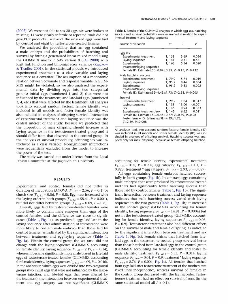
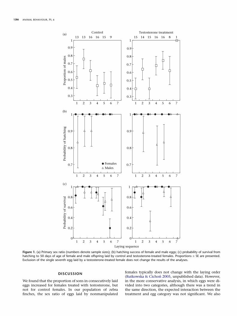
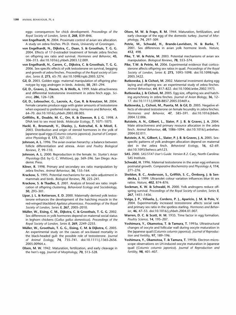
Overall, eggs laid by testosterone-treated females weremore likely to contain male embryos than eggs of thecontrol females, and the difference was close to signifi-cance (Table 1, Fig. 1a). As predicted, eggs laid late in thelaying sequence after administration of testosterone weremore likely to contain male embryos than those laid bycontrol females, as indicated by the significant interactionbetween treatment and egg-laying sequence (Table 1,Fig. 1a). Within the control group the sex ratio did notchange with the laying sequence (GLIMMIX accountingfor female identity, laying sequence: F1, 67¼ 2.19, P¼ 0.14),whereas it became significantly more male biased in late-laideggs of testosterone-treated females (GLIMMIX accountingfor female identity, laying sequence: F1, 81.3¼ 4.09, P¼ 0.046).In the analysis in which eggs were divided into two categoricalgroups (two initial eggs that were not influenced by the testos-terone injection, and late-laid eggs that were affected bythe treatment), the interaction between experimental treat-ment and egg category was not significant (GLIMMIX
accounting for female identity, experimental treatment:F1, 163¼ 0.02, P¼ 0.902; egg category: F1, 138 ¼ 0.01, P ¼0.923; treatment * egg category: F1, 152 ¼ 2.93, P ¼ 0.089).
All eggs containing female embryos hatched success-fully in both groups (Fig. 1b). In contrast, eggs containingmale embryos that were produced by testosterone-treatedmothers had significantly lower hatching success thanthose laid by control females (Table 1, Fig. 1b). The signif-icant interaction between treatment and laying sequenceindicates that male hatching success varied with layingsequence in the two groups (Table 1, Fig. 1b): it increasedin the control group (GLIMMIX accounting for femaleidentity, laying sequence: F1, 46.5 ¼ 14.81, P ¼ 0.0004) butnot in the testosterone-treated group (GLIMMIX account-ing for female identity, laying sequence: F1, 46.9 ¼ 0.03,P ¼ 0.9). Testosterone treatment also had different effectson the survival of male and female offspring, as indicatedby the significant interaction between treatment and sex(Table 1, Fig. 1c). Female chicks that hatched from late-laid eggs in the testosterone-treated group survived betterthan those hatched from late-laid eggs in the control group(GLIMMIX accounting for female identity and foster fe-male identity: treatment: F1, 68.7 ¼ 6.33, P ¼ 0.014; layingsequence: F1, 66.4¼ 0.01, P¼ 0.9; treatment * laying sequence:F1, 66.4¼ 8.76, P¼ 0.004; Fig. 1c). All females that hatchedfrom eggs laid after testosterone treatment of the mothers sur-vived until independence, whereas survival of females inthe control group decreased with the laying order. Testos-terone treatment had no effect on survival of sons (in thesame statistical model all P > 0.1).
Table 1. Results of the GLIMMIX analyses in which egg sex, hatchingsuccess and survival probability were examined in relation to exper-imental treatment and laying sequence
Source of variation df F P
Egg sexExperimental treatment 1, 158 3.69 0.056Laying sequence 1, 141 0.31 0.581Experimentaltreatment*laying sequence
1, 163 5.54 0.020
Female ID: Estimate�SE¼0.04�0.23; Z¼0.17, P¼0.432
Male hatching successExperimental treatment 1, 79.9 5.74 0.019Laying sequence 1, 95.2 8.46 0.004Experimentaltreatment*laying sequence
1, 95.2 9.83 0.002
Female ID: Estimate�SE¼4.45�1.73; Z¼2.58, P¼0.005
SurvivalExperimental treatment 1, 29.2 1.04 0.317Laying sequence 1, 133 13.00 <0.001Sex 1, 145 0.94 0.333Experimental treatment*sex 1, 145 6.62 0.011Female ID: Estimate�SE¼0.45�0.77; Z¼0.59, P¼0.28Foster female ID: Estimate�SE¼4.39�1.73;Z¼2.39, P¼0.008
All analyses took into account random factors: female identity (ID)was included in all models and foster female identity (ID) was in-cluded in analyses of offspring survival. Hatching success was ana-lysed only for male offspring, because all female offspring hatched.
ANIMAL BEHAVIOUR, 71, 61286
Prop
orti
on o
f m
ales
0.3
1 2 3 4 5 6 7
1 2 3 4 5 6 7
1 2 3 4 5 6 7 1 2 3 4 5 6 7
1 2 3 4 5 6 7
1 2 3 4 5 6 7
0.4
0.5
0.6
0.7
0.8
0.9
1
0.3
0.4
0.5
0.6
0.7
0.8
0.9
113 13 16 16 15 15 15 16 16 8 1149
(a)
Prob
abil
ity
of h
atch
ing
0.7
0.8
0.9
1
0.7
0.8
0.9
1
FemalesMales
(b)
Laying sequence
Prob
abil
ity
of s
urv
ival
0
0.2
0.4
0.6
0.8
1
0
0.2
0.4
0.6
0.8
1(c)
Control Testosterone treatment
Figure 1. (a) Primary sex ratio (numbers denote sample sizes); (b) hatching success of female and male eggs; (c) probability of survival fromhatching to 50 days of age of female and male offspring laid by control and testosterone-treated females. Proportions � SE are presented.
Exclusion of the single seventh egg laid by a testosterone-treated female does not change the results of the analyses.
DISCUSSION
We found that the proportion of sons in consecutively laideggs increased for females treated with testosterone, butnot for control females. In our population of zebrafinches, the sex ratio of eggs laid by nonmanipulated
females typically does not change with the laying order(Rutkowska & Cichon 2005, unpublished data). However,in the more conservative analysis, in which eggs were di-vided into two categories, although there was a trend inthe same direction, the expected interaction between thetreatment and egg category was not significant. We also
RUTKOWSKA & CICHON: ANDROGENS AND SEX RATIO 1287
found that elevated levels of androgens in eggs were asso-ciated with low hatching success of males but with im-proved survival of females.
A few studies have already manipulated maternalhormones to investigate their potential role in the de-termination of the primary sex ratio of offspring. In thedomestic chicken, Gallus gallus domesticus, females in-jected with progesterone produced more female offspring(Correa et al. 2005). In the Japanese quail, Coturnix japon-ica, females implanted with corticosterone also producedmore daughters, whereas implantation of testosteroneand 17-b-oestradiol had no significant effect on theprimary sex ratio (Pike & Petrie 2006). Similarly, no distor-tion of the primary sex ratio was observed in female zebrafinches injected with 17-b-oestradiol (von Engelhardtet al. 2004). Our experiment seems to corroborate thefinding on the spotless starling (Veiga et al. 2004), thatelevated maternal androgens may indeed result ina male-biased primary sex ratio.
Thus, androgens might constitute an important medi-ator of the mechanism of offspring sex determination.Exposure of the oocyte to elevated androgen levels mayaffect chromosome segregation during meiosis by influ-encing, for instance, the elasticity and movements of themeiotic spindles (Olsen 1942; Yoshimura et al. 1993a, b).Alternatively, it may affect levels of other hormones,both in the mother and in the egg, which could resultin nonrandom chromosome segregation. However,testosterone implants in Japanese quail do not alter thefemales’ corticosterone or oestradiol levels (Pike & Petrie2006).
Whether offspring sex is influenced more by hormonescirculating in the bloodstream of the mother or byhormones deposited in the yolk during egg productionis also unknown. In female Japanese quail, testosterone isapparently metabolized within a day of administration,whereas it is present in eggs laid on the next few days(Hackl et al. 2003). We can assume that in our experimentcirculating testosterone was also metabolized soon afterinjection, but it was significantly elevated in the yolk (Rut-kowska et al. 2005). This may indicate that elevated yolk,rather than circulating androgens, are responsible for themeiotic drive leading to a biased offspring sex ratio.
A number of studies have shown positive effects ofexperimentally elevated androgen levels in the eggs onoffspring performance, such as increased growth rate,begging rate and social status (Schwabl 1996; Lipar &Ketterson 2000; Eising et al. 2001; but see Sockman &Schwabl 2000 for negative effects of androgens in theAmerican kestrel, Falco sparverius). In consequence, ele-vated androgen deposition in eggs has often been consid-ered to reflect allocation of resources by the female to theoffspring (reviewed in Gil 2003). However, in these stud-ies offspring sex was not investigated. In our study, wehave shown that elevated androgen level had a positiveeffect on daughters but a negative one on sons. However,the effects of androgens on male and female offspringthat we observed should be interpreted with caution be-cause offspring sex was dependent on the hormonal treat-ment. Nevertheless, our finding corroborates a recentstudy in which in-ovo injection of testosterone in zebra
finches had opposite effects on the offspring of the twosexes, which was expressed as impaired growth of maleoffspring but enhanced growth of females (von Engel-hardt et al. 2006). Such sex-specific effects of androgenson juvenile birds was further supported by in-ovo injec-tion of Flutamide, which blocks androgen receptors, sothat offspring perceive reduced levels of androgens; thetreatment resulted in enhanced growth of male offspring,but reduced growth in female offspring in the black-headed gull, Larus ridibundus (Muller et al. 2005). Alto-gether, these findings show that elevated androgensseem to be beneficial for female but detrimental formale offspring. It suggests the existence of an importantconstraint on maternal hormone allocation. If an increasein yolk androgens within a range of natural concentra-tions has positive effects in one sex and negative effectsin the other, this would shape an optimal, probably sex-specific, maternal allocation of androgens. Given thatan elevated level of androgens has detrimental effectson male offspring, we conclude that if androgens mediatethe determination of offspring sex, the benefits of biasingthe sex ratio towards males by increasing the level of tes-tosterone may be limited.
In summary, our study shows that androgens mayconstitute an important maternal effect which maymediate the determination of offspring sex. However, ifandrogens were the sole factor responsible for a male-biased sex ratio we would expect a much stronger effect ofour treatment. We therefore suggest that some otherfactors, such as other hormones, must be involved inoffspring sex adjustment. Furthermore, as androgens hadnegative effects on male offspring we conclude that anandrogen-mediated mechanism of sex determinationseems to be costly and thus it may generally constrainmaternal hormone allocation.
Acknowledgments
We thank T. Wilk for help in taking care of the birds.J. Radwan, members of the Fajt klap and two anonymousreferees provided helpful comments on the manuscript.J. Rutkowska was supported by the Polish Ministry of Sci-ence and Information Technology in 2004–2006 and byEC Center of Excellence IBAES and the foundation forPolish Science.
References
Badyaev, A. V., Hill, G. E., Beck, M. L., Dervan, A. A., Duckworth,R. A., McGraw, K. J., Nolan, P. M. & Whittingham, L. A. 2002.
Sex-biased hatching order and adaptive population divergence in
a passerine bird. Science, 295, 316–318. doi:10.1126/
science.1066651.
Birenkott, G. P., Shoop, M. A., Cooper, K. & Wiggens, M. 1988.
Ovarian follicular growth and maturation in the domestic pigeonandguineafowl (Numidameleagris).PoultryScience,67,1783–1786.
Correa, S. M., Adkins-Regan, E. & Johnson, P. A. 2005. High pro-gesterone during avian meiosis biases sex ratios toward females.
Biology Letters, 1, 215–218. doi:10.1098/rsbl.2004.0283.
Eising, C. M., Eikenaar, C., Schwabl, H. & Groothuis, T. G. G.2001. Maternal androgens in black-headed gull (Larus ridibundus)
ANIMAL BEHAVIOUR, 71, 61288
eggs: consequences for chick development. Proceedings of the
Royal Society of London, Series B, 268, 839–846.
von Engelhardt, N. 2004. Proximate control of avian sex allocation.
A study on zebra finches. Ph.D. thesis, University of Groningen.
von Engelhardt, N., Dijkstra, C., Daan, S. & Groothuis, T. G. G.2004. Effects of 17-b-estradiol treatment of female zebra finches
on offspring sex ratio and survival. Hormones and Behavior, 45,306–313. doi:10.1016/j.yhbeh.2003.12.009.
von Engelhardt, N., Carere, C., Dijkstra, C. & Groothuis, T. G. G.2006. Sex-specific effects of yolk testosterone on survival, begging
and growth of zebra finches. Proceedings of the Royal society of Lon-
don, Series B, 273, 65–70. doi:10.1098/rspb.2005.3274.
Gil, D. 2003. Golden eggs: maternal manipulation of offspring phe-
notype by egg androgen in birds. Ardeola, 50, 281–294.
Gil, D., Graves, J., Hazon, N. & Wells, A. 1999. Male attractiveness
and differential testosterone investment in zebra finch eggs. Sci-
ence, 286, 126–128.
Gil, D., Leboucher, G., Lacroix, A., Cue, R. & Kreutzer, M. 2004.
Female canaries produce eggs with grater amounts of testosteronewhen exposed to preferred male song. Hormones and Behavior, 45,
64–70. doi:10.1016/j.yhbeh.2003.08.005.
Griffiths, R., Double, M. C., Orr, K. & Dawson, R. J. G. 1998. A
DNA test to sex most birds. Molecular Ecology, 7, 1071–1075.
Hackl, R., Bromundt, V., Diasley, J., Kotrschal, K. & Mostl, E.2003. Distribution and origin of steroid hormones in the yolk of
Japanese quail eggs (Coturnix coturnix japonica). Journal of Compar-
ative Physiology B, 173, 327–331.
Johnson, A. L. 1996. The avian ovarian hierarchy: a balance between
follicle differentiation and atresia. Avian and Poultry BiologicalReviews, 7, 99–110.
Johnson, A. L. 2000. Reproduction in the female. In: Sturkie’s AvianPhysiology (Ed. by G. C. Whittow), pp. 569–596. San Diego: Aca-
demic Press.
Kilner, R. 1998. Primary and secondary sex ratio manipulation byzebra finches. Animal Behaviour, 56, 155–164.
Krackow, S. 1995. Potential mechanisms for sex ratio adjustment inmammals and birds. Biological Reviews, 70, 225–241.
Krackow, S. & Tkadlec, E. 2001. Analysis of brood sex ratio: impli-cation of offspring clustering. Behavioral Ecology and Sociobiology,
50, 293–301.
Lipar, J. L. & Ketterson, E. D. 2000. Maternally derived yolk testos-
terone enhances the development of the hatching muscle in the
red-winged blackbird Agelaius phoeniceus. Proceedings of the Royal
Society of London, Series B, 267, 2005–2010.
Muller, W., Eising, C. M., Dijkstra, C. & Groothuis, T. G. G. 2002.
Sex differences in yolk hormones depend on maternal social statusin leghorn chickens (Gallus gallus domesticus). Proceedings of the
Royal Society of London, Series B, 269, 2249–2255.
Muller, W., Groothuis, T. G. G., Eising, C. M. & Dijkstra, C. 2005.
An experimental study on the causes of sex-biased mortality in
the black-headed gull: the possible role of testosterone. Journal
of Animal Ecology, 74, 735–741. doi:10.1111/j.1365-2656.2005.00964.x.
Olsen, M. W. 1942. Maturation, fertilization, and early cleavage inthe hen’s egg. Journal of Morphology, 70, 513–528.
Olsen, M. W. & Fraps, R. M. 1944. Maturation, fertilization, and
early cleavage of the egg of the domestic turkey. Journal of Mor-
phology, 74, 297–309.
Petrie, M., Schwabl, H., Brande-Lavridsen, N. & Burke, T.2001. Sex differences in avian yolk hormone levels. Nature,412, 498.
Pike, T. W. & Petrie, M. 2003. Potential mechanisms of avian sexmanipulation. Biological Reviews, 78, 553–574.
Pike, T.W. & Petrie, M. 2006. Experimental evidence that cortico-sterone affects offspring sex ratios in quail. Proceedings of the Royal
Society of London, Series B, 273, 1093–1098. doi:10.1098/rspb.
2005.3422.
Rutkowska, J. & Cichon, M. 2002. Maternal investment during egg
laying and offspring sex: an experimental study of zebra finches.
Animal Behaviour, 64, 817–822. doi:10.1006/anbe.2002.1973.
Rutkowska, J. & Cichon, M. 2005. Egg size, offspring sex and hatch-
ing asynchrony in zebra finches. Journal of Avian Biology, 36, 12–17. doi:10.1111/j.0908-8857.2005.03469.x.
Rutkowska, J., Cichon, M., Puerta, M. & Gil, D. 2005. Negative ef-fects of elevated testosterone on female fecundity in zebra finches.
Hormones and Behavior, 47, 585–591. doi:10.1016/jhbeh.
2004.12.006.
Rutstein, A. N., Gilbert, L., Slater, P. J. B. & Graves, J. A. 2004.
Mate attractiveness and primary resource allocation in the zebra
finch. Animal Behaviour, 68, 1086–1094. doi:10.1016/j.anbehav.2004.02.011.
Rutstein, A. N., Gilbert, L., Slater, P. J. B. & Graves, J. A. 2005. Sex-specific patterns of yolk androgen allocation depend on maternal
diet in the zebra finch. Behavioral Ecology, 16, 62–69.
doi:10.1093/beheco.arh123.
SAS. 2000. SAS/STAT User’s Guide. Version 8.2. Cary, North Carolina:
SAS Institute.
Schwabl, H. 1996. Maternal testosterone in the avian egg enhances
postnatal growth. Comparative Biochemistry and Physiology A, 114,
271–276.
Sheldon, B. C., Andersson, S., Griffith, S. C., Ornborg, J. & Sen-decka, J. 1999. Ultraviolet colour variation influences blue tit sexratios. Nature, 402, 874–876.
Sockman, K. W. & Schwabl, H. 2000. Yolk androgens reduce off-spring survival. Proceedings of the Royal Society of London, Series B,
267, 1451–1456.
Veiga, J. P., Vinuela, J., Cordero, P. J., Aparicio, J. M. & Polo, V.2004. Experimentally increased testosterone affects social rank
and primary sex ratio in the spotless starling. Hormones and Behav-
ior, 46, 47–53. doi:10.1016/j.yhbeh.2004.01.007.
Warren, D. C. & Scott, H. M. 1935. Time factor in egg formation.
Poultry Science, 14, 195–207.
Yoshimura, Y., Okamotoa, T. & Tamura, T. 1993a. Ultrastructural
changes of oocyte and follicular wall during oocyte maturation inthe Japanese quail (Coturnix coturnix japonica). Journal of Reproduc-
tion and Fertility, 97, 189–196.
Yoshimura, Y., Okamotoa, T. & Tamura, T. 1993b. Electron micro-scope observations on LH-induced oocyte maturation in Japanese
quail (Coturnix coturnix japonica). Journal of Reproduction and
Fertility, 98, 401–407.