Embed Size (px)
Citation preview

arX
iv:2
004.
0722
4v4
[q-
bio.
PE]
29
Sep
2020
Mathematical model of SARS-Cov-2 propagation
versus ACE2 fits COVID-19 lethality across age and
sex and predicts that of SARS, supporting possible
therapy
Ugo Bastolla(1)
(1) Centro de Biologia Molecular ”Severo Ochoa”
CSIC-UAM Cantoblanco, 28049 Madrid, Spain. email: [email protected]
Abstract
The fatality rate of Covid-19 escalates with age and is larger in men than women.I show that these variations correlate strongly with the level of the viral receptorprotein ACE2 in rat lungs, which is consistent with the still limited and apparentlycontradictory data on human ACE2. Surprisingly, lower levels of the receptor corre-late with higher fatality. However, a previous mathematical model predicts that thespeed of viral progression in the organism has a maximum and then declines withthe receptor level. Moreover, many manifestations of severe CoViD-19, such as se-vere lung injury, exacerbated inflammatory response and thrombotic problems mayderive from increased Angiotensin II (Ang-II) level that results from degradation ofACE2 by the virus. I present here a mathematical model based on the influence ofACE2 on viral propagation and disease severity. The model fits Covid-19 fatalityrate across age and sex with high accuracy (r2 > 0.9) under the hypothesis thatSARS-CoV-2 infections are in the dynamical regimes in which increased receptorslows down viral propagation. Moreover, rescaling the model parameters by theratio of the binding rates of the spike proteins of SARS-CoV and SARS-CoV-2 al-lows predicting the fatality rate of SARS-CoV across age and sex, thus linking themolecular and epidemiological levels. The presented model opposes the fear that an-giotensin receptor blockers (ARB), suggested as a therapy against the most adverseeffects of CoViD-19, may favour viral propagation, and suggests that Ang-II andACE2 are candidate prognostic factors for detecting population that needs strongerprotection.
The Covid-19 pandemics [1] is causing hundreds of thousands fatalities worldwide [2],creating a tremendous threat to global health. It presents a strong gradient of fatalitiesacross age and a sex bias with much higher severity in males than females. Modelling
1
studies that extrapolate the number of infections suggest that, at young age, most SARS-CoV-2 infections are asymptomatic and the fatality ratio is very low, whereas for theelderlies most infections are severe and a large fraction of them can be fatal [3]. Under-standing the biological reasons that underlie these striking differences is one of the mostpressing problems of CoViD-19 research, which might lead to better prediction of thedisease prognosis and possible treatments that approach the severity of the worst affectedgroups to that of the most protected ones.
Here I show that the case fatality ratio of Covid-19 across age and sex correlatesnegatively with the level of the protein Angiotensin converting enzyme 2 (ACE2), thecellular receptor both of SARS and SARS-CoV-2 virus [1, 4]. The correlation is verystrong with membrane-bound ACE2 protein in rat lungs, which decreases with age andis higher in old females than old males [5]. The same qualitative pattern is observed formembrane-bound ACE2 in mice [6], where all the anti-inflammatory axis of the Renin-Angiotensin-System (RAS) [7] to which ACE2 belongs decreases with age. In humans,ACE2 protein in serum [9] and mRNA [10–13] are lower in children than adults, butavailable data and current knowledge of the RAS supports that ACE2 as membraneprotein decreases through age [8], as hypothesized here and discussed later.
The negative correlation between ACE2 and lethality is surprising: higher levels ofthe receptor decrease the lethality exponentially. The paradox is only apparent becausehigh receptor levels do not necessarily favour viral propagation. A mathematical modelof viral infection [15], developed before the COVID-19 pandemics, computed how viralpropagation depends on the adsorption rate of viruses on cells. In terms of receptor level,this model predicts that the speed of viral propagation is a non-monotonic function thatincreases with the receptor level, reaches a maximum and then decreases.
The second mechanism that may underlie the negative correlation concerns the func-tion of ACE2 not as viral receptor but as key enzyme of the RAS [7]. This system,besides regulating blood pressure (BP) and electrolyte homeostasis in blood, plays a cen-tral role in inflammatory processes [17], immune response [18] and coagulation [19, 20],which characterize the most severe Covid-19 cases [29,30]. Its main player is the family ofpeptides derived from angiotensin I (Ang1-10), cleaved by the enzyme Renin from the pro-tein angiotensinogen. Its pro-inflammatory arm is constituted by angiotensin II (Ang1-8),cleaved from Ang1-10 by the angiotensin converting enzyme (ACE) homologous to ACE2.Ang1-8 bound to the receptor ATR-1 triggers a cascade of reactions leading not only tovasoconstriction and increased BP but also to activation of the transcription factor NFkBthat upregulates inflammatory cytokines (IL-1, TNF-α and IFN-γ among others), acti-vates white blood cells and platelets, and favours thrombotic processes [21]. The enzymeACE2 limits the level of Ang1-8 by converting its precursor Ang1-10 to Ang1-9 [22] thatis subsequently cleaved by ACE to Ang1-7, and by directly converting Ang1-8 to Ang1-7,which belongs to the anti-inflammatory arm of the RAS since it favours vasodilation,reduces BP and attenuates inflammation [23].
Upon viral entry the spike protein of SARS-CoV and probably also SARS-CoV-2cause the internalization and degradation of ACE2 [24] that critically contributes to lung
2

damage [25–27]. Decrease of ACE2 raises the severity of lung injury in other inflammatorydiseases [27] and in aging rats [28], which may be explained by the increase of Ang1-8and its adverse effects.
Here I develop a set of mathematical models of SARS-CoV-2 lethality versus the pre-infection level of ACE2 based on two aspects: the influence of ACE2 on viral progression[15] and the negative effect of its degradation. These models are fitted to the CFR ofSARS-CoV-2 across six classes of age and sex in Italy, Spain and Germany, and supportthe hypothesis that the receptor level slows down the virus propagation, which fits the datamuch better than the competing hypothesis. Furthermore, under the same hypothesis andby rescaling the parameters fitted to SARS-CoV-2 by the ratio between the binding ratesof the spike proteins of SARS-CoV and SARS-CoV-2, the model predicts well the CFR ofSARS-CoV further supporting the negative relationship between receptor level and viruspropagation.
Results
CoViD-19 lethality correlates negatively with ACE2 level
The level of the ACE2 protein in rat lungs were quantified across three age classes of thetwo sexes by Xie et al. [5], who found that it strongly decays with age and it is higher infemale than male rats, with largest difference in the oldest cohort where the expressionis almost double for females. Here I assume that human ACE2 membrane protein levelsacross age and sex are qualitatively similar to rat data, apart for multiplicative factors thatmay depend on the organ. This assumption is supported by mice data [6], ACE2 protein inhuman lungs [8] and ACE2 mRNA expression in the GTEx database [10], but it apparentlycontrasts with the observation that ACE2 in serum [9] and ACE2 mRNA [11–13] is higherin adults than in children. We discuss later that these discrepancies are in fact consistentwith the current knowledge of the RAS.
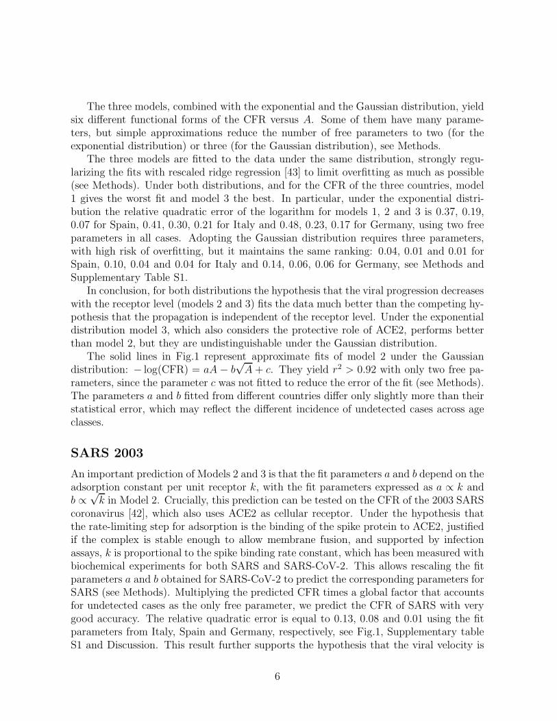
Strikingly, the profile of ACE2 in Ref. [5] is very strongly anti-correlated with thelethality of SARS-CoV-2. Fig.1 represents the level of the ACE2 protein in lung rats(horizontal axis, data from Fig.2 of [5]) versus the case fatality ratio (CFR) of CoVid-19registered in Italy [31], Spain [32] and Germany [33] in three uniform age classes (young,< 30, middle-age 30− 59 and old > 60; vertical axis) of each sex. Data strongly supportthe exponential decrease of mortality with ACE2 level, with r2 = 0.91, 0.97 and 0.89,respectively, suggesting that variations of ACE2 describe the largest part of the variationof the CFR.
As for other two-parameter fits tested in this work, the fitted exponents for Italy andGermany coincide within the error and the CFR differ only by a multiplicative factor,supporting the robustness of the data. Data from Spain present higher mortality in theyoung ages, which might be attributed to more frequent undetected cases in young agewith lower severity.
3

Old_MOld_F
Middle_M
Middle_F
Young_M
Young_F
Old_M Old_F
Mid_MMid_F
Young_M
Young_F
0 0.2 0.4 0.6 0.8 1Expression of ACE2 in rat lungs
0.0001
0.001
0.01
0.1
1
Cas
e-fa
talit
y ra
te o
f C
oVid
-19
per
age
and
gend
er
ItalySpainGermany
Italy model: CFR=exp(-7.6*ACE2+1.08*sqrt(ACE2)-0.52) r2=0.93
Spain model: CFR=exp(-6.85*ACE2+1.13*sqrt(ACE2)-0.85) r2=0.989
Germany model: CFR=exp(-8.7*ACE2+2.02*sqrt(ACE2)-1.42) r2=0.915
SARS 2003 HongKong
SARS Prediction: CFR=exp(-5.66*ACE2+0.93*sqrt(ACE2)+0.104) r2=0.87
SARS Prediction: CFR=exp(-5.09*ACE2+0.97*sqrt(ACE2)+0.01) r2=0.92
SARS Prediction: CFR=exp(-6.49*ACE2+1.74/sqrt(ACE2)-0.576) r2=0.90
Figure 1: Expression of the ACE2 protein in lung rats (horizontal axis), normalized sothat the highest expression is one, versus case fatality rate (vertical axis) of SARS-CoV-2(Circles: Italy; triangles: Spain; diamonds: Germany) and SARS 2003 (open squares) inthree age classes (young 0-29, middle-age 30-59 and old > 59) and two sexs (male andfemale). The solid lines represent fits to the mathematical model (see text), the dashedlines represent predictions that rescale the models fitted to SARS-CoV-2 with the ratiobetween the binding rate constants of SARS and SARS-CoV-2 (see text).
Mathematical model of covid-19 lethality
Mathematical models of viral growth consider three processes: virus adsorption into sus-ceptible cells, production of virus by infected cells after a delay time τ , and viral clearanceby the immune system [34]. I translate here this mathematical formalism in terms of re-ceptor density, exploiting that the adsorption rate is proportional to the receptor levelA expressed in susceptible cells times the association rate between the virus and thereceptor, kA ≡ kA.
The simple mean-field model that does not consider explicit space predicts a minimalreceptor density below which the virus does not grow and above which higher receptor lev-els accelerate the viral progression. Considering spatial diffusion modifies this situation.The analytical solution of a model of viral propagation in space was obtained in 2002 byFort and Mendez [35], who tested their model with experiments on the spread of bacterio-phages in lysis plaques. Their mathematical formulation is conceptually equivalent to thepresent setting and can be directly applied here. Varying the receptor level A (adsorptionrate in the original paper), three regimes appear: (1) When A is small the viral velocity
4

v increases with A less than linearly. (2) For intermediate kA, large with respect to 1/τbut small with respect to the rate of virus production, the viral velocity reaches a plateauwhere it is almost independent of A. (3) Although not explicitly discussed in Ref. [35],the formulas presented there remain valid in the regime where kA is larger than the rateof virus production. In this regime the viral progression slows down with receptor densityas v ∝ 1/
√kA.
The latter result is surprising: how can the virus progress more slowly for increasingreceptor level? Since this is a mathematical model, the answer is readily found: inthe model, viral particles are consumed when they enter a cell but the viral yield perinfected cell does not increase when a cell is infected multiple times. It was in factproposed that multiple viral entries in the same cell interfere with viral replication. Severalviruses such as HIV [36,37], measles [38], influenza [39] and hepatitis B [40] downregulatetheir own receptor, preventing multiple entries. The mathematical result that, after theinfection is established, very fast adsorption does not favour the virus, agrees with a recentstudy that demonstrated the protective effect of high adsorption rate through analyticcomputation, simulation and experiment [41]. In all, the mathematical model predictsthat viral progression in the organism declines above some receptor level.
Next, I consider two possible models of Covid-19 death. In the first one, death occurswhen the virus propagates through the upper respiratory tract or through endothelialcells, reaches the lungs and infects and destroys a critical fraction X of it, the samefor all patients. The second model considers the protective effect of ACE2, assumingthat the critical fraction X is a decreasing function of the pre-infection ACE2 level A,X = 1 − Ac/A, i.e. the larger is the initial level of ACE2, the more tolerant is assumedto be the organism to the viral infection.
Combining these two assumptions with the three regimes of viral propagation de-scribed above gives six mathematical models. For each of them, I compute the time tdafter which the virus causes death versus the ACE2 level A (see Methods). If the viralvelocity increases with ACE2 td is a decreasing function of A. This behaviour contradictsthe data and I shall not consider it further. In the regime in which the viral velocity isindependent of A td is an increasing function of A only for the model that considers theprotective effect of ACE2, which is called Model 1. Model 2 assumes that viral propaga-tion decreases with A but neglects the protective effect of ACE2 and Model 3 considersboth the decrease of viral propagation and the protective effect of ACE2.
I compute lethality as the probability that td is smaller than the time ti needed bythe immune system for clearing the virus, which I model as a random variable with twopossible distributions: (1) The exponential distribution, which is the distribution withmaximum entropy for given average value. (2) The Gaussian distribution, justified by thefact that ti is the sum of intermediate steps in the maturation of the immune responseand the limit of the sum of independent random variables is a Gaussian. I adopt thesame immune system parameters for all age and sex classes in order to limit the numberof free parameters and to test if variations of the receptor level are sufficient to explainthe lethality.
5
The three models, combined with the exponential and the Gaussian distribution, yieldsix different functional forms of the CFR versus A. Some of them have many parame-ters, but simple approximations reduce the number of free parameters to two (for theexponential distribution) or three (for the Gaussian distribution), see Methods.
The three models are fitted to the data under the same distribution, strongly regu-larizing the fits with rescaled ridge regression [43] to limit overfitting as much as possible(see Methods). Under both distributions, and for the CFR of the three countries, model1 gives the worst fit and model 3 the best. In particular, under the exponential distri-bution the relative quadratic error of the logarithm for models 1, 2 and 3 is 0.37, 0.19,0.07 for Spain, 0.41, 0.30, 0.21 for Italy and 0.48, 0.23, 0.17 for Germany, using two freeparameters in all cases. Adopting the Gaussian distribution requires three parameters,with high risk of overfitting, but it maintains the same ranking: 0.04, 0.01 and 0.01 forSpain, 0.10, 0.04 and 0.04 for Italy and 0.14, 0.06, 0.06 for Germany, see Methods andSupplementary Table S1.
In conclusion, for both distributions the hypothesis that the viral progression decreaseswith the receptor level (models 2 and 3) fits the data much better than the competing hy-pothesis that the propagation is independent of the receptor level. Under the exponentialdistribution model 3, which also considers the protective role of ACE2, performs betterthan model 2, but they are undistinguishable under the Gaussian distribution.
The solid lines in Fig.1 represent approximate fits of model 2 under the Gaussiandistribution: − log(CFR) = aA − b
√A + c. They yield r2 > 0.92 with only two free pa-
rameters, since the parameter c was not fitted to reduce the error of the fit (see Methods).The parameters a and b fitted from different countries differ only slightly more than theirstatistical error, which may reflect the different incidence of undetected cases across ageclasses.
SARS 2003
An important prediction of Models 2 and 3 is that the fit parameters a and b depend on theadsorption constant per unit receptor k, with the fit parameters expressed as a ∝ k andb ∝
√k in Model 2. Crucially, this prediction can be tested on the CFR of the 2003 SARS
coronavirus [42], which also uses ACE2 as cellular receptor. Under the hypothesis thatthe rate-limiting step for adsorption is the binding of the spike protein to ACE2, justifiedif the complex is stable enough to allow membrane fusion, and supported by infectionassays, k is proportional to the spike binding rate constant, which has been measured withbiochemical experiments for both SARS and SARS-CoV-2. This allows rescaling the fitparameters a and b obtained for SARS-CoV-2 to predict the corresponding parameters forSARS (see Methods). Multiplying the predicted CFR times a global factor that accountsfor undetected cases as the only free parameter, we predict the CFR of SARS with verygood accuracy. The relative quadratic error is equal to 0.13, 0.08 and 0.01 using the fitparameters from Italy, Spain and Germany, respectively, see Fig.1, Supplementary tableS1 and Discussion. This result further supports the hypothesis that the viral velocity is
6

slowed down by increasing receptor level.
NL63
It is natural to extend this analysis to the other human coronavirus that uses ACE2 asreceptor, NL63 that causes common cold and is not generally associated with pneumonia.Its spike protein contains a very stable receptor binding domain of 120 residues thatshowed high binding affinity for ACE2 [44]. However, the complete S1 domain of thespike (717 residues) is much less stable and its affinity for ACE2 is so small that it couldnot be measured with binding assays [45–47], and it was conjectured that it is 10-100-foldsmaller than that of SARS-CoV [46]. Since the CFR of SARS peaks for old females, whosenormalized ACE2 is equal to 22% of the maximum value, the model predicts that thisis the level at which SARS-CoV propagates fastest. If the binding affinity of the NL63spike is at least ten times smaller, the ACE2 level at which NL63 propagates fastest ismore than double the highest ACE2 level, implying that NL63 is in the regime in whichthe ACE2 level enhances its propagation.
This analysis agrees with the apparently surprising data reported in Fig.3A of Ref.[45], which shows that ACE2 overexpression in 293T cells enhances NL63 infection threetimes more than SARS-CoV infection, indicating that higher receptor levels accelerate thepropagation of NL63 more than that of SARS-CoV despite the latter has higher bindingaffinity.
Discussion
Since ACE2 is the SARS-CoV-2 receptor, we may expect that raising its level enhancesthe rate at which the virus propagates in the organism and worsens the outcome of theinfection. However, a mathematical model of viral progression presents a regime in whichthe increase of the receptor level slows down the virus propagation in the organism. Theobserved relation between SARS-CoV-2 lethality and ACE2 levels suggests that this isthe relevant regime of SARS-CoV-2 infections, as further supported by the prediction ofthe age- and sex-dependent lethality of 2003 SARS-CoV.
Human ACE2
An important limitation of the present work is that it uses data of ACE2 protein levelsin healthy rat lungs [5] since similar data are not available for humans. A recent clinicalstudy could not confirm significant differences of ACE2 expression between patients ofdifferent age with acute respiratory distress syndrome (ARDS) [48]. This clinical studymeasured the activity of RAS enzymes at only one time point for each patient, so thatthe dynamics of the RAS during ARDS may have obscured individual differences.
Consistent with rat data, decrease through age of membrane-bound ACE2 protein wasalso observed in mice, which show a general strengthening of the inflammatory arm of
7

the RAS with aging [6]. In humans, a recent preprint found that membrane-bound ACE2is more abundant in children than in adult lungs [8]. However, other publications andpreprints showed that ACE2 is higher in adults than in children in serum [9] and at themRNA level [11–13]. ACE2 is detached from the cellular membrane and shed to the serumby the metalloprotease ADAM17 [14]. The discrepancy between membrane-bound andserum levels suggests that ADAM17 is less active in children, consistent with our currentand still limited knowledge of the RAS. In fact, ADAM17 is upregulated by the binding ofAng1-8 to the angiotensin II type 1 receptor (ATR1). Ang1-8 increases with age [6], whilechildren express much more than adults the receptor ATR2 that competes with ATR1 andcounteracts its action [50]. Thus, children are expected to present lower binding of Ang1-8to ATR1 and lower activation of ADAM17. Regarding ACE2 mRNA, this starts beingexpressed after birth and reaches a maximum at young age [13]. The hypothesized lowershedding of ACE2 in children implies that the membrane protein reaches its maximum atyounger age than mRNA. Therefore, the higher expression of ACE2 as membrane proteinis not contradicted by these observations.
Rat data are also consistent with the observation that ACE2 mRNA expression de-creases through age in mice [51] and adult human cells [10], as also represented in Fig.3gof the preprint that reported higher ACE2 mRNA in adults [13]. Mechanistically, thisdecrease of ACE2 mRNA through age is probably related with the increase of Ang1-8 [6]that downregulates ACE2 expression [81].
Regarding sex differences, Ref. [10] found that ACE2 mRNA is higher in males thanfemales, as in rats. Ref. [13] reached the opposite conclusion, but this seems an artefactof the fact that smoking enhances ACE2 expression [12] and in their samples 50% ofmen were smokers compared to 25% of women. It has to be noted that the ACE2 geneis contained in the X chromosome, of which females have two copies. Although oneof these copies is epigenetically silenced, about 15% of the X-linked genes escape thisinactivation [52] and heterochromatin is known to dysregulate with age. It is interestingthat old female rats present almost exactly double ACE2 than males [5], as one wouldexpect if the epigenetic silencing fades at old age. Consistently, some of the sex differencesin human cardio-vascular diseases have been attributed to sex differences in the expressionof ACE2, which acts as protective factor [53].
The negative relation between ACE2 levels and severity of Covid-19 is supported byother risk and protective factors corrected for age, sex and other comorbidities in a largestudy in the UK [16]. Namely, being a current smoker constitute a curious protectivefactor (adjusted hazard ratio (AHR): 0.82−0.97), and smoking enhances ACE2 expression[12]. Contrary to single-factor analysis, diagnosed hypertension is a protective factor(AHR 0.85 − 0.93), which may be correlated with the fact that anti-hypertensive drugsenhance ACE2 expression [67]. Diabetes is a risk factor (AHR 1.72 − 2.09), and it hasbeen associated with reduced ACE2 expression [54]. Cardiovascular diseases and reducedkidney function are additional risk factors that are related with reduced ACE2 levels [7].Finally, Vitamin D deficiency is a risk factor for COVID-19 [55] that is also relatedwith low levels of ACE2 because Vitamin D inhibits the expression of Renin, which in
8

turn produces Ang1-10, the substrate from which is cleaved Ang1-8, which downregulatesACE2 [56]. Therefore, low levels of Vitamin D are expected to reduce ACE2. VitaminD levels are decreased in ethnic groups with dark skin pigmentation living at temperatelatitudes, perhaps due to high screening of solar radiation, providing a possible causalrelationship between ethnic status and Covid-19 (AHR 1.30 − 1.69 for Black people,discounting socio-economic factors), once again through ACE2. Therefore, other hazardfactors and protecting factors besides age and sex also support a negative correlationbetween ACE2 and Covid-19 lethality.
Finally, the GTEx database shows that, despite lungs are the organ that is moreseverely damaged by COVID-19, they do not present high expression of ACE2 mRNA [57],which is higher in tissues from reproductive organs, intestine, adipose tissue, kidney,hearth, thyroid, esophagus, breast, salivary glands and pancreas, among others. Someof these organs may be infected but they experience less damage, consistent with thenegative correlation between ACE2 levels in lungs and lethality.
Role of ACE2 for virus propagation and spike mutant D614G
The above evidence strongly supports the negative correlation between ACE2 protein lev-els and severity of CoViD-19. This in turn supports the mathematical model presentedhere, based on the hypothesis that increased ACE2 slows down viral propagation [35],which fits the CFR from Spain, Italy and Germany with r2 = 0.93, 0.79 and 0.83, re-spectively, using two free parameters. The same hypothesis predicts the lethality profileof the 2003 SARS virus across age and sex yielding r2 = 0.92, 0.87 and 0.99 using thefit parameters from Spain, Italy and Germany, respectively, and a single free parameter.This extrapolation from SARS-CoV-2 to SARS uses the ratio between the binding rateconstants of the spike proteins of the two viral species, bridging the molecular and theepidemiological level.
The above computation predicts that SARS has higher relative mortality for youngage with respect to old age (the observed ratio is 22% for SARS compared with 1.3% forSARS-CoV-2) due to the smaller binding affinity of the SARS spike for ACE2. It alsopredicts that mutations that decrease the binding of SARS-CoV-2 spike may generate astrain more severe for younger age.
Remarkably, the mutant D614G of the viral spike, which rapidly rose to almost fixationworld-wide [58], presents lower affinity for ACE2, propagates faster in cell cultures thanthe original spike [59] and its detected cases tend to be younger [60], in line with theabove prediction. A direct relation between D614G and disease severity could not beproven, but an indirect one seems to exist since D641G is associated with increased viralload and viral load is associated with hospitalization [60]. Nevertheless, the improvedpropagation of D614G was attribute to the higher population of the binding-competentopen configuration [59], thus other possible interpretations exist.
9

ARB and ACE2 as protective factors
SARS and probably also SARS-CoV-2 degrade ACE2 [24], with detrimental effects on thelungs on which ACE2 has a protective effect [25–27,70]. Several papers proposed that thedownregulation of ACE2 is a key factor for the severity of CoViD-19 and suggested thatACE inhibitors (ACE-I) and angiotensin receptor blockers (ARB) that limit the effectsof Ang1-8 may be beneficial for CoViD-19 patients [61–66]. A similar idea was alreadyproposed at the time of SARS, and a retrospective meta-analysis found that the use ofARB and ACE-I provides a consistent reduction in risk of pneumonia compared withcontrols [76]. In the context of CoViD-19, several studies found that ARB and ACE-Iprotect the lungs and mitigate the severity of COVID-19 for patients that already takethem against hypertension [71–75].
However, the possible protective role of ARB and ACE-I has been out-weighted bythe fear that they may favour viral propagation since they upregulate the viral receptorACE2 [67], and it was proposed that they should be discontinued [68,69]. Medical societiesfirmly opposed this suggestion due to lack of evidence [78–80]. The current consensus isthat available data are too limited to support either hypothesis that ACE-I and ARB maybe detrimental or beneficial in CoViD-19 infections, but withdrawal of anti-hypertensivedrugs in patients that need them may be harmful [74].
The negative correlation between ACE2 levels and lethality of SARS-Cov-2 foundhere, and the mathematical prediction that the receptor level does not enhance but slowsdown viral progression, contradict the fear that ARB and ACE-I may benefit the virusand suggests two complementary protective roles of high ACE2 levels. On one hand,they may slow down the propagation of the virus, an effect conceptually similar to thatobserved in recent experiments with soluble human ACE2 [77]. On the other hand, theyreduce the accumulation of Ang1-8, whose proinflammatory and prothrombotic effects arethought to underlie the most severe complications of CoViD-19. This work thus supportsthe idea that ARB and ACE-I used to treat high blood pressure may limit the mostadverse manifestations of CoViD-19.
A note of caution concerns the effect of these drugs on the bradykinin system. Thissystem is strongly coupled with the RAS and causes vasodilation, reduces blood pressureand increases vascular permeability. Its overactivity can lead to increased inflammation,thrombosis and angioedema in the lungs, and it was proposed that it mediates the severemanifestations of COVID-19 [84–86].
The bradykinin system consists of two axes. The first one is downregulated by ACE2,which degrades the signalling peptide des-Arg9-bradykinin (DABK) whose receptor BK1Ris upregulated by Ang1-8 (in turn downregulated by ACE2) bound to the receptor ATR1.Stimulation of this axis may lead to release of pro-inflammatory chemokines, lung inflam-mation and injury [87] and is expected to be reduced through ARB and ACE-I, whichwould exert a protective role.
The other axis is downregulated by ACE, which degrades the signalling peptide BK,whose receptor BK2R is in turn activated by Ang1-7 and Ang1-9 produced by ACE2,
10

and is stimulated by Ang1-8 bound to the receptor ATR2 [88]. Thus, ACE2, ATR1 re-ceptor blockers and even more ACE-I can upregulate the BK2R axis with pathologicalconsequences, as observed in severe side-effects of ACE-I [89], and their use should belimited in the presence of hypotension. Nevertheless, adverse effects have not been re-ported in studies of the effect of these drugs on COVID-19 patients. We speculate thatthe synergy between BK2R and ATR1 through heterodimerization [90] might generate anegative feedback that limits the effect of ARB on the BK2R axis.
Positive feedback loop
It is noteworthy that degradation of ACE2 increases the level of Ang1-8, which in turnbinds the ATR1 receptor and down-regulates ACE2 even further both at the mRNA andat the protein level [81]. Thus, the SARS-CoV-2 infection may trigger a dangerous positivefeedback loop that strongly raises Ang1-8, exacerbating inflammatory response [17,18] andcoagulation problems [19,20], frequent complications of severe CoViD-19 patients [30,82].Positive correlation between Ang1-8 levels and viral load has been reported in CoViD-19patients [83], supporting the link between severe CoViD-19 and dysregulation of the RAS.
Under this point of view, ARBs appear to be more favourable than ACE-I becausethey can interfere with the positive feedback loop of Ang1-8 and because Ang1-8 can begenerated by other proteases if ACE is inhibited [7].
Concluding remarks
Of course, clinical trials are necessary to establish whether ARB and ACE-I have a pos-itive, negative or neutral effect on CoViD-19 severity. To this aim, the clinical trialsNCT04312009 and NCT04311177 started in April 2020 at the University of Minnesota.The present work aims at stimulating others to join efforts to assess this promising treat-ment.
Finally, the results presented here suggest a prognostic role for the measurements ofACE2 levels in bronchial aspirated lavage samples and Ang1-8 in the serum, which maypredict the severity of the disease already at an early stage and may allow detecting riskgroups that need higher protection besides the elder, as supported by the associationbetween ACE2 and known risk and protecting factors against CoViD-19 [16]. We arecurrently investigating this possibility through retrospective studies.
Materials and Methods
Case-fatality-rates and expression data
Case fatality rates (CFR) were taken from Ref. [31–33] for CoViD-19 in Italy, Spain andGermany, respectively, and from Ref. [42] for the 2003 SARS outbreak in Hong-Kong.At the beginning of an outbreak, CFR underestimate the true fatality rate because their
11
calculation assumes that all people currently infected will recover, which unfortunatelyis not true. This effect may not be uniform across age-sex classes if patients of someclasses tend to die more rapidly, as assumed by the model. However, at a late epidemicstage this effect is expected to be small. On the other hand, CFR overestimate the truefatality rate because of undetected cases that tend to lower the denominator. Since age-sex classes with higher lethality also tend to have more severe cases and less undetectedcases, the overestimation is larger for classes with smaller lethality, with the consequenceof reducing the differences among classes for larger fraction of undetected cases. The datacurrently available do not allow correcting for this bias, which may account for some ofthe differences in the fit parameters.
Expression data presented in Ref. [5] were grouped in three age classes of 3 (young),12 (middle) and 24 months (old). CFR were presented in bins of 10 years, and I groupedthem in three equally spaced groups 0-29 (young), 30-59 (middle) and ≥ 60 years (old).Grouping the 20-29 years class with the middle age gave similar results with approximatelyexponential decrease of lethality with ACE2 expression.
For SARS CFR [42], ages were grouped differently: 0-44 (young), 45-74 (middle) and≥ 75 (old). To compare these groups with those of SARS-CoV-2, I interpolated expressiondata of ACE2 A for these groups as A(0 − 44) = 0.667A(0 − 29) + 0.333A(30 − 59),A(45 − 74) = 0.5A(30− 59) + 0.5A(≥ 60) and A(≥ 75) = 0.667A(≥ 60). Other schemesgave qualitatively similar results: The CFR decreases approximately exponentially withA and the exponent is smaller than for SARS-CoV-2, which are the two important pointsmade in the paper.
Mathematical model of viral propagation
The simplest mathematical model of viral propagation considers three populations: un-infected cells U(t), free virus V (t) that enter the cells with rate kAU(t)V (t) (adsorption)and are cleared with rate c, and infected cells I(t) that produce new virus at rate kV Y I(t)(Y stands for yield) after a delay τ called eclipse time, until they ultimately die [34]. HereI express the adsorption rate as a function of the receptor density A, kA = kA. The viralpopulation cannot grow below the minimum receptor density given by Amin = c/(kU0Y )(U0 is the initial concentration of susceptible cells). A spatially explicit version of thismodel in which virus diffuse through susceptible cells [15] was found amenable to ana-lytical solution that explicitly gives the viral velocity v as a function of the model pa-rameters [35]. Here I describe this solution in terms of the receptor density. The authorsdescribe two regimes: (1) For small A (A < 1/(kU0τY ), A < 1/(kU0kV )), the viral ve-
locity increases with A, but less then linearly, as v = 2√
D kAU0Y1+kAU0Y
. (2) For intermediate
receptor density 1/(kU0τY ) < A < 1/(kU0kV ) the viral velocity is given by v =√
2D/τ ,where D is the effective diffusion constant that depends on cell shape and density, andit is almost independent of A [35] so that the viral progression is not enhanced by theexpression of the receptor. (3) The formulas presented in the paper are also valid in the
12

third regime of very high receptor density, when kA is larger than the rate at which viralparticles are produced: A > 1/(kU0τY ), A > 1/(kU0kV ), although this regime was notexplicitly discussed. Counter-intuitively, in this regime the viral progression slows downwith receptor density as v =
√
kV /kAU0.The time that it takes for the virus to propagate through the upper respiratory tract
(URT) can be estimated as tU = lU/v. When the virus reaches the lungs, the number ofinfected cells grows with time as I(t) ∝ (vt)dF , where dF ≈ 2.35 is the fractal dimensionof the lungs [91], which are one of the classical examples of a fractal organ. As cells get
infected, the receptor density in the lungs decreases as A(t) = A(0)(
1− I(t)/ldFL
)
and it
reaches the critical level Ac after the time tL = (lL/v)(1−Ac/A0)1/dF ≈= lL
v
(
1− Ac
dF
1A(0)
)
.
I used the approximation Ac ≪ A(0), and A(0) is the receptor density at the beginning ofthe infection, which in the main text is simply denoted as A. Summing these two times,
I estimate the time at which death occurs as td ≈ (1/v)[
lU + lL
(
1− Ac
dF
1A
)]
.
I consider three situations: (1) v is independent of A; (2) v decreases as 1/√kA and
almost all the cells in the lungs must be infected to produce the death, i.e. Ac = 0; (3)v decreases with A and Ac > 0. In each situation, the death time td depends on A as
td ∝ 1− Ac
dF
1A(Model 1), td ∝
√k√A (Model 2), td ∝
√k(√
A− Ac
dF
1√
A
)
(Model 3).
In the model, death occurs if td is smaller than the time ti needed by the immunesystem to control the virus, which is modelled either as an exponential (E) or a Gaussian(G) random variable. In the first case, the probability that ti is larger than td can becomputed as Pd = exp(−td/T ), where T is the average value of ti. In the Gaussian case,the probability is well approximated as Pd = C exp (−(td − µ)2/(2σ2)). Combining theseexpressions with the three models of td versus A and grouping together terms with thesame power of A, we obtain six mathematical models of the lethality Pd as a function ofthe initial level of ACE2 A:
− ln(Pd) ≈
− aA+ b (1E) a
A2 − bA+ c (1G)
a√A+ b (2E) aA− b
√A+ c (2G)
a√A− b
√
A(3E) aA− b
√A+ c
√
A(3G)
a, b and c are positive fitting parameters. In Eq.(3G), there are five terms proportionalto A,
√A, 1/
√A, 1/A and constant, corresponding to five fitting parameters. In order to
fit only three parameters, the same number as for the other Gaussian fits, I neglected theterms proportional to 1/A and constant, obtaining Eq.(3G), while neglecting the termsproportional to 1/A and 1/
√A yields Eq.(2G).
Fits of the model
The fitting parameters a and b are determined through regularized fits performed withrescaled ridge regression [43], which minimizes the quadratic error plus the term Λ(a2+b2)that penalizes large values of the parameters. The regularization is necessary to avoid
13

amplifying the noise due to covariant explanatory variables, as in the present case, andit allows more robust parameter estimation, often avoiding that they acquire unphysicalvalues with incorrect sign, at the price of some bias. Rescaled ridge regression yieldsnon-vanishing parameters even in the limit of large Λ, overcoming a drawback of otherregularization schemes, and it fixes the parameter Λ based on an analogy with statisti-cal mechanics at the transition between the phase dominated by the noise and the onedominated by the bias.
For ridge regression there is no analytic formula to determine the statistical error ofthe fitting parameters, therefore I applied a bootstrap approach, repeating the fit whileeliminating each of the data points and computing the standard deviation of the fittingparameters numerically.
Models (1G,2G,3G) coupled to the Gaussian distribution depend on three parametersbut only the parameters a and b were fitted while c was fixed, fitting − ln(Pd) − c =aA − b
√A. The parameter c was determined not by minimizing the fit error but by
selecting the value of c that yields 50% relative error on the parameters a and b. Theplots shown in Fig.1 were obtained in this way.
Prediction for SARS
The models fitted to SARS-CoV-2 were rescaled in order to apply them to SARS-CoV,adopting the ratio between the binding rates of the spike proteins of both viruses to ACE2.The most precise measures available in the literature are kSARS−2 = (2.3±1.4)105nM−1s−1
and kSARS = (1.7±0.7)105nM−1s−1 (table 1 in [93]). Although the error bars are huge, thegreater rate constant of SARS-CoV-2 agrees with the more precisely determined bindingaffinity from the same table (KSARS−2 = (1.2±0.1)nM and KSARS = (5.0±0.1)nM), andfrom Ref. [92] that indicates that the spike protein of SARS-CoV-2 has greater affinityfor ACE than the one of SARS-CoV. In that paper only one experiment was performedinstead of five in Ref. [93] and the binding rate constant was greater for SARS-CoV, whichis consistent with the large statistical errors measured in Ref. [93]. Thus, although theavailable data is quite noisy, the best available evidence suggests that the binding rateof SARS-CoV-2 spike protein is on the average faster than for SARS and the binding ismore stable, which may also contribute to faster adsorption giving the virus more timeto perform membrane fusion.
For predicting the CFR of the 2003 SARS outbreak, I used the parameters of SARS-CoV-2 and rescaled them with the ratio between the kinetic binding constant kon of thetwo spike proteins: aSARS = aSARS−2/1.35 and bSARS = bSARS−2/
√1.35. I obtained the
lethality profile as exp(−aSARSA+ bSARS
√A), where A is the ACE2 level of each sex and
age class, and multiplied it times a constant factor that accounts for the different fractionof undetected cases, which was the only free parameter determined through a fit.
14

Acknowledgements
This work is supported by the Spanish Research Council (CSIC) under the grant 202020E165.I would like to thank medical doctors, nurses, police and other public employees thatfight against the virus at the risk of their lives. This work is dedicated to my father, whopassed away few days before the coronavirus outbreak became manifested in Italy, and mymother, who suffered cardiovascular diseases. I acknowledge useful discussions with sev-eral colleagues, in particular Patrick Chambers, Esther Lazaro, Manuel Fresno, AlbertoPascual-Garcia, David Abia, Alberto Rastrojo, Mario Mencia, Laura Garcia-Bermejo andFatima Sanchez.
References
[1] Zhou P, Yang XL, Wang XG, Hu B, Zhang L, Zhang W, et al. (2020) A pneumoniaoutbreak associated with a new coronavirus of probable bat origin. Nature. 579(7798): 270273. doi:10.1038/s41586-020-2012-7
[2] https://github.com/CSSEGISandData/COVID-19/tree/master/csse covid 19 data
[3] Davies, N.G., Klepac, P., Liu, Y. et al. Age-dependent effects in the trans-mission and control of COVID-19 epidemics. Nat Med 26, 12051211 (2020).https://doi.org/10.1038/s41591-020-0962-9
[4] Hamming I, Timens W, Bulthuis ML, Lely AT, Navis G, van Goor H (June 2004)Tissue distribution of ACE2 protein, the functional receptor for SARS coronavirus.A first step in understanding SARS pathogenesis. The Journal of Pathology. 203(2): 6317. doi:10.1002/path.1570
[5] Xie X, Chen J, Wang X, Zhang F, Liu Y (2006) Age- and sex-related difference ofACE2 expression in rat lung. Life Sci. 78:2166-71.
[6] Yoon HE, Kim EN, Kim MY, et al. Age-Associated Changes in the VascularRenin-Angiotensin System in Mice. Oxid Med Cell Longev. 2016;2016:6731093.doi:10.1155/2016/6731093
[7] Paz Ocaranza, M., Riquelme, J.A., Garcia, L. et al. Counter-regulatory renin-angiotensin system in cardiovascular disease. Nat Rev Cardiol 17, 116-129 (2020).https://doi.org/10.1038/s41569-019-0244-8
[8] Ortiz Bezara ME, Thurman A, Pezzulo AA, et al. Heterogeneous expression ofthe SARS-Coronavirus-2 receptor ACE2 in the human respiratory tract. Preprint.bioRxiv. 2020; 2020.04.22.056127. doi:10.1101/2020.04.22.056127.
15

[9] Pavel, A.B., Wu, J., Renert-Yuval, Y., Del Duca, E., Glickman, J.W., Miller, R.L.,Paller, A.S., Krueger, J.G. and Guttman-Yassky, E. (2020), SARS-CoV-2 recep-tor ACE2 protein expression in serum is significantly associated with age. Allergy.doi:10.1111/all.14522
[10] Chen, J.; Jiang, Q.; Xia, X.; Liu, K.; Yu, Z.; Tao, W.; Gong, W.; Han, J.J. Individ-ual Variation of the SARS-CoV2 Receptor ACE2 Gene Expression and Regulation.Preprints 2020, 2020030191
[11] Bunyavanich S, Do A, Vicencio A. Nasal Gene Expression of Angiotensin-ConvertingEnzyme 2 in Children and Adults [published online ahead of print, 2020 May 20].JAMA. 2020;323(23):2427-2429. doi:10.1001/jama.2020.8707
[12] NS Sharif-Askari, FS Sharif-Askari, M Alabed, MH Temsah, S Al Heialy, Q Hamid,R Halwani (2020) Airways Expression of SARS-CoV-2 Receptor, ACE2, and TM-PRSS2 Is Lower in Children Than Adults and Increases with Smoking and COPD.Mol Ther - Meth Clin Devel 18, 1-6.
[13] C Muus et al. Integrated analyses of single-cell atlases reveal age, sex, and smokingstatus associations with cell type-specific expression of mediators of SARS-CoV-2viral entry and highlights inflammatory programs in putative target cells. bioRxiv2020.04.19.049254; doi: https://doi.org/10.1101/2020.04.19.049254
[14] Xu J, Sriramula S, Xia H, et al. Clinical Relevance and Role of Neuronal AT1Receptors in ADAM17-Mediated ACE2 Shedding in Neurogenic Hypertension. CircRes. 2017;121(1):43-55. doi:10.1161/CIRCRESAHA.116.310509
[15] Fort J, Mendez V (2002) Time-delayed spread of viruses in growing plaques. PhysRev Lett. 89:178101.
[16] Williamson, E.J., Walker, A.J., Bhaskaran, K. et al. Factors associ-ated with COVID-19-related death using OpenSAFELY. Nature (2020).https://doi.org/10.1038/s41586-020-2521-4
[17] Agarwal D, Dange RB, Raizada MK, Francis J. Angiotensin II causes imbalancebetween pro- and anti-inflammatory cytokines by modulating GSK-3 in neuronalculture. Br J Pharmacol. 2013 169:860-74. doi: 10.1111/bph.12177.
[18] Satou R, Penrose H2, Navar LG Inflammation as a Regulator of the Renin-Angiotensin System and Blood Pressure. Curr Hypertens Rep. 2018 20:100. doi:10.1007/s11906-018-0900-0.
[19] GY Lip (2000) Hypertension and the prothrombotic state. J Hum Hypertens.14:687-90.
16

[20] Remkova A, Remko M (2010) The role of renin-angiotensin system in prothromboticstate in essential hypertension. Physiol Res. 59:13-23.
[21] Brasier AR. The nuclear factor-kappaB-interleukin-6 signalling pathway mediatingvascular inflammation. Cardiovasc Res. 2010;86:211-218. doi:10.1093/cvr/cvq076
[22] Donoghue M, Hsieh F, Baronas E, Godbout K, Gosselin M, Stagliano N, DonovanM,Woolf B, Robison K, Jeyaseelan R, Breitbart RE, and Acton S. A Novel Angiotensin-Converting Enzyme-Related Carboxypeptidase (ACE2) Converts Angiotensin I toAngiotensin 1-9. Circulation Research. 87 (5): e1-e9. doi:10.1161/01.RES.87.5.e
[23] Chappell, M.C.; Al Zayadneh, E.M. Angiotensin-(17) and the Regulation of Anti-Fibrotic Signaling Pathways. J. Cell Signal 2017, 2, 134.
Keywords: Angiotensin II; Angiotensin IV; Cardiac function; Hypertrophy; Fibrob-last proliferation; AT4 receptor
[24] Kuba K, Imai Y, Rao S, Gao H, Guo F, Guan B, Huan Y, Yang P, Zhang Y, DengW, Bao L, Zhang B, Liu G, Wang Z, Chappell M, Liu Y, Zheng D, Leibbrandt A,Wada T, Slutsky AS, Liu D, Qin C, Jiang C, Penninger JM (2005) A crucial role ofangiotensin converting enzyme 2 (ACE2) in SARS coronavirus-induced lung injury.Nat Med. 11:875-9.
[25] Imai Y, Kuba K, Rao S, Huan Y, Guo F, Guan B, Yang P, Sarao R, Wada T, Leong-Poi H, Crackower MA, Fukamizu A, Hui CC, Hein L, Uhlig S, Slutsky AS, Jiang C,Penninger JM (2005) Angiotensin-converting enzyme 2 protects from severe acutelung failure. Nature 436:112-6.
[26] Imai Y, Kuba K, Penninger JM (2008) The discovery of angiotensin-convertingenzyme 2 and its role in acute lung injury in mice. Experimental Physiology. 93 (5):543-8. doi:10.1113/expphysiol.2007.040048.
[27] Jia H (2016) Pulmonary Angiotensin-Converting Enzyme 2 (ACE2)and Inflammatory Lung Disease. Shock. Augusta, Ga. 46 (3): 239-48.doi:10.1097/SHK.0000000000000633.
[28] Schouten LRA, Helmerhorst HJF, Wagenaar GTM, Haltenhof T, Lutter R, RoelofsJJ, et al. Age-dependent changes in the pulmonary renin-angiotensin system areassociated with severity of lung injury in a model of acute lung injury in rats. CritCare Med. 2016;44:e1226-35.
[29] NE. Ingraham, AG. Barakat, R Reilkoff, T Bezdicek, T Schacker, JG. Chipman,CJ. Tignanelli, MA. Puskarich Understanding the Renin-Angiotensin-Aldosterone-SARS-CoV-Axis: A Comprehensive Review. Eur Resp J 2020, 2000912; DOI:10.1183/13993003.00912-2020
17

[30] Diamond B. The renin-angiotensin system: An integrated view of lung dis-ease and coagulopathy in COVID-19 and therapeutic implications. J Exp Med.2020;217(8):e20201000. doi:10.1084/jem.20201000
[31] Task force COVID-19 del Dipartimento Malattie Infettive eServizio di Informatica, Istituto Superiore di Sanita. EpidemiaCOVID-19, Aggiornamento nazionale: 30 marzo 2020 (in Italian)https://www.epicentro.iss.it/coronavirus/bollettino/Bollettino-sorveglianza-integrata-COVID-19 2-aprile-2020.pdf
[32] Centro de Coordinacion de Alertas y Emergencias Sanitarias (Spain) Actualizacion76. Enfermedad por el coronavirus (COVID-19). 15.04.2020
[33] Coronavirus Disease 2019(COVID-19) Daily Situation Report of the Robert KochInstitute 28/04/2020 UPDATED STATUS FOR GERMANY.
[34] Smith, A.M.; Perelson, A.S. Influenza A virus infection kinetics: Quantitative dataand models. Syst. Biol. Med. 2011, 3, 429445.
[35] V. Ortega-Cejas, J. Fort, V. Mendez and D. Campos (2004) Approximate solutionto the speed of spreading viruses. Phys Rev E 69, 031909.
[36] Michel N1, Allespach I, Venzke S, Fackler OT, Keppler OT (2005) The Nef proteinof human immunodeficiency virus establishes superinfection immunity by a dualstrategy to downregulate cell-surface CCR5 and CD4. Curr Biol. 15:714-23.
[37] Bour S. Geleziunas R. Wainberg M.A. (1995) The human immunodeficiency virustype 1 (HIV-1) CD4 receptor and its central role in promotion of HIV-1 infection.Microbiol. Rev. 59: 63-93
[38] Schneider-Schaulies J. Schnorr J.J. Brinckmann U. Dunster L.M. Baczko K. LiebertU.G. Schneider-Schaulies S. ter Meulen V. (1995) Receptor usage and differentialdownregulation of CD46 by measles virus wild-type and vaccine strains. Proc. Natl.Acad. Sci. USA. 92: 3943-3947
[39] Marschall M, Meier-Ewert H, Herrler G, Zimmer G, Maassab HF (1997) The cellreceptor level is reduced during persistent infection with influenza C virus. ArchVirol. 142:1155-64.
[40] Breiner K.M. Urban S. Glass B. Schaller H (2001) Envelope protein-mediated down-regulation of hepatitis B virus receptor in infected hepatocytes. J. Virol. 2001; 75:143-150
[41] Eriksen RS, Svenningsen SL, Sneppen K and Mitarai N (2018) A growing micro-colony can survive and support persistent propagation of virulent phages. PNAS115, 337-342.
18

[42] J. Karlberg, D. S. Y. Chong, and W. Y. Y. Lai (2004) Do Men Have a HigherCase Fatality Rate of Severe Acute Respiratory Syndrome than Women Do? Am JEpidemiol 159, 229-231 DOI: 10.1093/aje/kwh056
[43] Bastolla U, Dehouck Y. (2019) Can conformational changes of proteins be repre-sented in torsion angle space? A study with rescaled ridge regression. J Chem InfModel 59:4929-4941. doi:10.1021/acs.jcim.9b00627
[44] Kailang Wu, Weikai Li, Guiqing Peng, and Fang Li (2009) Crystal structure ofNL63 respiratory coronavirus receptor-binding domain complexed with its humanreceptor PNAS 106 19970-19974; https://doi.org/10.1073/pnas.0908837106
[45] Hofmann H, Pyrc K, van der Hoek L, Geier M, Berkhout B and PohlmannS (2005) Human coronavirus NL63 employs the severe acute respiratorysyndrome coronavirus receptor for cellular entry. PNAS 102: 7988-7993.https://doi.org/10.1073/pnas.0409465102
[46] Mathewson, A. C., A. Bishop, Y. Yao, F. Kemp, J. Ren, H. Chen, X. Xu, B.Berkhout, L. van der Hoek, and I. M. Jones. 2008. Interaction of severe acute respi-ratory syndrome-coronavirus and NL63 coronavirus spike proteins with angiotensinconverting enzyme-2. J. Gen. Virol. 89:2741-2745.
[47] Glowacka I, Bertram S, Herzog P, Pfefferle S, Steffen I, Muench MO, SimmonsG, Hofmann H, Kuri T, Weber F, Eichler J, Drosten C, Pohlmann S (2010) Dif-ferential downregulation of ACE2 by the spike proteins of severe acute respiratorysyndrome coronavirus and human coronavirus NL63. J Virol. 2010 84:1198-205. doi:10.1128/JVI.01248-09.
[48] Schouten, L.R., van Kaam, A.H., Kohse, F. et al. Age-dependent differences inpulmonary host responses in ARDS: a prospective observational cohort study. Ann.Intensive Care 9, 55 (2019). https://doi.org/10.1186/s13613-019-0529-4
[49] de Gasparo M, Catt KJ, Inagami T, Wright JW, Unger T (2000) International unionof pharmacology. XXIII. The angiotensin II receptors. Pharmacological Reviews. 52:41572.
[50] Kaschina E, Namsolleck P, Unger T. AT2 receptors in cardiovascular and renaldiseases. Pharmacol Res. 2017;125(Pt A):39-47. doi:10.1016/j.phrs.2017.07.008
[51] Sina Booeshaghi, Lior Pachter. Decrease in ACE2 mRNA expression in aged mouselung. Preprint doi: https://doi.org/10.1101/2020.04.02.021451
[52] Carrel L, and Willard HF. X-inactivation profile reveals extensive variability inX-linked gene expression in females. Nature. 2005; 434:400-4.
19

[53] Gupte M, Thatcher SE, Boustany-Kari CM, Shoemaker R, Yiannikouris F, ZhangX, Karounos M, Cassis LA. Angiotensin converting enzyme 2 contributes to sexdifferences in the development of obesity hypertension in C57BL/6 mice. ArteriosclerThromb Vasc Biol 32: 1392-1399, 2012. doi:10.1161/ATVBAHA.112.248559.
[54] Batlle D, Jose Soler M, Ye M. ACE2 and diabetes: ACE of ACEs?. Diabetes.2010;59(12):2994-2996. doi:10.2337/db10-1205
[55] Biesalski HK. Vitamin D deficiency and co-morbidities in COVID-19 patients. Afatal relationship?. NFS Journal. 2020;20:10-21. doi:10.1016/j.nfs.2020.06.001
[56] Ajabshir S, Asif A, Nayer A. The effects of vitamin D on the renin-angiotensinsystem. J Nephropathol. 2014;3(2):41-43. doi:10.12860/jnp.2014.09
[57] https://gtexportal.org/home/gene/ACE2
[58] Korber, B., Fischer, W.M., Gnanakaran, S., Yoon, H., Theiler, J., Abfalterer, W.,Hengartner, N., Giorgi, E.E., Bhattacharya, T., Foley, B., et al. (2020). TrackingChanges in SARS-CoV-2 Spike: Evidence that D614G Increases Infectivity of theCOVID-19 Virus. Cell 182, 812827.e19.
[59] Yurkovetskiy, L., Wang, X., Pascal, K.E., Tomkins-Tinch, C., Nyalile, T., Wang,Y., Baum, A., Diehl, W.E., Dauphin, A., Carbone, C., Veinotte, K., Egri, S.B.,Schaffner, S.F., Lemieux, J.E., Munro, J., Rafique, A., Barve, A., Sabeti, P.C.,Kyratsous, C.A., Dudkina, N., Shen, K., Luban, J. Structural and FunctionalAnalysis of the D614G SARS-CoV-2 Spike Protein Variant, Cell (2020), doi:https://doi.org/10.1016/j.cell.2020.09.032.
[60] Wagner, C., Roychoudhury, P., Hadfield, J., Hodcroft, E.B., Lee, J., Moncla, L.H.,Mller, N.F., Behrens, C., Huang, M.L., Mathias, P., et al. Comparing viral load andclinical outcomes in Washington State across D614G mutation in spike protein ofSARS-CoV-2. Github. https://github.com/blab/ncov-wa-d614g
[61] Sun, M. L., Yang, J. M., Sun, Y. P., Su, G. H. (2020). Inhibitors of RAS might bea good choice for the therapy of COVID?19 pneumonia. Zhonghua Jie He He Hu XiZa Zhi, 43, E014. https://doi.org/10.3760/cma.j.issn.1001-0939.2020.0014
[62] D. Gurwitz (2020) Angiotensin receptor blockers as tentative SARS-CoV-2 thera-peutics. Drug development research. https://doi.org/10.1002/ddr.21656
[63] Verdecchia P, Cavallini C, Spanevello A and Angeli F (2020) The pivotal link be-tween ACE2 deficiency and SARS-CoV-2 infection. Eur J Intern Med. 2020 Apr 20doi: 10.1016/j.ejim.2020.04.037
20

[64] Ciaglia E, Vecchione C, Puca AA (2020) COVID-19 Infection and CirculatingACE2 Levels: Protective Role in Women and Children. Frontiers in Pediatrics 8:206https://www.frontiersin.org/article/10.3389/fped.2020.00206
[65] Offringa A, Montijn R, Singh S, Paul M, Pinto YM, Pinto-Sietsma SJ. The mech-anistic overview of SARS-CoV-2 using angiotensin-converting enzyme 2 to enterthe cell for replication: possible treatment options related to the renin-angiotensinsystem. Eur Heart J Cardiovasc Pharmacother. 2020. doi:10.1093/ehjcvp/pvaa053
[66] Annweiler C, Cao Z, Wu Y, et al. Counter-regulatory ’Renin-Angiotensin’ System-based Candidate Drugs to Treat COVID-19 Diseases in SARS-CoV-2-infected pa-tients. Infect Disord Drug Targets. 2020; doi:10.2174/1871526520666200518073329
[67] C.M. Ferrario, J. Jessup, M.C. Chappell, D.B. Averill, K.B. Brosnihan,E.A. Tallant, D.I. Diz and P.E. Gallagher (2005) Effect of Angiotensin-Converting Enzyme Inhibition and Angiotensin II Receptor Blockers onCardiac Angiotensin-Converting Enzyme 2. Circulation 111:2605-2610.https://doi.org/10.1161/CIRCULATIONAHA.104.510461
[68] Fang L, Karakiulakis G, Roth M (2020) Are patients with hypertension and diabetesmellitus at increased risk for COVID-19 infection? Lancet Respir Med Availablefrom: http://dx.doi.org/10.1016/S2213-2600(20)30116-8
[69] Diaz JH (2020) Hypothesis: angiotensin-converting enzyme inhibitors and an-giotensin receptor blockers may increase the risk of severe COVID-19. Journal ofTravel Medicine. doi:10.1093/jtm/taaa04
[70] Nicholls J, Peiris M (2005) Good ACE, bad ACE do battle in lung injury, SARS.Nature Med. 11: 821-2. doi:10.1038/nm0805-821.
[71] Anti-hypertensive Angiotensin II receptor blockers associated to mitigation of dis-ease severity in elderly COVID-19 patients Yingxia Liu, Fengming Huang, Jun Xu,Penghui Yang, Yuhao Qin, Mengli Cao, Zhaoqin Wang, Xiaohe Li, Shaogeng Zhang,Lu Ye, Jingjun Lv, Jie Wei, Tuxiu Xie, Hong Gao, Kai-Feng Xu, Fusheng Wang,Lei Liu, Chengyu Jiang doi: https://doi.org/10.1101/2020.03.20.20039586
[72] Angiotensin II Receptor Blockers and Angiotensin-Converting Enzyme InhibitorsUsage is Associated with Improved Inflammatory Status and Clinical Outcomesin COVID-19 Patients With Hypertension. G Yang et al. MedRxiv preprinthttps://doi.org/10.1101/2020.03.31.20038935
[73] G.P. Rossi, V. Sanga, M. Barton (2020) Potential harmful effects of discontinuingACE-inhibitors and ARBs in COVID-19 patients eLife, available online
21
[74] M. Vaduganathan, O. Vardeny, T. Michel, J.J.V. McMurray, M.A. Pfeffer and S.D.Solomon (2020) Renin-Angiotensin-Aldosterone System Inhibitors in Patients withCovid-19. N Eng J Med, available online.
[75] T.C. Hanff, M.O. Harhay, T.S. Brown, J.B. Cohen, A.M. Mohareb (2020) Is Therean Association Between COVID-19 Mortality and the Renin-Angiotensin System-aCall for Epidemiologic Investigations. Clin Infect Dis 2020 Mar 26 (available online)
[76] Caldeira D, Alarcao J, Vaz-Carneiro A, Costa J (2012). Risk of pneumonia as-sociated with use of angiotensin converting enzyme inhibitors and angiotensinreceptor blockers: systematic review and meta-analysis. BMJ. 345: e4260.doi:10.1136/bmj.e4260.
[77] V. Monteil et al. (2020) Inhibition of SARS-CoV-2 infections in engineered humantissues using clinical-grade soluble human ACE2. Cell, available online
[78] Position Statement of the ESC Council on Hypertension on ACE-Inhibitors andAngiotensin Receptor Blockers. European Society of Cardiology (ESC). 13 March2020. Medscape
[79] EMA advises continued use of medicines for hypertension, heart or kidney diseaseduring COVID-19 pandemic. European Medicines Agency (EMA). 27 March 2020.Medscape.
[80] HFSA/ACC/AHA Statement Addresses Concerns Re: Using RAAS Antagonists inCOVID-19. American College of Cardiology (ACC). 27 March 2020. Medscape.
[81] Deshotels MR, Xia H, Sriramula S, Lazartigues E, Filipeanu CM. Angiotensin IImediates angiotensin converting enzyme type 2 internalization and degradationthrough an angiotensin II type I receptor-dependent mechanism. Hypertension.2014;64(6):1368-1375. doi:10.1161/HYPERTENSIONAHA.114.03743
[82] Tseng YH, Yang RC, Lu TS. Two hits to the renin-angiotensin system mayplay a key role in severe COVID-19. Kaohsiung J Med Sci. 2020;36(6):389-392.doi:10.1002/kjm2.12237
[83] Liu, Y., Yang, Y., Zhang, C. et al. Clinical and biochemical indexes from 2019-nCoV infected patients linked to viral loads and lung injury. Sci. China Life Sci. 63,364-374 (2020). https://doi.org/10.1007/s11427-020-1643-8
[84] Nicolau LAD, Magalhes PJC, Vale ML. What would Srgio Ferreira say to yourphysician in this war against COVID-19: How about kallikrein/kinin system? [pub-lished online ahead of print, 2020 May 30]. Med Hypotheses. 2020;143:109886.doi:10.1016/j.mehy.2020.109886
22

[85] van de Veerdonk FL, Netea MG, van Deuren M, et al. Kallikrein-kinin blockadein patients with COVID-19 to prevent acute respiratory distress syndrome. Elife.2020;9:e57555. doi:10.7554/eLife.57555
[86] Garvin MR, Alvarez C, Miller JI, et al. A mechanistic model and therapeuticinterventions for COVID-19 involving a RAS-mediated bradykinin storm. Elife.2020;9:e59177. doi:10.7554/eLife.59177
[87] Sodhi C.P., Wohlford-Lenane C., Yamaguchi Y., Prindle T., Fulton W.B., WangS. Attenuation of pulmonary ACE2 activity impairs inactivation of des-Arg9bradykinin/BKB1R axis and facilitates LPS-induced neutrophil infiltration. Am JPhysiol-Lung Cell Mol Physiol. 2018;314:L17L31. doi: 10.1152/ajplung.00498.2016.
[88] S. Kurisu, R. Ozono, T. Oshima, M. Kambe, T. Ishida, H. Sugino, et al. Cardiacangiotensin II type 2 receptor activates the kinin/NO system and inhibits fibrosis.Hypertension, 41 (2003), pp. 99-107
[89] Wood R. Bronchospasm and cough as adverse reactions to the ACE inhibitors cap-topril, enalapril and lisinopril. A controlled retrospective cohort study. Br J ClinPharmacol. 1995;39:265270. doi: 10.1111/j.1365-2125.1995.tb04447.x
[90] Quitterer U, AbdAlla S. Vasopressor meets vasodepressor: The AT1-B2 receptor heterodimer. Biochem Pharmacol. 2014;88(3):284-290.doi:10.1016/j.bcp.2014.01.019
[91] E.R. Weibel (1991) Fractal geometry: A design principle for living organisms, AmJ Physiol 261 L361-L369.
[92] D. Wrapp, N. Wang, K.S. Corbett, J.A. Goldsmith, C-L. Hsieh, O. Abiona, B.S.Graham, J.S. McLellan (2020) Cryo-EM structure of the 2019-nCoV spike in theprefusion conformation. Science 367, 1260-1263. DOI: 10.1126/science.abb2507
[93] Walls AC1, Park YJ1, Tortorici MA2, Wall A3, McGuire AT4, Veesler D (2020)Structure, Function, and Antigenicity of the SARS-CoV-2 Spike Glycoprotein. Cell.pii: S0092-8674(20)30262-2. doi: 10.1016/j.cell.2020.02.058
23