Embed Size (px)
Citation preview
Journal of Surgical Case Reports, 2016;5, 1–3
doi: 10.1093/jscr/rjw088Case Report
C A S E R E PORT
Mature brain tissue in the sacrococcygeal regionBinod Bade Shrestha1,*, Pradeep Ghimire1, Dilasma Ghartimagar2,Bishnu Jwarchan3, Subita Lalchan4, and Mikesh Karmacharya1
1Department of Surgery, Manipal College of Medical Sciences, Phulbari-11, Pokhara, Nepal, 2Department ofPathology, Manipal College of Medical Sciences, Phulbari-11, Pokhara, Nepal, 3Department of Medicine,Manipal College of Medical Sciences, Phulbari-11, Pokhara, Nepal, and 4Department of Radiology, ManipalCollege of Medical Sciences, Phulbari-11, Pokhara, Nepal
*Correspondence address. Department of Surgery, Manipal College of Medical Sciences, P.O. Box 341, Phulbari-11, Pokhara, Kaski, Nepal.Tel: +9841308692; Fax: +00977-061-527862; E-mail: [email protected] / [email protected]
AbstractComplete mature brain tissue in sacrococcygeal region is a rare congenital anomaly in a newborn, which usually is misdiag-nosed for sacrococcygeal teratoma. Glial tumor-like ependymoma is also common in sacrococcygeal area but mostlyappears later in life. We present a case of complete heterotopic brain tissue in the sacrococcygeal region. The patient under-went total excision of mass with coccygectomy. To our knowledge it is the second case being reported.
INTRODUCTIONSacrococcygeal area is a common site for the rare developmen-tal anomalies in a newborn, especially for germ cell tumors.Sacrococcygeal teratoma is one of the commonest diagnosis inthis region and are benign 75% of the time, malignant and life-threatening 12% of the time. But we hereby report a very rarecase of heterotopic brain tissue mimicking mature sacrococcy-geal teratoma. So far only one case has been reported bySugathadasa et al. [1].
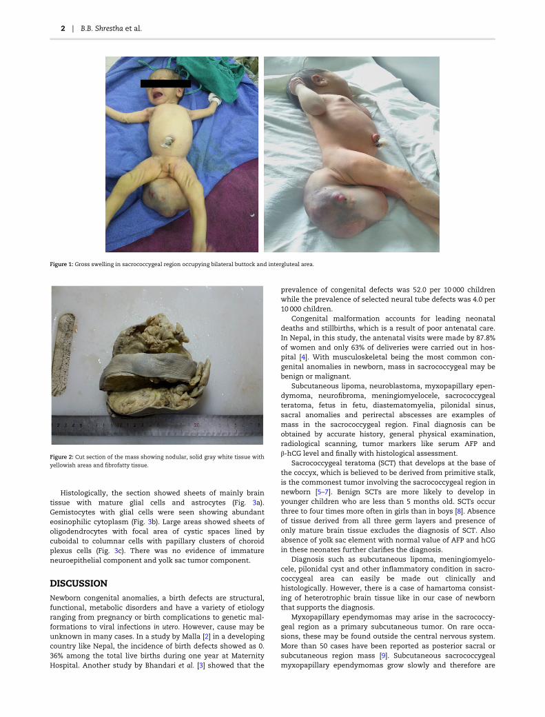
CASE REPORTA term newborn baby girl had a swelling of the sacrococcygealregion. She had no other congenital anomalies and neuro-logical deficit. Physical examination revealed a soft to solid ovalmobile mass occupying the coccygeal, bilateral gluteal andintergluteal fold that measured 15 × 10 cm size. Skin surfaceshowed slight bosselation of the surface without any ulcer-ation. However, there was bluish to red discoloration of skin in
posterior aspect of the mass (Fig. 1). On doing a rectal examin-ation the mass could not be felt extending to the pelvis.
Computed tomography (CT) and magnetic resonanceimaging (MRI) of pelvis revealed a large mass of 14.5 × 9.5 cmin coccygeal, buttock and intergluteal fold without extensionto adjacent structures. It showed a cystic to solid componentwith areas of hemorrhage. Unfortunately the CT and MRI filmswere lost on follow up. Echocardiography showed patent duc-tus arteriosus with small left to right shunt and mild pulmon-ary arterial hypertension with patent foramen ovale, which issupposed to be physiological. Serum markers like α-fetopro-tein (AFP) and human chorionic gonadotropin (hCG) levelwere within normal limit. No preoperative tumor biopsy wasdone.
Total excision of tumor with coccygectomy was done undergeneral anesthesia on the sixth day of life and the post-operative course was uneventful. Cut section surface showednodular, solid gray white tissue with yellowish areas of fibro-fatty tissue and small areas of hemorrhage (Fig. 2).
Received: March 24, 2016. Accepted: April 29, 2016
Published by Oxford University Press and JSCR Publishing Ltd. All rights reserved. © The Author 2016.This is an Open Access article distributed under the terms of the Creative Commons Attribution Non-Commercial License (http://creativecommons.org/licenses/by-nc/4.0/), which permits non-commercial re-use, distribution, and reproduction in any medium, provided the original work is properly cited.For commercial re-use, please contact [email protected]
1
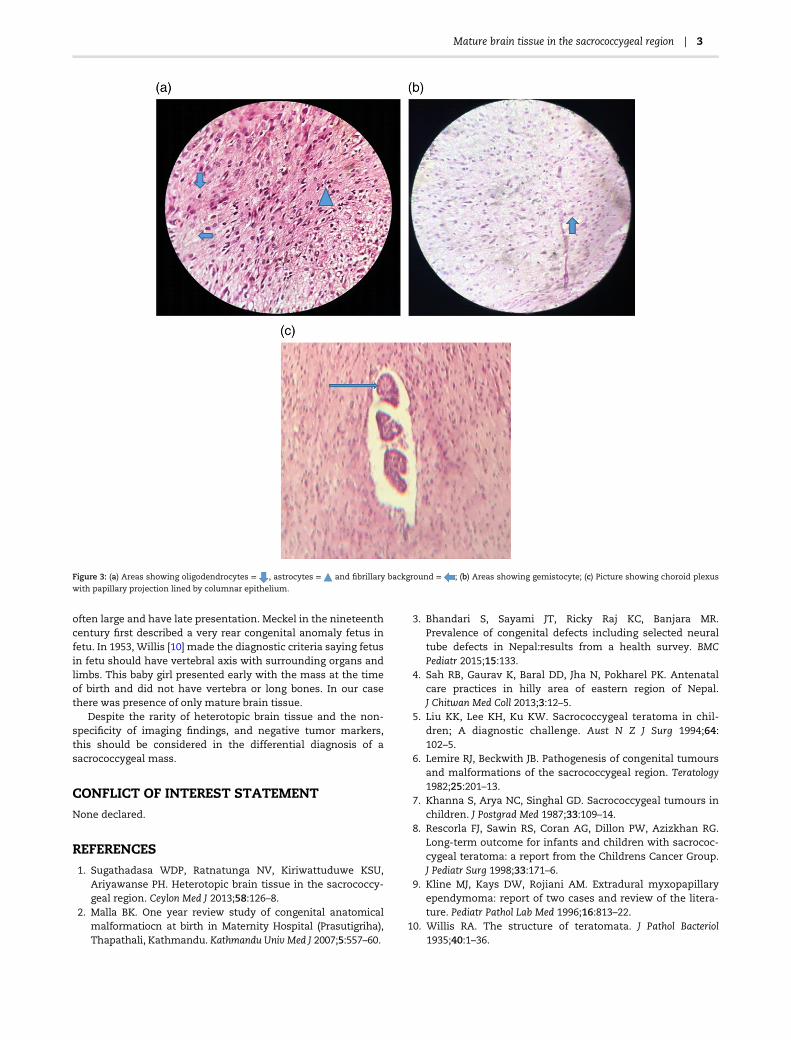
Histologically, the section showed sheets of mainly braintissue with mature glial cells and astrocytes (Fig. 3a).Gemistocytes with glial cells were seen showing abundanteosinophilic cytoplasm (Fig. 3b). Large areas showed sheets ofoligodendrocytes with focal area of cystic spaces lined bycuboidal to columnar cells with papillary clusters of choroidplexus cells (Fig. 3c). There was no evidence of immatureneuroepithelial component and yolk sac tumor component.
DISCUSSIONNewborn congenital anomalies, a birth defects are structural,functional, metabolic disorders and have a variety of etiologyranging from pregnancy or birth complications to genetic mal-formations to viral infections in utero. However, cause may beunknown in many cases. In a study by Malla [2] in a developingcountry like Nepal, the incidence of birth defects showed as 0.36% among the total live births during one year at MaternityHospital. Another study by Bhandari et al. [3] showed that the
prevalence of congenital defects was 52.0 per 10 000 childrenwhile the prevalence of selected neural tube defects was 4.0 per10 000 children.
Congenital malformation accounts for leading neonataldeaths and stillbirths, which is a result of poor antenatal care.In Nepal, in this study, the antenatal visits were made by 87.8%of women and only 63% of deliveries were carried out in hos-pital [4]. With musculoskeletal being the most common con-genital anomalies in newborn, mass in sacrococcygeal may bebenign or malignant.
Subcutaneous lipoma, neuroblastoma, myxopapillary epen-dymoma, neurofibroma, meningiomyelocele, sacrococcygealteratoma, fetus in fetu, diastematomyelia, pilonidal sinus,sacral anomalies and perirectal abscesses are examples ofmass in the sacrococcygeal region. Final diagnosis can beobtained by accurate history, general physical examination,radiological scanning, tumor markers like serum AFP andβ-hCG level and finally with histological assessment.
Sacrococcygeal teratoma (SCT) that develops at the base ofthe coccyx, which is believed to be derived from primitive stalk,is the commonest tumor involving the sacrococcygeal region innewborn [5–7]. Benign SCTs are more likely to develop inyounger children who are less than 5 months old. SCTs occurthree to four times more often in girls than in boys [8]. Absenceof tissue derived from all three germ layers and presence ofonly mature brain tissue excludes the diagnosis of SCT. Alsoabsence of yolk sac element with normal value of AFP and hCGin these neonates further clarifies the diagnosis.
Diagnosis such as subcutaneous lipoma, meningiomyelo-cele, pilonidal cyst and other inflammatory condition in sacro-coccygeal area can easily be made out clinically andhistologically. However, there is a case of hamartoma consist-ing of heterotrophic brain tissue like in our case of newbornthat supports the diagnosis.
Myxopapillary ependymomas may arise in the sacrococcy-geal region as a primary subcutaneous tumor. On rare occa-sions, these may be found outside the central nervous system.More than 50 cases have been reported as posterior sacral orsubcutaneous region mass [9]. Subcutaneous sacrococcygealmyxopapillary ependymomas grow slowly and therefore are
Figure 1: Gross swelling in sacrococcygeal region occupying bilateral buttock and intergluteal area.
Figure 2: Cut section of the mass showing nodular, solid gray white tissue with
yellowish areas and fibrofatty tissue.
2 | B.B. Shrestha et al.
often large and have late presentation. Meckel in the nineteenthcentury first described a very rear congenital anomaly fetus infetu. In 1953, Willis [10] made the diagnostic criteria saying fetusin fetu should have vertebral axis with surrounding organs andlimbs. This baby girl presented early with the mass at the timeof birth and did not have vertebra or long bones. In our casethere was presence of only mature brain tissue.
Despite the rarity of heterotopic brain tissue and the non-specificity of imaging findings, and negative tumor markers,this should be considered in the differential diagnosis of asacrococcygeal mass.
CONFLICT OF INTEREST STATEMENTNone declared.
REFERENCES1. Sugathadasa WDP, Ratnatunga NV, Kiriwattuduwe KSU,
Ariyawanse PH. Heterotopic brain tissue in the sacrococcy-geal region. Ceylon Med J 2013;58:126–8.
2. Malla BK. One year review study of congenital anatomicalmalformatiocn at birth in Maternity Hospital (Prasutigriha),Thapathali, Kathmandu. Kathmandu Univ Med J 2007;5:557–60.
3. Bhandari S, Sayami JT, Ricky Raj KC, Banjara MR.Prevalence of congenital defects including selected neuraltube defects in Nepal:results from a health survey. BMCPediatr 2015;15:133.
4. Sah RB, Gaurav K, Baral DD, Jha N, Pokharel PK. Antenatalcare practices in hilly area of eastern region of Nepal.J Chitwan Med Coll 2013;3:12–5.
5. Liu KK, Lee KH, Ku KW. Sacrococcygeal teratoma in chil-dren; A diagnostic challenge. Aust N Z J Surg 1994;64:102–5.
6. Lemire RJ, Beckwith JB. Pathogenesis of congenital tumoursand malformations of the sacrococcygeal region. Teratology1982;25:201–13.
7. Khanna S, Arya NC, Singhal GD. Sacrococcygeal tumours inchildren. J Postgrad Med 1987;33:109–14.
8. Rescorla FJ, Sawin RS, Coran AG, Dillon PW, Azizkhan RG.Long-term outcome for infants and children with sacrococ-cygeal teratoma: a report from the Childrens Cancer Group.J Pediatr Surg 1998;33:171–6.
9. Kline MJ, Kays DW, Rojiani AM. Extradural myxopapillaryependymoma: report of two cases and review of the litera-ture. Pediatr Pathol Lab Med 1996;16:813–22.
10. Willis RA. The structure of teratomata. J Pathol Bacteriol1935;40:1–36.
Figure 3: (a) Areas showing oligodendrocytes = , astrocytes = and fibrillary background = ; (b) Areas showing gemistocyte; (c) Picture showing choroid plexus
with papillary projection lined by columnar epithelium.
Mature brain tissue in the sacrococcygeal region | 3