Embed Size (px)
Citation preview

Max Brinsmead MB BS PhDMay 2015
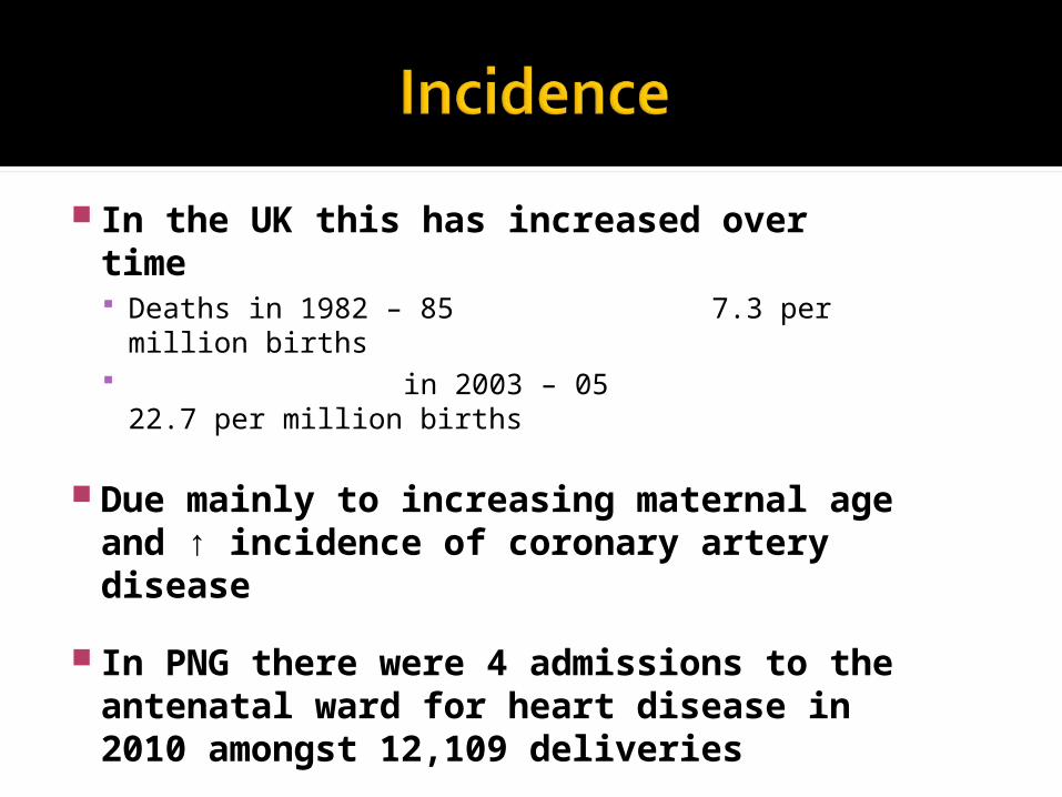
In the UK this has increased over time Deaths in 1982 – 85 7.3 per million births in 2003 – 05 22.7 per million
births
Due mainly to increasing maternal age and ↑ incidence of coronary artery disease
In PNG there were 4 admissions to the antenatal ward for heart disease in 2010 amongst 12,109 deliveries
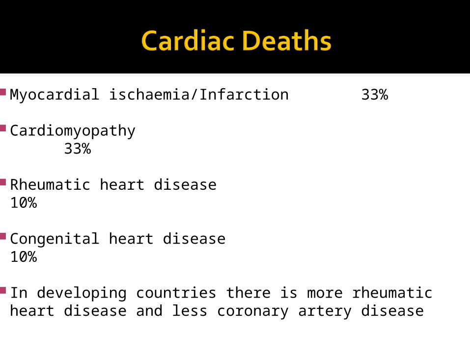
Myocardial ischaemia/Infarction 33%
Cardiomyopathy 33%
Rheumatic heart disease 10%
Congenital heart disease 10%
In developing countries there is more rheumatic heart disease and less coronary artery disease
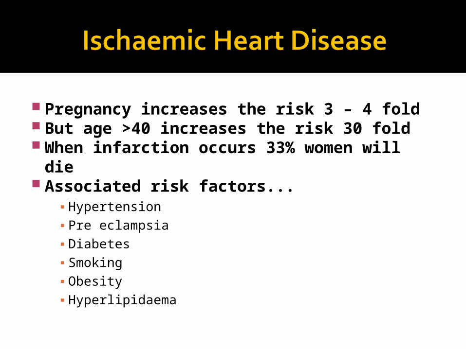
Pregnancy increases the risk 3 – 4 fold But age >40 increases the risk 30 fold When infarction occurs 33% women
will die Associated risk factors...
▪ Hypertension▪ Pre eclampsia▪ Diabetes▪ Smoking▪ Obesity▪ Hyperlipidaema
A high index of suspicion in patients at risk
If they develop chest pain then early recourse to...▪ ECG▪ Serum tropinins▪ CT or MRI▪ Angiography if required
Unknown aetiology and no known risk factors
25% will be associated with hypertension
Sometimes due to viral myocarditis
Can occur any time in the antenatal period and up to 6 months postpartum
A patient who complains of increasing dyspnoea
Especially nocturnal orthopnoea Investigate by...
▪ ECG▪ CXR▪ Echocardiography
Mitral stenosis is the most common And most serious
But it is difficult to detect
So early referral and echocardiography is recommended when... Any diastolic murmur is detected There is any history suggestive of
rheumatic fever

Usually associated with systolic hypertension So keep this controlled
Also a complication of Marfan’s Sydrome The spider people With dislocated lens May be a family history But 30% are spontaneous mutations Risk of aortic dissection is low if the aortic
root diam is <40 mm
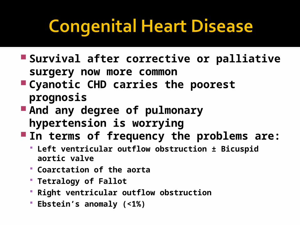
Survival after corrective or palliative surgery now more common
Cyanotic CHD carries the poorest prognosis
And any degree of pulmonary hypertension is worrying
In terms of frequency the problems are: Left ventricular outflow obstruction ± Bicuspid aortic
valve Coarctation of the aorta Tetralogy of Fallot Right ventricular outflow obstruction Ebstein’s anomaly (<1%)
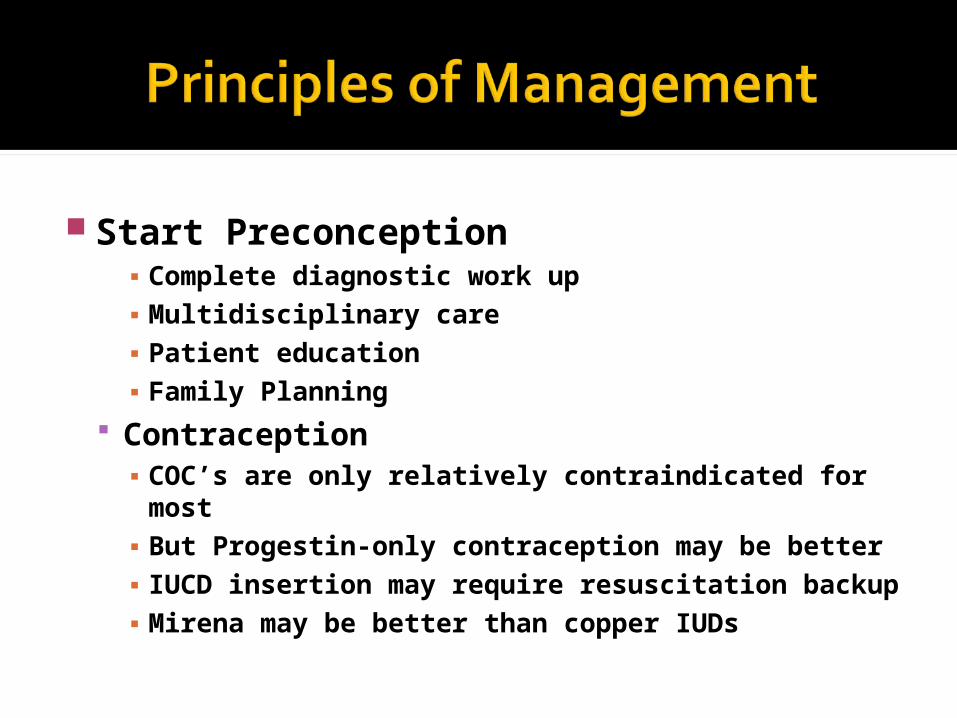
Start Preconception▪ Complete diagnostic work up▪ Multidisciplinary care▪ Patient education▪ Family Planning
Contraception▪ COC’s are only relatively contraindicated for
most▪ But Progestin-only contraception may be better▪ IUCD insertion may require resuscitation
backup▪ Mirena may be better than copper IUDs
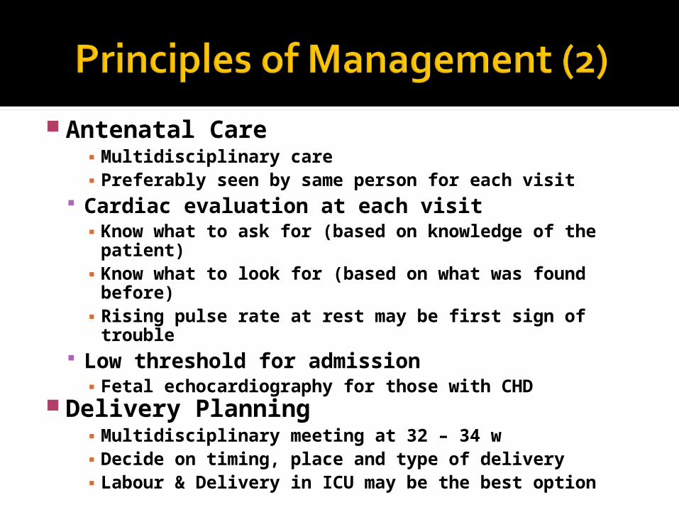
Antenatal Care▪ Multidisciplinary care▪ Preferably seen by same person for each visit
Cardiac evaluation at each visit▪ Know what to ask for (based on knowledge of the
patient)▪ Know what to look for (based on what was found
before)▪ Rising pulse rate at rest may be first sign of
trouble Low threshold for admission▪ Fetal echocardiography for those with CHD
Delivery Planning▪ Multidisciplinary meeting at 32 – 34 w▪ Decide on timing, place and type of delivery▪ Labour & Delivery in ICU may be the best option
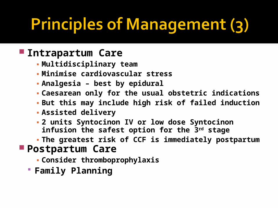
Intrapartum Care▪ Multidisciplinary team▪ Minimise cardiovascular stress▪ Analgesia – best by epidural▪ Caesarean only for the usual obstetric
indications▪ But this may include high risk of failed induction▪ Assisted delivery▪ 2 units Syntocinon IV or low dose Syntocinon
infusion the safest option for the 3rd stage▪ The greatest risk of CCF is immediately
postpartum Postpartum Care
▪ Consider thromboprophylaxis Family Planning
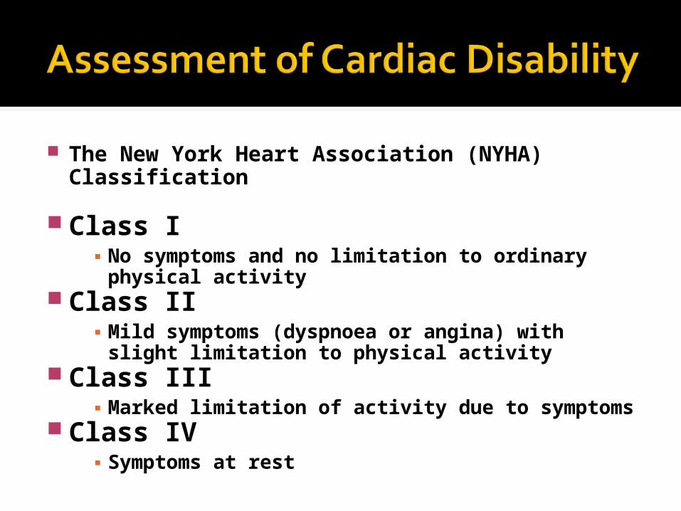
The New York Heart Association (NYHA) Classification
Class I▪ No symptoms and no limitation to ordinary
physical activity Class II
▪ Mild symptoms (dyspnoea or angina) with slight limitation to physical activity
Class III▪ Marked limitation of activity due to symptoms
Class IV▪ Symptoms at rest

Please leave a note on the Welcome Page of this website