Embed Size (px)
Citation preview
UNIVERSIDADE FEDERAL DE MINAS GERAIS PROGRAMA DE PÓS GRADUAÇÃO LATO SENSU
NEUROCIÊNCIAS E SUAS FRONTEIRAS
Paula Juliene Teles Alves
MAXILLOFACIAL INJURIES ASSOCIATED WITH
TRAUMATIC BRAIN INJURY: A SYSTEMATIC REVIEW
Belo Horizonte – MG, Brazil 2018
Paula Juliene Teles Alves
MAXILLOFACIAL INJURIES ASSOCIATED WITH
TRAUMATIC BRAIN INJURY: A SYSTEMATIC REVIEW
Belo Horizonte – MG, Brazil
2018
Monography to the post-graduation program lato sensu Neurosciences and Its Frontiers of the Federal University of Minas Gerais, as part of the specialization course in Neurosciences.
Advisor: PhD. Aline Silva de Miranda

Para minha amada família: Helias, Ivis, Pedro e Vera.

Agradeço a Deus pelo esforço recompensado; à minha família pelo imenso
apoio; à minha orientadora Aline, pela paciência e confiança; Obrigada, Carol,
pela amizade e constante ajuda e a todos que indiretamente contribuíram para
a conclusão deste trabalho.

"The good surgeon knows how to operate;
A better surgeon knows when to operate;
The best surgeon knows when not to operate "

Raymond Maurice Kirk
LIST OF TABLES AND ILLUSTRATIONS
List of acronyms and abbreviations ...........................................................................07
Flowchart 1 ................................................ .................................................. ........... 15
Table 1 ................................................ .................................................. .................. 38

LIST OF ACRONYMS AND ABBREVIATIONS
TBI: Traumatic Brain Injury
MFF: Maxillofacial Fractures
MFI: Maxillofacial Injury
MFT: Maxillofacial Trauma
PRISMA: Main Items to Report Systematic reviews and Meta-analyzes
pTBI : penetrating Traumatic Brain Injury
cTBI: closed Traumatic Brain Injury
MACE: Military Acute Concussion Evaluation
ISS: Injury Severity Score
GCS: Glasgow Coma Scale
CT: Computed tomography
CSF: Cerebrospinal fluid
ABSTRACT
The high rate of morbidity and mortality due to traumatic brain injury (TBI) is a
major public health problem, since the treatment and follow-up of the affected
patients have a significant social and economic impact. The bone complexity of the
face and maxillofacial injury (MFI) that may sometimes be associated with TBI
necessitates the present review that systematically discusses the evidence available
in the literature associated with the potential relationships between the TBI and the
MFI and its implications diagnostic and therapeutic. The research based on the
PRISMA guideline was carried out by two researchers (Alves, PJT, Machado, CA) in
the PubMed and Scopus databases, which presented a total of 27 competent articles
to the topic of the 149 researched ones. Of this selection, 13 are retrospective
studies, 9 prospective studies and 5 are cohorts. The preliminary result is a flowchart
based on the PRISMA protocol detailing the search and inclusion and exclusion of
articles. It is important to train all the emergency room professionals for the
recognition of TBI and adequate initial management of the patients. The use of
several TBI classifications makes it difficult to associate with MFIs. Further studies
are needed to highlight the relationship between TBI and MFI and to stress the
importance of concomitant management of both lesions and multidisciplinary
conduction in the hospital settingfor in a better prognosis.
KEYWORDS: TRAUMATIC BRAIN INJURY; MAXILLOFACIAL INJURIES; HEAD
INJURIES.
SUMMARY
1 INTRODUCTION................................................................................................
2 OBJECTIVES.....................................................................................................
10
12
3 METHODS ......................................................................................................... 13
3.1 Design........................................................................................................ 13
3.2 Inclusion criteria......................................................................................... 13
3.3 Search methods for identification of studies .............................................. 13
3.4 Selection of studies ................................................................................... 13
4 RESULTS...........................................................................................................
4.1 Study characteristics..................................................................................
4.2 TBI classification.........................................................................................
4.3 Causes of TBI and MFI...............................................................................
4.4 TBI features ..............................................................................................
4.5 MFI classification........................................................................................
4.6 MFI features...............................................................................................
4.7 MFI with TBI ..............................................................................................
4.8 TBI and MFI Management..........................................................................
15
16
16
17
18
19
20
20
20
5 DISCUSSION...............................................................................................
6 FINAL CONSIDERATIONS..............................................................................
23
28
REFERENCES.................................................................................................... 29
ATTACHMENT................................................................................................... 38

12
1 INTRODUCTION
Traumatic Brain Injury (TBI) is an injury caused by external mechanical forces
that can lead to anatomical and functional dysfunctions of both the skull and the
brain. (MAGALHÃES et al, 2018). TBI lesions may arise in the direct impact area and
elsewhere in the brain (PEIXOTO et al, 2015). Cranial lesions may promote
lacerations, abrasions, avulsions, contusions, fractures, intracranial bleeding in
addition to hypoxia and cerebral ischemia (MILORO et al, 2004). The high rate of
morbidity and mortality due to TBI is a major public health problem, since the
treatment and follow-up of the affected patients have a significant social and
economic impact (OLIVEIRA et al, 2010).
Given the bone diversity of the viscerocranium, there are several types of
classification for maxillofacial fractures (MFF) that can be associated with TBI.
Maxillofacial Injury (MFI) may, for example, cause bleeding and secretions, such as
cerebrospinal fluid; obturation of the pharynx by loosening of the base of the tongue,
obstruction airways by fracture of the midface or by avulsion of teeth or dental
prosthesis, factors incompatible with life if not treated in time (MILORO et al., 2004).
It is a routine of large emergency departments to evaluate and manage
trauma-related facial injuries (DAVIDOFF et al., 1988). The early recognition of TBI
and concomitant lesions in the initial assessment and in the treatment of patients for
the reduction of morbidity and mortality is a priority (SCHEYERER et al, 2015).
Understanding the source of the lesion and / or mechanism, severity level and
dimension of facial trauma and concomitant lesions aid in the optimization of initial
clinical treatment and correct definition of the time to involve the oral surgeon.
(SCHEYERER et al, 2015).
13
The oral and maxillofacial surgeon should be aware of the occurrence of
concomitant brain injury and have an understanding of the pathophysiology of TBI at
initial recognition and treatment. Whatever patient is affected by maxillofacial injury,
with or without associated fractures, he will be at risk of traumatic brain injury
(CHOONTHAR et al, 2016). Thus, all patients with maxillofacial lesions should be
under neurosurgical observation and continuous follow-up (RAJANDRAM et al,
2013).
The relationship between MFI due to TBI is limited in the literature considering
thecomplex multidisciplinary nature, its high rates of morbidity and mortality and the
need for joint scientific knowledge of the specialties in recognition, early diagnosis
and rigorous cases of TBI. With the current debate about the exact definition of
complications and their categorization (SALENTIJN et al, 2014), this study intends to
make a systematic review in order to discuss the available evidence associated with
the potential relationships between TBI and MFI and implications.
The medical and dental specialties should have joint access to the maximum
of parameters in the diagnosis of TBI for the broadest benefit of the patients. Early
intervention and rapid diagnosis is essential in the prevention of morbidity and
mortality, especially with regard to the prevention of TBI, in the short-term duration of
hypoxia and edema that may progress to permanent neurological and psychiatric
deficits (RAJANDRAM et al, 2013).
This systematic review aims to dicuss emerging evidence regarding the impact
of facial lesions resulting from TBI and to provide information that may contribute to
clinical decision making, as well as to alert the multidisciplinary approach of
polytrauma patients.
14
2 OBJECTIVES
The present review aims to discuss emerging evidence on the relationship
between each type of TBI and MFI, as well as its diagnostic and therapeutic
implications.
15
3 METHODS
3.1 Design
This systematic review was conducted according to the guidelines of
the Preferred Reporting Items for Systematic Reviews and Meta Analyses (PRISMA)
(Moher et al, 2009).
3.2 Inclusion criteria
Observational studies as well as cross-sectional and cohort studies, all in the
English language, were eligible for inclusion. Studies excluded from this review
included: non-English articles, case reports, reviews, pediatric studies, laboratory /
experimental / animal studies, sports, pathological lesions of non-traumatic origin and
articles dealing with brain trauma and individual maxillofacial lesions.
3.3 Search methods for identification of studies
An electronic search for relevant articles was performed independently by two
authors (Alves, P.J.T.; Machado, C.A.) using PUBMED and SCOPUS without period
limitation. The search terms were TBI, MFI, head injuries, only in English language.
The search combination used was: “TRAUMATIC BRAIN INJURY” AND
“MAXILLOFACIAL INJURIES” AND “HEAD INJURIES”.
3.4 Selection of studies
Two researchers independently (Alves,P.J.T.; Machado, C.A.) reviewed the
eligibility of the studies and analyzed their characteristics, quality, and accuracy.
Studies were initially extracted for abstract screening and those found to be relevant

16
were fully retrieved for a detailed review. Disagreements on eligibility were resolved
by discussion between authors. Once the eligible studies were established, data
were extracted by authors.
.
17
4 RESULTS
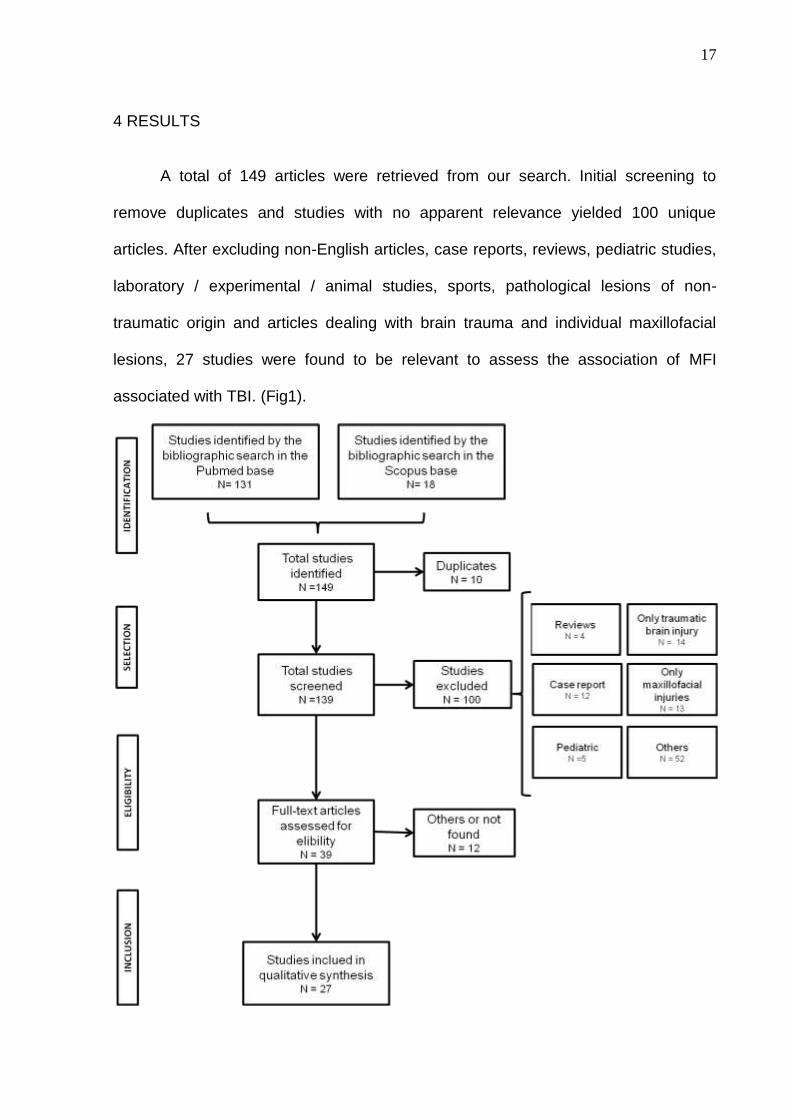
A total of 149 articles were retrieved from our search. Initial screening to
remove duplicates and studies with no apparent relevance yielded 100 unique
articles. After excluding non-English articles, case reports, reviews, pediatric studies,
laboratory / experimental / animal studies, sports, pathological lesions of non-
traumatic origin and articles dealing with brain trauma and individual maxillofacial
lesions, 27 studies were found to be relevant to assess the association of MFI
associated with TBI. (Fig1).

18
Fig. 1: PRISMA flow diagram for observational studies of correlation between the MFI
associated with TBI.
4.1 Study characteristics
The selected studies (n = 27) investigated in this systematic review was 13
retrospective studies, 9 prospective studies and 5 cohorts, that presenting had very
diverse populations with different age groups (from 1 to 93 years) and with a data
from a period of 24 hours to 10 years.
4.2 TBI classification
It is generally known in the literature that the classification of TBI, produced by
the Glasgow coma scale (GCS), is mild, moderate and severe. Of the articles, 8
mention this classification (RAJANDRAM et al, 2013; PULJULA et al, 2012;
SALENTIJN et al, 2014; CAVALCANTE et al ,2012; HUANG et al 2012;
CZERWINSKI et al, 2008; RAZAK et al, 2017; HAMMOND et al, 2018). Another 9
articles use other minor and major brain injuries, penetrating TBI (pTBI) and closed
TBI (cTBI), concussion, concussion with or without less consciousness,
mild/moderate/severe head injuries, anterior/middle/posterior fossa injuries(GRANT
et al, 2012; FARES et al, 2014; PAPPACHAN et al, 2006; HOLMGREN et al, 2004;
WANYURA et al, 2014; ALADELUSI et al, 2014; SOBIN et al, 2016; GÖNÜL et al,
2005; HAMMOND et al, 2018). 12 articles showed only the characteristics of the TBI
as concussion, skill fractures, hemorrage, haematoma, edema, diffuse injuries, focal
injuries, contusion, amnesia, loss of consciousness, skin laceration, cerebral edema,
pneumocephalus, neurologic injuries, ophthalmologic injuries and pneumocranium
(VEERAMUTHU et al, 2016; ŽIVKOVIĆ et al, 2014; CHU et al, 2011; PAPPACHAN
19
et al, 2006; COSSMAN et al, 2014; SCHEYERER et al, 2015; LOPES
ALBUQUERQUE et al, 2014; HUANG et al, 2017; STEPHENS et al, 2016;
HOLMGREN et al, 2004; WANYURA et al, 2014; CAI ET AL, 2017), 3 articles shows
the types and features (RAJANDRAM et al, 2013; CZERWINSKI et al, 2008;
ALADELUSI et al, 2014), while 3 articles did not present any characteristics or
evidenced some type of TBI (ŽIVKOVIĆ et al, 2014; KRAUS et al, 2003; CHU et al,
2011).
4.3 Causes of TBI and MFI
The most common causes of these lesions were, respectively, road traffic
accidents (RAJANDRAM et al,2013; GRANT et al, 2012; PULJULA et al, 2012;
SALENTIJN et al, 2014; CAVALCANTE et al, 2012, KRAUS et al, 2003;
PAPPACHAN et al, 2006; COSSMAN et al, 2014; SCHEYERER et al, 2015; HUANG
et al, 2017; CZERWINSKI et al, 2008; STEPHENS et al, 2016; WANYURA et al,
2014; SOBIN et al, 2016; RAZAK et al, 2017; CAI et al, 2017; VEERAMUTHU et al,
2016; LOPES ALBUQUERQUE et al, 2014; HOLMGREN et al, 2004), falls
(RAJANDRAM et al, 2013; GRANT et al, 2012; SALENTIJN et al, 2014;
CAVALCANTE et al, 2012; KRAUS et al, 2003; COSSMAN et al, 2014;
SCHEYERER et al, 2015; HUANG et al, 2017; LOPES ALBUQUERQUE et al, 2014;
HOLMGREN et al, 2004; ŽIVKOVIĆ et al, 2014), and Assault (RAJANDRAM et al,
2013; GRANT et al, 2012; SALENTIJN et al, 2014; SCHEYERER et al, 2015; SOBIN
et al, 2016; CAI et al, 2017 ). Other causes associated with TBI were Sport injury
(RAJANDRAM et al, 2013; GRANT et al, 2012; PULJULA et al, 2012; SOBIN et al,
2016; RAZAK et al, 2017), Industrial injury (RAJANDRAM et al, 2013; RAZAK et al,
20
2017), Gunshot (KELLER et al, 2015; CZERWINSKI et al, 2008), Munitions (FARES
et al, 2014; GÖNÜL et al, 2005), Handmine and Blast (KELLER et al, 2015),
Earthquake (CHU et al, 2011).
4.4 TBI Features
The most cited TBI features were skull fractures (KRAUS et al, 2003; CHU et
al, 2011; PAPPACHAN et al, 2006; COSSMAN et al, 2014; HUANG et al, 2017;
STEPHENS et al, 2016; ALADELUSI et al, 2014; GÖNÜL et al, 2005; CAI et al,
2017), Hemorrhage (RAJANDRAM et al, 2013; CHU et al, 2011; PAPPACHAN et al,
2006; SCHEYERER et al, 2015; HUANG et al, 2017; CZERWINSKI et al, 2008; CAI
et al, 2017), Hematoma (ŽIVKOVIĆ et al, 2014; CHU et al, 2011; HUANG et al, 2017;
CZERWINSKI et al, 2008; WANYURA et al, 2014; CAI et al, 2017) and concussion
(RAJANDRAM et al, 2013; PAPPACHAN et al, 2006; HOLMGREN et al, 2004;
WANYURA et al, 2014; SOBIN et al, 2016; HAMMOND et al, 2018). Other
characteristics related to TBI were loss of consciousness (VEERAMUTHU et al,
2016; LOPES ALBUQUERQUE et al, 2014; HOLMGREN et al, 2004), Contusion
(RAJANDRAM et al, 2013; PAPPACHAN et al, 2006; CAI et al, 2017), skin
lacerations (FARES et al, 2014; PAPPACHAN et al, 2006), diffuse injuries
(RAJANDRAM et al, 2013; CZERWINSKI et al, 2008), amnesia (VEERAMUTHU et
al, 2016; LOPES ALBUQUERQUE et al, 2014), Edema (HUANG et al, 2017), focal
injuries (Rajandram et al, 2013), neurologic injuries (Cossman et al, 2014; LOPES
ALBUQUERQUE et al, 2014), ophtalmologic injuries (COSSMAN et al, 2014),
Pneumocranium (WANYURA et al, 2014), cerebral edema, pneumocephalus and
cephalophyma (CHU et al, 2011) and pTBI and cTBI (FARES et al, 2014).
21
4.5 MFI classification
Facial fractures are basically classified as Le Fort I, II and III or carry the name
of the affected bone (OCHS et al, 2005). Of the 27 articles, 19 used the classification
by name of fractured maxillofacial bones (RAJANDRAM et al, 2013; GRANT et al,
2012; KELLER et al, 2015; PULJULA et al, 2012; ŽIVKOVIĆ et al, 2014; KRAUS et
al, 2003; CHU et al, 2011; HUANG et al, 2012; PAPPACHAN et al, 2006; COSSMAN
et al, 2014; SCHEYERER et al, 2015; HUANG et al, 2017; CZERWINSKI et al,2008;
STEPHENS et al, 2016; HOLMGREN et al, 2004; WANYURA et al, 2014; SOBIN et
al, 2016; GÖNÜL et al, 2005; CAI, et al, 2017), 3 articles used the upper / middle /
lower third face classification (RAJANDRAM et al, 2013; LOPES ALBUQUERQUE et
al, 2014; HUANG et al, 2017), 2 articles presented the classification according to the
affected facial bone and other facial lesions (LOPES ALBUQUERQUE et al, 2014;
ALADELUSI et al, 2014) and 1 article did not mention any classification for facial
injuries (HAMMOND et al, 2018). The Le Fort fracture, when mentioned in 4 articles,
appears concomitantly with other lesion classifications (GRANT et al, 2012.
PAPPACHAN et al, 2006; SCHEYERER et al, 2015; HOLMGREN et al, 2004). The
other facial lesions mentioned were: Tissue or muscle and mixed injuries
(VEERAMUTHU et al, 2016), nerve injury, visual deficiency, soft tissue damage
(SALENTIJN et al,2014; FARES et al, 2014), occlusal disturbance, hemorrhage
(SALENTIJN et al, 2014), lip injury, eyelid injury (CAVALCANTE et al, 2012),
abrasion, contusion/haematoma and laceration ( RAZAK et al, 2017) and tooth
lesions (SCHEYERER et al, 2015).
22
4.6 MFI features
Of the 23 articles that associates facial fractures with TBI, 19 articles correlate
multiple fractures (RAJANDRAM et al, 2013; GRANT et al, 2012; KELLER et al,
2015; VEERAMUTHU et al, 2016; PULJULA et al, 2012; SALENTIJN et al, 2014;
KRAUS et al, 2003; CHU et al, 2011; PAPPACHAN et al, 2006; SCHEYERER et al,
2015; LOPES ALBUQUERQUE et al, 2014; HUANG et al, 2017; CZERWINSKI et al,
2008; STEPHENS et al, 2016; HOLMGREN et al, 2004; WANYURA et al, 2014;
ALADELUSI et al, 2014; SOBIN et al, 2016; CAI. et al, 2017) and 4 articles reported
incidence of isolated facial fractures (ŽIVKOVIĆ et al, 2014; HUANG et al, 2012;
COSSMAN et al, 2014; GÖNÜL et al, 2005). Soft tissue injury were reported in 8
articles (VEERAMUTHU et al, 2016; SALENTIJN et al, 2014; CAVALCANTE et al,
2012; FARES et al, 2014; SCHEYERER et al, 2015; HUANG et al, 2017;
ALADELUSI et al, 2014; RAZAK et al,2017).
4.7 MFI with TBI
Of the 27 studies selected, 9 articles reported that an association between the
two lesions and stated that MFI increases the risk of TBI (RAJANDRAM et al, 2013;
GRANT et al, 2012; KRAUS et al, 2003; CHU et al, 2011, PAPPACHAN et al, 2006;
SCHEYERER et al, 2015; HUANG et al, 2017; CZERWINSKI et al, 2008;
ALADELUSI et al, 2014). It was verified the relation of the TBI with: frontal sinus
fracture (STEPHENS et al, 2016), orbital lesions (ŽIVKOVIĆ et al, 2014; HUANG et
al, 2012; COSSMAN et al, 2014; HOLMGREN et al, 2004; GÖNÜL, et al, 2005), the
midface fracture (HUANG et al, 2012; LOPES ALBUQUERQUE et al, 2014) and
23
isolated fracture of the mandible (CZERWINSKI et al, 2008; SOBIN et al, 2016;
HAMMOND et al, 2018). Also, three articles recommends the reduction of fractures
to the cranium, thus preventing cerebral spinal fluid (CSF) overflow, meningitis and
infections (KELLER et al, 2015; HOLMGREN et al, 2004; GÖNÜL et al, 2005). Five
articles assert that mild TBI should be suspected in patients with facial injuries and
lower face lacerations (GRANT et al, 2012; VEERAMUTHU et al, 2016; RAZAK et al,
2017; HAMMOND et al, 2018). In addition, there’s a high suspicion for concussion
when accompanied mild TBI associated with isolated mandibular fractures (SOBIN et
al, 2016; HUANG et al, 2012). Two article reveals that the use of motorcycle helmet
prevents the MF but not the TBI (LOPES ALBUQUERQUE et al, 2014; RAZAK et al,
2017). Seven articles recommend the use of skull and face CT (HUANG et al, 2012;
SCHEYERER et al, 2015; HUANG et al, 2017; CZERWINSKI et al, 2008;
HOLMGREN et al, 2004; GÖNÜL et al, 2005; HAMMOND et al, 2018), two of which
advocate the use of CT for better investigation of orbital fractures (HOLMGREN et al,
2004; GÖNÜL et al, 2005). To aid the Glasgow Coma Scale(GCS), 2 articles made
use of the scales: Military Acute Concussion Evaluation(MACE) (SOBIN et al, 2016)
and Injury Severity Score (ISS) (HOLMGREN et al, 2004). Four articles advocate
multidisciplinary treatment (PULJULA et al, 2012; COSSMAN et al, 2014;
SCHEYERER et al, 2015; CHU et al, 2011).
4.8 TBI and MFI Management
Of the 27 articles, three approach the treatment and complications from TBI
and MFI (SALENTIJN et al, 2014; CHU et al, 2011; COSSMAN et al, 2014). The
most common complications are: Nerve injury iInfection/ inflammation, occlusal
disturbance, visual deficiency, soft tissue damage, facial bone defect, psychiatric /

24
psychological disorder, neurological deficit, hemorrhage / blood loss and
physiological dysregulation (SALENTIJN et al, 2014). Others complications found are
upper respiratory tract obstruction, haematocele and parapharyngeal soft tissue
swelling and pharyngeal stenosis (CHU et al, 2011). One study made a very
interesting correlation. Severe TBI is mainly related with physiological dysregulation,
infection/inflammation, psychiatric/psychologic disorders and neurological deficits. in
the incidence of moderate TBI, it is more associated with facial fratures, nerve
injuries and soft tissue damages while that not were observed, development of
complications in mild TBI (SALENTIJN et al, 2014).
One article listed the 13 treatments used for complications of TBI and MFI.
They are: conservative treatment, decompression, antibiotic treatment, secondary
facial correction, foreign body removal, specialist consultation, pharmacological
treatment (non-antibiotic), cooling / sedation, surgical intervention, compression,
blood transfusion and ventilatory support. Of these, the most common treatment
modality was pharmacological (non-antibiotic) treatment, followed by antibiotic
treatment, conservative treatment, and decompression therapy in the cases of
haematoma (SALENTIJN et al, 2014). Another article mentioned that the most
applied treatments to the orbital roof fractures treatment is bone reduction,
absorbable mesh, plate and split calvaria (COSSMAN et al, 2014). Soft tissue injury
debridement with antibiotics and ocular enucleation are other modalities of the
treatments found (CHU et al, 2011).
25
5 DISCUSSION
The TBI is an injury due to mechanical force against the brain leading to the
anatomical and functional impairment of the soft parts, such as scalp and meninges
and the brain itself, and hard tissues such as bones of the skull. This condition can
lead to both transient and permanent neuropsychiatric changes (MACEDO et al,
2006).
One of the most accepted ways to classify TBI is to perform the GCS, which
assesses the level of consciousness, aiding in therapeutic and diagnostic decision-
making, sorting the TBI is mild, moderate or severe, and also evaluating the
prognosis of TBI was mentioned in nineteen articles (RAJANDRAM et al, 2013;
GRANT et al, 2012; VEERAMUTHU et al, 2016; PULJULA et al, 2012; SALENTIJN
et al, 2014; CAVALCANTE et al, 2012; HUANG et al, 2012; PAPPACHAN et al,
2006; COSSMAN et al, 2014; SCHEYERER et al, 2015; HUANG et al, 2017;
CZERWINSKI et al, 2008; STEPHENS et al, 2016; HOLMGREN et al, 2004;
ALADELUSI et al, 2014; GÖNÜL et al, 2005; RAZAK et al, 2017; HAMMOND et al,
2018; CAI et al, 2017). Eight articles did not use the GCS given the goal of their
studies, as autopsy study, maxillofacial studies, surgical study, motorcycle helmets
and ammunition study (KELLER et al, 2015; ŽIVKOVIĆ et al,2014; KRAUS et al ,
2003; CHU et al, 2011; FARES et al, 2014; LOPES ALBUQUERQUE et al, 2014;
WANYURA et al , 2014; SOBIN et al, 2016). The ISS is an established medical score
to assess trauma severity. It correlates with mortality, morbidity and hospitalization
time after trauma, made by the Committee of the Association for the Advancement of
Automotive Medicine that designed and promotes improvements in the scale
(BAKER et al, 1974; COPES et al, 1988). The MACE is a medical screening and
26
documentation measure that is used to gauge the severity of symptoms and
cognitive deficits after a diagnosis of a concussion has been made. It has been
distributed to US military and care for veterans and active-service members who
have sustained a TBI (KENNEDY et al, 2013). The ISS and MACE accessory scales
were used respectively in tomographic studies of the lesions and in the study of
isolated jaw fractures associated with concussion (HOLMGREN et al, 2004; SOBIN
et al, 2016). The use of classifications that do not contemplate the classic
classification GCS (mild, moderate and severe), as seen in item 4.2, makes the
correlation between MFI and its implications unfeasible.
The main causes of TBI are road traffic accidents, physical assault and falls,
and most of them occurred in young adult males (ANDRADE et al, 2009;
RAJANDRAM et al, 2013; GRANT et al, 2012; PULJULA et al, 2012; SALEJIN et al,
2014; CAVALCANTE et al, 2012; ŽIVKOVIĆ et al, 2014; KRAUS et al, 2003;
PAPPACHAN et al, 2006; COSSMAN et al, 2014; SCHEYERER et al, 2015; LOPES
ALBUQUERQUE et al, 2014; HUANG et al, 2017; CZERWINSKI et al, 2008;
STEPHENS et al, 2016; HOLMGREN et al, 2004; ALADELUSI et al, 2014; RAZAK et
al, 2017; CAI et al, 2017). Although the use of a helmet reduces the risk of MFF, it
does not prevent TBI. Therefore the most serious accidents occur on a motorcycle
(KRAUS et al, 2003; LOPES ALBUQUERQUE et al, 2014; CAVALCANTE et al,
2012). This finding is worrying because this patients can stare at long-time
reabilitation and permanent neurological problems.
The facial region when traumatized usually presents soft tissue, teeth, jaw,
maxilla, zygomatic, naso-orbito-etmoidal complex and periorbital and paranasal
sinuses lesions. There are numerous nomenclatures for the classification of FBMF,
27
such as Le Fort I, II, III; nasal, mandibular, naso-orbito-etmoidal fracture, roof and
orbital blowout among others (OCHS et al, 2005).
The MFFs are usually associated with other lesions, such as soft tissue,
eyeball, and encephalon (CARVALHO et al, 2010; VEERAMUTHU et al, 2016;
SALENTIJN et al, 2014; SCHEYERER et al, 2015; HUANG et al, 2017; ALADELUSI
et al, 2014). When considering the individual facial bones, the central midfacial bone
is associated more commonly with TBI, be related the fragility of the region by
accommodating the maxillary sinuses (PAPPACHAN et al, 2006). Although that
almost 66,67% of the orbital roof fractures are associated with TBI, the orbital
fractures are commonly overlooked (HOLMGREN et al, 2004; CZERWINSKI et al,
2008). It requires a secondary scan for accurate diagnosis, not being diagnosed by
the cranial CT and demanding the use of facial CT, considered adequate for the
accurate diagnosis for the diagnosis of these fractures. (COSSMAN et al, 2014;
HUANG et al, 2017; HOLMGREN et al, 2004; CZERWINSKI et al, 2008;
SCHEYERER et al, 2015; GÖNÜL et al, 2005).
The association between MFI increases the risk of TBI is due to the fact that
facial bones, which, in addition to storing the eyeballs, primordial to the eyesight are
part of the upper airways which, when obstructed by fractures, edema, hemorrhage
or CSF, are a risk for the maintenance of life when not treated (RAJANDRAM et al,
2013; GRANT et al, 2012; KRAUS et al, 2003; CHU et al, 2011; PAPPACHAN et al,
2006; SCHEYERER et al, 2015; HUANG et al, 2017; CZERWINSKI et al, 2008;
ALADELUSI et al, 2014).
The articles did not always discriminate each type of TBI and correlated its
relation with the MFI, a factor that signals the need for further investigation. When
reported, the mild, moderate and severe TBI were interesting: The severe TBI was
28
correlated in coma patients with secondary bone healing in mandibular condyle and
mandibular fractures (HUANG et al, 2012). Some hypotheses were drawn and the
most relevant is the possibility of a proliferation of humoral factors osteogenic due to
damage of the blood-brain barrier. The moderate TBI was related to most motorcycle
accidents (CAVALCANTE et al, 2012), given that the prevalence group is young
men, and the treatment and rehabilitation of these patients is usually prolonged and
strenuous. The mild TBI that frequently remains unrecorded in subjects with MFI
(GRANT et al, 2012; PULJULA et al, 2012), was related to the higher severity of MFI,
facial contusion, haematoma and inferior facial laceration, and midface
fractures.(RAZAK et al, 2017). Another aspect of the mild TBI is that the affected
patients appear to treat MFI with maxillofacial surgeons and are often not referred for
TBI investigation (HAMMOND et al, 2018). This fact raises the importance of
knowledge and preparedness to suspect potential incidence of TBI and MFI.
It is of utmost importance that multi-disciplinary patient care should be
effectively collaborated for the survival and better functional recovery of the victim.
(CARVALHO et al, 2010; GÖNÜL et al, 2005). Ideally, every pacient with craniofacial
fracture should be examined by a neurologist or a neurosurgeon, but this is not
always possible. Four articles report the importance of multidisciplinary management
that is strongly important for the treatment and rehabilitation of the patients
(PULJULA et al, 2012; COSSMAN et al, 2014; SCHEYERER et al, 2015). Therefore,
regular updating seminars of signs and symptoms of TBI should be mandatory for all
emergency room personnel (PULJULA et al, 2012).
The best treatment of maxillofacial lesions may be compromised in some
cases, when deference for neurosurgical concerns is required (SALENTIJN et al,
2014; SCHEYERER et al, 2015). MFI are often associated with a risk of other serious
29
concomitant injuries, the TBI, particularly. Even though emergency operations are
only necessary in rare cases, diagnosis and treatment of such concomitant injuries
have the potential to be overlooked or delayed in severely injured patients. A variety
of complications frequently arise which often necessitate further treatment and
prolonged hospitalisation (SALENTIJN et al, 2014; SCHEYERER et al, 2015)
Both diagnoses for TBI and MFI are performed clinically and by imaging.
Imaging research for CT is widely advocated for both cranial and MFF, especially
orbital fractures and the anterior skull base (RAJANDRAM et al, 2013; HUANG et al,
2017; CZERWINSKI et al, 2008; HOLMGREN et al, 2004). The presence of MFI
worsens the prognosis of TBI (RAZAK et al, 2017) because the facial skeleton can
transmit forces directly to the neurocranium, resulting in severe brain injury
(ALADEUSI et al, 2014). On the other hand, other authors argue that facial fractures
present a low risk of traumatic brain injury, since they affirm that the bones of the
face act as brain protectors when crushed in an episode of trauma, a phenomenon
known as the Crumple Zone (PAPPACHAN et al, 2006; CAI et al, 2017).
Patient care with TBI and MF involves several steps that determine the
success of the prognosis (CARVALHO et al, 2010; WANYURA et al, 2014). The
direct relationship between the severity of maxillofacial injury and head,
ophtalmologic injury and polytrauma, emphasizes the need for thorough examination
of patients, especially in patients who suffered high impact trauma.( ALADELUSI et
al, 2014)
The management of patients with TBI and MF is a major challenge with regard to
optimal treatment. Care with these patients requires engaged multidisciplinary
involvement and requires skills such as airway stabilization, expertise ilimited to
30
performing tracheotomy, ability to facial fracture repair, tecidual repair, and control of
epistaxis and infection control (KELLER et al, 2015).
31
7 FINAL CONSIDERATIONS
According to the literature found, the major causes of TBI is the road traffic
accident, assault and falls, as described in the literature. Facial lesions that require
surgical treatment are often neglected in detriment of neurological injuries or
unawareness of their existence. Few studies directly correlate mild, moderate and
severe TBI with MFI incidence. CT is considered adequate for the accurate diagnosis
of TBI and MFI. An interesting question raised was the need to better prepare the
multidisciplinary team in the care and conduct of trauma cases in a multidisciplinary
way. The evidence on the relationship between each type of TBI and MFI, as well as
its diagnostic and therapeutic implications are camouflaged by the lack of
standardization in the description of TBI. More studies are needed to show the
relationship between each type of TBI and MFI to proof the importance of
concomitant and multidisciplinary management of both lesions for a better prognosis.
32
REFERENCES
ALADELUSI, T.; AKINMOLADUN, V.; OLUSANYA, A.; AKADIRI,O.; FASOLA,A.
Analysis of Road Traffic Crashes–Related Maxillofacial Injuries Severity and
Concomitant Injuries in 201 Patients Seen at the UCH, Ibadan. Craniomaxillofac
Trauma Reconstruction. 2014. v.7. p.284–289. DOI http://dx.doi.org/10.1055/s-0034-
1378183. ISSN 1943-3875.
ANDRADE, A.F.; PAIVA, W.S.; AMORIM, R.L.O.; FIGUEIREDO, E.G.; NETO, E.R.;
TEIXEIRA, M.J. Mecanismos de lesão cerebral no traumatismo
cranioencefálico. Rev Assoc Med Bras.2009. v.55(1). p.75-81. DOI:
http://dx.doi.org/10.1590/S0104-42302009000100020
BAKER SP, O'Neill B, Haddon W, Long WB (1974). The Injury Severity Score: a
method for describing patients with multiple injuries and evaluating emergency
care. The Journal of Trauma. Lippincott Williams & Wilkins. 14 (3): 187–196.
doi:10.1097/00005373-197403000-00001. PMID 4814394.
CAI SS, MOSSOP C, DIACONU SC, HERSH DS, ALFADIL S, RASKO YM,
CHRISTY MR, GRANT MP, NAM AJ. The "Crumple Zone" hypothesis:
Association of frontal sinus volume and cerebral injury after craniofacial
trauma. J Craniomaxillofac Surg. 2017 Jul;45(7):1094-1098. doi:
10.1016/j.jcms.2017.04.005. Epub 2017 Apr 25. Erratum in: J Craniomaxillofac Surg.
2017 Nov;45(11):1907. PubMed PMID: 28551409.
CARVALHO, M.F.; HERRERO, R.K.R.; MOREIRA, D.R.; URBANO, E.S.; REHER,
P. Principles of Manegement of Hospitalized Patients in Oral and Maxilo Facial
Surgery. Rev. Cir. Traumatol. Buco-Maxilo-Facial., Camaragibe v.10, n.4, p. 79-84,
out./dez.2010. ISSN 1808-5210 (versão online).

33
CAVALCANTE JR, OKA SC, DE SANTANA SANTOS T, DOURADO E, DE
OLIVEIRA E SILVA ED, GOMES AC. Influence of helmet use in facial trauma and
moderate traumatic brain injury victims of motorcycle accidents. J Craniofac
Surg. 2012 Jul;23(4):982-5. doi: 10.1097/SCS.0b013e31824e5b04. PubMed PMID:
22777446.
CHOONTHAR, M.M.; RAGHOTHAMAN, A.; PRASAD, R.; PRADEEP, S.; PANDYA,
K. Head Injury: A maxillofacial surgeon’s perspective.Journal of Clinical and
Diagnostic Research. 2016, Jan, Vol-10(1). DOI: 10.7860/JCDR/2016/16112.7122
CHU ZG, YANG ZG, DONG ZH, CHEN TW, ZHU ZY, DENG W, XIAO JH. Features
of cranio-maxillofacial trauma in the massive Sichuan earthquake: analysis of
221 cases with multi-detector row CT. J Craniomaxillofac Surg. 2011
Oct;39(7):503-8. doi: 10.1016/j.jcms.2010.10.022. Epub 2010 Nov 26. PubMed
PMID: 21112795.
COPES, W.S.; H.R. Champion; W.J. Sacco; M.M. Lawnick; S.L. Keast; L.W. Bain
(1988). The Injury Severity Score revisited. The Journal of Trauma. Lippincott
Williams & Wilkins. 28 (1): 69–77. doi:10.1097/00005373-198801000-00010. PMID
3123707.
COSSMAN JP, MORRISON CS, TAYLOR HO, SALTER AB, KLINGE PM,
SULLIVAN SR. Traumatic orbital roof fractures: interdisciplinary evaluation and
management. Plast Reconstr Surg. 2014 Mar;133(3):335e-343e. doi:
10.1097/01.prs.0000438051.36881.e0. PubMed PMID: 24572878.
CZERWINSKI M, PARKER WL, WILLIAMS HB. Algorithm for head computed
tomography imaging in patients with mandible fractures. J Oral Maxillofac Surg.
2008 Oct;66(10):2093-7. doi: 10.1016/j.joms.2008.04.011. PubMed PMID: 18848107.
34
DAVIDOFF, G.; JAKUBOWSKI, M.; THOMAS, D.; ALPERT, M. The spectrum of
closed-head injuries in facial trauma victims: incidence and impact. Ann Emerg
Med. 1988. n.17, p.6-9. DOI: https://doi.org/10.1016/S0196-0644(88)80492-X
FARES Y, FARES J, GEBEILY S. Head and facial injuries due to cluster
munitions. Neurol Sci. 2014 Jun;35(6):905-10. doi: 10.1007/s10072-013-1623-2.
Epub 2014 Jan 4. PubMed PMID: 24389857.
GÖNÜL E, ERDOĞAN E, TAŞAR M, YETIŞER S, AKAY KM, DÜZ B, BEDÜK A,
Timurkaynak E. Penetrating orbitocranial gunshot injuries. Surg Neurol. 2005
Jan;63(1):24-30; discussion 31. PubMed PMID: 15639513.
GRANT, A.L.; RANGER, A.; YOUNG, G.B.; YAZDANI,A. Incidence of Major and
Minor Brain Injuries in Facial Fractures. The Journal of Craniofacial Surgery.
2012. v..23. n.5. p.1324-1328. DOI: 10.1097/SCS.0b013e31825e60ae.
HAMMOND D, WELBURY R, SAMMONS G, TOMAN E, HARLAND M, RICE S. How
do oral and maxillofacial surgeons manage concussion? Br J Oral Maxillofac
Surg. 2018 Feb;56(2):134-138. doi: 10.1016/j.bjoms.2017.12.014. Epub 2018 Jan 8.
PubMed PMID: 29325790.
HOLMGREN EP, DIERKS EJ, HOMER LD, POTTER BE. Facial computed
tomography use in trauma patients who require a head computed tomogram. J
Oral Maxillofac Surg. 2004 Aug;62(8):913-8. PubMed PMID: 15278853.
HUANG LK, WANG HH, TU HF, FU CY. Simultaneous head and facial computed
tomography scans for assessing facial fractures in patients with traumatic
braininjury Injury. 2017 Jul;48(7):1417-1422. doi:10.1016/j.injury.2017.04.046.
Epub 2017 Apr 24. PubMed PMID: 28455003.
35
HUANG W, Li Z, Li Z, YANG R. Does traumatic brain injury result in accelerated
mandibular fracture healing? J Oral Maxillofac Surg. 2012 Sep;70(9):2135-42.
doi:10.1016/j.joms.2012.04.016. PubMed PMID: 22907111.
KELLER, M.W.; HAN, P.P.; GALARNEAU, M.R.; GABALL, C.W. Characteristics of
Maxillofacial Injuries and Safety of In Theater Facial Fracture Repair in Severe
Combat Trauma. Military Medicine. 2015. v. 180.n. 3. p.315. DOI: 10.7205/MILMED-
D-14-00345.
KENNEDY, Carrie H.; Moore, Jeffrey L. (2010). Military Neuropsychology. Springer
Publishing Company. pp. 115–. ISBN 9780826104496. Retrieved 30 April 2013.
KRAUS JF, RICE TM, PEEK-ASA C, MCARTHUR DL. Facial trauma and the risk
of intracranial injury in motorcycle riders. Ann Emerg Med. 2003 Jan;41(1):18-26.
PubMed PMID: 12514678.
LOPES ALBUQUERQUE CE, NOGUEIRA ARCANJO FP, CRISTINO-FILHO G,
MONT'ALVERNE LOPES-FILHO A, CESAR DE ALMEIDA P, PRADO R, PEREIRA-
STABILE CL. How safe is your motorcycle helmet? J Oral Maxillofac Surg. 2014
Mar;72(3):542-9. doi: 10.1016/j.joms.2013.10.017. Epub 2013 Oct 31. PubMed
PMID: 24326016.
MACEDO, K.C. Características clínicas e epidemiológicas de crianças e
adolescentes com traumatismo cranioencefálico leve e análise de fatores
associados à fratura de crânio e lesão intracraniana. 2006. 122f.Dissertação.
Faculdade de Medicina.Universidade Federal de Minas Gerais. Belo Horizonte.
2006.
MAGALHÃES,A.L.G.; DE SOUZA L.C.; Faleiro,R.M.; TEIXEIRA,A.L.;
MIRANDA,A.S. Epidemiology of Traumatic Brain Injury in Brazil.Rev Bras Neurol.
v.53, n 2 p.15-22, 2017.

36
MOHER, D., Liberati, A., TETZLAFF, J., Altman, D.G., GROUP, P., 2009. Preferred
reporting items for systematic reviews and meta-analyses: the PRISMA
statement. PLoS Med. 6 (7), e1000097.
http://dx.doi.org/10.1371/journal.pmed.1000097.
OCHS, M.W.; TUCKER,M.R. Tratamento das Fraturas Faciais. In: PETERSON, L J;
ELLIS III, E; HUPP,J.R.; TUCKER,M.R. Cirurgia Oral e Maxilofacial
Contemporânea.Rio de Janeiro. Elsevier, 2005.cap.24,p. 559-590. ISBN 85-352-
1539-5.
OLIVEIRA, I.B.; OLIVEIRA, A.B.A.; GOES,K.O.; MELO, L.A. Traumatismo
cranioencefálico: considerações anatomofuncionais e clínicas. Revista Saúde e
Pesquisa. Jan./abr.2010. v. 3. n. 1. p. 99-106. ISSN 1983-1870.
PAPPACHAN, B.; ALEXANDER, M. Correlating Facial Fractures and Cranial
Injuries. Journal Oral Maxillofacial Surgery.2006. v. 64. p.1023-1029
DOI:10.1016/j.joms.2006.03.021.
PEIXOTO, R. M. Lesões encefálicas: Definições, perspectivas e
repercussão social. 2015. 27f. Manuscrito. Pós Graduação em Neurociências.
Universidade Federal de Minas Gerais. 2015.
PERVEZ, M.; KITAGAWA, R.S.; CHANG, T.R.. Definition of raumatic Brain Injury,
Neurosurgery, Trauma Orthopedics, Neuroimaging, Psychology, and
Psychiatry in Mild Traumatic Brain Injury. Neuroimag Clin. 2018. v.28. P.1-13.
DOI: https://doi.org/10.1016/j.nic.2017.09 .010.
POWERS,M.P; SCHERER,M.S. Initial Management of the Trauma Patient. In:
MILORO,M..; GHALI,E.; LARSEN, P. E.; WAITE, P. D.. Peterson's Principles Of Oral
And Maxillofacial Surgery. 2 ed. BC Decker Inc ,2004.cap. 18, p. 328-356. ISBN 1-
55009-234-0

37
PULJULA J, CYGNEL H, MÄKINEN E, TUOMIVAARA V, KARTTUNEN V,
KARTTUNEN A, HILLBOM M. Mild traumatic brain injury diagnosis frequently
remains unrecorded in subjects with craniofacial fractures. Injury. 2012
Dec;43(12):2100-4. doi: 10.1016/j.injury.2012.04.010. Epub 2012 May 1. PubMed
PMID: 22552039.
RAJANDRAM R.K.; SYED OMAR S.N.; RASHDI M.F.N.; ABDUL JABAR M.N.
Maxillofacial injuries and traumatic brain injury-: a pilot study. Dent Traumatol.
2014. v.30(2).p.128–32. DOI: 10.1111/edt.12052
RAZAK,N.A.; NORDIN,R.; ABD RAHMAN, N.A.; RAMLI, R. A retrospective
analysis of the relationship between facial injury and mild traumatic brain
injury. Dental Traumatology. 2017. v.33. p.400–405. DOI:10.1111/edt12355.
SALENTIJN, E.G.; Collin, J.D.; BOFFANO, P.; FOROUZANFAR, T. A ten year
analysis of the traumatic maxillofacial and brain injury patient in Amsterdam:
Complications and treatment. Journal of Cranio-Maxillo-Facial Surgery. 2014.
v.42.p.1717-1722. DOI: http://dx.doi.org/10.1016/j.jcms.2014.06.005.
SCHEYERER, M.J.; DÖRING, R.; FUCHS, N.; METZLER, P.; SPRENGEL,K.;
WERNER, C.M.L.; SIMMEN, H.P.; GRÄTZ, K.; GUIDO, A. Maxillofacial injuries in
severely injured patients. Wanner.Journal of Trauma Management & Outcomes,
2015. DOI 10.1186/s13032-015-0025-2.
SOBIN L, KOPP R, WALSH R, KELLMAN RM, HARRIS T. Incidence of
Concussion in Patients With Isolated Mandible Fractures. JAMA Facial Plast
Surg. 2016 Jan-Feb;18(1):15-8. doi: 10.1001/jamafacial.2015.1339. PubMed PMID:
26448310.
STEPHENS JR, HOLMES S, BULTERS D, EVANS BT. The effect of direction of
force to the craniofacial skeleton on the severity of brain injury in patients with
38
a fronto-basal fracture. Int J Oral Maxillofac Surg. 2016 Jul;45(7):872-7.
doi:10.1016/j.ijom.2016.01.016. Epub 2016 Mar 10. PubMed PMID: 26972160.
VEERAMUTHU V, HARIRI F, NARAYANAN V, TAN LK, RAMLI N, GANESAN D.
Microstructural Change and Cognitive Alteration in Maxillofacial Trauma and
Mild Traumatic Brain Injury: A Diffusion Tensor Imaging Study. J Oral Maxillofac
Surg. 2016 Jun;74(6):1197.e1-1197.e10. doi: 10.1016/j.joms.2016.01.042.
WANYURA H, KAMIŃSKI A, STOPA Z. Three-layered osteodural plasty for
severe anterior skull base and facial injuries. Report of eleven cases. Neurol
Neurochir Pol. 2014 Jan-Feb;48(1):8-14. doi: 10.1016/j.pjnns.2013.03.003. Epub
2014 Jan 23. PubMed PMID: 24636764.
ŽIVKOVIĆ V, NIKOLIĆ S. Head injuries in falls from a standing height: do
fractures of the orbital roof matter? A prospective autopsy study. Forensic
SciMed Pathol. 2014 Dec;10(4):483-6. doi: 10.1007/s12024-014-9583-2. Epub 2014
Jul16. PubMed PMID: 25027415.
39
ATTACHMENT
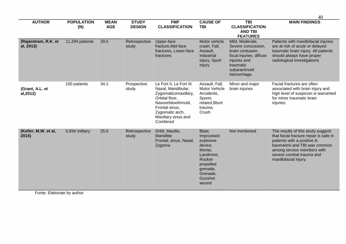
Table 1: Studies that associated maxillofacial injuries associated with traumatic brain injury.
40
Fonte: Elaborate by author
AUTHOR POPULATION (N)
MEAN AGE
STUDY DESIGN
FMF CLASSIFICATION
CAUSE OF TBI
TBI CLASSIFICATION
AND TBI FEATURES
MAIN FINDINGS
(Rajandram, R.K. et al, 2013)
11,294 patients
29.5
Retrospective study
Upper-face fracture,Mid-face fractures, Lower-face fractures
Motor vehicle crash, Fall, Assault, Industrial injury, Sport injury
Mild, Moderate, Severe concussion, brain contusion, focal injuries, diffuse injuries and traumatic subarachnoid hemorrhage.
Patients with maxillofacial injuries are at risk of acute or delayed traumatic brain injury. All patients should always have proper radiological investigations
(Grant, A.L. et al,2012)
100 patients
34.1
Prospective study
Le Fort II, Le Fort III Nasal, Mandibular, Zygomaticomaxillary, Orbital floor, Nasoorbitoethmoid, Frontal sinus, Zygomatic arch, Maxillary sinus and Combined
Assault, Fall, Motor Vehicle Accidents, Sports related,Blunt trauma, Crush
Minor and major brain injuries
Facial fractures are often associated with brain injury and high level of suspicion is warranted for minor traumatic brain injuries.
(Keller, M.W. et al, 2015)
5,934 military
25.0
Retrospective study
Orbit, Maxilla, Mandible Frontal, sinus, Nasal, Zygoma
Blast, Improvised explosive device, Mortar, Landmine, Rocket-propelled grenade, Grenade, Gunshot wound
Not mentioned
The results of this study suggest that facial fracture repair is safe in patients with a positive A. baumannii and TBI was common among service members with severe combat trauma and maxillofacial injury.
41
AUTHOR POPULATION (N)
MEAN AGE
STUDY DESIGN
FMF CLASSIFICATION CAUSE OF TBI
TBI CLASSIFICATION
AND TBI FEATURES
MAIN FINDINGS
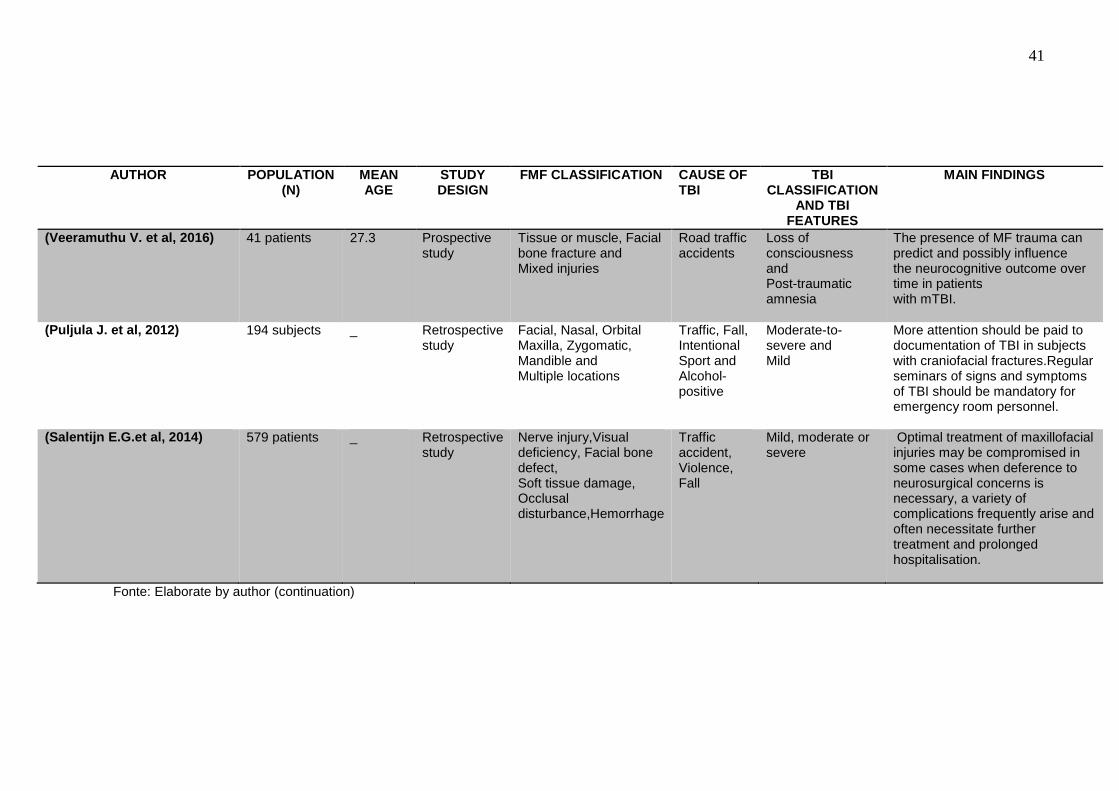
(Veeramuthu V. et al, 2016)
41 patients
27.3
Prospective study
Tissue or muscle, Facial bone fracture and Mixed injuries
Road traffic accidents
Loss of consciousness and Post-traumatic amnesia
The presence of MF trauma can predict and possibly influence the neurocognitive outcome over time in patients with mTBI.
(Puljula J. et al, 2012)
194 subjects
_
Retrospective study
Facial, Nasal, Orbital Maxilla, Zygomatic, Mandible and Multiple locations
Traffic, Fall, Intentional Sport and Alcohol-positive
Moderate-to-severe and Mild
More attention should be paid to documentation of TBI in subjects with craniofacial fractures.Regular seminars of signs and symptoms of TBI should be mandatory for emergency room personnel.
(Salentijn E.G.et al, 2014)
579 patients
_
Retrospective study
Nerve injury,Visual deficiency, Facial bone defect, Soft tissue damage, Occlusal disturbance,Hemorrhage
Traffic accident, Violence, Fall
Mild, moderate or severe
Optimal treatment of maxillofacial injuries may be compromised in some cases when deference to neurosurgical concerns is necessary, a variety of complications frequently arise and often necessitate further treatment and prolonged hospitalisation.
Fonte: Elaborate by author (continuation)

42
Fonte: Elaborate by author (continuation)
AUTHOR POPULATION (N)
MEAN AGE
STUDY DESIGN
FMF CLASSIFICATION CAUSE OF TBI
TBI CLASSIFICATION AND TBI FEATURES
MAIN FINDINGS
(Cavalcante, J.R. et al, 2012)
272 patients
_
Prospective study
Lip injury, Eyelid injury, Facial injury and Extensive injury in the face
Fall on dirt road, Fall on paved road, Collision with car Collision with motorcycle, Collision with bicycle, Collision with animal, Collision with pedestrian
Moderate traumatic brain injury
Individuals in the most productive age group are most affected and It is important to alert the population regarding the severity of injuries likely to occur with motorcycle accidents.
(Živković, V. et al, 2014)
50 cases
70.0
Prospective study
Orbital roof fractures
Ground level falls
Periorbital hematoma, Conjunctival hemorrhages, contusions, Fractures of skull, Subdural and Epidural hematoma; Skin lacerations.
In autopsy in cases of fall,the occurrence of orbital roof fractures was not significantly associated with concomitant skull fractures and brain injuries.
(Kraus, J.F. et al, 2003)
5,790 motorcycle riders
28.7
Cohort
Mandible, Maxilla, Nose Zygoma, Orbit, All bones
Traffic accident and Violence
Skull fracture
The findings support previous research demonstrating an association between facial fractures and traumatic brain injury.

43
AUTHOR POPULATION (N)
MEAN AGE
STUDY DESIGN
FMF CLASSIFICATION
CAUSE OF TBI
TBI CLASSIFICATION AND TBI FEATURES
MAIN FINDINGS
(Chu, Z.G. et al, 2011)
221 cases
35.0
Retrospective study
Mandible: Symphysis, Parasymphysis, Body, Ramus, Condyle, Maxilla: Posterolateral wall of maxillary sinus, Anterior wall of maxillary sinus, Medial wall of maxillary sinus, Palatine process Zygoma: Body, Arch Orbit: Lateral wall, Medial wall, Inferior wall, Superior wall
Earthquake
Frontal, Parietal,Temporal,Occipital and Sphenoid bone; Intracranial injury:hematoma, haemorrhage,Pneumocephalus, Cerebral edema,Cerebral contusion and laceration.
Our results indicate that the cranio-maxillofacial trauma arising from the massive Sichuan earthquake had some characteristic features, and a significant number of individuals had the potential for combined cranial and maxillofacial injuries, successful management of which required a multidisciplinary approach.
(Huang, W. et al, 2012)
45 patients
27.9
Cohort
Condyle-ramus
Not mentioned
Severe Tramatic Brain Injury
The patients with severe TBI and mandibular fractures exhibit increased fracture healing as evidenced by early callus formation.
(Fares, Y. et al, 2014)
417 casualties
27.0
Prospective study
Facial nerve,Eyes, Skin,soft-tissue lesions
Cluster munitions
pTBI, cTBI and scalp
Due to cluster bombs can inflict injuries to the head and face regions upon and patients suffer from multiple injuries at the same time. Psychological tribulations are often strong post incidence.
44
Fonte: Elaborate by author (continuation)
AUTHOR POPULATION (N)
MEAN AGE
STUDY DESIGN
FMF CLASSIFICATION CAUSE OF TBI
TBI CLASSIFICATION AND TBI FEATURES
MAIN FINDINGS
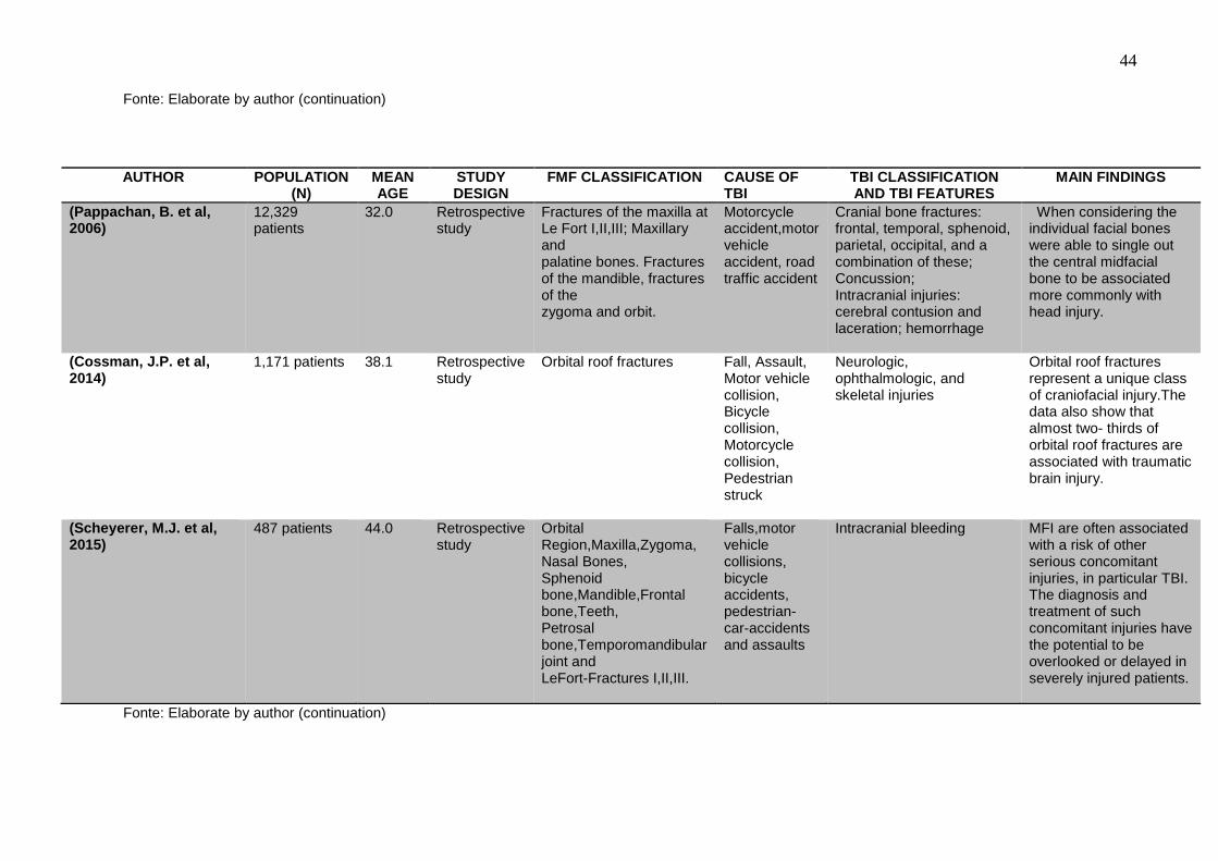
(Pappachan, B. et al, 2006)
12,329 patients
32.0
Retrospective study
Fractures of the maxilla at Le Fort I,II,III; Maxillary and palatine bones. Fractures of the mandible, fractures of the zygoma and orbit.
Motorcycle accident,motor vehicle accident, road traffic accident
Cranial bone fractures: frontal, temporal, sphenoid, parietal, occipital, and a combination of these; Concussion; Intracranial injuries: cerebral contusion and laceration; hemorrhage
When considering the individual facial bones were able to single out the central midfacial bone to be associated more commonly with head injury.
(Cossman, J.P. et al, 2014)
1,171 patients
38.1
Retrospective study
Orbital roof fractures
Fall, Assault, Motor vehicle collision, Bicycle collision, Motorcycle collision, Pedestrian struck
Neurologic, ophthalmologic, and skeletal injuries
Orbital roof fractures represent a unique class of craniofacial injury.The data also show that almost two- thirds of orbital roof fractures are associated with traumatic brain injury.
(Scheyerer, M.J. et al, 2015)
487 patients
44.0
Retrospective study
Orbital Region,Maxilla,Zygoma, Nasal Bones, Sphenoid bone,Mandible,Frontal bone,Teeth, Petrosal bone,Temporomandibular joint and LeFort-Fractures I,II,III.
Falls,motor vehicle collisions, bicycle accidents, pedestrian-car-accidents and assaults
Intracranial bleeding
MFI are often associated with a risk of other serious concomitant injuries, in particular TBI. The diagnosis and treatment of such concomitant injuries have the potential to be overlooked or delayed in severely injured patients.
Fonte: Elaborate by author (continuation)

45
AUTHOR POPULATION (N)
MEAN AGE
STUDY DESIGN
FMF CLASSIFICATION CAUSE OF TBI
TBI CLASSIFICATION AND TBI FEATURES
MAIN FINDINGS
(Lopes Albuquerque, C.E. et al, 2014)
253 motorcyclists
29.64
Cohort
Mandible, midface, upper face and facial lacerations
Motorcycle accident
Loss of consciousness, loss of memory, alteration in mental state, focal neurologic deficits transient or not.
The large part of motorcyclists had MF and TBI, and crash helmets did not always offer adequate protection against MFI, especially open-face helmets.
(Huang L.K. et al, 2017)
1,649 patients
53.1
Cohort
Facial wound, Tooth rupture, Epistaxis, Fractures of the Upper face, Midface,Lower face, Orbital fracture, Fracture of the Nasal bone, Zygoma, Maxilla, Mandible.
Fall from elevation, Motorbike collision
Intracranial hemorrhage, Skull fracture, Facial swelling/hematoma
TBI patients with risk factors have a higher probability of concomitant MF. Fractures of the lower face and orbit are easily overlooked but often require surgical intervention. Therefore, imultaneous head and facial CT scans are suggested in TBI patients.
(Czerwinski, M. et al, 2018)
181 medical records
35.0
Retrospective study
Mandible fractures: parasymphyseal and symphyseal, condylar, subcondylar, condyle, and condylar neck.
Motor-vehicle collision, fall from height, struck by vehicle, gunshot wound.
Mild, moderate and severe TBI, hemorrhage, hematoma, diffuse axonal injury.
Was suggest an algorithm, based on the results of the study, to help determine the need for head CT scans in trauma patients who sustain a mandibular fracture.
Fonte: Elaborate by author (continuation)
46
AUTHOR POPULATION (N)
MEAN AGE
STUDY DESIGN
FMF CLASSIFICATION
CAUSE OF TBI
TBI CLASSIFICATION AND TBI FEATURES
MAIN FINDINGS
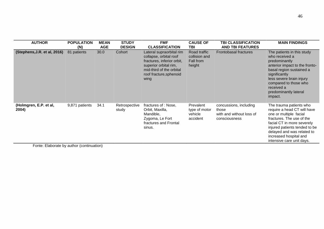
(Stephens,J.R. et al, 2016)
81 patients
30.0
Cohort
Lateral supraorbital rim collapse, orbital roof fractures, inferior orbit, superior orbital rim, mid-third of the orbital roof fracture,sphenoid wing
Road traffic collision and Fall from height
Frontobasal fractures
The patients in this study who received a predominantly anterior impact to the fronto-basal region sustained a significantly less severe brain injury compared to those who received a predominantly lateral impact.
(Holmgren, E.P. et al, 2004)
9,871 patients
34.1
Retrospective study
fractures of : Nose, Orbit, Maxilla, Mandible, Zygoma, Le Fort fractures and Frontal sinus.
Prevalent type of motor vehicle accident
concussions, including those with and without loss of consciousness
The trauma patients who require a head CT will have one or multiple facial fractures. The use of the facial CT in more severely injured patients tended to be delayed and was related to increased hospital and intensive care unit days.
Fonte: Elaborate by author (continuation)

47

48
AUTHOR POPULATION (N)
MEAN AGE
STUDY DESIGN
FMF CLASSIFICATION
CAUSE OF TBI
TBI CLASSIFICATION AND TBI FEATURES
MAIN FINDINGS
(Wanyura, H. et al, 2014)
11 patients
NM
Prospective study
Frontal sinus, supraorbital bar, frontal bone
Impaction with wood in sawmill,Beating – wound from sharp object, Beating, Car accident, Crushed by wooden ball, Beat by horse head, Beat by brunch of a tree.
Concussion, Pneumocranium, LCR leak, epidural haematoma, subdural haematoma.
The three-layer osteodural plasty of severe anterior skull base injuries with the use of autologous bone grafts for the reconstruction of craniofacial skeleton resulted in a good final functional, morphological and aesthetic outcome in all patients.
(Aladelusi, T. et al, 2014)
201 patients
30.3
Prospective study
Soft tissue ,Maxilla, Mandible, Zygoma, Multiple fracture,Naso-orbito-ethmoidal
Road traffic crashes
Mild, moderate, severe head injuries and Cranial fracture.
The study showed a direct relationship between the severity of maxillofacial injury and head, ocular and polytrauma and further emphasizes the need for thorough examination of patients presenting with road traffic crashes related maxillofacial injuries.
(Sobin, L. et al, 2016)
562 patients
27.5
Prospective study
Mandible fractures: body, symphyseal, parasymphyseal, subcondylar, angle, ramus.
Assault, all-terrain vehicle,bike, Sport.
Concussion
The concussive symptoms have a high index of suspicion for mTBI and are associated with isolated mandible fractures was identified.
Fonte: Elaborate by author (continuation)

49
AUTHOR POPULATION (N)
MEAN AGE
STUDY DESIGN
FMF CLASSIFICATION CAUSE OF TBI TBI CLASSIFICATION AND TBI FEATURES
MAIN FINDINGS
(Gönül, E. et al, 2005)
402 patients
20.0
Prospective study
Orbital bone
Gunshot
Anterior cranial fossa,middle fossa and posterior fossa injuries
The prognosis of the injury depends on the course of the bullet or shrapnel fragment and the interdisciplinary care. An extensive preoperative evaluation of penetrating orbital trauma and a combined ophthalmic and neurosurgical approach are recommended to minimize the morbidity of the patients.
(Razak, N. A. et al, 2017)
361 medical records
33.37
Retrospective study
Facial injury: Abrasion, Contusion/Haematoma, Laceration
Road Traffic Crashes,Sports, Industrial, Domestic, Fall and Assault
mTBI
Mild traumatic brain injury should be suspected in patients with facial injuries and particularly those with lower face lacerations, midface fractures, moderate to severe facial injury and presence of multiple injuries.
(Hammond, D. et al, 2018)
500 patients
NM
Retrospective study
Not mentioned
Not mentioned
Concussion and severe TBI
Awareness of concussion and itsclinical relevance should also be covered for oral and maxillofacial surgeons trainees.
Fonte: Elaborate by author (continuation)
50
AUTHOR POPULATION (N)
MEAN AGE
STUDY DESIGN
FMF CLASSIFICATION
CAUSE OF TBI
TBI CLASSIFICATION AND TBI FEATURES
MAIN FINDINGS
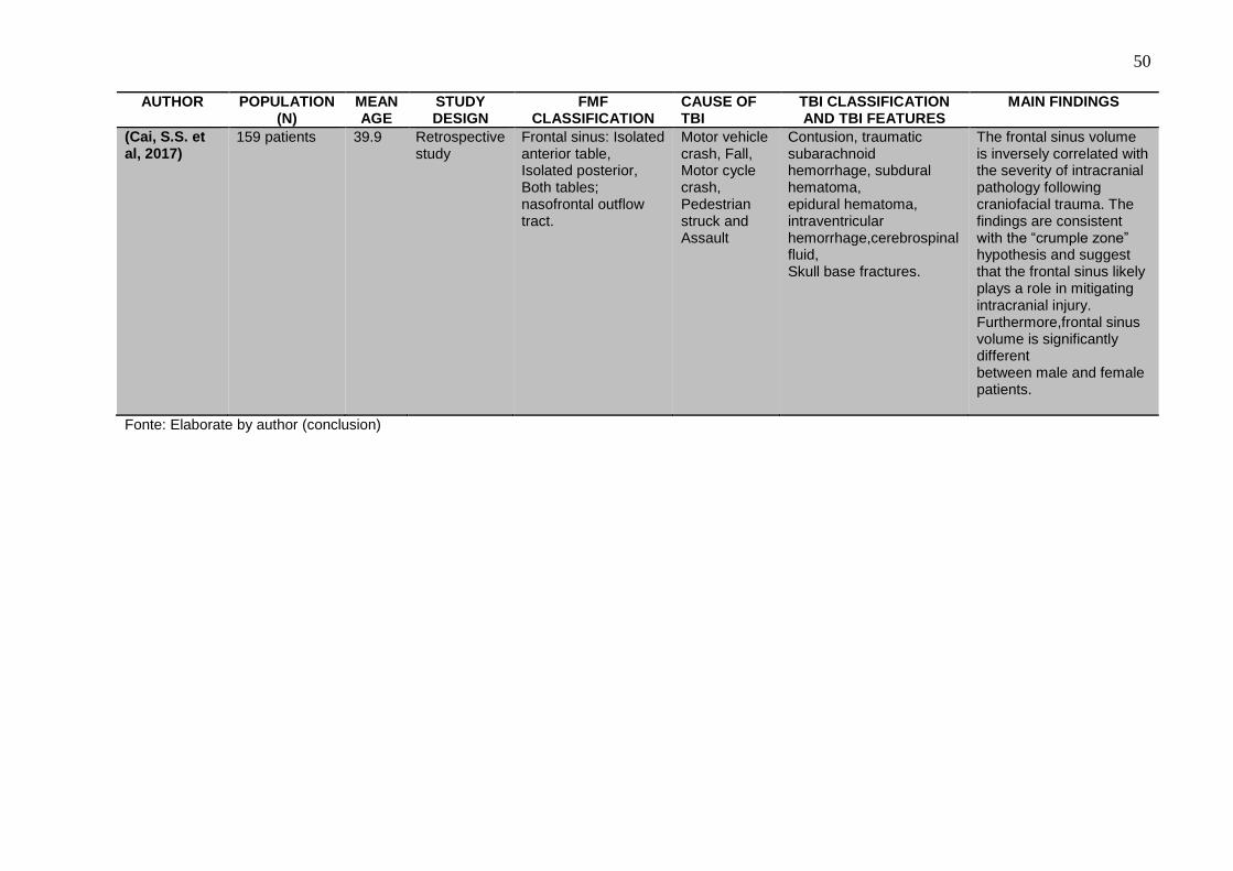
(Cai, S.S. et al, 2017)
159 patients
39.9
Retrospective study
Frontal sinus: Isolated anterior table, Isolated posterior, Both tables; nasofrontal outflow tract.
Motor vehicle crash, Fall, Motor cycle crash, Pedestrian struck and Assault
Contusion, traumatic subarachnoid hemorrhage, subdural hematoma, epidural hematoma, intraventricular hemorrhage,cerebrospinal fluid, Skull base fractures.
The frontal sinus volume is inversely correlated with the severity of intracranial pathology following craniofacial trauma. The findings are consistent with the “crumple zone” hypothesis and suggest that the frontal sinus likely plays a role in mitigating intracranial injury. Furthermore,frontal sinus volume is significantly different between male and female patients.
Fonte: Elaborate by author (conclusion)