Embed Size (px)
DESCRIPTION
Pashubandha eBulliten
Citation preview

Newsletter Date : 31st May 2013 Volume No : 02 Issue : 05
Veterinary College, Bengaluru Monthly e-Bullletin
Pashubandha 2013 Volume No : 02 Issue : 05
Raveendra Hegde, B.M.chandranaik, P. Giridhar and M.D. Venkatesha ([email protected])
Institute of Animal Health and Veterinary Biologicals, KVAFSU, Hebbal, Bangalore
Malignant catarrhal fever (MCF) is a frequently fatal disease of certain ruminant species, caused by a herpesvirus, to which these ruminant species are poorly adapted. The disease is characterized by inflammation, ulceration, and exudation of the oral and upper respiratory mucous membranes, and sometimes eye lesions and nervous system disturbances. The causative viruses exist in nature as subclinical infections in other species that serve as carriers, to which they are well-adapted. Two major epidemiologic forms of MCF are recognized, defined by the reservoir ruminant species from which the causative virus arises. One, known as the African form, is referred to as wildebeest-associated MCF (WA-MCF). The other is referred to as sheep-associated MCF (SA-MCF).
Etiology: Malignant catarrhal fever is caused by several viruses in the genus Rhadinovirus of the family Herpesviridae (subfamily Gammaherpesvirinae). The MCF subgroup of viruses (MCFV or type 1 RuRV) contains at least ten members, five of which are known to cause disease. The best characterised causative agents of MCF are the two γ-herpesviruses, alcelaphine herpesvirus 1 (AlHV-1) and ovine herpesvirus 2 (OvHV-2). AlHV-1 is present in wildebeest and is a known cause of WA-MCF. OvHV-2 is present in sheep and is a known cause of SA-MCF. Caprine herpesvirus 2 (CpHV-2) is endemic in most domesticated goats and can cause MCF in cervids. A virus of unknown origin, currently called MCFV-WTD, has also been associated with MCF in white-tailed deer. An interesting feature of MCF is that the natural reservoir species (wildebeest and sheep) do not exhibit any clinical signs of infection, whereas the disease is usually fatal in MCF-susceptible species, which are phylogenetically related to reservoir hosts (eg:cattle).
Species Affected: MCF viruses are carried asymptomatically by their reservoir hosts, but can cause disease in other species.
Fodder Crop
Siratro
Varieties -
Season June-July
Oct-Dec
Duration Perennial
Seed rate/acre
3kg/acre
Space Broadcasting
Irrigation
At sowing &
Once in 15days
Stage of
harvest
Flowering Stage
Yield 250 Qtls/year
Protein content
18%
No. of cutting
6 cutting/year
Fodder/animal/day
5kgs
FODDER BOX

The effect of heavy metals on
Pashubandha 2013 Volume No : 02 Issue : 05
All or most wildebeest in the wild and in zoos appear to be infected by AlHV-1.virus. Most sheep are infected with OvHV-2, and most goats are infected with CpHV-2. The normal host for MCFV-WTD is not known. Other exotic ruminants known to have MCFV include ibex, musk ox, gemsbok/ South African oryx, aoudads, hartebeest, topi and roan antelope. There are excellent animal models of MCF. Rabbits and hamsters can be infected with AlHV-1 or OvHV-2 and develop a MCF-like syndrome that is very similar to that seen in species naturally susceptible to MCF. A feature of MCF, with respect to cattle, is that many outbreaks are sporadic, with single or only a few individuals in a herd being affected. However, occasionally there are more serious outbreaks that can affect up to 40 per cent of a herd. The reasons for this are not known. Certain species of deer, (e.g., Père David's), Bali cattle and bison appear to be particularly susceptible to MCF in terms of rapid death following clinical signs and the proportion of animals affected in closed herds. Clinical disease occurs in even-toed ungulates. Most susceptible species are cattle, bison, water buffalo and exotic ruminants such as antelope, guar, banteng , deer, reindeer, moose, but other species such as giraffes and pigs are also affected. Susceptibility varies with the virus and the host. Most cattle are susceptible to AlHV-1 but are relatively resistant to the sheep-associated (OvHV-2) form of MCF. Water buffalo and some species of deer are more susceptible to OvH-2, and bison, Père David's deer, white-tailed deer, axis deer and Bali cattle are highly susceptible to this virus. Sheep-associated MCF has also been reported in pigs and lesions have been reported in goats. CpHV-2 associated disease has been reported in cervids including moose, roe deer, sika deer and white-tailed deer.
Geographic Distribution:
MCF is present anywhere either of the two principal carrier hosts, the domestic sheep or wildebeest, are present. Since wildebeest are present in Africa and elsewhere only in zoos, game farms or zoological gardens, the determining factor for most of the world’s MCF is the presence or absence of domestic sheep, which are universally infected with one of the causative viruses.
Sheep exist in virtually all countries, thus the distribution of MCF is worldwide. Reports exist which document its presence in North and South America, Africa, virtually all countries of Europe, Indochina, Japan, Australia, New Zealand, Indonesia, Israel, Russia, the Philippines, and many other countries. It safely can be assumed that the distribution of the disease is virtually universal. Recently the disease has been confirmed in Karnataka state indicating the circulation of virus both in bison and sheep.
Morbidity and Mortality:
In cattle, malignant catarrhal fever usually occurs sporadically in one or to a few animals, but larger outbreaks may also be seen. Morbidity rates of 28 to 45 percent have been reported in some outbreaks. The mortality rate is classically described to be 90-100 per cent in symptomatic animals, but a few studies have suggested that up to 35 per cent of cattle may recover from disease. Subclinical and chronic cases, including cattle with skin disease, have also been reported. Residual corneal opacity is often seen in recovered cattle, but one cow without ocular lesions was reported to make a complete recovery.
The clinical course is usually shorter in bison, however the morbidity and mortality rate can be very high. During a recent outbreak in the U.S., approximately half of the bison (800 animals) died in one feedlot. In another herd pastured more than a kilometer from a sheep feedlot, nearly all of the bison died over two years. The incidence of clinical disease in wild cervids is unknown, but the sero-prevalence rate in wild deer, moose and ruminants usually ranges from 0 to 9 per cent.

The effect of heavy metals on
Pashubandha 2013 Volume No : 02 Issue : 05
Pigs may recover from malignant catarrhal fever. In one pig farm, the most severe cases and deaths were reported early in the outbreak; pigs infected later had milder signs and tended to recover without treatment.
Transmission:
Like other herpesviruses, MCF virus establishes lifelong, latent infections. AlHV-1 is transmitted mainly by wildebeest calves, which can become infected in utero, by direct contact with other wildebeest, or in aerosols during close contact. Contamination of pastures may also contribute to transmission. Infected calves, particularly animals one to two months of age, shed the virus in nasal and ocular secretions. Wildebeest calves over the age of six months rarely shed virus. In these animals and in adult wildebeest, AlHV-1 occurs mainly in a cell-associated, rarely transmitted form; however, cell-free virus can be isolated from the nasal secretions of some animals that are stressed or given corticosteroids. Most cases of wildebeest-associated MCF are seen when susceptible animals are exposed to parturient wildebeest or young calves. Close contact is usually necessary, but transmission has been reported when the animals were separated by at least 100 meters. Cell-associated MCFV is very fragile, and infectivity disappears after 72 hours in the environment; however, cell-free virus can survive for more than 13 days in humid environments. MCF viruses are inactivated quickly by sunlight.
OHV-2 appears to be transmitted mainly by the respiratory route, probably in aerosols. This virus is shed intermittently in nasal secretions, particularly by 6 to 9 month old lambs. OHV-2 DNA has also been reported in the semen of rams. OHV-2 is rarely transmitted transplacentally or in colostrum or milk; most lambs do not become infected until they are at least two months of age. Susceptible animals usually become infected when they are in close contact with sheep, but cases have been reported when sheep and cattle were separated by 70 meters, as well as in bison herds up to 5 km from a lamb feedlot. Cattle-to-cattle, bison-to-bison, or deer-to-deer transmission is rare, and these species are considered dead end hosts once infected with OHV-2 or AHV-1.
Clinical Signs:
The transmission of one of the virulent strains of MCF viruses from their carrier hosts to clinically-susceptible ruminants can initiate the syndrome of classic malignant catarrhal fever, which is an acute polysystemic disease characterized by lymphoproliferation and inflammation oriented toward mucosal surfaces and blood vessels. The clinical signs depend to some extent on the species infected, the virus and how long the animal survives after the onset of clinical signs. Experimental infections have an incubation period of 9 to 77 days, but the incubation period is unknown for natural infections. Some animals are subclinically infected and develop disease when they become stressed. Malignant catarrhal fever is a broad syndrome with a variety of symptoms in different species. Clinical MCF is very similar whether it is caused by OvH-2 or AlHV-1.
MCF clinical presentations were divided into 4 “forms” by Gotze ( 21 ): 1) peracute, 2) head and eye, 3) alimentary, and 4) mild. It should be borne in mind that rather than being distinct and clearly delineated, there is much overlap between the ‘forms’. Sudden death can occur in the peracute form. The head and eye form is the most common expression of disease in cattle. It progresses through the early signs of fever, reddened mucosa, and enlarged prescapular lymph nodes. Eventually the lesions become necrotic and death can occur. The intestinal form has the same early signs as the head and eye form, but the animal dies of severe diarrhea before the lesions become necrotic.
The disease presentation is sudden, with little preliminary indications of illness. Although there are some rather constant signs, there is also considerable variability in presentation. Typical signs in cattle include sudden fever, drop in milk production, inappetance, serous discharge from the eyes and nose with matting of facial hair.

The effect of heavy metals on
Pashubandha 2013 Volume No : 02 Issue : 05
Temperature may spike to 106 oF for a day or two before declining to 103 to 104 oF. Within a day or two of initial signs, corneal edema appears, typically starting around the limbus and spreading centrally. Episcleral injection, lid swelling and sensitivity to light are common. Deep corneal inflammation frequently progresses to blindness within 4 to 5 days. Prolonged inflammation of the cornea can terminate in perforation and herniation of the iris. Nasal exudate becomes mucoid in character within a few days, with mucopurulent discharges from the nose, stertorous breathing, and often dyspnea. Muzzle epithelium is initially inflamed and later necrotic, resulting in encrustation, cracking and often dislodging of affected epithelial patches to reveal the underlying inflamed subepithelium. At this stage, animals are usually severely depressed, and may separate from herd mates and stand immobile with head hanging. CNS involvement is common, however, and can lead to hyperexcitability, aggressiveness, twitching, incoordination, nystagmus, and muscular tremors.
Skin lesions are common in cattle and deer with the ovine strain of MCFV, and with the recently-described caprine strain, but less so with the wildebeest strains. Areas of erythema and exudation may be found in any area of the body, including the bulbs of the heels and between the digits. Small elevated circumscribed areas may be present on the udder or vulva. The skin of the teats and udder may become inflamed, then dry, thickened and may crack, leading to fissure formation and scabbing
Bison frequently dies of acute MCF without developing purulent rhinitis or keratoconjunctivitis. Unlike cattle, which develop prominently enlarged lymph nodes, lymphadenomegaly is usually minimal in Bison. Recumbent animals generally die within a few hours. Unusually, skin lesions have been the primary complaint in some deer infected with CpHV- 2,with lesions of widespread alopecia; thickening, crusting, hyperkeratosis, and focal ulceration of the skin; weight loss; and impaired vision. Deer and antelope may have minimal lesions or be less specific than cattle or bison, but many of the same signs occur.
Fever, anorexia, dyspnea, foul-smelling nasal discharge, erosions on the nasal and oral mucosa, abortions, stillbirths, smaller-than-normal litters, reddened foci on the skin, and sudden death have been reported in pigs. Neurological signs including ataxia, tremors, convulsions and hyperesthesia have also been seen. Most cases in swine are acute or peracute, but chronic cases have been reported in some outbreaks. Although younger pigs may be affected, most cases of MCF have been reported in gilts and sows. In one outbreak, only pregnant animals on the farm were af-fected. Post mortem lesions: In hosts with clinical disease, lesions can be found in any organ. The actual severity depends on the course of the illness. With sudden death, hemorrhagic enterocolitis in the infected animal may be the only sign. Erosions and necrotic areas appear throughout the omasum and intestinal tract. Gross findings at postmortem examination include petechial haemorrhages on the tongue, buccal mucosa, in the gastrointestinal and respiratory tracts and urinary bladder. The gastrointestinal tract can be affected, starting in the oral cavity ( erosions on the tongue and the hard and soft palate of a cow). he muzzle is often raw and encrusted with a serous, mucopurulent or purulent nasal discharge. Hyperemia, edema and small focal erosions or ulcers may be found on the nasal mucosa. Generalized exudation, crusting and matting of the hair is common on the ventral thorax and abdomen, inguinal region, perineum and sometimes the head. Skin ulcers may also be found. The lymph nodes are usually markedly enlarged in cattle. On cut surface, they may be firm and white, hemorrhagic or necrotic.

The effect of heavy metals on
Pashubandha 2013 Volume No : 02 Issue : 05
Commonly, there will be raised pale foci on the surfaces of the kidneys and these may extend into the cortex. Ecchymotic hemorrhages, hyperemia and edema are common in the mucosa of the urinary bladder. In more chronic cases, the small arteries in the subcutaneous tissues, thorax, abdomen and central nervous system are very prominent and tortuous, with thickened walls. Fibrinous polyarthritis is also common. Diagnosis: Diagnosis of MCF depends on a combination of clinical signs, post-mortem findings, histopathology and detection of virus-specific antibodies in blood or DNA in blood or tissue samples. Clinical: Based on the clinical signs described, MCF should be suspected in susceptible animals if they have been exposed to sheep, goats, antelope or wildebeest, particularly around parturition. Animals that suddenly die or have a fever and erosions of the mucosa, nasal and lacrimal discharge, or bilateral corneal opacity should be tested for MCF. Laboratory tests The similarity of the clinical signs to other enteric or vesicular diseases, the lack of unique disease-specific clinical diagnostic features and the variability in the presentation of the disease make laboratory confirmation of a clinical diagnosis of MCF is must. Histopathological analysis of postmortem samples should give a clear diagnosis. The World Organisation for Animal Health (OIE) recognises histopathology as the definitive diagnostic test. PCR. Because some MCF viruses cannot be isolated from infected animals, polymerase chain reaction (PCR) tests have become the diagnostic method of choice. PCR can detect both AHV-1 and OHV-2, as well as other MCF viruses. The use of PCR allows sensitive confirmation of the presence of MCF viruses in infected animals and may also be useful for phylogenetic and epidemiological studies in both natural and MCF-susceptible hosts. EDTA-anticoagulated blood should be submitted from live animals for PCR on peripheral blood leukocyte DNA. Animals that are ill from MCF will have sufficient levels of viral DNA in their leukocytes and tis-sues to detect readily by PCR. Careful PCR primer selection by the lab is important, as PCR tests are generally specific for a given strain of virus. Serology. A broad array of serological assays has been used to detect antibody against MCF viral antigens. Serologic tests that have been used include viral neutralization, complement fixation, indirect immunofluorescence or immunohistochemistry, direct-binding ELISA, and competitive-inhibition ELISA (CI-ELISA). Viral neutralization tests utilize the Alcelaphine virus for a neutralization target, and are reliably specific for the MCF group of viruses. Virus isolation: AlHV-1 infections, can be confirmed by virus isolation in bovine thyroid cells or other susceptible cell lines. The virus can be recovered from peripheral blood leukocytes, lymph nodes or other affected tissues. Viable cells are necessary, as the virus cannot be isolated from dead cells. Cell-free virus is usually found only in nasal swabs from wildebeest calves. Isolation of OvHV-2 is unsuccessful.

The effect of heavy metals on
Pashubandha 2013 Volume No : 02 Issue : 05
Samples to collect: In order to isolate the virus, 10-20 mls of blood should be collected and put in an EDTA tube. The virus is quickly inactivated in dead animals so spleen, lung, lymph nodes, and adrenal glands should be collected as soon as possible, refrigerated (NOT FROZEN), and shipped on ice to laboratory for virus isolation, fluorescent antibody, or immunoperoxidase tests. In order to run a PCR test, peripheral blood, fresh tissues, or paraffin-embedded tissue samples must be used. Samples of lung, liver, lymph nodes, skin (if lesions are present), kidney, adrenal gland, eye, oral epithelium, esophagus, Peyer’s patches, urinary bladder, carotid rete, thyroid and heart muscle (and, if practical, brain) should be submitted for histopathology in affected cattle. Samples for histopathology should be fixed in 10% neutral buffered formalin. As some animals have antibodies to the virus, paired sera should be used to identify an infection. Single samples are of limited value and serum should be taken 3 to 4 weeks apart, if possible, for serology. Differential diagnosis: Malignant catarrhal fever must be differentiated from bovine virus diarrhea (BVD) mucosal disease, bluetongue, rinderpest, infectious bovine rhinotracheitis, vesicular diseases such as foot and mouth disease and vesicular stomatitis, ingestion of caustic materials, and some poisonous plants and mycotoxins. This disease should also be included among the differentials in susceptible ruminants with an undiagnosed subacute or chronic disease, particularly when alopecia or weight loss is present. Treatment: Mortality in clinically ill animals is nearly 100 per cent and survival in other exposed animals is rare. Supportive therapy (fluids) and antibiotics for secondary bacterial infections can be tried for valuable animals. If recovery occurs, animals will likely remain virus carriers and could spread infection. Stress reduction can help prevent disease in subclinically or mildly affected animals. Prevention and Control: If a case of MCF is suspected, the state authorities should be notified immediately. Animals suspected with MCF should be isolated, and the farm should be quarantined until definitive diagnosis is determined. Should an epidemic occur, clinical and carrier animals should be separated from susceptible species. As domestic sheep and goats are asymptomatic carriers, they should be kept separated from cattle at all costs, especially during parturition. African wildlife, wildebeests, hartebeests, and topi, should also be kept separated from cattle to limit the spread of infection. Zoological parks should only introduce seronegative animals and follow strict quarantine restrictions of newly acquired animals. There is no vaccine currently available, but experimental evidence in cattle has shown some protection from challenge inoculation. Transmission on fomites must be avoided, particularly when the species is highly susceptible. MCF viruses are susceptible to most commonly available disinfectants. Cell-associated MCFV is very fragile, and infectivity disappears after 72 hours in the environment. Cell-free virus can survive for more than 13 days in humid environments, but disappears quickly when the humidity is low. Conclusions: Malignant catarrhal fever is an important and fascinating disease with many unanswered questions concerning transmission, the sporadic occurrence of the disease and pathogenesis. The importance of MCF as a pathogen of farmed deer and bison, as well as cattle, is driving research for improved diagnostic tools and development of effective vaccines. The recent confirmation of MCF in Karnataka needs further detailed serological study and research in this area.

The effect of heavy metals on
Pashubandha 2013 Volume No : 02 Issue : 05
B. N. Nagaraja, Sangeeta Jadhav, Ramesh Rathod, D. R. Manjunath and A. S. Patil Department of Veterinary Surgery, Veterinary College, Hebbal, Bangalore.
([email protected] ) Introduction: Caecal dilatation and torsion represents a common and economically important abdominal disorder that affects mainly dairy cattle. Caecotomy is indicated in cattle suffering from dilation and torsion of the caecum and ansa proximalis of the colon. In some cases only dilation is present, but usually torsion also occurs; this may be clockwise or counterclockwise seen from the animal's right side. Anorexia, not passing dung and distension of right flank with decreased milk production are prominent features. Dilatation, displacement and torsion of caecum occur due to atony of caecum, accumulation of ingesta and gas inside the caecum. Prognosis for cattle with caecal dilatation and volvulus is good following medical therapy or decompression at surgery unless there was severe vascular compromise of the caecum. Etiology: 1. Excessive feeding of grains plays significant role as this results in increased production of volatile fatty
acids and gas due to fermentation of undigested grains.
2. Free end of Ceacum in cattle is devoid of mesentery and thus prone to rotation.
3. History of parturition.
4. It may also be due to difficult calving or iatrogenic rectal perforation
Clinical signs:
Anorexia, reduced milk yield, passing scanty dung, kicking at the abdomen with signs of colic, Per rectal examination revealed mucus mixed scanty dung. Distension of right paralumbar fossa, upon auscultation and percussion tympanic resonance will be heard. Clinical signs match with that of intestinal obstruction. Temperature, heart rate and respiration are normal in cases of simple dilatation but in advanced cases hypothermia and tachycardia are noticed. Reduction of milk yield is more pronounced and feces are absent or very sparse and of dry consistency covered with mucus.
Diagnosis:
History, clinical signs, tympanic resonance sound is heard on auscultation and percussion, The distended cecum is identified through rectal examination. Biochemical and haematological examination of blood shows rise in blood urea, total proteins, creatinine, inorganic phosphorus and packed cell volume with decreased levels of Serum chloride and potassium levels along with metabolic alkalosis.
Differential Diagnosis: intestinal obstruction, intussusceptions.
Treatment:
1. Conservative treatment consisted of a continuous drip infusion containing neostigmine and potassium chloride.
2. Administration of purgatives for caecal dilatation and tortion such as liquid paraffin, sometimes in combination with sodium sulphate.
3. Surgery - Right flank laparotomy
The effect of heavy metals on
Pashubandha 2013 Volume No : 02 Issue : 05
Cecomtomy (typhlotomy) and evacuation of intestinal contents is usually the surgical treatment of choice. Cecal amputation is indicated only in cases of recurrence or in cases of devitalization of the cecal wall. Typhlotomy:
Surgery is performed through a right flank approach, preferably in the standing animal under local anaesthesia. The abdomen is opened through a 25 cm long incision starting dorsally about 8 cm below the lateral processes of the lumbar vertebrae and 8 cm cranial to the tuber coxae, extending slightly oblique in a cranioventral direction parallel to the internal oblique abdominal muscle. The abdomen is then thoroughly explored and the cecum, PLAC, and spiral colon positions identified. The cecum and as much of the PLAC as possible are exteriorized by gently pushing from the inside toward the outside of the abdomen with the palm(s) of one or both hands in order to reduce the risk of rupture and/or perforation of the distended bowel. The apex of the cecum is isolated from the rest of the abdomen, and a typhlotomy is performed at the most ventral location. Digesta are first passively drained from the extraabdominal part of the cecum and then gently milked out from the intraabdominal part of the cecum and the PLAC to the incision site. The exteriorized cecum is rinsed with copious amounts of prewarmed 0.9% saline solution and the incision site closed with a simple inverting continuous or an inverting seromuscular suture pattern (i.e. Cushing or Lembert), using size USP 3-0 monofilament resorbable suture material. The exteriorized sections are again copiously rinsed and placed back into their physiologic position within the supraomental recess. The cecum is evaluated again 10 minutes later; and if it has refilled, a second typhlotomy is done to relieve the cecum and PLAC of digesta that may have accumulated within these segments by propulsion from the ileum or reflux from the spiral colon. The typhlotomy site is finally oversewn twice. Closure of the abdominal wall is performed in a routine manner. Postoperatively, bethanechol (0.07 mg/kg Bwt, SC, TID, for 2 days) may be administered to help restore intestinal motility.Antimicrobials (for example sodium penicillin, 30,000 IU/kg Bwt, IV) are administered perioperatively. If necessary, intravenous or oral rehydration, correction of electrolyte imbalances, and treatment of ketosis should be performed. Operated cows are set on a restrictive diet for 24 to 48 hours, followed by a medium coarse forage ration of increasing quantity to finally reach the normal ration within 5 to 7 days.
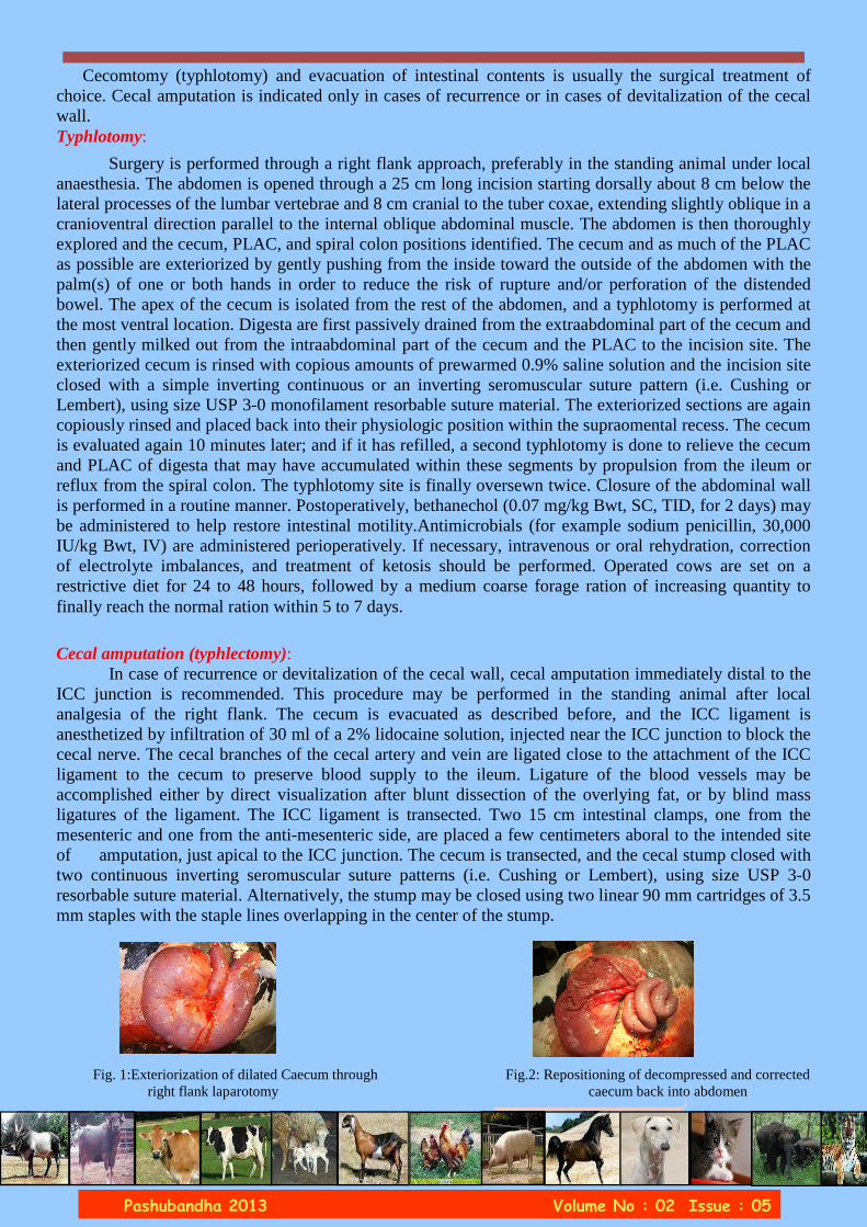
Cecal amputation (typhlectomy): In case of recurrence or devitalization of the cecal wall, cecal amputation immediately distal to the ICC junction is recommended. This procedure may be performed in the standing animal after local analgesia of the right flank. The cecum is evacuated as described before, and the ICC ligament is anesthetized by infiltration of 30 ml of a 2% lidocaine solution, injected near the ICC junction to block the cecal nerve. The cecal branches of the cecal artery and vein are ligated close to the attachment of the ICC ligament to the cecum to preserve blood supply to the ileum. Ligature of the blood vessels may be accomplished either by direct visualization after blunt dissection of the overlying fat, or by blind mass ligatures of the ligament. The ICC ligament is transected. Two 15 cm intestinal clamps, one from the mesenteric and one from the anti-mesenteric side, are placed a few centimeters aboral to the intended site of amputation, just apical to the ICC junction. The cecum is transected, and the cecal stump closed with two continuous inverting seromuscular suture patterns (i.e. Cushing or Lembert), using size USP 3-0 resorbable suture material. Alternatively, the stump may be closed using two linear 90 mm cartridges of 3.5 mm staples with the staple lines overlapping in the center of the stump. Fig. 1:Exteriorization of dilated Caecum through Fig.2: Repositioning of decompressed and corrected right flank laparotomy caecum back into abdomen

The effect of heavy metals on
Pashubandha 2013 Volume No : 02 Issue : 05
Dr. M.C. Anilkumar, Dr. Ansar Kamran and Dr. P.T. Ramesh Dept. of Veterinary Medicine, Veterinary College, Hebbal, Bengaluru
Synonyms: Black leg, Emphysematous gangrene. Definition:
It is the infectious disease of cattle, buffaloes and sheep caused by Clostridium Chauvoei and is
characterized by high fever, emphysematous serohaemorrhagic swelling of heavy muscles and lameness.
Etiology:
• It is caused by Clostridium Chauvoei.
• It is Gram positive bacilli. It is motile by peritrichous flagella
• It is anaerobic and spore forming bacteria.
• The spores are highly resistant to the environmental conditions and persist in the soil for many years as it is soil borne organism.
Occurrence: It is world wide in occurrence and causes severe economic loss.
Cattle and sheep are commonly affected. Cattle of age group 6 months to 2 years and rapidly
growing on high plane of nutrition are highly susceptible. Sheep of any age group can be affected.
BQ commonly occurs every year immediately after the onset of monsoon rains where there will be
sudden change in the weather condition which will act as a stress on the body.
Transmission:
It is soil borne infection. The portal route of entry is through ingestion of contaminated feed and
water in cattle and through the wounds in sheep due to shearing, docking and lambing.
Clinical signs:
In per acute form the animal may die suddenly without showing any clinical signs. There can be
oozing of blood tinged fluid or blood from natural orifices which will clot immediately.
In acute form, very high fever (106 – 108 F), anorexia, decreased milk production, acute lameness
is seen. In the heavy muscles, there will be swelling which is hot and painful in the beginning and soon
becomes cold and painless. Crepitating sounds can occur upon palpation of this region. It can be
generalized where in entire body can be affected. The affected skin will be discoloured and becomes dry
and cracked. Later sloughing of skin and muscles can occur. During the final stages the animal will be
recumbent and coma and death occurs.
Necropsy findings:
Incision of affected muscle reveals dark, discoloured, swollen tissue with rancid odour, a metallic
sheen on the cut surface and excess of thin sero-sanguinous fluid containing bubbles of gas.

The effect of heavy metals on
Pashubandha 2013 Volume No : 02 Issue : 05
Diagnosis:
1. History of occurrence of BQ immediately after the onset of monsoon rains
2. By clinical signs of high fever, lameness and crepitating swellings of heavy muscles.
3. Do muscle biopsy, then make a smear, air dry and heat fix and perform Gram’s staining. If good
numbers of Gram positive bacilli are present then it is BQ.
4. Serological tests like FAT, ELISA can be used.
Treatment:
1. To overcome etiological agent administer penicillin @ 10,000-20,000 IU/kg b.wt i/m for 5-7 days
Metronidazole is effective against anaerobes can be administered along with penicillin @ 5-10 mg/kg
b.wt i/v.
2. To prevent effect of histamines administer antihistamines.
3. To reduce pyrexia, administer antipyretics.
4. To overcome toxemia, administer dexamethasone @ 0.5-2 mg/kg b.wt i/v
Prevention and control:
1. Good hygienic measures should be followed.
2. Isolate the affected animals and treat them.
3. Vaccinate the healthy animals with BQ vaccine (IAH & VB). It is formaline killed vaccine.
Dose: Cattle and buffaloes : 5-10 ml S/c
Sheep, goats and calves 2-3 ml S/c
Vaccinate before the onset of monsoon rains.
2. Raksha HS+BQ vaccine: Dose is 3 ml s/c
The above vaccine has to be given to calves at 6 months and ab ove of age and repeated annually.
In endemic areas repeat every 6 months.

The effect of heavy metals on
Pashubandha 2013 Volume No : 02 Issue : 05
Dr. Suchitra B R ([email protected])
Assistant Professor, Department of Veterinary Gynaecology and Obstetrics, Veterinary College, Hassan
Definition: Uterine torsion is defined as the rotation or twisting of the entire length of the uterus on its longitudinal axis. Classification of uterine torsion:
1. Based on degree of rotation of the uterus
90°, 180°, 270°, 360° torsion
90-180° rotation usually occurs during last few months of gestation and become evident at the time
of parturition while 180 - 360° uterine torsion is a severe condition often associated with obstruction of
the blood supply to the uterus and finally death of the fetus.
2. Based on the point of uterine torsion:
• Post-cervical (vaginal) torsion: occurs caudal to the cervix.
• Pre cervical uterine torsion occurs cranial to the cervix.
3. Based on the side of the rotation:
• Right side uterine torsion (clock wise)
• Left side uterine torsion (anti-clock wise)
Pre disposing factors: i. Instability of the bovine uterus, ii. An overlarge foetus iii. Confinement of the animal iv. Sloping pastures v. Possibly low calcium levels, vi. Manner in which the cow rises vii. Sudden push from another cow viii. Lack of tone to the uterus ix. Lack of fetal fluids x. Sudden fall xi. Energetic movements of the fetus during first stage labor.
Clinical symptoms: When uterine torsion occurs at the time of parturition the first clinical finding noticed by animal owners is non-progression of labor. Continuous straining, colic-like signs such as lying down and violent rolling, raised tail head, depressed feed intake, displaced vulval commissure, constipation, depressed lumbo sacral spine, restlessness, depression, tachycardia, non appearance of water bags or fetus through the vulva.
Diagnosis: 1. Based on clinical symptoms as mentioned above. 2.On per-vaginal examination spiral folds are palpable if the uterine torsion is post cervical.

The effect of heavy metals on
Pashubandha 2013 Volume No : 02 Issue : 05
3. Per-rectal examination can facilitate diagnosis through palpation of the broad ligaments which can be felt as a tight band.
Treatment: Generally treatment for uterine torsion can be divided into:
1. Correction of uterine torsion
2. Post correction management
1. Correction methods:
There are 3 different methods of relieving uterine torsion
1. Manual correction per vaginum
2. Rolling
a. Rolling alone
b. Rolling with per vaginum manipulation
c. Rolling with a plank - ‘Schäfers’ method
3. Surgical method/ caesarean section
2. Post correction management: Following correction, further obstetrical assistance is commonly required for delivery of the calf if
complete cervical dilatation has not taken place. Leave the cow for up to three hours to allow second stage
parturition to proceed. During this waiting period supplement the cow with 400-450 ml of calcium and
2-3 liters of DNS intra venously to minimise the chances of uterine inertia and also to hasten the cervical
dilatation. Secondary bacterial infections can be minimized by administering antibiotics.
Failure to deliver the calf at this stage would indicate surgical approach/caesarean section.
Rolling with a plank - ‘Schäfers’ method

The effect of heavy metals on
Pashubandha 2013 Volume No : 02 Issue : 05
Prognosis: Survival rate in torsion affected cows declines linearly (from 87 to 43%) with an increase in the
duration of torsion.
E. g: (90º-360º) of uterine torsion of short duration (<12-36 h) - good
(180º-360º) of torsion of long duration (>36-72 h) – poor
Long standing (>72 h) torsion of >180º- the prognosis will be gaurded
Calf mortality is typically high with uterine torsions, especially if the delay in consultation to clinics is beyond 36 hours, already separated placentomes, resulting in lack of blood supply to the uterus due to torsion and subsequent hypoxia.
Displacement of the vulval commissure seen in a case of uterine torsion (left: pre-correction; right: post-correction). Note discolouration around the anus
and increased skin tension between the anus and vulva.
Conclusion:
Uterine torsions are a common cause of veterinary attended dystocia associated with high calf mortality and dam culling rate. Further work should be targeted towards establishing risk factors for the condition so that management practices may be implemented to decrease the incidence of the condition.
Contact :
Dept of Veterinary and Animal Husbandry Extension Education
Veterinary College, Hebbal Bangalore
email: [email protected]
monthly e-Bulletin
Published and circulated by Veterinary College, Hebbal Bengaluru
Editor: Associate Editior:
Dean, Veterinary College, Hebbal, Bengaluru Head,Dept of Vety & Animal Husbandry Extension Education
Dr.S.Yathiraj (Ex-Officio) Dr.K.Satyanarayana (Ex-Officio)


![Engineering Mechanics[May2013]](https://img.pdfslide.net/doc/110x75/55cf8c955503462b138dea30/engineering-mechanicsmay2013.jpg)