Embed Size (px)
Citation preview

MCPHERSON EYE RESEARCH INSTITUTE 2014 ANNUAL REPORT 2015 CALENDAR
VISION SIGHT SIGHTEDNESS EYESIGHT SEEING EYE OCULUS LAMP PEEPER ORB WINKER OPTIC CONCEIVE ENVISAGE
ENVISION FANCY FEATURE IMAGE IMAGINE REALIZE VISUALIZE VISIONAL SEEABLE VISIBLE OPTICAL VISUAL OCULAR
VIEWABLE VISIBILITY VISUALITY VIEW SEE BEHOLD DISCERN DISTINGUISH ESPY MARK MIND NOTE NOTICE OBSERVE
PERCEIVE PERSPECTIVE OUTLOOK VIEW VISTA VISION REVELATION ORACLE PROPHECY DAYDREAM FANTASY DREAM
VISIONARY SEER DREAMER IDEALIST UTOPIAN FORESEE PREVISION PROPHESY BLIND SIGHTLESS VISIONLESS
DARK DIM-SIGHTED PURBLIND UNSEEING UNSEEABLE INVISIBLE BLIND SPOT BLINDSIGHT SHADOW
CLOUD OCCLUDE
DIM OBSCURE ADUM
BRATE BEFOG SIGHT MAKE OUT EXAM
INE SCAN SCRUTINIZE PENETRATE PIERCE PROBE APPRAISE
FIX EYES ON HOLD IN VIEW TAKE NOTICE OF TURN ONE’S EYES TO TAKE A GANDER LAY EYES ON LOOK W
ATCH GAZE GLARE
PEEP PEEK STARE PEER GLANCE GLIMPSE SPOT OGLE GAW
K GOGGLE VISUAL MEM
ORY VISUAL ATTENTION
VISUAL AWARENESS VISUAL IM
AGERY VISUAL DYNAMICS AM
BIENT OPTIC ARRAY OPTIC FLOW VISUAL PATHW
AYS CORNEA
AQUEOUS HUMOR PUPIL IRIS LENS CILIARY MUSCLES VITREOUS HUMOR RETINA PHOTORECEPTORS OPTIC NERVE
OPTIC CHIASM BINOCULAR VISION STEREOSCOPIC VISION COLOR VISION COLOR BLINDNESS DEPTH PERCEPTION COMPUTER VISION

Dear Friends, By all accounts this has been a momentous year for the McPherson Eye Research Institute (MERI). We witnessed dramatic growth in many key areas, including facilities and resources, collaborative research initiatives, and grant opportunities. Most importantly, we continued to attract the best and brightest investigators, who work collectively to advance vision research and help those with sight-threatening diseases. This annual report highlights these recent accomplishments and provides a sense of what is yet to come.
Early in 2014, we celebrated the formal opening of our state-of-the-art research laboratories and gallery space on the 9th floor of the Wisconsin Institutes for Medical Research (WIMR II). The new and fully operational MERI labs, led by Nader Sheibani (Ophthalmology and Visual Sciences), Chris Sorenson (Pediatrics), and Jeremy Rogers (Biomedical Engineering), are expanding efforts to monitor, study, and treat eye disorders such as diabetic retinopathy, retinopathy of prematurity, and age-related macular degeneration. The Mandelbaum & Albert Family Vision Gallery, which showcases the breadth of MERI’s mission bridging the science and art of vision, has hosted two highly publicized exhibits and recently added an engaging sculpture, Man Alone, donated by Mildred and Marv Conney.
Critical to the McPherson ERI’s growing influence has been our ability to facilitate collaborative research initiatives and provide new scientific equipment (see highlights below). We were particularly pleased to partner with the Morgridge Institute for Research and the Department of Ophthalmology and Visual Sciences to sponsor grant competitions aimed at fast-tracking research on macular degeneration. We received numerous excellent applications and awarded three $25,000 seed grants. Opportunities like this help us fulfill MERI’s mission to promote cutting edge vision science and demonstrate how we can pool resources to maximize the impact of research dollars.
The discoveries and hard work of the now 150+ researchers who comprise the Institute continue to extend awareness of the McPherson ERI within the wider vision research community. World-renowned visiting scientists such as Dr. David Williams (University of Rochester) and Dr. Paul Sieving (Director of the National Eye Institute) were impressed by the quality and depth of vision research at UW-Madison. Both initiated further dialogue with MERI members and proposed novel collaborative projects that capitalize on the diversity and strength of our Institute.
As you peruse this calendar, I hope you enjoy reading – and seeing – how the McPherson ERI is achieving its aspiration of becoming one of the top vision research institutes in the world. Even in the face of a difficult funding climate, we continue to accelerate research efforts and pursue breakthroughs by sharing knowledge, resources, and skills. These are the hallmarks of the McPherson Eye Research Institute, and it is how the tough problems are solved.
David M. Gamm, MD, PHD RRF Emmett A. Humble Distinguished Director
Sandra Lemke Trout Chair in Eye Research
Nader Sheibani, PhD Jeremy Rogers, PhD Christine Sorenson, PhD
McPherson ERI Director Dr. David Gamm with Dr. Alice McPherson in Lausanne, Switzerland. Dr. McPherson received the prestigious Gonin Medal in 2014, honoring her outstanding work in ophthalmology and her dedication to research advocacy and support.

MERI now provides shared access to critical new vision research equipment that will expand our members’ research capacity and help secure future grant funding. This year the Institute purchased an electroretinography (ERG) machine—a device that records the electrical properties of the retina—which is housed in the WIMR II vision core facility directed by members Aki Ikeda and Bikash Pattnaik. MERI also supported a successful bid by Gillian McLellan to obtain an optical coherence tomography (OCT) machine, which provides ultra-high resolution retinal images in living subjects.
Jeremy Rogers (back), Biomedical Engineering; Kevin Eliceiri (front), Director, Laboratory for Optical and Computational Instrumentation
Joe Carroll (left) and Alfredo Dubra (right), Co-Directors, Advanced Ocular Imaging Program, Medical College of Wisconsin
Gillian McLellan (Veterinary Medicine; Ophthalmology and Visual Sciences)
David R. Williams, PhD
Bikash Pattnaik (left), Pediatrics; Aki Ikeda (right), MERI Associate Director, Medical Genetics
New scientific collaborations capitalize on the ingenuity, resources and diversity of our research members. UW-Madison MERI members Jeremy Rogers and Kevin Eliceiri are collaborating with new associate MERI members Joe Carroll and Alfredo Dubra (Medical College of Wisconsin) to develop better tools to image the retina. By blending expertise in adaptive optics—a tool capable of imaging individual photoreceptors (on screen above) in patients—with expertise in computational analysis and optical scattering in tissue, the team will explore new quantitative methods to study the retina in health and disease.
As the 2014 McPherson ERI Endowed Lecturer, Dr. David Williams spoke about “Imaging Single Cells in the Living Eye.” He is a world-renowned vision scientist and engineer who developed a now-famous method to measure and correct optical defects of the eye far more accurately than previously possible. This adaptive optics development is a key technology in a camera that can take the sharpest pictures ever of the retina inside the living human eye.
MCPHERSON EYE RESEARCH INSTITUTE UNIVERSITY OF WISCONSIN-MADISON

MARSHALL FLAX, MS, CLVT, COMSDIRECTOR, VISION REHABILITATION SERVICES WISCONSIN COUNCIL OF THE BLIND & VISUALLY IMPAIRED
The ability to travel independently is basic to being human— but how do you get around if you can’t see? When fully sighted people think of suddenly having no vision, often their first concern is, “I can’t move. It’s not safe.” And the idea of crossing streets without vision is often terrifying. Yet people who are blind or have vision impairments travel independently on a daily basis.
Learning how to travel with limited or no useful vision requires adaptive skills and techniques. Orientation and mobility (O&M) is the discipline that teaches people with vision impairments how to travel safely and efficiently. A certified orientation and mobility specialist (COMS) with the Wisconsin Council of the Blind and Visually Impaired, Marshall Flax works with people across the life span and in a variety of settings to provide one-on-one O&M instruction in the “real world” (as opposed to a clinical setting). This forces the student to address the endless variations that can occur on a routine trip: construction or maintenance blocking the sidewalk, weather, crowds, and traffic.
With recent developments in artificial vision (such as the Argus II retinal implant) and with stem cell and gene therapies, researchers are moving a group of people with little or no useful vision into the world of low vision. Even when post-treatment changes may be impossible to measure objectively on a visual acuity chart, subjects may report improved functional vision. Assessment of orientation and mobility can provide insight into these gains. Marshall has been involved in O&M assessment of subjects in clinical trials with the Argus II (Second Sight Medical Products) and in gene therapy trials for people with Leber Congenital Amaurosis (CCMT Phase 3 Protocol for LCA2: Evaluating Functional Mobility & Activity of Daily Living Performance in Patients with Gene Therapy).
GILLIAN MCLELLAN, BVMS, PHD, DACVOOPHTHALMOLOGY AND VISUAL SCIENCES, SMPH SURGICAL SCIENCES, VETERINARY MEDICINE
Loss of vision in glaucoma, a leading cause of irreversible blindness worldwide, results from damage to the optic nerve. Despite conventional treatments that lower eye pressure, inexorable vision loss continues unabated in many glaucoma patients. In her clinical work as a veterinary ophthalmologist at UW-Madison’s School of Veterinary Medicine, Dr. Gillian McLellan routinely diagnoses and treats glaucoma in dogs and cats and has identified a form of inherited glaucoma in animals.
As a clinician-scientist, Dr. McLellan is committed to improving animal and human health through research. Working with collaborators in Iowa, she has identified the gene mutation responsible for this form of inherited glaucoma in cats. In children, mutations in this same gene cause severe glaucoma, as well as lens abnormalities. Mounting evidence points to an important role for chemicals known as growth factors, including Transforming Growth Factor Beta (TGF-ß), in processes leading to blindness in glaucoma. The McLellan lab, in the Department of Ophthalmology and Visual Sciences within the School of Medicine and Public Health, has measured high levels of TGF-ß in the front of the eye in affected animals.
In ongoing research, Dr. McLellan is examining how TGF-ß might influence development and progression of damage to the optic nerve. A recent Grant-in-Aid from Fight for Sight has funded her study examining patterns of gene expression in the optic nerve as glaucoma progresses. Study results will aid in confirmation and discovery of genes and pathways that contribute to optic nerve damage in glaucoma—knowledge that will help to establish new treatment strategies to preserve sight in human as well as veterinary glaucoma patients.
HEATHER L. KIRKORIAN, PHDHUMAN DEVELOPMENT AND FAMILY STUDIES SCHOOL OF HUMAN ECOLOGY
Heather Kirkorian, assistant professor in the Human Development and Family Studies Department of the School of Human Ecology, seeks to better understand how viewers of different ages (infants, children, and adults) watch and learn from screen media. With specialized, non-invasive cameras that record exactly where a person is looking – for example, recording moment-to-moment changes in visual attention while young children watch television and play video games – she and her research group can observe human visual attention in a natural setting.
Dr. Kirkorian’s research has demonstrated that older viewers (especially adults) look at video in a systematic way. For example, adult TV-viewers tend to look at the same things at the same time as one another, to process information more quickly, and to “look ahead” in order to anticipate what will happen next. All of these factors likely make it easier for adults to understand and learn from video. Infants, on the other hand, are less systematic. They process visual information more slowly and are less prone to look at the same things as other infants. Instead of anticipating what is going to happen next, infants respond to things that have already happened—likely making it harder for infants to comprehend and learn from video.
Recently, Dr. Kirkorian has used eye-tracking technology to understand how toddlers learn from interactive media, discerning that they learn more when they interact with a touchscreen tablet than when they passively view a video, but only when the application – unlike many on the market – helps children pay attention to the right information at the right time. Interactive media may improve learning by young children when it actively guides visual attention. This line of research may eventually help media creators to develop content that is educationally valuable, and may help parents to select beneficial content if they choose to let their toddlers use screen media.
FEATURED FACULTY AND SCIENTISTS OF THE MCPHERSON EYE RESEARCH INSTITUTE

BARBARA A. BLODI, MDMEDICAL DIRECTOR, FUNDUS PHOTOGRAPH READING CENTER OPHTHALMOLOGY AND VISUAL SCIENCES, SMPH
Dr. Barbara Blodi’s research at the Fundus Photograph Reading Center (FPRC) focuses on retinal image analysis in clinical trials of retinal diseases such as macular degeneration, diabetic retinopathy, and retinal vein occlusion. She is currently the reading center principal investigator for a retinal vein occlusion trial, SCORE2. SCORE2 (Study of COmparative Treatments in REtinal Vein Occlusion 2) is an NIH sponsored, investigator-initiated clinical trial comparing new treatments for patients with macular edema due to central retinal vein occlusion (CRVO). SCORE2 will compare the efficacy of two anti-VEGF agents, bevacizumab and aflibercept – which can limit the leakiness of blood vessels in the eye – in 360 participants with CRVO across 84 centers in the United States. This comparative effectiveness trial for CRVO incorporates state-of-the-art retinal imaging that will be analyzed at the FPRC, a core lab for retinal image analysis in the Department of Ophthalmology and Visual Sciences.
In the SCORE2 trial, all patients undergo monthly imaging with optical coherence tomography (OCT) scans. Each scan is analyzed for the thickness and anatomic features of the retina. In addition, new OCT software is used that allows for evaluation of the individual layers of the retina. The photoreceptor layer is of particular interest, since there is evidence to suggest that changes in photoreceptor layer thickness in eyes with CRVO may be a surrogate marker for visual function.
Another novel component of the SCORE2 trial is the ultra-wide-field angiography images that provide a panoramic (200°) view of the retina. FPRC research staff evaluate these wide angle angiograms in order to identify retinal leakage and retinal ischemia throughout the retina (see figure). This allows Dr. Blodi to correlate the anatomic features of retinal imaging with the visual acuity and long-term prognosis of patients with central retinal vein occlusion.
BAS ROKERS, PHDDEPARTMENT OF PSYCHOLOGY COLLEGE OF LETTERS AND SCIENCE
Research in the Rokers Vision Laboratory, under the direction of assistant professor Bas Rokers in the Department of Psychology, focuses on the neural mechanisms underlying visual perception. One of their goals is to understand the consequences of amblyopia (‘lazy eye’) on the human visual system in an effort to improve clinical treatment.
Amblyopia, a developmental disorder caused by poorly coordinated binocular input, occurs in approximately 5% of the normal population. Individuals with amblyopia are subject to reduced visual acuity and impairments in binocular depth perception. It has been well established that these visual deficits are neuronal in nature – visual input leads to normal responses in the eye, but subsequent neural activity in the visual cortex is reduced. Accordingly, amblyopia is of interest not only as a clinical disorder, but also as a human model of the interplay between sensory input and neural development.
In a recent study, Dr. Rokers and his group used diffusion-weighted magnetic resonance imaging (DWI) to identify the pathways that relay visual input from the eyes to specific visual brain areas (shown in image). When investigating these pathways, they found that connections between the thalamus and the visual cortex were abnormal in amblyopes, suggesting that reduced activity in the visual cortex is directly associated with structural changes in these visual pathways.
They are currently exploring whether these structural abnormalities can be reduced with behavioral treatment using video games. Study results could lead to new clinical interventions that eliminate the visual deficits associated with amblyopia.
NADER SHEIBANI, PHDRRF ALICE R. MCPHERSON RESEARCH CHAIR OPHTHALMOLOGY AND VISUAL SCIENCES, SMPH
The formation of new blood vessels (angiogenesis) occurs under both normal and pathological conditions. Professor Nader Sheibani and his lab are working to learn how angiogenesis is regulated in the eye, focusing specifically on the role of two proteins that our body makes normally in order to keep this process in check—thrombospondin-1 (TSP1) and pigment epithelium-derived factor (PEDF)—proteins that naturally inhibit angiogenesis. The Sheibani group has shown that the levels of these proteins change under various pathological conditions including diabetic retinopathy, age-related macular degeneration (AMD), and ocular tumor formation. Most interestingly, they have used animal models to demonstrate that transgenic expression of these proteins has significant protective effects in these eye diseases, with the potential to stop new blood vessel growth.
An area of current investigation in the Sheibani lab is the development of novel mimetic peptides from these protein molecules for treatment of exudative (wet) AMD. This is a multidisciplinary endeavor among three universities including UW-Madison, Northwestern University, and the University of Nebraska—each of which is providing unique expertise to accomplish the objectives of this translational project. Dr. Sheibani’s goal is to have multiple candidates from these molecules identified and tested in preclinical models of wet AMD, for subsequent initiation of human clinical trials.
Dr. Sheibani is hopeful that these new compounds will have significant advantages over current treatments. As a class of drugs that mimic the body’s natural defenses, these therapeutic candidates will potentially be more effective, with fewer side effects. Along with novel methods of delivery, they may lead to better and safer treatment of wet AMD.
MCPHERSON EYE RESEARCH INSTITUTE UNIVERSITY OF WISCONSIN-MADISON

ANDREA MASON, PHDDEPARTMENT OF KINESIOLOGY SCHOOL OF EDUCATION
Andrea Mason, associate professor in Kinesiology, studies how people use visual information to plan and perform movements with their hands in both natural and virtual environments. Her lab has investigated differences in young and older adults as they perform simple reaching movements to grasp and pick up objects in a three-dimensional, table-top virtual environment. In an initial study, they demonstrated that young, healthy adults utilized very simple visual feedback of their fingertips to improve motor performance when compared to a condition in which no visual feedback of self was present. In contrast, senior adults could not make use of this limited visual feedback and tended to rely on a feed-forward (pre-planned) strategy, in addition to using a very large grasp configuration to compensate for task uncertainty.
To follow up on these observations, Mason’s lab manipulated the luminance contrast level of the graphical representations shown in the virtual environment, to determine whether this would influence performance depending on age. Under conditions of very low-contrast visual feedback, they found that neither younger nor older adults made effective use of visual feedback of their hand movements during task completion. However, as luminance contrast was increased, age-related performance differences were found—with young adults improving performance when going from low to moderate contrast, while senior adults, on the contrary, improved significantly only under high contrast conditions.
These results support the idea that efficient performance in virtual environments may be realized through the age-specific use of visual information presented at the correct luminance contrast levels. Potential applications relate to performance enhancement and rehabilitation of elderly individuals using computer based technologies, and suggest that the design process for these technologies must account for the age of the targeted user when considering parameters of visual scene rendering. Failure to account for age differences in sensorimotor processing may result in ineffective technology design.
JANIS T. EELLS, PHDDEPARTMENT OF BIOMEDICAL SCIENCES UNIVERSITY OF WISCONSIN-MILWAUKEE
Small organelles called mitochondria play a key role in cellular energy metabolism and intracellular signaling processes; they are involved in many complex signaling cascades regulating both cell survival and cell death. Importantly, mitochondrial dysfunction and the resulting oxidative stress are central in the pathogenesis of retinal aging and retinal degenerative disease. In Professor Janis Eells’ laboratory, research is directed at understanding the mitochondrial signaling pathways that regulate the processes of cellular aging and degeneration in the retina, with the long-term goal of learning how to protect the retina against these degenerative processes.
The Eells’ laboratory has pioneered investigations into the mechanism of action and therapeutic efficacy of photobiomodulation in retinal injury and disease. Evidence is growing that exposure of cells to low-energy photon irradiation in the near-infrared (NIR) range of the spectrum, collectively termed photobiomodulation (PBM), can restore the function of damaged mitochondria, upregulate the production of cytoprotective factors, and prevent cell death. Photobiomodulation studies by Dr. Eel ls and her col leagues have documented retinoprotection in animal models of retinal injury, diabetic retinopathy, and retinal degeneration. They have shown that NIR photons are absorbed by the mitochondrial photoacceptor molecule, cytochrome c oxidase— triggering intracellular signaling pathways that culminate in improved mitochondrial energy metabolism, increased cytoprotective factor production, and cell survival.
Recent studies in a rodent model of retinitis pigmentosa have demonstrated that photobiomodulation improves retinal mitochondrial function and protects against photoreceptor cell loss. Dr. Eells anticipates that this research will provide important insights into the role of mitochondrial dysfunction in the pathogenesis of retinal degenerative disease and will also demonstrate the potential of photobiomodulation for retinal disease treatment.
BARBARA E. K. KLEIN, MD, MPH RONALD KLEIN, MD, MPHOPHTHALMOLOGY AND VISUAL SCIENCES, SMPH
Professors Barbara and Ronald Klein have devoted their careers to decreasing blindness and impaired vision due to diabetic retinopathy, age-related macular degeneration (AMD), and cataract. They are principal investigators of two landmark population-based epidemiological cohort studies, the Wisconsin Epidemiologic Study of Diabetic Retinopathy (WESDR) and the Beaver Dam Eye Study (BDES).
The WESDR, begun in 1980, was the earliest population based study of diabetic retinopathy, a common and potentially devastating complication of diabetes. At that time, the importance of optimal control of blood sugar levels in reducing the incidence of diabetic retinopathy had not yet been established, and there were no uniform guidelines for ophthalmologic care for persons with diabetes. The study cohort of 2,370 persons was examined 7 times over a period of 33 years—yielding unparalleled information on the long-term complications of both type 1 and type 2 diabetes.
The Beaver Dam Eye Study was launched in 1987. At that time, little was known about the prevalence, incidence, and risk factors for AMD and cataract and their effects on quality of life. A cohort of 4,926 persons 43-86 years of age was identified by a private census and examined 5 times over a 20-year period (a sixth examination is currently underway). The BDES revealed the importance of smoking as a major risk factor for AMD and cataract. It also found and described the differences in risk factors among the three most common forms of age-related cataract.
Insights from both the WESDR and the BDES have been incorporated into preventive guidelines for diabetic retinopathy, cataract, and AMD patients around the world. The methodology, protocols, and grading approaches developed for these studies by the Drs. Klein have been highly influential and have been adapted for many other epidemiologic studies and clinical trials worldwide.
FEATURED FACULTY AND SCIENTISTS OF THE MCPHERSON EYE RESEARCH INSTITUTE
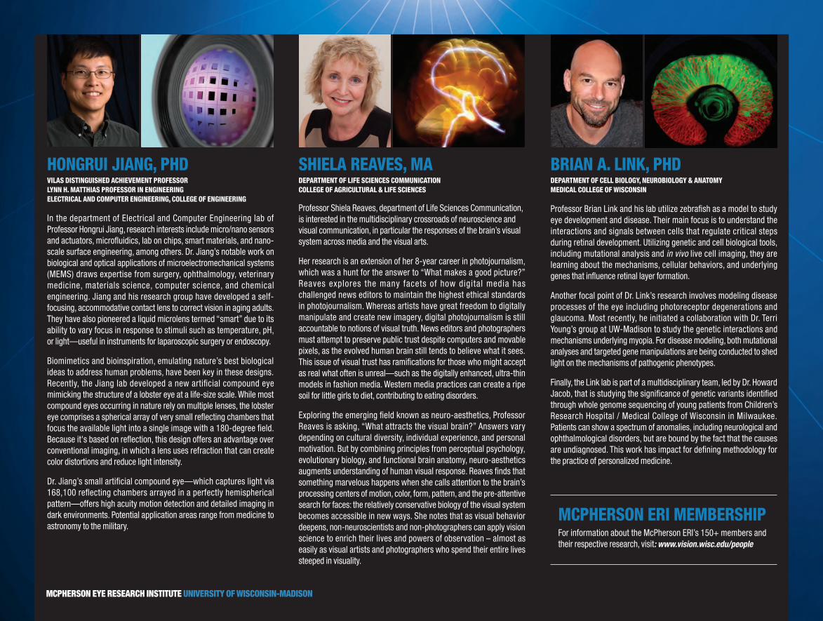
BRIAN A. LINK, PHDDEPARTMENT OF CELL BIOLOGY, NEUROBIOLOGY & ANATOMY MEDICAL COLLEGE OF WISCONSIN
Professor Brian Link and his lab utilize zebrafish as a model to study eye development and disease. Their main focus is to understand the interactions and signals between cells that regulate critical steps during retinal development. Utilizing genetic and cell biological tools, including mutational analysis and in vivo live cell imaging, they are learning about the mechanisms, cellular behaviors, and underlying genes that influence retinal layer formation.
Another focal point of Dr. Link’s research involves modeling disease processes of the eye including photoreceptor degenerations and glaucoma. Most recently, he initiated a collaboration with Dr. Terri Young’s group at UW-Madison to study the genetic interactions and mechanisms underlying myopia. For disease modeling, both mutational analyses and targeted gene manipulations are being conducted to shed light on the mechanisms of pathogenic phenotypes.
Finally, the Link lab is part of a multidisciplinary team, led by Dr. Howard Jacob, that is studying the significance of genetic variants identified through whole genome sequencing of young patients from Children’s Research Hospital / Medical College of Wisconsin in Milwaukee. Patients can show a spectrum of anomalies, including neurological and ophthalmological disorders, but are bound by the fact that the causes are undiagnosed. This work has impact for defining methodology for the practice of personalized medicine.
HONGRUI JIANG, PHDVILAS DISTINGUISHED ACHIEVEMENT PROFESSOR LYNN H. MATTHIAS PROFESSOR IN ENGINEERING ELECTRICAL AND COMPUTER ENGINEERING, COLLEGE OF ENGINEERING
In the department of Electrical and Computer Engineering lab of Professor Hongrui Jiang, research interests include micro/nano sensors and actuators, microfluidics, lab on chips, smart materials, and nano-scale surface engineering, among others. Dr. Jiang’s notable work on biological and optical applications of microelectromechanical systems (MEMS) draws expertise from surgery, ophthalmology, veterinary medicine, materials science, computer science, and chemical engineering. Jiang and his research group have developed a self-focusing, accommodative contact lens to correct vision in aging adults. They have also pioneered a liquid microlens termed “smart” due to its ability to vary focus in response to stimuli such as temperature, pH, or light—useful in instruments for laparoscopic surgery or endoscopy.
Biomimetics and bioinspiration, emulating nature’s best biological ideas to address human problems, have been key in these designs. Recently, the Jiang lab developed a new artificial compound eye mimicking the structure of a lobster eye at a life-size scale. While most compound eyes occurring in nature rely on multiple lenses, the lobster eye comprises a spherical array of very small reflecting chambers that focus the available light into a single image with a 180-degree field. Because it's based on reflection, this design offers an advantage over conventional imaging, in which a lens uses refraction that can create color distortions and reduce light intensity.
Dr. Jiang’s small artificial compound eye—which captures light via 168,100 reflecting chambers arrayed in a perfectly hemispherical pattern—offers high acuity motion detection and detailed imaging in dark environments. Potential application areas range from medicine to astronomy to the military.
SHIELA REAVES, MADEPARTMENT OF LIFE SCIENCES COMMUNICATION COLLEGE OF AGRICULTURAL & LIFE SCIENCES
Professor Shiela Reaves, department of Life Sciences Communication, is interested in the multidisciplinary crossroads of neuroscience and visual communication, in particular the responses of the brain’s visual system across media and the visual arts.
Her research is an extension of her 8-year career in photojournalism, which was a hunt for the answer to “What makes a good picture?” Reaves explores the many facets of how digital media has challenged news editors to maintain the highest ethical standards in photojournalism. Whereas artists have great freedom to digitally manipulate and create new imagery, digital photojournalism is still accountable to notions of visual truth. News editors and photographers must attempt to preserve public trust despite computers and movable pixels, as the evolved human brain still tends to believe what it sees. This issue of visual trust has ramifications for those who might accept as real what often is unreal—such as the digitally enhanced, ultra-thin models in fashion media. Western media practices can create a ripe soil for little girls to diet, contributing to eating disorders.
Exploring the emerging field known as neuro-aesthetics, Professor Reaves is asking, “What attracts the visual brain?” Answers vary depending on cultural diversity, individual experience, and personal motivation. But by combining principles from perceptual psychology, evolutionary biology, and functional brain anatomy, neuro-aesthetics augments understanding of human visual response. Reaves finds that something marvelous happens when she calls attention to the brain’s processing centers of motion, color, form, pattern, and the pre-attentive search for faces: the relatively conservative biology of the visual system becomes accessible in new ways. She notes that as visual behavior deepens, non-neuroscientists and non-photographers can apply vision science to enrich their lives and powers of observation – almost as easily as visual artists and photographers who spend their entire lives steeped in visuality.
MCPHERSON ERI MEMBERSHIPFor information about the McPherson ERI’s 150+ members and their respective research, visit: www.vision.wisc.edu/people
MCPHERSON EYE RESEARCH INSTITUTE UNIVERSITY OF WISCONSIN-MADISON

ENDOWED PROFESSORSHIPS AND CHAIRS AT THE MCPHERSON EYE RESEARCH INSTITUTE
David M. Gamm, MD, PhD Director, McPherson Eye Research Institute
Retina Research Foundation Emmett A. Humble Distinguished Directorship
Modeling and Treating Retinal Disease with Human Induced Pluripotent Stem Cells (hiPSCs)
Sandra Lemke Trout Chair in Eye Research
Applications of stem cell technology to the study and treatment of age-related macular degeneration
Jeremy Rogers, PhD
Retina Research Foundation Edwin and Dorothy Gamewell Professor
Optical Instrumentation and Technology Platforms for the Study and Screening of Retinal Disease
Nansi J. Colley, PhD
Retina Research Foundation M. D. Matthews Research Professor
Molecular Genetic Studies of Retinal Degeneration in Drosophila
Akihiro Ikeda, DVM, PhD Associate Director, McPherson Eye Research Institute
Retina Research Foundation Walter H. Helmerich Research Chair
Identification of Genetic Factors Affecting Aging of the Retina
Aparna Lakkaraju, PhD
Retina Research Foundation Rebecca Meyer Brown Professor
Insight into the Cellular Basis of Retinal Degenerative Diseases
Christine M. Sorenson, PhD
Retina Research Foundation Daniel M. Albert Chair
Apoptosis in Retinal Vascular Development and Disease
Arthur S. Polans, PhD
Retina Research Foundation Kathryn and Latimer Murfee Chair
New Agents for the Treatment of Ocular Tumors and Neovascular Diseases of the Eye
MCPHERSON EYE RESEARCH INSTITUTE ADVISORY BOARD, 2013-2014
Daniel M. Albert, MD, MS Founding Director, McPherson ERI
Rose Barroilhet Chair, McPherson ERI Advisory Board
Darrell Behnke, JD
Petros E. Carvounis, MD
Erik Christianson
Kenneth Frazier
Donald R. Gray, PhD
Bruce E. Harville
Emmett A. Humble
Alice R. McPherson, MD
Sharon Madnek
Alan R. Morse, JD, PhD
Nell R. Ray
Harry Roth, MD
David G. Walsh, JD
Marilyn Vanderhoof
HONORARY ADVISORY BOARD MEMBERS
Oscar C. Boldt
Patricia Boldt
Derilyn Cattelino
Monroe Trout, MD
Sandra Trout
MCPHERSON EYE RESEARCH INSTITUTE LEADERSHIP COMMITTEE, 2013-2014
Nansi Jo Colley, PhD School of Medicine and Public Health
Richard R. Dubielzig, DVM School of Veterinary Medicine
Kevin W. Eliceiri, MS Graduate School
David M. Gamm, MD, PhD, Director School of Medicine and Public Health
Akihiro Ikeda, DVM, PhD, Associate Director School of Medicine and Public Health
Andrea H. Mason, PhD School of Education
Margaret J. McFall-Ngai, PhD School of Medicine and Public Health
Gillian McLellan, BVMS, PhD, DACVO School of Veterinary Medicine School of Medicine and Public Health
Shiela I. Reaves, MA College of Agricultural and Life Sciences
Vanessa R. Simmering, PhD College of Letters and Science
Xiaojin (Jerry) Zhu, PhD College of Letters and Science
Alice R. McPherson, MD
Founder, President, and Scientific Director Retina Research Foundation
Dr. Monroe Trout and Sandra Trout
McPherson ERI Visionaries Endowed the Sandra Lemke Trout Chair in Eye Research
WITH GRATITUDE TO . . .
Ikeda Lab

Family members in whose honor the gallery is named include (from left) Eleanor Albert, Dr. Daniel Albert, David Mandelbaum and Karen Mandelbaum.
The Mandelbaum & Albert Family Vision Gallery, adjacent to the McPherson ERI’s new laboratory space on the 9th floor of the Wisconsin Institutes for Medical Research, was dedicated on May 8th, 2014. Named for the related families of Dr. Daniel Albert, the McPherson ERI’s Founding Director, and David & Nathan Mandelbaum, key supporters of the Institute since its inception, the gallery brings an exciting new exhibit space to the medical campus.
Two exhibits were featured in 2014: The Delighted Eye, a show of exceptional artwork from UW-Madison’s Tandem Press, and Cool Science Images 2014, featuring winners of the annual campus-wide science images contest sponsored by the online UW magazine The Why Files. Exhibits in this McPherson ERI gallery, which is open to the public during building hours, will rotate 2-3 times per year. We encourage you to visit!
The Delighted Eye exhibition, Tandem Press
2014 Cool Science Image exhibition, The Why Files
MANDELBAUM & ALBERT FAMILY VISION GALLERY
Man Alone, a stone sculpture by Zimbabwean Shona artist Damian Manuhwa, was donated to the Mandelbaum & Albert Family Vision Gallery by McPherson ERI supporters Mildred and Marv Conney.
MCPHERSON EYE RESEARCH INSTITUTE UNIVERSITY OF WISCONSIN-MADISON

MERI DISTINGUISHED VISITING SCIENTIST
NATIONAL EYE INSTITUTE DIRECTOR
DR. PAUL SIEVING APRIL 10-15, 2014
MERI member participants and attendees were involved with Dr. Sieving in the following events:
■ campus-wide symposium with Sieving keynote lecture & six faculty speakers Navigating the Waters: Basic & Translational Research from Bench to Clinic
■ vision science roundtable MERI Leadership Committee
■ Grand Rounds presentation on NEI’s Audacious Goal: “Replace photoreceptors & regenerate ganglion cell axons”
■ small group research forums:
■ multi-scale quantitative imaging
■ glaucoma research
■ image quantification
■ translational applications, stem cell technology for retinal disease
CYCLE FOR SIGHTOur March 2014 indoor cycling fundraiser expanded this year, taking place at two UW campus locations and at the Princeton Club in Madison. More than 150 riders participated in raising funds for vision research—and had a great workout near the end of a long Madison winter.
Cycle for Sight 2015 will be held on campus as well as at the Princeton Club on Madison’s west side.
SIGN UP NOW FOR CYCLE FOR SIGHT 2015
SATURDAY MARCH 14TH, 2015!Visit Cycleforsight.wisc.edu or contact Michael Chaim ([email protected])
WITH THANKS AND APPRECIATION TO THE FOLLOWING $50+ CONTRIBUTORS TO THE MCPHERSON EYE RESEARCH INSTITUTE:
■ Marcia & Robert Achenbach
■ Sanjay Agarwal
■ Daniel M. & Eleanor Albert
■ Nancy Alexander
■ Kathryn A. Allen
■ Roxanne & Brad Anderson
■ Christine Antonuzzo & Wade DallaGrana
■ Mike Archer
■ Robert Ayers
■ Eric Bangerter
■ Neal Barney
■ Rose Barroilhet
■ Anne Bauer
■ Audrey Bauman
■ Mary Ellen & Goff Beach Family Foundation
■ Darrell & Michelle Behnke
■ Linda Bergren
■ Ronald Bilchik
■ Oscar C. & Patricia Boldt
■ The Boldt Company
■ Jill Bradshaw
■ Rainy Broomfield
■ Robin Broomfield
■ Cynthia Brower
■ Ryan Brower
■ Brian J. Brown
■ Derilyn & Anthony Cattelino
■ Michael Chaim
■ Samuel S. Charles
■ Emily Chew
■ Koeun Choi
■ Erik & Rachel Christianson
■ Robert Chritton
■ Bradley Clark
■ Mildred & Marv Conney
■ Ann Conroy
■ John Court
■ Juan Torres Cubillos
■ Amy Cushman
■ Cyc Fitness
■ William Dalton
■ Chandra Darjatmoko
■ Laura S. Darjatmoko
■ Soesiawati Darjatmoko
■ Matthew & Nancy Davis
■ Shannon DeMarte
■ Lori Devine
■ Direct Fitness Solutions
■ Dean Dischler
■ Chris Doering
■ Gary Doering
■ Juanita & Norman Doering
■ Richard Dortzbach
■ C. Thomas Dow
■ Richard & Doris Dubielzig
■ Neva Dyer
■ Michael T. Eckhardt
■ Janis Eells
■ Erik’s Bike Shop
■ Joel Favre
■ Marshall & Lisa Flax
■ Anna Floch
■ Susan G. Frackman
■ Jeanne M. Frank
■ Kenneth Frazier
■ Amy Fulton
■ B’Ann T. Gabelt
■ David & Marilyn Gamm
■ Janet Geraths
■ Angela Gerach
■ Natalie Geyer
■ David Goode
■ Kannan Govindasamy
■ Niki Graham
■ Judy & Steve Grahovak
■ Cathy Guckeen
■ Louise Guerin
■ Bea C. Haney
■ Judy Harms
■ Bruce Harville
■ Robert R. M. Holloway
■ Emmett A. Humble
■ Akihiro & Sakae Ikeda
■ Brad & Kimberley Ingersoll
■ Naveen Irudarayaj
■ Jody & Gary Jensen
■ John H. Jenson
■ Paul Jobst
■ Theodore C. Johnson
■ Sherry F. Kaiman
■ Kim G. Kalepp
■ Mackenzie L. Kalepp
■ Jean Kalscheur
■ Christy & Chip Kaufman
■ Kristin Kaufman
■ Luke Kelly
■ Deborah Kirkorian
■ Bonnie Kleczka
■ Knights of Columbus Black Hawk Council 3099
■ Karin E. Konrad
■ Aparna Lakkaraju
■ Allan Lewis
■ Lions Club of Lancaster
■ Yao Liu
■ Wanda Lomprey
■ Alfred Maccoun
■ McFarland Lioness Club
■ Colette McGuinness
■ Eugene McNurlen
■ Alice McPherson, MD
■ Madison Central Lions Club
■ Madison Evening Lions Club
■ Madison Monona Lioness Club
■ Ruth Madnek
■ Sharon Madnek
■ Julie & Henry Majerus
■ David & Karen Mandelbaum
■ Nathan & Sheree Mandelbaum
■ Julie Mares
■ Jennifer & Tony Marusic
■ Andrea & Scott Mason
■ Sigurd H. Midelfort
■ Nancy Mills
■ Jon Moldenhauer
■ Alan R. Morse
■ Carolyn & Charles Mowbray
■ Charles Naatz
■ Barbara & Stephen Napier
■ Angela Nechvatal
■ Robert W. Nickells
■ Christine Nicometo
■ Nikon Instruments
■ T. Michael Nork
■ Randy Nornes
■ Susan S. North
■ Thomas O’Connor
■ Ronald L. Ohlsen
■ Oregon-Brooklyn Lions Club
■ Jill & Bill Oswald
■ Marjorie Oswald
■ Samuel Packer
■ Scott Pasternak
■ Michael Pastor
■ Bikash Pattnaik
■ Rashmi Payyanadan
■ Tracy & Todd Perkins
■ Mark Perona
■ Judy & Dan Peterson
■ Vassil D. Peytchev
■ Rose M. Oswald Poels
■ Arthur S. Polans & Myra Schultz
■ The Princeton Club, Madison
■ Patricia Rasmussen
■ Paul J. Rathouz
■ Nell & Harmon Ray
■ Division of Recreational Sports, UW-Madison
■ Mark D. Redsten
■ Retina Research Foundation
■ Cheryl M. Retzlaff
■ Toni & Donald Richards
■ Elizabeth M. Roberts
■ Thomas Roepke
■ Jeremy Rogers
■ Kristine Ross
■ Harry & Karen Roth
■ Katie Howarth Ryan
■ Rebecca J. Ryan
■ Saris Cycling Group
■ Alexandra Mostaza Scallon
■ Gregory S. Scallon
■ Katie Scallon
■ Ramona L. Scallon
■ Rita Scallon
■ John H. Schloemer
■ Kim Schmid
■ Diane Schumacher
■ Jane & Bill Shepard
■ Kathy Shepard
■ Marilyn & Stanley Shepard
■ The Shopko Foundation
■ Jason Sicotte
■ Jonathan Skarie & Emily Exten
■ Christine M. Sorenson
■ Harry & Bonnie Spiegelberg
■ Leslie & Greg Stilson
■ Gail & Richard Stirr
■ Jodi & Alan Stout
■ Sarah F. Sullivan
■ Tammy Switow
■ Marcia Thomas
■ Bradley Thuro
■ Kimberly Ann Toops
■ Laura Toops
■ Daniel Tormey
■ Jerrill & David Tormey
■ Jim Tormey
■ Sandra & Monroe Trout
■ Cathleen M. Trueba
■ US Bancorp
■ David Uvodich
■ Brett Valentyn
■ Connor Jon Valentyn
■ Nancy & Tim Valentyn
■ Paul & Elise Van Ginkel
■ Ellen VanPay
■ James N. Ver Hoeve
■ Martina Verre
■ Dennis Vogel
■ Victor S. Wahl
■ David & Nancy Walsh
■ Laurence & Frances Weinstein Foundation
■ Lauri L. Weis
■ Esther Weiss
■ Wisconsin Distributors
■ Robert & Carol Witt
■ Gerard V. Xavier
■ Xiaojin Zhu
■ David & Karen Zimmerman
■ Janet & Rolland Zimmerman
July 1, 2013 – November 15, 2014
The McPherson Eye Research Institute is grateful for contributions at all levels of support and acknowledges all donors on our website: www.vision.wisc.edu
MCPHERSON EYE RESEARCH INSTITUTE UNIVERSITY OF WISCONSIN-MADISON
“VISION IS THE ART OF SEEING WHAT IS INVISIBLE TO OTHERS.” JONATHAN SWIFT
For more information on how to partner with the McPherson Eye Research Institute in support of research, education and treatment advances in the visual sciences, please contact us.
T (608) 265-4023 E [email protected] W www.vision.wisc.edu A McPherson Eye Research Institute | 9431 WIMR
1111 Highland Avenue | Madison WI 53705
© 2014-2015 The Board of Regents of the University of Wisconsin System MERI logo design by H. Adam Steinberg, www.artforscience.com Calendar design by Malin Nordlund, www.malinnordlund.com
MISSION STATEMENT:
The McPherson Eye Research Institute is a multidisciplinary community of scholars working to gain critical knowledge about the science and art of vision and apply it to the prevention of blindness.