Embed Size (px)
Citation preview
Research Article
Received: 28 January 2009, Revised: 5 March 2009, Accepted: 5 March 2009 Published online in Wiley Interscience: 14 May 2009
(www.interscience.wiley.com) DOI 10.1002/bmc.1241
Copyright © 2009 John Wiley & Sons, Ltd. Biomed. Chromatogr. 2009; 23: 1186–1190
11
86
John Wiley & Sons, Ltd.
Measurement of bisphenol A and bisphenol B levels in human blood sera from healthy and endometriotic womenMeasurement of bisphenol A and bisphenol B levels in human blood sera
Luigi Cobellis,a Nicola Colacurci,a Elisabetta Trabucco,a Carmen Carpentierob and Lucia Grumettob*
ABSTRACT: A sensitive HPLC method with fluorescence detection was developed for the determination of bisphenol A (BPA)and bisphenol B (BPB) in human blood serum. The detection limits of the method were 0.18 and 0.20 ng/mL for BPA and BPB,respectively. A single-step liquid–liquid extraction was used for the pre-treatment of serum samples. The recoveries of BPAand BPB spiked to sera were 85.6 and 87.7%, respectively. The analyses of sera from both healthy and endometriotic womenemphasized the absence of bisphenols in all the control cases (11 women), whereas BPA was found in 30 sera (51.7%) and BPBwas found in 16 sera (27.6%) in the group of 58 patients with endometriosis; in nine of such sera BPA and BPB were presentsimultaneously. Only relatively to the sera quantitated, BPA concentrations ranged from 0.79 to 7.12 ng/mL (mean concentration2.91 ±±±± 1.74 ng/mL), whereas BPB concentrations ranged from 0.88 to 11.94 ng/mL (mean concentration 5.15 ±±±± 4.16 ng/mL).Therefore, the presence of at least one of the two bisphenols was verified in a percentage as high as 63.8% in the sera fromendometriotic women, suggesting the existence of a relationship between endometriosis and BPA and/or BPB exposure.Indeed, it is well known that bisphenols can work as xenoestrogens, owing to their structural similarity to natural and syntheticestrogens (e.g. estradiol and dietilstilbestrol). However, further studies are necessary to confirm this hypothesis and to assessthe actual dose at which exposures to bisphenols are able to increase the sensitivity of the endometriotic cells to estradiol.Copyright © 2009 John Wiley & Sons, Ltd.
Keywords: bisphenol A; bisphenol B; HPLC; serum levels; endometriosis
Introduction
Human endometrium changes during fertile life, pregnancy
and menopause are regulated by cytokines and growth factors
under the control of steroid hormones. Endometriosis is a com-
mon gynecological disorder, affecting at least 10% of women,
and often causes pain or infertility. The disease is characterized
by growth of endometrial tissue outside the uterus, with a
spread that involves mainly ovaries, but also peritoneum, utero-
sacral ligaments and recto-vaginal septum. Several theories
have been proposed to explain the pathogenesis: retrograde
menstrual flow through the fallopian tubes implants on the
peritoneal surfaces, celomic metaplasia and hematologic and
lymphatic spread (Sampson, 1940; Oral and Arici, 1997; Jenkins
et al., 1986).
Emerging evidences have proposed a putative role for
ubiquitous environmental contaminants in the occurrence of
endometriosis. The mechanism of action of environmental sub-
stances may be carried out through the interaction with steroi-
dal receptors, mimicking an estrogenic effect. More recently, the
global concerns about the endocrine disruptors that mimic the
effects of natural estrogens have also focused on bisphenols
and their congeners (Gallart-Ayala et al., 2007).
Bisphenol A [2,2-bis(4-hydroxyphenyl)propane; BPA] is a small
monomer (MW = 228.29) that is polymerized to produce poly-
carbonate, epoxy resins and other plastics. They are extensively
employed for polycarbonate bottles and containers, food can
linings and white dental fillings and sealants. Human exposure
to BPA may arise through BPA leaching from these materials into
foods (Sampson, 1940; Oral and Arici, 1997) and/or saliva (Jen-
kins et al., 1986; Krishnan E et al., 1993). BPA is also used as an
additive in other types of plastics, such as polyvinyl chloride
(PVC), used in medical tubing, toys and water pipes, and
polyethylene terephthalates (PET). Bisphenol B [2,2-bis(4-
hydroxyphenyl)butane; BPB] is a BPA derivative (MW = 242.32)
with similar estrogen-like activity used in the manufacture of
phenolic resins. Since a weak estrogen-like activity of BPA was
reported by Krishnan et al. (1993), its effects on human health
have become of growing concern. BPA shows estrogenic activity
towards cell lines, such as estrogen-responsive breast cancer cell
line MCF-7 cells and endocrine-disrupting effects in vivo (Rier
and Foster, 2002; Zeyneloglu et al., 1997; Cobellis et al., 2003;
Ashby et al., 1998; Ashby and Odum, 2004; Bergeron et al., 1999;
Kim et al., 2002; Matthews et al., 2001).
The interaction of BPA and/or BPB with estrogenic receptor
produces the activation of the same transcriptional-factor
* Correspondence to: L. Grumetto, Dipartimento di Chimica Farmaceutica e
Tossicologica, Università di Napoli Federico II, Via D. Montesano 49, I-80131
Naples, Italy. E-mail: [email protected]
a Department of Gynaecology, Obstetrics and Reproductive Medicine, Second
University of Naples, Largo Madonna delle Grazie 1, I-80130 Naples, Italy
b Department of Pharmaceutical and Toxicological Chemistry, Università degli
Studi di Napoli Federico II, Via D. Montesano, 49, I-80131 Naples, Italy
Abbreviations used: BPA, bisphenol A; BPB, bisphenol B.
Measurement of bisphenol A and bisphenol B levels in human blood sera
Biomed. Chromatogr. 2009; 23: 1186–1190 Copyright © 2009 John Wiley & Sons, Ltd. www.interscience.wiley.com/journal/bmc
11
87
(CREB) as 17-β-estradiol (Tinwell et al., 2000; Quesada et al.,2002), located in proximal promoter II of aromatase gene. There-
fore, probably, this mechanism determines a greater activity of
aromatase and, therefore, a greater production of estrogens,
favoring the proliferative and inflammatory characteristics of
endometriosis. Furthermore, previous studies have shown a rela-
tionship between BPA (and its metabolites) serum concentration
increase, altered secretion of gonadotropic hormones and increase
of androgenic hormones (Takeuchi et al., 2004, 2006).
It is useful to remember that androgenic hormones are the
substrate of enzymes, codified from the genes CYP17 and CYP19
(Mlynarcikova et al., 2005), involved as agents in charge of the
conversion of androstenedione and testosterone in estrone and
estradiol, respectively; therefore, androgenic hormones are also
likely to be involved in the onset of a mainly estrogenic climate.
In conclusion, the way bisphenols work is still not clear, but it is
assumable that they basically produce an alteration of the
endocrine system that regulates the proliferation, the interac-
tion and the cellular differentiation priming mechanisms favor-
ing adverse health effects. Therefore, endometriosis having a
multifactorial complex etiology, research in future should seek
the meeting point between genetic predisposition and environ-
mental factors able to modulate phenotypic expression.
Exposure estimates by the European Commission for BPA
from various sources amount to less than 30 μg per day for an
adult (or 0.05 mg/kg body weight) (Dekant and Volkel, 2008).
However, it is now widely recognized that toxic effects of
bisphenols can arise from chronic exposure to doses much
lower than those reported for acute exposure by chemical cor-
porations and regulatory agencies (vom Saal et al., 2006).
Indeed, following a near-continuous daily exposure, and due to
the high lipophilicity (clog P are 3.32 and 4.20 for BPA and BPB,
respectively; clog P for Windows Version 2.0, Biobyte Corp., Clar-
emont, CA, USA), they can accumulate in the adipose tissue, giv-
ing rise to persistent, although low, serum levels. Therefore,
highly sensitive and selective analytical methods are needed for
determination of low levels of BPA and BPB in serum samples.
The aim of the present study has been to set up a method for
the simultaneous determination of BPA and BPB, for routine
monitoring of serum levels in endometriotic women. Therefore,
particular attention was paid to setting up a method character-
ized by a fast and easy extraction procedure of the active com-
pounds from the biological matrix. In the present work we have
used a simple one-step extraction procedure and a reversed-
phase high-performance liquid chromatographic (RP-HPLC)
technique with fluorescence detection. In order to avoid com-
plex extraction procedures, with consequent possible poor
recoveries, we selected as stationary phase a monolithic column
because it is hardly affected by the presence of residual biological
components of the samples.
Finally, we measured the concentration of both BPA and BPB
in sera of women affected by endometriosis vs healthy controls,
in order to establish a possible relationship between bisphenol
presence in the serum and endometriosis.
Experimental
Reagents and Chemicals
Bisphenol A standard (minimum purity ≥99%) was purchased from
Sigma-Aldrich (UK) and bisphenol B standard (minimum purity
≥99%) from TCI Europe (Zwijndrecht, Belgium). All chemicals
and reagents were of either analytical or HPLC-grade and were
purchased from Carlo Erba (Italy). Water was deionized, distilled
and passed through a water purification system (Sartorius,
Germany).
Subjects
The study was approved by the institutional review board of
Second University of Naples; informed consent was obtained
from the participants. A group of fertile women (n = 69), referred
to the Department of Gynaecology, Obstetrics and Reproductive
Medicine of the same university was enrolled. The patients were
submitted to diagnostic or operative laparoscopy for the evi-
dence of ovarian cysts or to investigate chronic pelvic pain and
dysmenorrhea.
At the end of the operative procedure two different groups
were established: endometriotic patients (n = 58; age 32.8 ± 6.7
years, mean ± SD; age range 21–42) and a control group (n = 11;
age 34.5 ± 4.1 years, mean ± SD; age range 18–44). The latter
group did not show any macroscopic evidence of disease and
served in the study as age-matched controls. The endometriosis
diagnosis was confirmed by histological examination of the
endometriotic lesions, and patients were classified according to
the revised American Fertility Society classification of endometriosis
(Buttram 1985). Blood samples were collected in Vacu-test®
tubes from the antecubital vein of each volunteer (5 mL each
time). Glass syringes and glass tubes were employed throughout
the sampling to avoid a contamination of BPA and BPB. Vacu-test®
tubes were centrifuged at 3000 rpm, 20 min, promptly, then
they were transferred to clean glass vials, labelled with a code
identifying the patient and immediately frozen and stored at
–20°C until assayed within one week after collection.
HPLC System
The HPLC system consisted of a pump (LC 10ADVP, Shimadzu,
Japan), a 7725 injector with a 20 μL loop (Rheodyne, Cotati, USA)
and a fluorescence detector (Waters 470; excitation wavelength
273 nm, emission wavelength 300 nm). The signal was recorded
using PC software (Chromatoplus 2007, Shimadzu-Corporation,
Kyoto, Japan). The mobile phase was a mixture of acetonitrile
and phosphate buffer at pH 6.0 (35:65 v/v). The stainless-steel
column was a reversed-phase Onyx Monolithic C18 column
(100 × 4.6 mm i.d.; Phenomenex, USA). Chromatography was
performed at a flow rate of 1.0 mL/min at room temperature
(20 ± 2°C). Sample volumes of at least three fold the loop vol-
ume (20 μL) were injected into the chromatograph and concen-
trations of BPA and BPB were estimated on the basis of peak
area from the calibration curve. The retention times (tr) were
9.57 ± 0.30 min for BPA and 12.01 ± 0.40 min for BPB.
Mass Spectrometry Measurements
The mass spectrometric analysis was performed on samples
obtained by collecting the chromatographic peaks correspond-
ing to either BPA (tr range 9.00–10.00 min) or BPB (tr range
11.50–12.50 min) in 20 runs. Mass spectra were acquired using
an API 2000 triple-quadrupole mass spectrometer equipped
with an electrospray ionization (ESI) source (Applied Biosystem,
MDS Sciex, Foster City, CA, USA).
All the mass spectra were recorded by infusion into the ESI
source using acetonitrile as the solvent. For each analysis the

L. Cobellis et al.
www.interscience.wiley.com/journal/bmc Copyright © 2009 John Wiley & Sons, Ltd. Biomed. Chromatogr. 2009; 23: 1186–1190
11
88
full-scan spectrum (mass range 100–600 m/z, scan time 1 s) was
acquired for identification purposes. The ESI-MS was operated in
the negative ion mode under the following conditions: ionspray
voltage was kept at 5000 V; turbo gas temperature, 250°C; nebu-
lizer gas (compressed air), 55 psi; curtain gas (N2), 20 psi; declus-
tering potential, 40 V; focusing potential, 200 V; entrance
potential, 10 V.
Drug Analysis
The analyses of both standard solutions and serum samples
were performed in triplicate according to the following proce-
dure: 300 μL serum was added to 150 μL mobile phase; 150 μL
perchloric acid 25% w/v was added to precipitate proteins and
the mixture was vortex-mixed for 10 s and centrifuged at
3000 rpm for 5 min. The filtered supernatant was collected for
the analysis.
The samples of control patients were considered as blanks;
they were used (i) to confirm the assignment of peak identity to
BPA and BPB, (ii) to verify the absence of interfering peaks, (iii) to
assess the accuracy of the method and (iv) to determine the
recovery.
Recoveries were calculated by comparing the peak areas of
the spiked sera at 2.0, 4.0 and 10.0 ng/mL, subjected to the pro-
cedure for the sample pre-treatment, with those of standard
solutions at the same concentrations of BPA and BPB. Both the
analytes showed similar recoveries: 85.6 and 87.7% for BPA and
BPB, respectively.
BPA and BPB methanol standard solutions were both at the
following final concentrations: 0.5, 1.0, 5.0, 10.0 and 20.0 ng/mL.
Calibration curves of peak area vs concentration (ng/mL) were
obtained by injecting into the chromatograph the standard
solutions (at least three fold the loop volume), corresponding to
bisphenol amounts ranging from 10 to 400 pg. Straight lines
(r2 = 0.989 for BPA and r2 = 0.985 for BPB) were observed within
the range of concentrations considered. The limits of detection
(LOD) of the analytical procedure are the lowest concentrations
of analytes that can be measured with definable statistical cer-
tainty in a sample, and were calculated from the levels of the
various analytes equivalent to three times the standard devia-
tion of noise on analysis, whereas the limits of quantitation
(LOQ) were calculated from the concentration of the analytes
that provided signals equal to 10 times the noise signal of analy-
sis. The LOQ of synthetic solutions were 0.50 and 0.60 ng/mL, for
BPA and for BPB, respectively; the LOD were 0.15 and 0.18 ng/mL
for BPA and BPB, respectively.
Standard solutions of BPA and BPB were injected in the HPLC
immediately after preparation. No degradation product was
observed in the HPLC analysis of standard solutions. Calibration
procedures were repeated every two weeks. Within this period
the coefficient of variation (CV%) was <0.5%.
Statistical Analysis
A commercially available statistical package for personal com-
puter (Microsoft Excel 2000) was used. Data are expressed as
mean ± standard deviation (mean ± SD).
Results and Discussion
The HPLC method we set up allowed us to perform rapid analy-
ses of BPA and BPB in the sera with good detectability and quan-
tification. Moreover, the extraction step of bisphenols from the
biological matrix was very rapid and easy, due the use of a mon-
olithic stationary phase. No interference from biological compo-
nents was verified, since no signal interfering with the signals of
BPA and BPB was observed, and other compounds and metabo-
lites of serum samples were eluted within 4 min in chromato-
grams. The possible presence of either BPA or BPB in the sera
was confirmed by comparison with the chromatograms of the
same samples spiked with the respective pure compounds and
through mass spectrometric analyses. For quantitative analysis,
calibration procedures were performed on BPA and BPB stand-
ard solutions and repeated every 2 weeks; within this period the
coefficient of variation (CV) was <0.5%. The solutions were
injected in the HPLC immediately after preparation and no deg-
radation product was observed in their analysis.
The method, after validation on spiked sera, was performed in
the analyses of 69 sera from healthy and endometriotic women.
Neither BPA nor BPB was found in any of the 11 control serum
samples; for the 58 samples from endometriotic women, BPA
was detected in the serum of 30 subjects (51.7%), but it was
quantified in the serum of only 15 subjects, being under the
LOQ in the other 15 subjects, i.e. in the subjects had serum con-
centrations under 0.50 ng/mL (this range accounts for both
recovery percentage and serum dilution before HPLC analysis).
In the 15 sera quantified, the mean concentration of BPA was
2.91 ± 1.74 ng/mL (mean ± SD), the minimal concentration being
0.79 ng/mL and the maximal concentration being 7.12 ng/mL.
BPB was detected in the serum of 16 subjects (27.6%), but it was
quantified in only 10 subjects, being under LOQ in six subjects,
i.e. under 0.60 ng/mL. In the 10 sera quantified, the mean con-
centration of BPB was 5.15 ± 4.16 ng/mL (mean ± SD), with a
minimal concentration of 0.88 ng/mL and a maximal concentra-
tion of 11.94 ng/mL. Since BPA and BPB were simultaneously
present in nine serum samples, only 21 samples (36.2%) did not
show the presence of BPA and/or BPB. The results are summarized
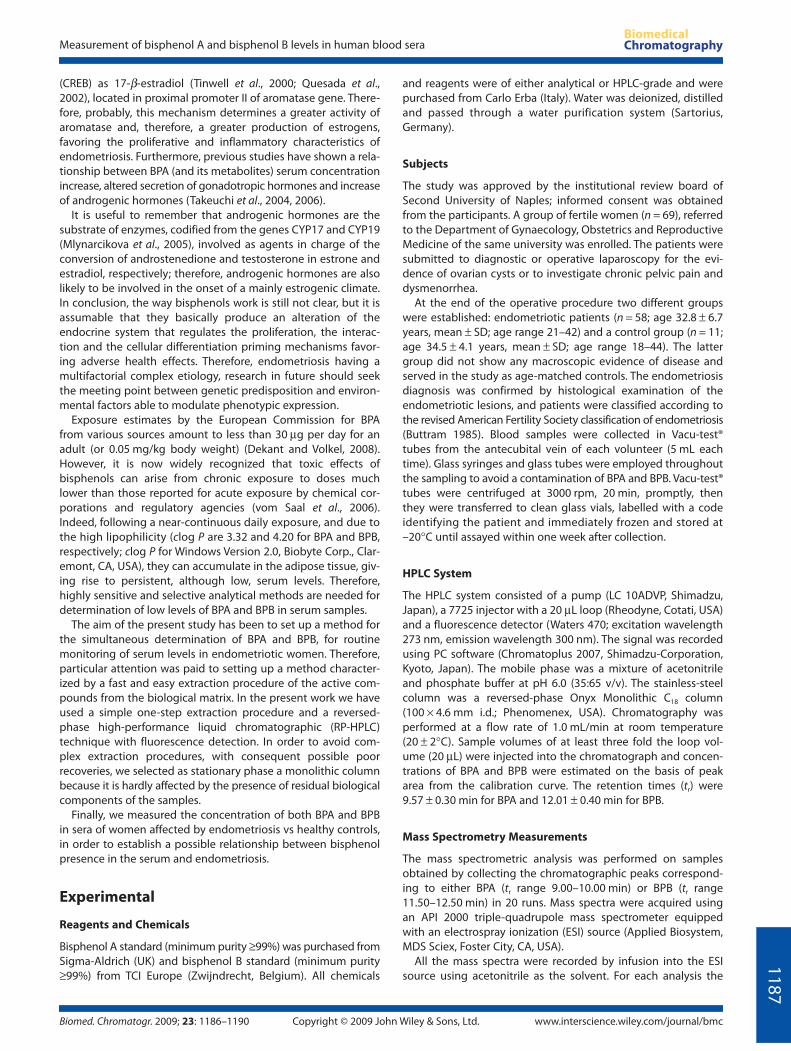
in Table 1, whereas Fig. 1 shows the representative chromatograms
Table 1. Results of the analyses for BPA and BPB on serum samples of endometriotic women
BPA BPB
Number of serum samples containing bisphenol 30 16
Percentage of serum samples containing bisphenol 51.7% 27.6%
Percentage of serum samples quantitated 25.9% 17.2%
Range of concentration (ng/mL) 0.79–7.12 ng/mL 0.88–11.94 ng/mL
Mean concentration ± SD (ng/mL) 2.91 (±1.74) ng/mL 5.15 (±4.16) ng/mL
Measurement of bisphenol A and bisphenol B levels in human blood sera
Biomed. Chromatogr. 2009; 23: 1186–1190 Copyright © 2009 John Wiley & Sons, Ltd. www.interscience.wiley.com/journal/bmc
11
89
obtained from (i) a standard solution, (ii) a reagent blank and (iii)
a blood serum sample containing both BPA and BPB.
Precision is the measure of how close results are to one
another, and it is evaluated by making repetitive measurements
for the entire method and examined on serum samples. Very
good inter-day precision data (n = 6) and intra-day precision
data (n = 3) were obtained on samples of control patients spiked
with 3.0 ppb of either BPA or BPB. The RSD (relative standard
deviation) was 3.9 and 4.2%, respectively. The RSD% was calcu-
lated by dividing the standard deviation by the mean, and
multiplying the value by 100. Figure 2(a, b) shows the mass
spectra of two chromatographic peaks of a real sample,
eluting at the retention times of BPA and BPB, respectively.
The correspondence of the signals of molecular ions to those
of BPA and BPB standards (m/z 227 [M−H]− for BPA and m/z 241
[M−H]− for BPB), together to the fragmentation pattern, con-
firmed their identity. Although the sensitivity of the proposed
method is slightly lower than that of the other analytical
methods reported in the literature for BPA in biological samples
(Gallart-Ayala et al., 2007; Sun et al., 2001; Kuroda et al., 2003;
Gallart-Ayala et al., 2008; Padmanabhan et al., 2008), the main
advantage of the proposed method is that, differently from
HPLC-MS methods; it does not require the use of non-routine
procedures.
Conclusion
To date, the etiology of the endometriosis, an estrogen-
dependent pathology, is still not clear, although in recent
years many studies have addressed the possible implications of
environmental factors in its pathogenesis. Increasing attention
has been focused on some substances, defined as ‘endocrine
disruptors’, that are able to interact with steroidal receptors and
to reproduce such the endocrine responsiveness. BPA and BPB
are two compounds that adequately reflect these properties,
their ability to interact with estrogen receptors being fully
justified by their structural similarity to the natural estrogens.
Therefore, their implication in the estrogen-dependent
pathologies is possible and the availability of rapid and simple
analytical methods may be of great usefulness in the future
investigations, which are expected to involve screenings on
larger scale.
In the present study we have set up a HPLC method for the
simultaneous determination of BPA and BPB in the serum. The
method has been applied in an investigation about the possible
relationship between serum levels of bisphenols and the occur-
rence of endometriosis.
The results can be summarized as follows: neither BPA nor BPB
was found in any of the sera from healthy women (the control
cases); in contrast, at least one of the bisphenols was found in
63.8% of sera of 58 patients with endometriosis. These findings
strongly suggest the existence of a relationship between occur-
rence of endometriosis and the presence of BPA and/or BPB in
the serum. Moreover, it is also important to underline that it is
the first time that the sera of endometriotic women have been
investigated for the presence of BPB. Its recognition (although
less frequent than BPA, i.e. 27.6 vs 51.7%) supports its possible
role in the occurrence of estrogen-dependent pathologies. The
hypothesis that bisphenols are involved in the pathogenesis of
endometriosis needs to be further confirmed by monitoring
larger number of cases; however, our results are consistent with
the hypothesis (Golden et al., 1998) that bisphenols enhance the
sensitivity of the endometriotic cells to estradiol, working as
xenoestrogens.
Figure 1. Chromatograms corresponding to (A) a standard solution of
BPA and BPB 10.0 ng/mL; (B) blood serum of a healthy women; and (C) serum
of an endometriotic woman containing both BPA and BPB. Figures on the
axes are: time (min) on the abscissa, absorbance units on the ordinate.

L. Cobellis et al.
www.interscience.wiley.com/journal/bmc Copyright © 2009 John Wiley & Sons, Ltd. Biomed. Chromatogr. 2009; 23: 1186–1190
11
90
References
Ashby J and Odum J. Gene expression changes in the immature ratuterus: effects of uterotrophic and sub-uterotrophic doses ofbisphenol A. Toxicological Sciences 2004; 82: 458–467.
Ashby J and Tinwell H. Uterotrophic activity of bisphenol A in theimmature rat. Environmental Health Perspectives 1998; 106: 719–720.
Bergeron RM, Thompson TB, Leonard LS, Pluta L and Gaido KW.Estrogenicity of bisphenol A in a human endometrial carcinoma cellline. Molecular and Cellular Endocrinology 1999; 150: 179–187.
Buttram VC Jr. Evolution of the revised American Fertility Societyclassification of endometriosis. Fertility and Sterility 1985; 43: 347–350.
Cobellis L, Latini G, De Felice C, Razzi S, Paris I, Ruggieri F, Mazzeo P andPetraglia F. High plasma concentrations of di-(2-ethylhexyl)-phthalatein women with endometriosis. Human Reproduction 2003; 18: 1512–1515.
Dekant W and Volkel W. Human exposure to bisphenol A bybiomonitoring: methods, results and assessment of environmentalexposures. Toxicology and Applied Pharmacology 2008; 228: 114–134.
Gallart-Ayala H, Moyano E and Galceran MT. Liquid chromatography/multi-stage mass spectrometry of bisphenol A and its halogenatedderivatives. Rapid Communications in Mass Spectrometry 2007; 21:4039–4048.
Gallart-Ayala H, Moyano E and Galceran MT. Liquid chromatography/tandem spectrometry (highly selective selected reaction monitoring)for the analysis of isopropyithioxanthone in packaged food. Journal ofChromatography A 2008; 1208: 182–188.
Golden RJ, Noller KL, Titus-Ernstoff L, Kaufman RH, Mittendorf R, StillmanR and Reese EA. Environmental endocrine modulators and humanhealth: an assessment of the biological evidence. Critical Reviews inToxicology 1998; 28: 109–227.
Jenkins S, Olive DL and Haney AF. Endometriosis: pathogeneticimplications of the anatomic distribution.Obstetrics and Gynecology1986; 67: 335–338.
Kim EJ, Kim JW, Lee SK. Inhibition of oocyte development in Japanesemedaka (Oryzias latipes) exposed to di-2-ethylhexyl phthalate.Environment International 2002; 28: 359–365.
Krishnan AV, Stathis P, Permuth SF, Tokes L, Feldman D. Bisphenol-A: anestrogenic substance is released from polycarbonate flasks duringautoclaving. Endocrinology 1993; 132: 2279–2286.
Kuroda N, Kinoshita Y, Sun Y, Wada M, Kishikawa N, Nakashima K,Makino T and Nakazawa H. Measurement of bisphenol A levels inhuman blood serum and ascitic fluid by HPLC using a fluorescent
labeling reagent. Journal of Pharmaceutical and Biomedical Analysis2003; 30: 1743–1749.
Matthews JB, Twomey K and Zacharewski TR. In vitro and in vivointeractions of bisphenol A and its metabolite, bisphenol Aglucuronide, with estrogen receptors alpha and beta. ChemicalResearch in Toxicology 2001; 14: 149–157.
Mlynarcikova A, Fickova M, Scsukova S. Ovarian intrafollicular processesas a target for cigarette smoke components and selected environmentalreproductive disruptors. Endocrine Regulations 2005; 39: 20–31.
Oral E and Arici A. Pathogenesis of endometriosis. Obstetrics andGynecology Clinics of North America 1997; 24: 219–233.
Padmanabhan V, Siefert K, Ransom S, Johnson T, Pinkerton J, AndersonL, Tao L and Kannan K. Maternal bisphenol-A at delivery: a loomingproblem? Journal of Perinatology 2008, 28: 258–263.
Quesada I, Fuentes E, Viso-León MC, Soria B, Ripoll C and Nadal A. Lowdoses of the endocrine disruptor bisphenol-A and the native hormone17beta-estradiol rapidly activate transcription factor CREB. FASEBJournal 2002; 16: 1671–1673.
Rier S and Foster WG. Environmental dioxins and endometriosis.Toxicological Science 2002; 70: 161–170.
Sampson JA. The development of the implantation theory for the originof peritoneal endometriosis. American Journal of Obstetrics andGynecology 1940; 40: 549–557.
Sun Y, Wada M, Kuroda N, Hirayama K, Nakazawa H and Nakashima K.Simultaneous determination of phenolic xenoestrogens by solid-phase extraction and high-performance liquid chromatography withfluorescence detection. Analytical Sciences 2001; 17: 697–702.
Takeguchi T, Tsutsumi O, Ikezuki Y, Takai Y and Taketani Y. Positiverelationship between androgen and the endocrine disruptor,bisphenol A, in normal women and women with ovarian dysfunction.Endocrine Journal 2004; 51: 165–169.
Takeuchi T, Tsutsumi O, Ikezuki Y, Kamei Y, Osuga Y, Fujiwara T, Takai Y,Momoeda M, Yano T and Taketani Y. Elevated serum bisphenol Alevels under hyperandrogenic conditions may be caused bydecreased UDP-glucuronosyltransferase activity. Endocrine Journal2006; 53: 485–491.
Tinwell H, Joiner R, Pate I, Soames A, Foster J and Ashby J. Uterotrophicactivity of bisphenol A in the immature mouse. Regulatory Toxicologyand Pharmacology 2000; 32: 118–126.
vom Saal FS and Welshons WV. Large effects from small exposures. II.The importance of positive controls in low-dose research onbisphenol A. Environmental Research 2006; 100: 50–76.
Zeyneloglu H, Arici A and Olive DL. Environmental toxins and endometriosis.Obstetrics and Gynecology Clinics of North America 1997; 24: 307–329.
Figure 2. Mass spectra of BPA (a) and BPB (b) in a real sample of serum of endometriotic patients, acquired in full-scan ESI-MS in negative mode.


![Bisphenol A Diglycidyl Ether of Bisphenol A Method · PDF file4 of 18 Diglycidyl Ether of Bisphenol A13 synonyms: 2,2-bis[4-(glycidyloxy)phenyl]propane, 4,4′-isopropylidenediphenol](https://img.pdfslide.net/doc/110x75/5a76e9947f8b9a93088d7abf/bisphenol-a-diglycidyl-ether-of-bisphenol-a-method-4-of-18-diglycidyl-ether.jpg)