Embed Size (px)
Citation preview
S38 J O U R N A L O F T H E A M E R I C A N C O L L E G E O F C A R D I O L O G Y , V O L . 6 7 , N O . 1 6 , S U P P L S , 2 0 1 6
measurement the number of patients treated by provisional stentingwas 24 for both groups (relatively equal to the QCA decision beforestarting the procedure). From them in 8 patients (group A StentysDES) and in 10 (group B Stentys X-position) was placed. Patientstreated just with SB stenting according to FFR after preparation of theSB lesion were 3 for any of both groups (150% increase respect theinitial QCA decision in group A; 300%increase respect the initial QCAbased decision in group B). Final complex treatment was performed in8 patients from the both groups (20% reduction from the initial QCAbased decision). Types of complex treatment for both group isdemonstrated in table 2. No difficulties was found to cross both MBand SB with the Acist RXi system based on NAVVUS microcatheter inany of the 35 patients from group B. The mean procedure time, X-raytime and contrast usage in the both groups (table 3) shows reductionin the all variables using Acist RXi system based on NAVVUS micro-catheter FFR measurement.CONCLUSION Our result clearly confirmed:
1. That the treatment of a bifurcation based only on angiography leadsto overestimation of the complexity and excessive usage of stents.
2. Performing fractional flow reserve measurement of the both bifur-cation branches permits identifying of the significant lesion or at leastexcluding the vessel with not significant stenosis.
3. The pre-treatment FFR evaluation is of crucial importance to estab-lish the PCI strategy (one, two or dedicated stent).
4. We demonstrate that in many cases the mid procedure FFR mea-surement permits to confirm or to change the strategy, in this settingshifting from one stent to more complex treatment including twostents or dedicated stent.
5. Using the new Acist RXi FFR measurement with NAVVUS micro-catheter that can be carried on the existing coronary wires placed inMB and SB facilitates the evaluation of the FFR during the wholetreatment process maintaining the wires in position.
6. Using the new Acist RXi FFR measurement with NAVVUS micro-catheter that can be carried on the existing coronary wires placed inMB and SB reduce significantly the number of used wires, the pro-cedure time, the X-ray time and the contrast usage.
7. Moreover, the new Acist RXi FFR measurement with NAVVUSmicrocatheter that can be carried on the existing coronary wiresused to cross the struts of placed stents permits to evaluate thecoronary reserve at the end of the procedure and to confirm thefinal result.
INVASIVE IMAGING (IVUS, OCT, SPECTROSCOPY, ETC.)(TCTAP A-087 TO TCTAP A-093)
TCTAP A-087Small Caliber Coronary Arteries in Indian Female Diabetics Fact or Fiction?Insights from Intravascular Ultrasound Guided Percutaneous CoronaryIntervention
Debdatta Bhattacharyya,1 Ayan Kar,2 Siddhartha Mani11Rabindranath Tagore International Institute of Cardiac Sciences, India;2NH-RTIICS, India
BACKGROUND It is a well known fact that the outcomes of PCI indiabetics with small vessel disease are poor. In this subset binaryrestenosis rates are high (>25%) despite the use of modern DrugEluting Stents (DES) and optimal Dual Antiplatelet Therapy (DAPT).Successive trials have shown Coronary Artery Bypass Grafting (CABG)to be a superior compared to PCI in diabetics especially where theproximal LAD is involved. Indian women with an overall smaller bodyhabitus are thought to have small caliber coronary arteries. Largestudies like the Coronary Artery Surgery Study (CASS) have repeatedlyshown that patients with “small vessels” present a higher risk foradverse outcomes after PCI with a higher incidence of restenosis andincreased (Major Adverse Cardiovascular Events) MACE rates. In factthey show that perioperative mortality significantly increases whenmeasured LAD diameters are smaller than 2.5mm. Based on our pre-vious experience of Intravascular Ultrasound (IVUS) in PCI in proximalLAD where the proximal LAD appeared to be > 3.50mm in the majorityof patients, we surmised that it was possible that due to the diffusenature of atherosclerotic disease in diabetic patients we could beunderestimating the LAD by Quantitative Coronary Angiography(QCA). Our hypothesis was that the LAD appeared to be small due toincreased plaque burden and the vessels per se were of a largercaliber. We set out to test this idea by doing IVUS studies in a series ofsix diabetic female patients who presented with severe diffuse smallvessel Coronary Artery Disease (CAD) involving the Left Anterior
Descending Artery (LAD) with disease extending into the proximalsegment.
METHODS Six female diabetic patients with diffuse LAD disease whounderwent IVUS guided PCI were included in the study. Routine de-mographic data, angiographic profile, quantitative coronary angio-gram (QCA) and IVUS pullback studies with measurement of theinternal diameter of the LAD (proximal mid and distal) pre and poststenting were recorded and compared. Here we present an illustrativecase (Case 3) which may be regarded as typical of all the six cases donein this series. We performed IVUS to answer two crucial questions: (i)the diameter of the distal landing zone. We positioned the IVUS probewhich by angiography appeared to us to be a suitable distal landingsite and measured the lumen diameter by IVUS at this location. Wedetermined this to be the diameter of our distal stent and (ii) Tomeasure the diameter of the proximal LAD where we were going toland the proximal part of our stent. In three of our six cases the dis-ease was right up to the ostium of the LAD and here we took thediameter of our stent to be media-to-media instead of the lumendiameter. We also took multiple measurements along the course ofthe LAD especially at branch points to optimize our strategy for
J O U R N A L O F T H E A M E R I C A N C O L L E G E O F C A R D I O L O G Y , V O L . 6 7 , N O . 1 6 , S U P P L S , 2 0 1 6 S39
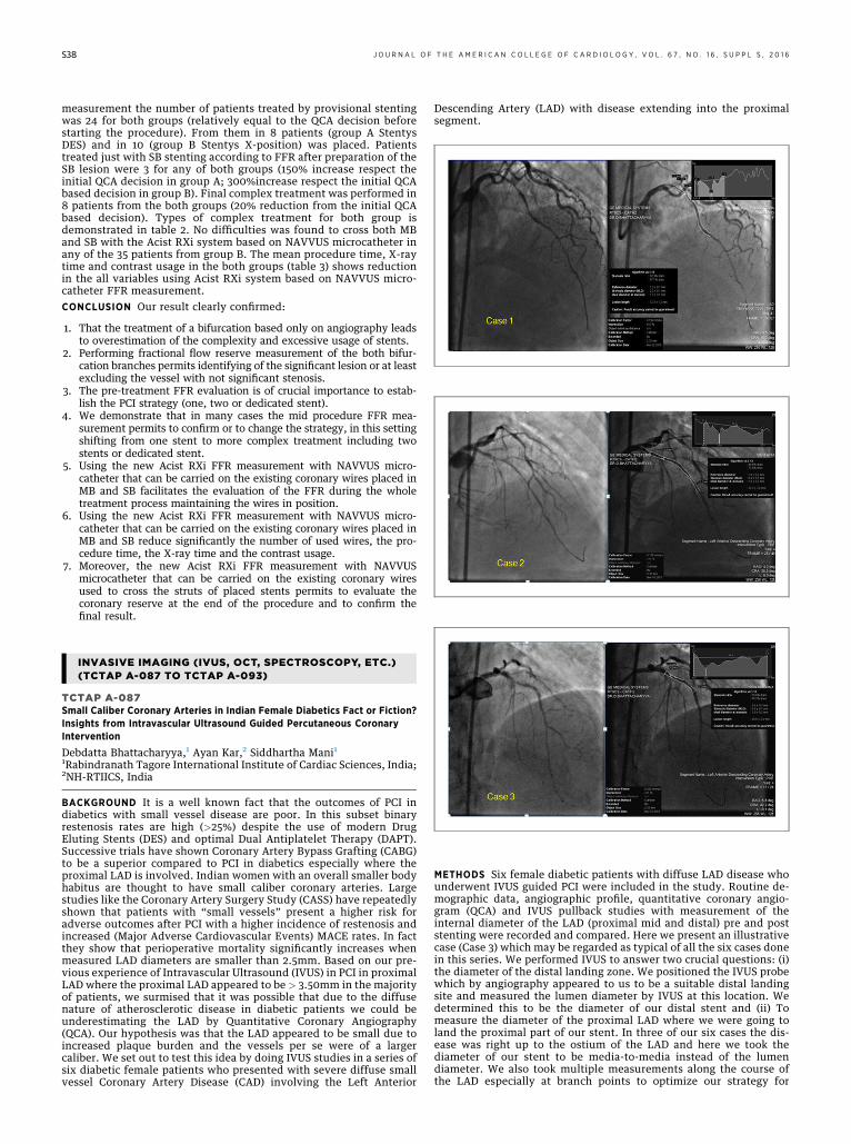
graded balloon dilatations. After initial deployment of those stents werepeated IVUS and optimized our results by IVUS guidance going forhigher balloon diameters where media was visible circumferentiallysignifying that the artery could be stretched further.
RESULTS Four of our six patients were above the age of 55 yrs andtwo were in their forties. Their average height was 152 cms with thetallest being 157 cms and shortest 148 cms. Body weight varied be-tween 42 to 57 kgs with a mean of 49 kgs. Body surface area variedbetween 1.32 to 1.58 m2 with a mean of 1.48m2. Body mass Indexranged from 16.63 to 23.12 kg/m2 with a mean of 19.5kg/m2. All sixwere diabetic with three of them being insulin dependent. All patientshad normal ejection fractions and hemoglobin and creatinine levelswere within normal limits. In all of the above patients the proximalLAD as defined earlier measured 3.5mm or above when measuredfrom media to media. The distal landing zone which at the time ofstent implantation appeared between 2.25-2.5mm invariably required
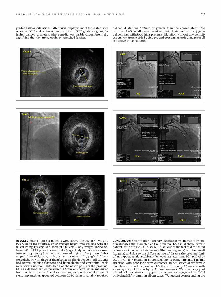
balloon dilatations 0.25mm or greater than the chosen stent. Theproximal LAD in all cases required post dilatation with a 3.5mmballoon and withstood high pressure dilatation without any compli-cation. We present side by side pre and post angiographic images of allthe above three patients.
CONCLUSION Quantitative Coronary Angiography dramatically un-derestimates the diameter of the proximal LAD in diabetic femalepatients with diffuse LAD disease. This is due to the fact that the distalreference diameter in this vessels (the landing zone) is often small(2.25mm) and due to the diffuse nature of disease the proximal LADoften appears angiographically between 2.5-2.75 mm. PCI guided byQCA invariably results in undersized stents being implanted in thissituation with poor long term outcomes. In our series of six femalediabetics we found the proximal LAD to be invariably 3.5mm and witha discrepancy of >1mm by QCA measurements. We invariably postdilated all our stents to 3.5mm or above as suggested by IVUSachieving MLA > 7mm2 in all our cases. We present corresponding pre
S40 J O U R N A L O F T H E A M E R I C A N C O L L E G E O F C A R D I O L O G Y , V O L . 6 7 , N O . 1 6 , S U P P L S , 2 0 1 6
and post IVUS cross sections of a typical case whose angiographicimages (Case 1) we have shown above to illustrate our point. Toconclude, in our experience, small vessel disease in Indian diabeticfemales is due to high plaque burden and the use of IVUS is beneficialin optimizing stent expansion and is likely to improve long termoutcomes in this subset.
TCTAP A-088In-Stent Restenosis: Insights and Outcomes Emerging from IntravascularUltrasound Guided Percutaneous Coronary Intervention
Debdatta Bhattacharyya,1 Ayan Kar21Rabindranath Tagore International Institute of Cardiac Sciences, India;2NH-RTIICS, India
BACKGROUND Despite the use of newer generation drug elutingstents there is a relatively high rate of recurrence of luminal narrow-ing due to In-Stent Re-stenosis (ISR). Data regarding factors respon-sible for ISR in Indians remain scarce.ISR remains one of the most annoying complications of contempo-
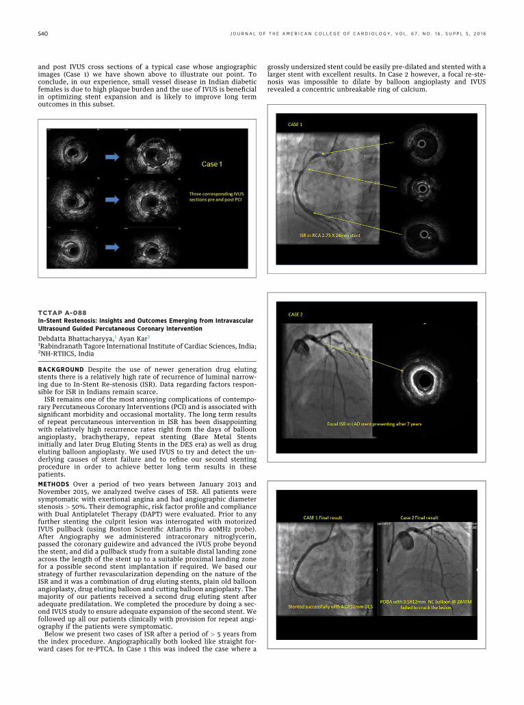
rary Percutaneous Coronary Interventions (PCI) and is associated withsignificant morbidity and occasional mortality. The long term resultsof repeat percutaneous intervention in ISR has been disappointingwith relatively high recurrence rates right from the days of balloonangioplasty, brachytherapy, repeat stenting (Bare Metal Stentsinitially and later Drug Eluting Stents in the DES era) as well as drugeluting balloon angioplasty. We used IVUS to try and detect the un-derlying causes of stent failure and to refine our second stentingprocedure in order to achieve better long term results in thesepatients.METHODS Over a period of two years between January 2013 andNovember 2015, we analyzed twelve cases of ISR. All patients weresymptomatic with exertional angina and had angiographic diameterstenosis > 50%. Their demographic, risk factor profile and compliancewith Dual Antiplatelet Therapy (DAPT) were evaluated. Prior to anyfurther stenting the culprit lesion was interrogated with motorizedIVUS pullback (using Boston Scientific Atlantis Pro 40MHz probe).After Angiography we administered intracoronary nitroglycerin,passed the coronary guidewire and advanced the iVUS probe beyondthe stent, and did a pullback study from a suitable distal landing zoneacross the length of the stent up to a suitable proximal landing zonefor a possible second stent implantation if required. We based ourstrategy of further revascularization depending on the nature of theISR and it was a combination of drug eluting stents, plain old balloonangioplasty, drug eluting balloon and cutting balloon angioplasty. Themajority of our patients received a second drug eluting stent afteradequate predilatation. We completed the procedure by doing a sec-ond IVUS study to ensure adequate expansion of the second stent. Wefollowed up all our patients clinically with provision for repeat angi-ography if the patients were symptomatic.Below we present two cases of ISR after a period of > 5 years from
the index procedure. Angiographically both looked like straight for-ward cases for re-PTCA. In Case 1 this was indeed the case where a
grossly undersized stent could be easily pre-dilated and stented with alarger stent with excellent results. In Case 2 however, a focal re-ste-nosis was impossible to dilate by balloon angioplasty and IVUSrevealed a concentric unbreakable ring of calcium.