Embed Size (px)
Citation preview
J. exp. Biol. (1982), 98, 239-267 2 3 9
MVith 20 figures
^Printed in Great Britain
MECHANICAL DESIGN OF SPICULE-REINFORCEDCONNECTIVE TISSUE: STIFFNESS
BY M. A. R. KOEHL
Department of Zoology, University of California, Berkeley, California 94720
(Received 15 July 1981)
SUMMARY
Many animals from different phyla have, embedded in their pliable con-nective tissues, small bits of stiff material known as spicules. The tensilebehaviour of spicule-reinforced connective tissues from various cnidariansand sponges, as well as of model spiculated 'tissues', is here investigated inorder to elucidate the effects on mechanical properties of spicule size andshape, and of their packing density and orientation within a tissue. The mainconclusions are:
1. Spicules increase the stiffness of pliable connective tissues probably bymechanisms analogous to those by which filler particles stiffen deformablepolymers - local strain amplification, and interference with molecular re-arrangement in response to a load.
2. The greater the volume fraction of spicules, the stiffer the tissue.3. The greater the surface area of spicules per volume of tissue, the stiffer
the tissue. Thus, a given volume of spicules of high surface-area-to-volume-ratio (S/V) have a greater stiffening effect than does an equal volume ofspicules of low S/V. Furthermore, a high volume fraction of large spiculesin a tissue can have the same stiffening effect as a lower volume fraction ofsmaller spicules.
4. Spicules that are anisometric in shape have a greater stiffening effectparallel to their long axes.
5. Spicules with very high aspect ratios appear to act like reinforcingfibres - stress is transferred by shearing from the pliable matrix to the stifffibres, which thus bear in tension part of the load on the composite.
6. Spicule-reinforced tissues exhibit stress-softening behaviour, which ismore pronounced in heavily spiculated tissues.
INTRODUCTION
Small pieces of calcareous or siliceous material are embedded in the connectivetissues of numerous animals from different phyla. Examples of such rigid particles insoft biological materials include the spicules of sponges, the sclerites of cnidarians,the ossicles of echinoderms, the spicules in some chitons (molluscs), stalked barnacles(arthropods), and ascidians (urochordates), and even the mineral deposits in theconnective tissues of humans suffering from ailments such as osteoarthritis, calciferoustendonitis and bursitis, and gout. For ease of discussion, I shall refer to all small, stiff
i l lusions in soft biological materials as 'spicules'.

240 M. A. R. KOEHL
Spicules have been described as playing some role in mechanical support and/odas serving as predator-deterrents (e.g. Currey, 1970; Wainwright et al. 1976; Jackson,1977; Berquist, 1978). In spite of their broad occurrence in the animal kingdom,however, the mechanical functions of spicules are poorly understood. In animalswhere spicules abut on each other, they are no doubt basically compressive skeletalelements. In some systems, such as starfish (Eylers, 1976), the small rigid elementsare associated with muscles as well as connective tissue and essentially function assmall bones. In other systems, such as lychnisc sponges (Reif & Robinson, 1976) andMelithaea (octocoral) stalks (Muzik & Wainwright, 1977), spicules appear to act likethe struts of braced frameworks; the rigidity or flexibility of such braced frameworksdepends on the arrangement of struts and on the deformability of the material joiningthem together (Wainwright et al. 1976).
What, if any, mechanical roles do spicules play in tissues in which they do not abuton each other? The structure of such spiculated tissues suggests that their mechanicalbehaviour may be analogous to that of man-made 'filled polymers', pliable materialsreinforced with small, rigid inclusions.
The purpose of this study was to describe the tensile mechanical behaviour ofspiculated tissues and to determine whether such tissues display the mechanicalproperties characteristic of filled polymers. A further purpose of this study was toexplore the mechanical effects of spicule size, shape, and packing within connectivetissues.
MATERIALS AND METHODS
Mechanical testing of spifiulated tissues
Mechanical tests were conducted on fresh spiculated tissue from the sponges andcnidarians listed in Table 1. The animals were kept in aerated sea water aquaria at10 °C (animals from Scotland) or at 20 °C (animals from the Red Sea). The Alcyoniumdigitatum were fed brine shrimp nauplii twice weekly; all other animals were not fed,but were used within a week of being collected. Alcyonacean colonies were anaesthe-tized overnight at 10 °C (or 20 °C) in solutions of 20% MgSO^HjjO one to onewith sea water (Pantin, 1964) until polyps pinched with forceps did not contract.Strips of spicule-containing connective tissue (' coenenchyme') were cut from colonieswith a scalpel and kept in anaesthetic at 10 °C (or 20 °C) for several hours until sub-jected to mechanical testing. Strips cut with a scalpel from sponges were kept in seawater at 10 °C until tested.
The cross-sectional area of each strip was determined by cutting a section off eachend of the strip, mounting the sections in anaesthetic on microscope slides, tracingtheir perimeters and holes onto paper using a camera lucida on a compound micro-scope, cutting out and weighing the paper 'cross-sections', converting the weights toareas (to the nearest o-oi mm2) and taking the mean of the two areas (the standarderror, S.E., for such pairs of cross sections was < + 5 %). When such area determina-tions were repeated for sections at three successive 3 h intervals, no significant changein area with time was observed.
Tissue strips were gripped in spring-loaded clamps for mechanical testing and thelength of each specimen between the grips was measured to the nearest o-1 mm

Spiculecomposition*
CC
C
C
sscc
Volume fractionof spicules±s.D., n
o-i2±o-o4, 6o-i2±o-oa, 6
oi8±o-O2, 3
o-36±o-i6, 6
oi4±o-O3, 3o-os±o-oi, 3
0-02±0'OO2, 3o-o6±o-oi, 3
Mechanical design of spicule-reinforced connective tissue 241
Table 1. Spiculated tissues
Source of tissue
Phylum CnidariaClass Athozoa, Order Alcyonacea
Alcyomum digitatum (L.)Outer coenenchymeInner coenenchyme
Dendronephtya (Dendronephyta) hempriclri (Klunzinger)tSterile stalk
Sarcopliyton glaucum (Quoy and Gaimard)"|"Sterile stalk
Phylum PoriferaClass Demospongiae, Subclass Monoaxnida
Suberitet domuncula (Olivi)Hymeniacidon tanguinea (Grant)
Class CalcareaSycon ciliatum (Fabricius)Grantia compreua (Fabricius)
• C = calcium carbonate, S = silicon dioxide; (Brown, 1975).•(• Collected from the Red Sea. (All other animals were collected from the Firth of Clyde, Scotland.)
callipers. The surfaces of each specimen that would have come in contact with thegrips were protected by squares of paper towel glued to the specimen with cyano-acrylate contact cement (Loctite Superbinder 496).
Strips of tissue were pulled at various rates in an Instron Universal TestingInstrument (Model TM-M) that simultaneously measured force (s.E. = ± 0-5 %) andextension (s.E. = + 0-25 %). Data from specimens extended at a rate of 50 cm.min"1
was recorded by photographing the tracing on an oscilloscope (Tetronix 5103Noscilloscope system with a 5A22N differential amplifier for the force signal and a5B10N time base amplifier for extension). Data from specimens pulled more slowlywas recorded using the Instron's pen recorder. Plots of stress (<r, force per cross-sectional area) versus extension ratio (A, ratio of extended length of the specimen toits original length) were made. The slope of the straight portion of each such stress-extension curve was taken to be the modulus of elasticity (E, a measure of stiffness)for that specimen.
Cyclic stress-extension tests were also performed using the Instron. Specimenswere pulled at a given rate to a given load, immediately returned at the same rate totheir original length, immediately pulled again to a greater load and returned, andso on.
Stress-relaxation tests were performed using the Instron. Specimens were rapidlystretched (strain rate, e = 0-3 s"1, where e = M,/tLQ, where AL is the change inlength of the specimen at time t, and Lo is the original length of the specimen) toknown lengths and then held; the force required to hold the specimen at that lengthwas measured through time. Time-dependent elastic moduli (E(t)) were calculated,

242 M. A. R. KOEHL
where cr(t) is the stress in the tissue at time t. During stress-relaxation tests, specimenswere submerged in a bath of the anaesthetic solution kept between 7 and 10 °C by anice-water jacket.
Constant loads were applied to specimens and their extensions were measuredthrough time using an apparatus similar to that diagrammed by Alexander (1962).Recovery tests, during which the loads were removed and decreases in specimenlength were measured through time, were also conducted using that apparatus. Thecompliance (D, an index of extensibility) of a specimen was calculated,
where bJJj) is the change in length of the specimen at time t, and <r(t) is the stressin the specimen at time t (calculated assuming the specimens were of constant volume).Specimens were submerged in anaesthetic; thymol was added to the anaesthetic oftissues tested for longer than 6 h to minimize bacterial decay of the specimens (therewas no significant difference between the behaviour during the first several hours ofextension tests of tissues in baths with and without thymol).
Mechanical testing of model tissues
The mechanical behaviour of a spiculated tissue must certainly depend upon themechanical properties of the material in which the spicules are embedded as well asupon the nature of the attachment between this material and the spicules. In order toeliminate these two possible differences between tissues, models of spiculated tissueswere constructed which all had the same type of material surrounding the spicules.Because spicule type and volume fraction in such models could be controlled, themodels provided a system in which the mechanical effects of spicule size and shape,and of spicule packing density could be tested independently.
Spicules used in the models had been isolated from various animals. Fresh animaltissues were boiled in concentrated solutions of potassium hydroxide until they dis-integrated. The spicules thereby released were cleaned by repeated rinsing in waterand decanting, were then soaked for several weeks in a solution of sodium hypo-chlorite (10-14%, w/v, available chlorine) to remove any remaining organic material,and were again repeatedly rinsed. Siliceous sponge spicules were then soaked ini"5%HCl for several weeks to eliminate any calcareous contaminants, and thenrepeatedly rinsed. Cleaned Sarcophyton spicules were sorted into two size classes bywashing them through a 300 /*m pore sieve, and Dendronephthya spicules were sortedinto three size classes by passing them through first a 500 /im and then a 300 /imsieve. Some models were also prepared using reagent grade CaCO3 powder as the'spicules'.
Model tissues were prepared by embedding these spicules in gelatin. Spicules wereweighed out into square dishes (22 x 22 mm) and equal volumes of melted gelatin(Maid Marian, raspberry) and of water were added to each to give spicule volumefractions of o-oi, 0-05, and 0-15 (and 0-32 when possible, i.e. when spicules hadindices of packing (described below) greater than 0-32). These mixtures were gentlyheated by floating the dishes on hot water and were slowly stirred to remove air
Mechanical design of spicule-reinforced connective tissue 243
bubbles and to evenly disperse the spicules. The dishes were then rapidly chilled onice. The models were unmoulded, cut into strips, and kept in greased, covered Petridishes until tested (within 24 h of their preparation). All specimens were visuallyinspected; only those specimens with evenly-dispersed spicules and no visible airbubbles or surface imperfections were used in mechanical tests.
Model tissues at room temperature were gripped and subjected to stress-extensiontests as described above.
MorphologySpicules for morphometric analysis were isolated as described above. In order to
minimize sampling bias, vials containing spicules were inverted repeatedly beforesamples of spicules were removed for study. Some spicules were mounted in water onmicroscope slides; the length and width of spicules encountered along transects acrossthe slides were measured to the nearest 10 /<m using an eyepiece graticule in a com-pound microscope. Other spicules were mounted on stubs with double-sided cello-phane tape, coated with gold (in a Polaron Equipment Ltd, SEM Coating UnitE5000) and photographed in a scanning electron microscope (Cambridge Stereoscan600). Vernier callipers were used to measure the dimensions of various features of thespicules in such photographs. The surface areas and volumes of these spicules werecalculated by modelling each spicule as a combination of cylinders, cones, hemispheres,and prolate spheroids. The 'index of packing', an estimate of the ability of spiculesof various shapes to be packed together, was obtained by taking the ratio of the actualvolume of spicules (calculated from their weight, using a density of 2-6 g.cm~3 forcalcareous spicules (Jones, 1970) and of 2-2g.cm~3, the density of amorphoushydrated SiO2 (Weast & Astle, 1978) for siliceous spicules) to the volume theyoccupied in a vial (measured to the nearest 001 cm3) which had been tapped untilthe height of the spicules no longer changed with respect to marks on the vial.
The volume fraction of spicules in animal tissues was determined. The volume ofa piece of tissue was ascertained by measuring with a pipette the volume of waterdisplaced by the tissue when put into a filled volumetric flask. (Volume measurementswere made three times and were repeatable to the nearest o-oi cm3.) The tissue wasthen blotted on filter paper and its wet weight was measured. (All weight mesaure-ments were made three times using a Mettler B6 balance and were repeatable to thenearest 0001 g.) The specimen was then dried for 3 h in an oven at n o °C and itsdry weight measured. The tissue was then ashed in a muffle furnace at 500 °C for3 h (see Paine, 1971) and the ash weight was measured. Known weights of sea waterwere also dried, ashed, and weighed. The volume fraction (y) of spicules in a tissuewas calculated,
y Pv
where p is the density (g.cm~3) of the spicules, V is the volume (cm3) of the tissue,and At is the ash weight (g) due to the spicules,
244 M. A. R. KOEHL
where AT is the total ash weight (g) of the specimen, Aw is the ash weight of a given!wet weight, Ww, of sea water, Ws is the wet weight of the specimen, and D is the drjrweight of the specimen.
The structure of the tissues containing spicules was examined using s.E.M. Smallblocks of tissue (which had been anaesthetized as described above) were fixed in 4 %formaldehyde in sea water and prepared for S.E.M. using the procedure outlined byMariscal (1974). Specimens were critical-point dried in CO2, and then coated andphotographed as described above.
The orientation of spicules in the cnidarians was measured. The angle (to thenearest io°) between the longest axis of a spicule and the vertical axis of a colony wasmeasured using a rotating eyepiece on a dissecting microscope for the first fiftyspicules encountered in a tissue (n = 3 per species). Because the spicules inA. digitatum were smaller and more difficult to see than those of the other species, theywere stained with alizarin red and the surrounding tissue was stained with Alcian blueand cleared with trypsin (Wassersug, 1976; Dinerkus & Uhler, 1977). The orientationof fibres seen in S.E.M.'s of A. digitatum coenenchyme was measured as described byKoehl (1977 a).
Biologically relevant extensions and stresses in A. digitatum
The diurnal expansion and retraction of A. digitatum colonies was monitored.Seven colonies in aquaria at the Millport Marine Laboratory were photographed attimed intervals for 72 h. The ratio of the maximum height of an expanded colony toits minimum height when retracted should be equivalent to the longitudinal Amax ofthe inner coenenchyme when the colony undergoes such a shape change. (The A ofthe outer coenenchyme was difficult to measure accurately on the photographs becauseof the increase in diameter during colony expansion of the polyps perforating thistissue).
Drag forces (to the nearest 0-05 N) on A. digitatum colonies towed through seawater were measured using a Salter spring balance. Colonies were towed with theirwidest axis both perpendicular and parallel to the flow direction. The deflection of thetip of a calibrated plastic cantilever 1 cm long was used to measure the velocities(to the nearest 0-2 m.s"1) at which colonies were towed. The projected areas of thecolonies when orientated perpendicular and parallel to the flow direction weremeasured by tracing the colonies' perimeters on to mm2 graph paper and countingthe squares contained within the outlines; such tracings were made immediately afterthe drag measurements were taken. Stresses in each colony were calculated by assum-ing the colony was a cantilevered beam bearing the drag force as a load on its freeend (see Koehl,
RESULTS AND DISCUSSION
Morphology
The volume fractions of spicules in the types of tissues subjected to mechanicaltesting are listed in Table 1. Morphological features of the spicules used in the modeltissues are listed in Table 2 and S.E.M.'s of these spicules are presented in Fig. 1. Theorientations of spicules and fibres in the cnidarian tissues are presented in Fig.
Journal of Experimental Biology, Vol.
A
5 cm
Fig. 2
3 mm
0-5 mm
5 cm
1 mm
5 cm
1 mm
M. A. R. K O I L H L

Journal of Experimental Biology, Vol. 98 Fig. 1
Fig. 1. SEMS's of spicules used in the model tissues. (A) D. liemprichi, large, (B) medium,and (C) small. (D) S. glaucum large, and (E) small. (F) A. digitatum outer, and (G) inner.(H) H. sanguinea. (I) S. domuncula. (J) CaCO, crystals.
M. A. R. KOEHL (Facing p. 244)

Mechanical design of spicule-reinforced connective tissue
Table 2. Spicule morphology
245
Spicule type
A. digitatum, outerA. digitatum, innerD. htmprichi, largeD. hemprichi, mediumD. hemprichi, smallS. glaucwn, largeS. glaucum, smallCaCO, crystals5. domunculaH. sanguinca
Length (mm)±S.D.
(« = 82)
007 ±0-03O-2I ±0091 72 ±0-451 23 ±0-36O'2O±OO9o-go±oiso-34±o-i8005 ±003*023 ±0-090-3210-09
Length/width±S.D.
(n = 12)
1 3 ±0-350 ±1-495 ±2-88-7±3'5
IO-O±2-844 ±0-77-4 ±2-8I-7 + O-7*
498 ±n-8488+HI
Surface area/volume
(mm'/mm1)±S.D.
(» = 3)
787±i70227 ±47
I7±235±278±2433±7
105 ± 5064a±i53#
762 ±143472 ± S4
Index of packing±S.D.
(« = 2)
O-2S±OO"22±O-OIo-28±o-oio-2s±o-o6O-32 ±O-O3O'32±o-o6o-34±o0-3410-03o-is±oo-o6±o-oi
• CaCOj crystals tended to form clumps (Fig. iF). These measurements were made on clumpsrather than individual crystals. Surface area was estimated assuming individual crystals to be cubeswith half of their surface area exposed when in a clump.
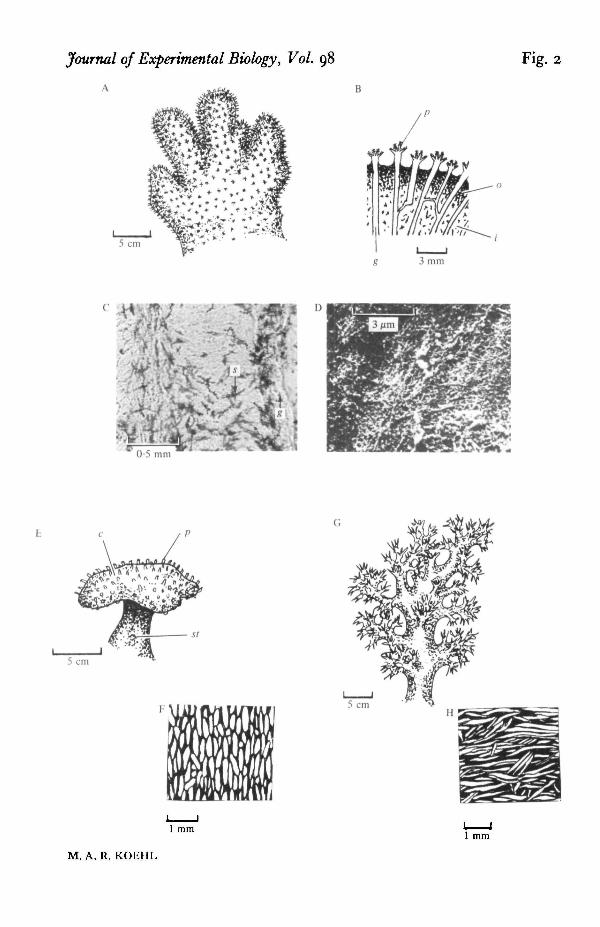
The fleshy colonies of the soft coral A. digitatum, 'dead man's fingers' (Fig. 2 Aand B), are composed of connective tissue (coenenchyme) penetrated by the cylin-drical gastrovascular cavities of the polyps and some fine strands of cells (see Hickson,1895, 1901). The coenenchyme near the surface of the colony (to which I will refer as'outer coenenchyme') contains small (Fig. 1 F) closely-packed spicules. In contrast,the 'inner coenenchyme' contains larger (Fig. 1 G), more dispersed (Fig. 2C) spicules.The coenenchyme of A. digitatum is fibrous (Fig. 2D) and the fibres appear to be inintimate association with the spicules, although the nature of the attachment, if any,of the fibres to the spicules has not yet been worked out. The longest arms of spiculesof the inner coenenchyme tend to be orientated at low angles with respect to thevertical axis of the colony (Fig. 3 A), whereas the fibres do not show any preferredorientation (Fig. 3 B). The connective tissue of the coenenchyme stains with Alcianblue, which is specific for mucopolysaccharides (Dingerkus & Uhler, 1977). If thistissue is similar in composition to the cnidarian connective tissues that have beenmore extensively analysed (for a review, see Koehl, 1977 a), it is composed of collagenfibres in a amorphous mucopolysaccharide matrix.
Fig. 2. Alcyonaceans whose spiculated connective tissues were used for mechanical testing.All colonies and sections of tissues shown have their vertical axis parallel to the vertical axis ofthe page. (A) Diagram of a colony of A. digitatum. (B) Diagram of a longitudinal sectionthrough the tip of a branch of an A. digitatum. The gastrovascular cavities (g) of the polyps(p) extend into the fleshy coenenchyme. The spicules, which are indicated by dark spots,are more densely packed in the outer coenenchyme (o) than in the inner coenenchyme (i).(C) Micrograph of a longitudinal section (i mm thick) of the inner coenenchyme of a clearedA. digitatum with stained spicules (s). The gastrovascular cavity (g) of a polyp is shown.(D) SEM of the connective tissue in between the spicules of the inner coenenchyme of anA. digitatum. Note the feltwork of fibres in the tissue. (E) Diagram of a colony of S. glaucum,showing the polyp (p) -covered capitulum (c) supported on the stalk (si). (F) DiagTam of alongitudinal section through the stalk of a S. glaucum. The spicules are shown as white andthe surrounding connective tissue as black. (G) Diagram of a colony of D. hemprichi. The stalkand branches of the colony are hollow, thin-walled, and hydrostatically supported. (H) Diagramof a surface view of the stalk wall of a D. hemprichi. The spicules arc shown as white and thesurrounding connective tissue as black.

246 M. A. R. KOEHL
1 5 n A
•S 1 0 -
1!E3
5 -
Illl.10-1
5-
I I I0° 30° 60° 90°
25 -1 C
2 0 -
I3Z
10 -
5 -
0 IL.0° 30° 60° 90°
20-i D
15 -
° I O -
E3
Z 5 ^
0
0° 30° 60° 90° 0° 30° 60° 90'
1 T 1 10° 30° 60° 90°
0° 30° 60° 90°
0° 30° 60° 90°
1.10° 30° 60° 90°
!•• ••0° 30° 60° 90°
Jill0° 30° 60° 90° 0° 30° 60° 90° 0° 30° 60° 90°
Angle with respect to vertical axis of colony or branch
Fig. 3. Histograms of orientations of (A) spicules in A. digitatum inner coenenchyme, (B) fibresin A. digitatum inner coenenchyme, (C) spicules in S. glaucum stalk, and (D) spicules inD. hemprichi branch wall. Each histogram represents a separate specimen.

Mechanical design of spicule-reinforced connective tissue 24720 -1
1-5
10-zo
0-5-
Outer
Inner
I I 1-2 1-3 1-4 1-5 1-6 1-7
Fig. 4. Stress (<r) - extension (A) curves for inner and outer coenenchyme of A. digitatumpulled at « = 0-04 a"1. The portions of the curves between the arrows were used to calculateE. The curves end where the specimens broke.
Colonies of the soft coral Sarcophyton glaucum are mushroom-shaped, with a broad,polyp-covered capitulum supported on a stalk (Fig. 2E). The stalk contains large(Fig. 1 D, E), closely-packed spicules which tend to be orientated at low angles withrespect to the vertical axis of the stalk (Figs. 2F and 3C).
The tree-like branching colonies of the soft coral Dendronephtya hemprichi arehydrostatically supported. The cylindrical branches (Fig. 2 G) of these colonies arecomposed of a layer of spiculated connective tissue (one spicule layer thick) surround-ing a fluid-filled space. The large spicules (Fig. 1 A, B) in the connective tissue tendto be orientated at large angles with respect to the longitudinal axis of a branch(Figs. 2H and 3D). The small spicules (Fig. 1C) isolated from this species are foundin the polyps.
Mechanical properties of spiculated connective tissues
In the following section the behaviour of spiculated tissues when subjected tovarious mechanical tests will be described and compared to the behaviour of filledpolymers. The mechanics of filled polymers, which will only be briefly summarizedhere, is described in much greater depth in articles and reviews such as those byChristensen (1979), Dickie (1977), Farris (1972), Fedors & Landel (1975), Ferry(1970), Kotani & Sternstein (1972), Kraus (1965), Lepie & Adicoff (1972), Mark(1970), Mascia (1974), Mullins (1963, 1980), Titow & Lanham (1975) and Wake
Stiffness
Typical stress-extension curves for the lightly spiculated inner coenenchyme andheavily spiculated outer coenenchyme of A. digitatum are shown in Fig. 4. The
248 M. A. R. KOEHL
6-0 n
5 0 -
4-0-
tp'sz 3 0 •
s
20-
1 0 -
i
1 **01 0-2 0-3 0-4 0-5
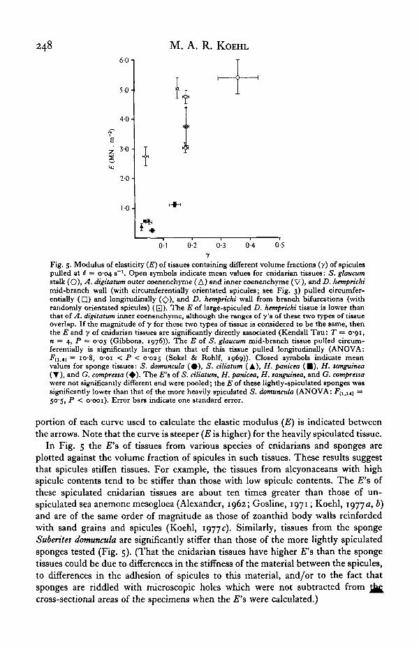
Fig. 5. Modulus of elasticity (E) of tissues containing different volume fractions (y) of spiculespulled at £ — 0-04 s~\ Open symbols indicate mean values for cnidarian tissues: S. glaucumstalk (O), A. digitatum outer coenenchyme (A) and inner coenenchyme (V), and D. hemprichimid-branch wall (with circumferentially orientated spicules; see Fig. 3) pulled circumfer-entially ( • ) and longitudinally (<£>), and D. hemprichi wall from branch bifurcations (withrandomly orientated spicules) (Q). The E of large-spiculed D. hemprichi tissue is lower thanthat of A. digitatum inner coenenchyme, although the ranges of y's of these two types of tissueoverlap. If the magnitude of y for these two types of tissue is considered to be the same, thenthe E and y of cnidarian tissues are significandy direcdy associated (Kendall Tau: T •= o-oi,n = 4, P = 005 (Gibbons, 1976)). The E of 5. glaucum mid-branch tissue pulled circum-ferentially is significantly larger than that of this tissue pulled longitudinally (ANOVA:•F[i.i] = I0"8» O'Oi < P < 0-025 (Sokel & Rohlf, 1969)). Closed symbols indicate meanvalues for sponge tissues: S. domuncula (# ) , S. ciliatum (A), H. panicea (B), H. tanguinea(T), and G. comprcssa ( • ) . The E't of S. ciliatum, H. panicea, H. tanguinea, and G. comprettawere not significantly different and were pooled; die E of diese lightly-spiculated sponges wassignificantly lower than that of the more heavily spiculated S. domuncula (ANOVA: Futltj =50-5, P < o-ooi). Error bars indicate one standard error.
portion of each curve used to calculate the elastic modulus (£) is indicated betweenthe arrows. Note that the curve is steeper (E is higher) for the heavily spiculated tissue.
In Fig. 5 the E's of tissues from various species of cnidarians and sponges areplotted against the volume fraction of spicules in such tissues. These results suggestthat spicules stiffen tissues. For example, the tissues from alcyonaceans with highspicule contents tend to be stiffer than those with low spicule contents. The E's ofthese spiculated cnidarian tissues are about ten times greater than those of un-spiculated sea anemone mesogloea (Alexander, 1962; Gosline, 1971; Koehl, 1977a, b)and are of the same order of magnitude as those of zoanthid body walls reinfordedwith sand grains and spicules (Koehl, 1977c). Similarly, tissues from the spongeSuberites domuncula are significantly stiffer than those of the more lightly spiculatedsponges tested (Fig. 5). (That the cnidarian tissues have higher E's than the spongetissues could be due to differences in the stiffness of the material between the spicules,to differences in the adhesion of spicules to this material, and/or to the fact thatsponges are riddled with microscopic holes which were not subtracted fromcross-sectional areas of the specimens when the £"s were calculated.)
Mechanical design of spicule-reinforced connective tissue 249
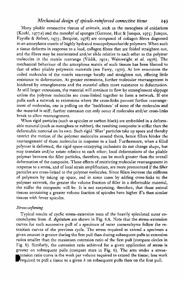
Many pliable connective tissues of animals, such as the mesogloea of cnidarians(Koehl, 1977 a) and the mesohyl of sponges (Garrone, Hue & Junqua, 1975; Junqua,Fayolle & Robert, 1975; Berquist, 1978) are composed of collagen fibres dispersedin an amorphous matrix of highly hydrated mucopolysaccharide polymers. When sucha tissue deforms in response to a load, collagen fibres that are folded straighten out,and the fibres may be reorientated and/or slide relative to each other as the polymermolecules in the matrix rearrange (Viidik, 1972; Wainwright et al. 1976). Themechanical behaviour of the amorphous matrix of such tissues has been likened tothat of other pliable polymeric materials (see Ferry, 1970). At low extensions thecoiled molecules of the matrix rearrange locally and straighten out, offering littleresistance to deformation. At greater extensions, further molecular rearrangement ishindered by entanglements and the material offers more resistance to deformation.At still larger extensions, the material will continue to flow by entanglement slippageunless the polymer molecules are cross-linked together to form a network. If onepulls such a network to extensions where the cross-links prevent further rearrange-ment of molecules, one is pulling on the ' backbones' of some of the molecules andthe material is stiff; further extension can only occur if molecules and/or cross-linksbreak to allow rearrangement.
When rigid particles (such as spicules or carbon black) are embedded in a deform-able material (such as mesogloea or rubber), the resulting composite is stiffer than thedeformable material on its own. Such rigid 'filler' particles take up space and therebyrestrict the motion of the polymer molecules around them, hence fillers hinder therearrangement of those molecules in response to a load. Furthermore, when a filledpolymer is deformed, the rigid space-occupying inclusions do not change shape, butmay translate and/or rotate relative to each other; local deformations of the pliablepolymer between the filler particles, therefore, can be much greater than the overalldeformation of the composite. These effects of restricting molecular rearrangement inresponse to a stress, and of local strain amplification, are more pronounced if the fillerparticles are cross-linked to the polymer molecules. Since fillers increase the stiffnessof polymers by taking up space, and in some cases by adding cross-links to thepolymer network, the greater the volume fraction of filler in a deformable material,the stiffer the composite will be. It is not surprising, therefore, that those animaltissues containing a greater volume fraction of spicules have higher E's than similartissues with fewer spicules.
Stress-softening
Typical results of cyclic stress-extension tests of the heavily spiculated outer co-enenchyme from A. digitatum are shown in Fig. 6A. Note that the stress-extensioncurves for each successive pull of a specimen of outer coenenchyme follow the re-traction curves of the previous cycle. The stress required to extend a specimen agiven amount is greater during the first pull than during subsequent pulls to extensionratios smaller than the maximum extension ratio of the first pull (compare circles inFig. 6). Similarly, the extension ratio achieved for a given application of stress isgreater on subsequent pulls (compare stars in Fig. 6). The area under a stress-
»tension ratio curve is the work per volume required to extend the tissue; less workrequired to pull a tissue to a given A on subsequent pulls than on the first pull.

250 M. A. R. KOEHL
Fig. 6. Stress (<r) - extension (A) curves for strips of A. digitatum coenenchyme subjected tocyclic tests (e = 0-04 s"1) until they broke. Tissues were being pulled when arrows pointupwards and to the right and were being returned to their original length when arrows pointdownwards and to the left. (A) Outer coenenchyme (results typical of 9 such tests run).(B) Inner coenenchyme (results typical of 5 such tests run). Stars and circles are explainedin the text.
The behaviour of outer coenenchyme when subjected to cyclic stress-extensiontesta is known as ' stress softening' (the ' Mullins effect') and is characteristic of filledpolymers. Various models have been proposed to explain this phenomenon (seereferences about filled polymers cited above). A basic feature of most of these modelsis that, because local strains in filled polymers can be quite high, local' failures' (suchas permanent slippage or breakage of polymer chains, or formation of voids betweenfiller particles and the polymer) can occur at low extensions of the composite. Onsubsequent pulls these chains or attachments no longer contribute to resisting thedeformation of the material, which therefore extends more easily. The slope of thestress-extension ratio curve of such a ' softened' material is low until it is pulled toextensions where molecules which were maximally extended, but not broken (ordetached, or slipped), on previous pulls again are maximally extended and thereforeoffering a great deal of resistance to further stretching of the composite. At such A'sthe slope of the stress-extension curve ratio shows a marked increase (Fig. 6A). AtA's greater than that of previous pulls, the slope of the stress-extension curve is thesame a9 it would be if the material had not been previously pulled. Typical results ofcyclic stress-extension tests of the lightly spiculated inner coenchyme of A. digitatumare given in Fig. 6B. The stress-softening behaviour described above is less pro-nounced in this material, as would be expected for a tissue containing fewer spiculesand therefore subjected to less local strain-amplification.
Both outer and inner coenenchyme often failed at lower stresses than those achievedon earlier pulls (9 of the 21 specimens of outer coenenchyme; 6 of the 21 specimensof inner coenenchyme). Such behaviour is consistent with the hypothesis that stress-softening in these tissues is due to local failures within the tissues.
Mechanical design of spicule-reinforced connective tissue4-0
30-
s20-
10-
Outer
1 10 1 L 10° 1 0 0° 1 h 1 0 0 0 ° 6 hTime (5)
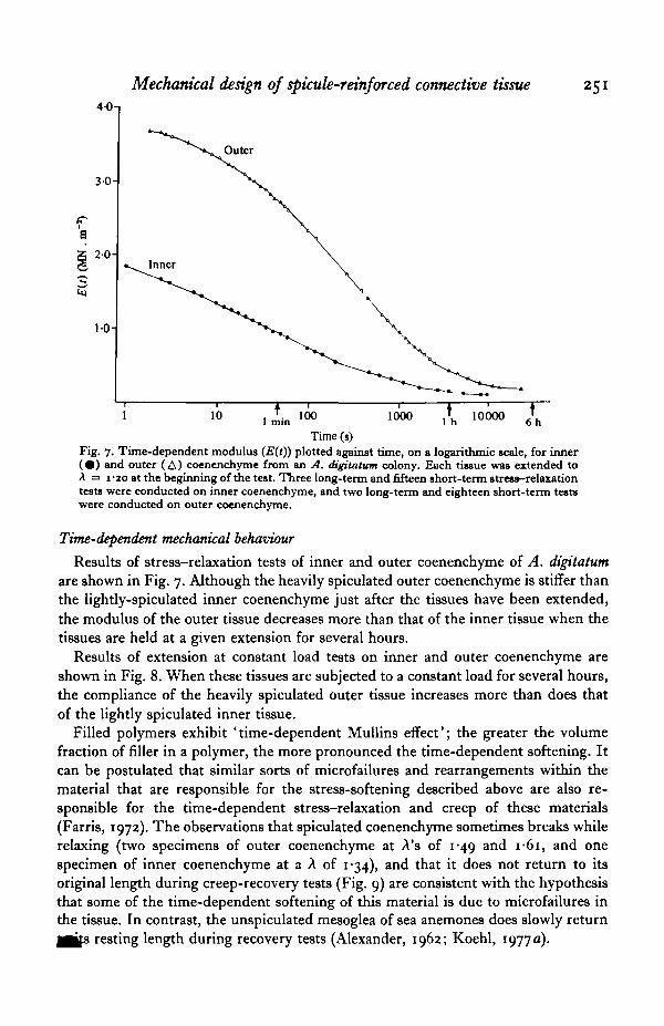
Fig. 7. Time-dependent modulus (E(t)) plotted against time, on a logarithmic scale, for inner( # ) and outer (A) coenenchyme from an A. digitatum colony. Each tissue was extended toA =3 1 -so at the beginning of the test. Three long-term and fifteen short-term stress-relaxationtests were conducted on inner coenenchyme, and two long-term and eighteen short-term testswere conducted on outer coenenchyme.
Time-dependent mechanical behaviour
Results of stress-relaxation tests of inner and outer coenenchyme of A. digitatumare shown in Fig. 7. Although the heavily spiculated outer coenenchyme is stiffer thanthe lightly-spiculated inner coenenchyme just after the tissues have been extended,the modulus of the outer tissue decreases more than that of the inner tissue when thetissues are held at a given extension for several hours.
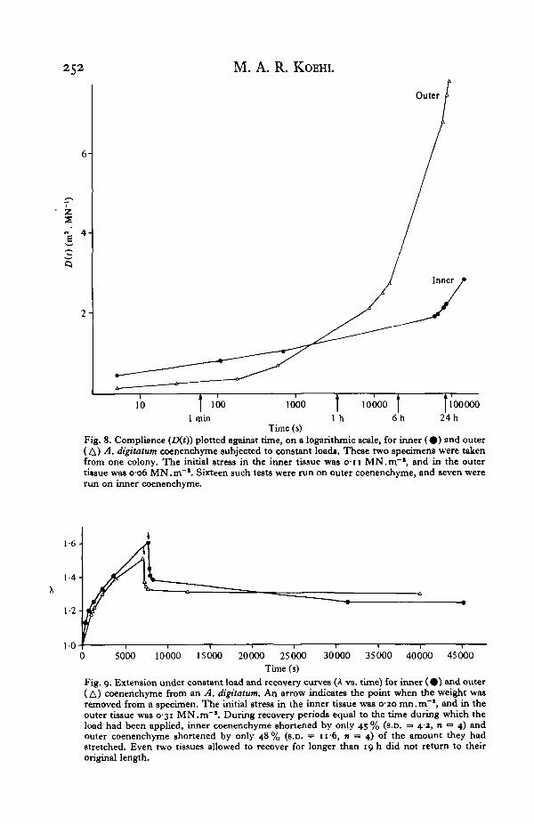
Results of extension at constant load tests on inner and outer coenenchyme areshown in Fig. 8. When these tissues are subjected to a constant load for several hours,the compliance of the heavily spiculated outer tissue increases more than does thatof the lightly spiculated inner tissue.
Filled polymers exhibit ' time-dependent Mullins effect'; the greater the volumefraction of filler in a polymer, the more pronounced the time-dependent softening. Itcan be postulated that similar sorts of microfailures and rearrangements within thematerial that are responsible for the stress-softening described above are also re-sponsible for the time-dependent stress-relaxation and creep of these materials(Farris, 1972). The observations that spiculated coenenchyme sometimes breaks whilerelaxing (two specimens of outer coenenchyme at A's of 1-49 and I-6I , and onespecimen of inner coenenchyme at a A of 1*34), and that it does not return to itsoriginal length during creep-recovery tests (Fig. 9) are consistent with the hypothesisthat some of the time-dependent softening of this material is due to microfailures inthe tissue. In contrast, the unspiculated mesoglea of sea anemones does slowly return
resting length during recovery tests (Alexander, 1962; Koehl, 1977 a).
252 M. A. R. KOEHL
6-
Z
"a 4
2-
Outer
10 ] 100 1000 I 10000 I [1000001 min 1 h 6 h 24 h
Time (s)Fig. 8. Compliance (ZKO) plotted against time, on a logarithmic scale, for inner ( • ) and outer(A) A. digitatum coenenchyme subjected to constant load*. These two specimens were takenfrom one colony. The initial stress in the inner tissue was o-n MN.m"1, and in the outertitsue was o-o6 MN.m"'. Sixteen such tests were run on outer coenenchyme, and seven wererun on inner coenenchyme.
1-6-
1-4-
1-2-
100 5000 10000 15000 20000 25000 30000 35000 40000 45000
Time (s)Fig. 9. Extension under constant load and recovery curves (A vs. time) for inner ( • ) and outer(A) coenenchyme from an A. digitatum. An arrow indicates the point when the weight wasremoved from a specimen. The initial stress in the inner tissue was 0-20 mn.m"1, and in theouter tissue was 0-31 MN.m"1. During recovery periods equal to the time during which theload had been applied, inner coenenchyme shortened by only 43 % (s.D. = 4-2, n = 4) andouter coenenchyme shortened by only 48 % (s.D. = 11 6, n = 4) of the amount they hadstretched. Even two tissues allowed to recover for longer than 19 h did not return to theiroriginal length.
Mechanical design of spicule-reinforced connective tissue 253
10-
f 5"E 4-
2-
I-0-01 01 1
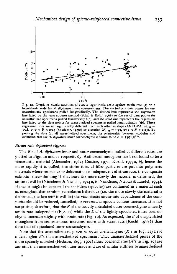
e (s"1)Fig. 10. Graph of elastic modulus (£) on a logarithmic scale against strain rate (4) on alogarithmic scale for A. digitatum inner coenenchyme. The x's indicate data points for un-anaesthetized specimens pulled longitudinally. The dashed line represents the regressionline fitted by the least squares method (Sokal & Rohlf, 1968) to the set of data points foranaesthetized specimens pulled transversely (O), and the solid line represents the regressionline fitted to the data points for anaesthetized specimens pulled longitudinally ( # ) . Theseregression lines are not significantly different from each other in slope (ANCOVA: F1-tt =i'48, o-io < P < 0-25 (Snedecor, 1956)) or elevation (Flitt = 179, o-io < P < 0-25). Bypooling the data for all anaesthetized specimens, the relationship between modulus andextension rate for A. digitatum inner coenenchyme is found to be E = 3-97 (e)011.
Strain-rate-dependent stiffness
The £"s of A. digitatum inner and outer coenenchyme pulled at different rates areplotted in Figs. 10 and 11 respectively. Anthozoan mesogloea has been found to be aviscoelastic material (Alexander, 1962; Gosline, 1971; Koehl, 1977 a, b), hence themore rapidly it is pulled, the stiffer it is. If filler particles are put into polymericmaterials whose resistance to deformation is independent of strain rate, the compositeexhibits 'shear-thinning' behaviour: the more slowly the material is deformed, thestiffer it will be (Nicodemo & Nicolais, 1974a, b; Nicodemo, Nicolas & Landel, 1974).Hence it might be expected that if fillers (spicules) are contained in a material suchas mesogloea that exhibits viscoelastic behaviour (i.e. the more slowly the material isdeformed, the less stiff it will be) the viscoelastic strain-rate dependence of the com-posite should be reduced, cancelled, or reversed as spicule content increases. It is notsurprising, therefore, that the E of the heavily spiculated outer coenenchyme is nearlystrain-rate-independent (Fig. 11) while the E of the lightly-spiculated inner coenen-chyme increases slightly with strain rate (Fig. 10). As expected, the E of unspiculatedmesogloea from sea anemones increases more with strain rate (Koehl, 19776) thandoes that of spiculated inner coenenchyme.
Note that the unanesthetized pieces of outer coenenchyme (X's in Fig. 11) havemuch higher £"s than anaesthetized specimens. That unanaesthetized pieces of themore sparsely muscled (Hickson, 1895, 1901) inner coenenchyme (X's in Fig. 10) are
stiff than unanaesthetized outer tissue and are of similar stiffness to anaesthetized
EXB 98

254 M. A. R. KOEHL
10-
tr 5-E 4 "Z 3 -
^ 2 -
• X
001 01 1
(Fig. 11. Graph of elastic modulus (E) on a logarithmic scale against strain rate (4) on a logarith-mic scale for A. digitatum outer coenenchyme. The line represents the regression line fittedto the set of data points for anaesthetized specimens. This line for outer coenenchyme issignificantly different in slope from that for inner coenenchyme (Fig. io) (ANCOVA:^"I.IU = 6"o6, o-oi < P < 0-025). The relationship between modulus and strain rate forA. digitatum outer coenenchyme is found to be E = 4-78 (4)0"04. The x's indicate data pointsfor unanaesthetized specimens.
inner coenenchyme suggests that contracting muscles are responsible for the higher£'8 of unanaesthetized outer coenenchyme. These observations should serve asreminders that many animals can actively control their stiffness by contracting muscles.
Isotopy or aniostropy of mechanical behaviour
The stiffness of a composite material composed of a pliable matrix containing longor continuous fibres, which bear in tension part of the load on the material, dependsupon the orientation of the fibres with respect to the axis along which the materialis pulled:
E oc S an cos V.where an is the proportion of fibres in a plane at angle <j> to the stress axis (Wainwrightet al. 1976). Furthermore, the deformation of the matrix in a material reinforced withelongate filler particles or short fibres is greatest for a given overall extension of thecomposite if it is pulled along axes parallel to the particles or fibres, hence such amaterial is stiffest along these axes (Gosline, 1971; Wainwright et al. 1976). Therefore,one would expect animal tissues having an anisometric arrangement of fibres and/orspicules to be mechanically anisotropic. A number of pliable biological materialsreinforced with fibres showing a preferred orientation have been found to be stiffestwhen pulled parallel to the fibres (Wainwright et al. 1976); one such material is themesogloea of sea anemones (Gosline, 1971; Koehl, 1977a).
The colonies of D. hemprichi and A. digitatum were wide enough that samples couldbe cut from them for transverse as well as longitudinal stress-extension testing.D. hemprichi coenenchyme is significantly stiffer when pulled circumferentially thanwhen pulled longitudinally (Fig. 5). The long spicules in this coenenchyme tend to h£
Mechanical design of spicule-reinforced connective tissue 255
Orientated at high angles with respect to the long axis of the colony (Fig. 3 D). Incontrast, inner coenenchyme from A. digitatum is not significantly stiffer when pulledlongitudinally than when pulled circumferentially (Fig. 10). The fibres of this tissueare arranged in a feltwork (Fig. 2D) and do not show a preferred orientation (Fig. 3 A).Although the spicules show a slight tendency to be orientated with their longest armsat small angles to the longitudinal axis of the colony (Fig. 3 A), the spicules are shortand only slightly.anisometric in shape (Table 2).
Mechanical behaviour of model tissues
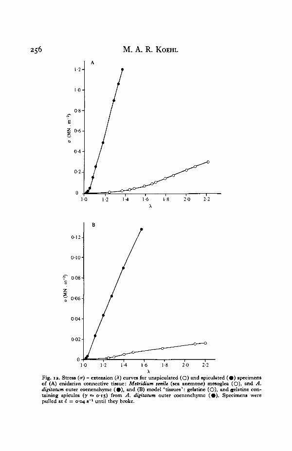
That the gelatine ' tissues' are reasonable models of cnidarian tissues is illustratedin Fig. 12. The stress-extension curves plotted up to the yield point for cnidarianconnective tissue without spicules (M. senile mesogloea) and with 0-15 volume fractionof spicules (A. digitatum outer coenenchyme) are presented in Fig. 12A. Stress-extension curves for gelatin tissue without spicules and with 0-15 volume fraction ofspicules (isolated from A. digitatum outer coenenchyme) are presented in Fig. 12 B.Note that, although the model tissues are about one tenth as stiff as the real tissues,the yield extensions of the models and real tissues are comparable, as are the stiffeningeffects of the spicules.
Volume fraction of spicules
The moduli of models containing different volume fractions of particular types ofspicules are plotted in Fig. 13. Of the models containing o-oi and 0-05 volume fractionof spicules, only those containing sponge spicules or CaCOs crystals are significantlystiffer than unspiculated models. All models containing 0-15 (and 0-32 in the two casespossible) volume fraction spicules, however, are significantly stiffer than the un-spiculated gelatin. The results plotted in Fig. 13 indicate that (1) the greater thevolume fraction of spicules, the stiffer the tissue, and (2) some types of spicules aremore stiffening than others.
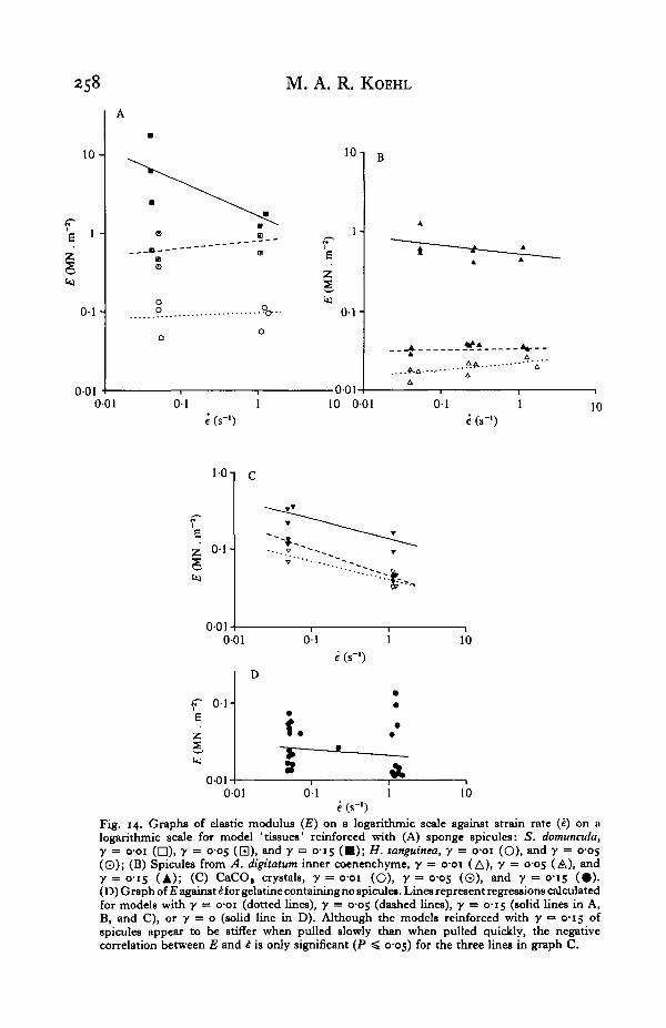
The moduli of various model tissues pulled at different strain rates are plotted inFig. 14. Note that the models either exhibit strain-rate-independent behaviour or arestiffer when pulled slowly than when pulled quickly. Note again that models contain-ing high volume fractions of spicules are stiffer than those with lower spicule contents.
Aspect ratio of spicules
The monaxon sponge spicules, which have a much greater stiffening effect than theother spicules tested, have a very high aspect ratio (length/diameter) compared withthe other spicules. There is no correlation, however, between the aspect ratios of theother spicules and the £"s of the models they reinforce (Fig. 15).
If a composite composed of discontinuous stiff fibres or rods in a pliable matrix ispulled, tensile stresses in the matrix are transmitted to the fibres via shear stresses atthe fibre-matrix interfaces. The length at each end of a fibre over which stresses aretransferred (i.e. shear stresses at the matrix-fibre interface decrease and tensilestresses in the fibre increase to some maximum value) is the ' transfer length' (lT),
lT = ro-F/r,
9-a
256 M. A. R. KOEHL
1-2-
1-0 1-2 1-4 1-6 1-8 20 2-2
012-
0-10
r 008E
0-06 H
0-04
002-
10 1-2 1-4 16 2-0 2-2
Fig. 12. Stress (<r) - extension (A) curves for unspiculated (O) and spiculated ( # ) specimensof (A) cnidarian connective tissue: Metridittm senile (sea anemone) mesoglea (O)i and A.digttatum outer coenenchyme ( • ) , and (B) model 'tissues': gelatine (O), and gelatine con-taining spicules {y = 0-15) from A. digitatum outer coenenchyme (#) . Specimens werepulled at i = 0-04 s~l until they broke.

Mechanical design of spicule-reinforced connective tissue 257
I
O 0-01 0-05 010 015 0-20 0-25 0-30
Fig. 13. Graph of the mean moduli of elasticity (if) of model 'tissues' containing differentvolume fractions (y) of spicules: the cnidarian spicules A. digitatum inner (A) and outer (A),S. glaucwn small (O) and large (9) . and D. hemprichi small (El), medium ( • ) , and large ( • ) ;the CaCO, crystals (x); and the sponge spicules S. domuncula (Y) and H. tanguinea (V). Thelines serve only to connect related points and do not necessarily indicate the true shapes of theE—y curves. Error bars indicate one standard error. Specimens were pulled at k = 0-04 s~l.The star indicates the mean modulus for unspiculated gelatine. Models containing y = o-oiof H. tanguinea spicules were significantly stifTer than gelatine (ANOVA: F(li l t] = 31-1,P < cool), as were models containing y = o-oi of CaCO, crystals (ANOVA: F( l i l t] = 10-7,0-005 < P < o-oi). The models reinforced with cnidarian spicules of y = 001 and y = 0-05were not significantly stifTer than gelatine, but did have significantly higher E'» than gelatinewhen y = 0-15: A. digitatum inner (ANOVA: F(1>14] = 288-6, P < o-ooi), and outer(ANOVA: F,,.,,] = 10766, P < 0001); 5. glaucum small (ANOVA: F t l i l l ] = 413-9,P < 0001), and large (ANOVA: FC11,, = 38-2, P < o-ooi); D. hemprichi small (ANOVA:•F[i.iil = 4929-6, P < o-ooi), medium (ANOVA: F(1 , t ) = 83-2, P < o-ooi), and large(ANOVA: F U i l i ) = 183-8, P < o-ooi).
where r is the fibre radius, aF is the fracture strength of the fibre, and T is the shearstress at the matrix-fibre interface (Wainwright et al. 1976). Fibres which are longrelative to their transfer length will bear in tension part of the tensile load imposedon the composite. Perhaps the aspect ratio of the sponge spicules is great enough thatthese spicules act as stiff reinforcing fibres, bearing in tension part of the load on thecomposite, whereas the other stubbier spicules merely act as filler particles occupyingspace.
Spicule size and surface area
Small spicules appear to be more stiffening than large ones (Fig. 16). If tissues arereinforced with a given volume fraction of evenly dispersed spicules, smaller expansesof matrix are present between numerous small spicules than between a few large ones.The smaller the spaces between filler particles, the greater will be the local strainamplification within the matrix, the hindrance to rearrangement of matrix molecules,and therefore the stiffness of the composite. Furthermore, small spicules have highersurface-area-to-volume ratios (S/V) than do large spicules; therefore, a given volume
of small spicules in a tissue presents more surface area for interaction with
258 M. A. R. KOEHL
10-
0 1 -
001
1-
0 1 -
001-
B
A
" —
t
A
- - • - * - * -
r—^—
6
• - A
001 01 1e (s-1)
10 001 0 1 10
'e
o-oi
rEZ
001
0 1 -
0 1 10
e (s-1)
0-01-001 0 1 1 10
Fig. 14. Graphs of elastic modulus (£) on a logarithmic scale against strain rate (i) on alogarithmic scale for model 'tissues' reinforced with (A) sponge spicules: S. domuncvla,y = o-oi (D)i 7 = o-o5 (Q), and y = 0-15 ( • ) ; H. sanguined, y = o-oi (O), and y = 0̂ 05(©); (B) Spicules from A. digitatttm inner coenenchyme, y = o-oi (A), 7 = °'OS (A), and7 = 0-15 (A); (C) CaCO, crystals, y = 001 (O), 7 = °-°5 (©). and y = 0-15 (#) .(D) Graph of E against HOT gelatine containing no spicules. Lines represent regressions calculatedfor models with y = o-oi (dotted lines), y = cos (dashed lines), y = C15 (solid lines in A,B, and C), or y = o (solid line in D). Although the models reinforced with y •= 0-15 ofspicules appear to be stiffer when pulled slowly than when pulled quickly, the negativecorrelation between E and £ is only significant (P ^ 0-05) for the three lines in graph C.

Mechanical design of spicule-reinforced connective tissue 259
2S0-3-
0-2-
011
8-92 ± 4-52 MN . i
4
10010Spicule l/d
Fig. 15. Graph of the mean moduli of elasticity (E) on a logarithmic scale against the meanspicule aspect ratio (l/d) on a logarithmic scale of models reinforced with y = 0-15 of varioustypes of spicules. Specimens were pulled at i = 0-04 a"1. Error bars indicate one standarderror. The point at a much higher E and l/d than the others is the mean for models reinforcedwith spicules from the sponge S. domunada. There is no significant association betweenspicule l/d and model E, however, for the other models, which contain spicules of low l/d(Kendall Tau: T = —0071, n =• 8, P > 08).
10 -
IT'p 0-5-
• 0-4-l o . 3 Hk)
0 -2 -
01
t
001 101Spicule length (mm)
Pig. 16. Graph of the mean elastic moduli (E) on a logarithmic scale against the mean spiculelengths (/) on a logarithmic scale of models reinforced with y = 0-15 of various types ofspicules. Specimens were pulled at i = 004 s-1. Error bars indicate one standard error.(Specimens reinforced with sponge spicules are not included because sponge spicules appearto act as fibres rather than filler particles.) There is a significant inverse association between/ and E (Kendall Tau: T = -0-50, n = 8, P = cos).
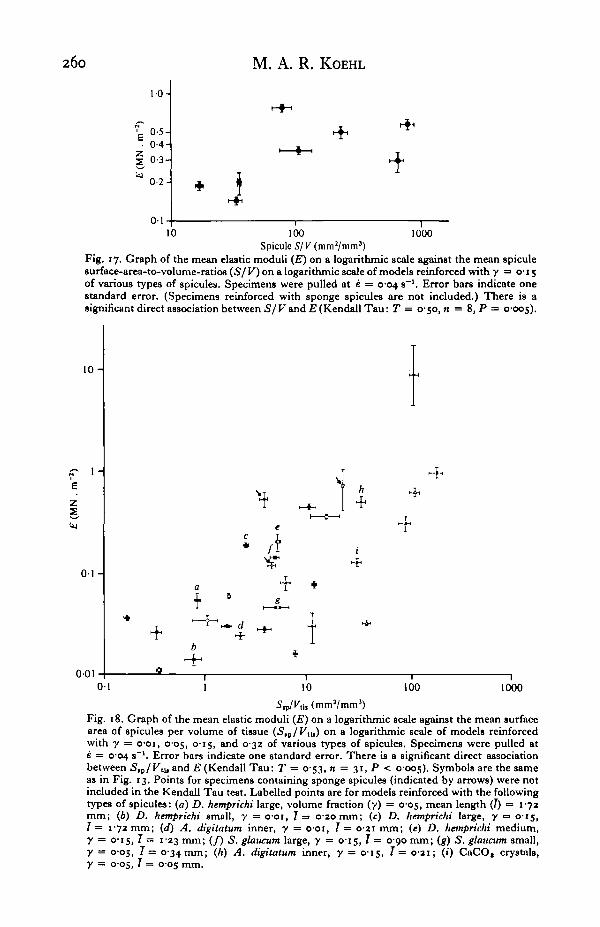
the matrix than does an equal volume fraction of large spicules. The warts, arms, andspines on spicules increase their S/V, and the E's of model tissues reinforced by agiven volume fraction of spicules tend to be greater when the S/V of the spicules islarge (Fig. 17). If the stiffening of a tissue depends upon the surface area of spiculesavailable for interacting with the matrix, a particular E can be achieved, for example,by a high volume fraction of large (low (S/V) spicules or by a small volume fractionof small (high S/V) spicules. Indeed, the E of models does increase as the surfacearea of spicules per volume of tissue (Ssp/VllB) increases (Fig. 18).
A greater volume fraction of large (low S/V) spicules is required than of small(high S/V) spicules to achieve a particular surface area of spicules per volume of
ue. Therefore, the spaces between large spicules in a tissue of a given Ssv/VilB are
260 M. A. R. KOEHL
1 0 -
0-4-
0-110 1000100
Spicule 5/K(mm2/mm3)Fig. 17. Graph of the mean elastic moduli (E) on a logarithmic scale against the mean spiculesurface-area-to-volume-ratios (S/ V) on a logarithmic scale of models reinforced with y = 0-15of various types of spicules. Specimens were pulled at e = 0-04 s"1. Error bars indicate onestandard error. (Specimens reinforced with sponge spicules are not included.) There is asignificant direct association between S/V and E (Kendall Tau: T = 0-50, n = 8, P = 0-005).
10-
1 -
Z3
0 1 -
0-01
T
1*
01 1 100 100010
Vti, (mm2/mm3)
Fig. 18. Graph of the mean elastic moduli (E) on a logarithmic scale against the mean surfacearea of spicules per volume of tissue (S,p/Vti,) on a logarithmic scale of models reinforcedwith y = o-oi, 005, 0-15, and 032 of various types of spicules. Specimens were pulled ate = 004 s~'. Error bars indicate one standard error. There is a significant direct associationbetween 5,P/Ktu and E (Kendall Tau: T = 0-53, n = 31, P < 0-005). Symbols are the sameas in Fig. 13. Points for specimens containing sponge spicules (indicated by arrows) were notincluded in the Kendall Tau test. Labelled points are for models reinforced with the followingtypes of spicules: (a) D. hemprichi large, volume fraction (y) = 0-05, mean length (7) = 1-72mm; (6) D. hemprichi small, y = o-oi, 7 = 020mm; (c) D. hemprichi large, y =• 0-15,T = 172 mm; (d) A. digitatum inner, y = 001, T= 0-21 mm; (e) D. hemprichi medium,y = 015, 7 = 123 mm; (/) S. glaucum large, y — 0-15, 7 = 0-90 mm; (g) S. glaucum small,y = 005, 7 = 0-34 mm; (h) A. digitatum inner, y = 0-15, 7 = 0 2 1 ; (i) CaCO, crystals,y = 0-05, 7 = 0-05 mm.
Mechanical design of spicule-reinforced connective tissue 261
Generally smaller than between small spicules in a tissue of the same Ssp/VUs. Hence,Tor a given Sep/V,le, large spicules should be more stiffening than small ones. Com-parison of points a and b, c and d, e, f, and g, and h and i in Fig. 18 illustrates thatthis is so. It appears, therefore, that both the volume spicules occupy in a tissue andthe surface area of interaction between spicules and matrix are important to thestiffening effect of the spicules.
Biological implications
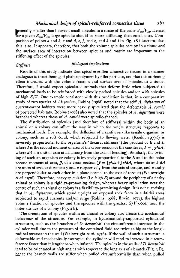
Results of this study indicate that spicules stiffen connective tissues in a manneranalogous to the stiffening of pliable polymers by filler particles, and that this stiffeningeffect increases with the volume fraction and surface area of spicules in a tissue.Therefore, I would expect spiculated animals that deform little when subjected tomechanical loads to be reinforced with closely packed spicules and/or with spiculesof high S/V. One example consistent with this prediction is that, in a comparativestudy of two species of Alcyonium, Robins (1968) noted that the stiff A. digitatum ofcurrent-swept habitats were more heavily spiculated than the deformable A. couchiof protected habitats. Robins (1968) also noted that the spicules of A. digitatum werebranched whereas those of A. couchi were spindle-shaped.
The distribution of spicules (and therefore of stiffness) within the body of ananimal or a colony can affect the way in which the whole structure responds tomechanical loads. For example, the deflexion of a cantilever-like sessile organism orcolony, such as a soft coral, when subjected to flowing water (Koehl, 19776) isinversely proportional to the organism's ' flexural stiffness' (the product of E and I,where / is the second moment of area of the cross-section of the cantilever, I = \y*dA,where dA is a unit of area at distance y from the axis of bending). Similarly, the twist-ing of such an organism or colony is inversely proportional to the E and to the polarsecond moment of area, J, of a cross section (J = $y2da + fxidA, where da and dAare units of area at distances y and x respectively from the axis of torque, and x and yare perpendicular to each other in a plane normal to the axis of torque) (Wainwrightet al. 1976). Therefore, heavy spiculation (i.e. high E) around the periphery of a fleshyanimal or colony is a rigidity-promoting design, whereas heavy spiculation near thecentre of such an animal or colony is a flexibility-permitting design. It is not surprisingthat in A. digitatum, which stand upright on exposed rock faces in subtidal areassubjected to rapid currents and/or surge (Robins, 1968; Erwin, 1977), the highestvolume fraction of spicules and the spicules with the greatest S/V occur near theouter surface of a colony (Fig. 2B).
The orientation of spicules within an animal or colony also affects the mechanicalbehaviour of the structure. For example, in hydrostatically-supported cylindricalstructures, such as the branches of D. hemprichi, the circumferential stresses in thecylinder wall due to the pressure of the contained fluid are twice as big as the longi-tudinal stresses in the wall (Wainwright et al. 1976). If the wall of such a structure isdeformable and mechanically isotropic, the cylinder will tend to increase in circum-ference faster than it lengthens when inflated. The spicules in the walls of D. hemprichitend to be orientated at high angles with respect to the long axis of a branch (Fig. 3 D),hence the branch walls are stiffer when pulled circumferentially than when pulled
262 M. A. R. KOEHL
longitudinally (Fig. 5), and colonies become taller when inflated. Another example (Ma structure containing anisotropically arranged spicules is the solid stalk of a mush-room-shaped S. glaucum colony. Such a stalk, which supports the polyp-bearingcapitulum in flowing water, is subjected to bending and/or tensile loads; tensilestresses in the stalk should therefore be greatest in the longitudinal direction, whichis the direction in which spicules tend to be orientated (Fig. 3 C). It is intriguing tospeculate about the possible role of mechanical stresses axes in determining theorientation of spicules in such organisms.
Although big spicules are less stiffening than small ones, many animals containlarge spicules. Might there be advantages to being reinforced with big spicules? Onemight argue that large spicules are more likely than small spicules to irritate themouths or guts of potential predators. Berquist (1978), however, presents evidenceindicating that spicules might not serve as predator-deterrents. The metabolic costsof secreting spicules and connective tissues are not yet known; if it is cheaper toprecipitate minerals than to synthesize proteins and mucopolysaccharides, a sessile orsedentary organism (for whom costs of locomotion are unimportant) can perhapsachieve a given stiffness at lower metabolic cost by reinforcing its connective tissuewith a high volume fraction of large spicules rather than a low volume fraction ofsmall spicules. Flexibility and extensibility can be features which minimize thebreakage of an organism by moving water (Koehl & Wainwright, 1977; Koehl, 1979);for a given volume fraction of spicules, large ones permit more deformation.
Spicule size, shape, and packing can affect the strength and toughness of a tissueas well as its stiffness, as will be discussed elsewhere (Koehl, in preparation). It shouldalso be pointed out that in many animals small spicules are interspersed between largeones; the mechanical consequences of such arrangements have yet to be worked out.
Stress-softening
The spiculated tissues of A. digitatum exhibit stress-softening, a behaviour which,has been observed in a few other biological tissues (Chu, 1972, 1973; Vincent, 1975,1976; Hawkes, 1976). In order to determine whether or not the stress-softeningbehaviour of a spiculated tissue is important biologically, one must compare themagnitude of the extensions and stresses at which the Mullins effect occurs for aparticular tissue with those it experiences in vivo.
A. digitatum colonies undergo diurnal (Hickson, 1892; Ceccatty, Buisson &Gargouil, 1963) and seasonal (Hartnoll, 1975) cycles of retraction and expansion;colonies expand as cilia of the siphonoglyphs pump water into the polyps' gastro-vascular cavities that extend deep into the coenenchyme. The mean A^* measuredfor A. digitatum inner coenenchyme as the colonies expanded and retracted was 1-29(S.D. = o-i6, n = 7); Hickson (1895) and Robins (1968) have reported that collectedcolonies shrink in length by 10-40%, corresponding to A's between I - I I and 1-67.Stress-softening can be noted in inner coenenchyme at such A's (Fig. 6B). The A'sof the outer coenenchyme when colonies undergo such size changes are probablylower than those of the inner coenenchyme because the diameters of the polypsperforating this tissue increases during colony expansion. Vincent (1975, 1976) hassuggested that stress-softening materials would be advantageous in biological st

Mechanical design of spicule-reinforced connective tissue 263
Velocity2 ([m.s"1]2)Fig. 19. Graph of drag force (D) against the square of velocity (o1) for A. digitatwn coloniestowed with their widest axis parallel (solid line) and perpendicular (dashed line) to the direc-tion of water flow. The Reynolds numbers for these colonies at such velocities ranged betweenio4 and io', hence drag force is given by
D = (i/3.)pv*ACD,where p is the density of the fluid, A is the projected area of the colony, and CD is the co-efficient of drag. When colony I (A) was parallel to the flow, A = 0738 x io~* m* andCD = 0-33 (s.D. = 0-08, n = 3), and when perpendicular to the flow, A = i'i6ox io~*m1
and CD = 0-44 (s.D. = 0-17, n = 3). When colony II (O) was parallel to the flow, A = 0-873x io"' m* and CD = 1*17 (s.D. = 0-53, n = 3), and when perpendicular to the flow, A =3-049 x io - > m1 and CD = 0-184 (s.D. = 0-30, n = 3). When colony III ( • ) was parallel tothe flow, A •= 1-497 x IO~* m* and CD = 0-99 (s.D. = 0-54, n = 3), and when perpendicularto the flow, A = 4-543 x io"1 m* and CD = 088 (s.D. = 0-08, n = 3). The diagram by eachline indicates the projected shape of the colony in that orientation.
tures which should maintain their shape, but which should be extendable by smallforces. The fleshy colonies of soft corals such as A. digitatum appear to be just suchstructures: they can be inflated by ciliary pumps, but stand up even when cut so thatno hydrostatic pressure can be maintained in their gastrovascular cavities.
Stresses in A. digitatum coenenchyme in situ can be estimated. A. digitatum occuron exposed rock faces in subtidal areas subjected to surge and tidal currents; velocitiesin the vicinity of A. digitatum have been measured to be 0-02-0-22 m.s"1 (Robins,1968), and A. digitatum are abundant on walls in areas where mainstream velocitiescan be as high as 3-5 m.s"1 (Erwin, 1977). Drag forces on A. digitatum colonies ofvarious sizes subjected to a range of flow velocities are plotted in Fig. 19. Small A.digitatum colonies are finger-like, whereas larger ones are shaped like planar bloatedhands. Although drag forces on and drag-induced stresses in such planar colonies aremuch lower when colonies are orientated parallel to the flow direction, it is likely thatA. digitatum, like many other planar organisms (Riedl, 1971), are orientated normalto the predominant current direction; the few colonies I have observed whileSCUBA diving were orientated in this way, and the very similar A. sidereum havebeen found to be normal to flow directions (Patterson, 1980). Perhaps this orientationminimizes torsion of the colonies (see Wainwright & Dillon, 1969) and/or enhancesthe food-capturing ability (see Leversee, 1976) of these planktivorous colonies|Eoughshady & Hansen, 1961). Estimates of the maximum tensile stresses in the

264 M. A. R. KOEHL
0-30-
0 0-5 3010 1-5Velocity (m . s"1)
Fig. 20. Graph of calculated estimates of maximum tensile stresses ( c r j in the coenenchymeof the A. digitatum colonies plotted in Fig. 19 against the flow velocities which would inducesuch stresses. Colonies are assumed to be solid beams orientated normal to the direction ofwater flow. Symbols are the same as those used in Fig. 19. Values for stresses at velocities of2 m.s"1 and 3 m.s"1 were calculated using drag forces estimated by extrapolating the dragvs. velocity1 lines in Fig. 19.
coenenchyme of A. digitatum colonies when normal to the flow are plotted in Fig. 20.Even the maximum stresses in colonies exposed to rapid currents are more than an
order of magnitude lower that stresses which break coenenchyme (Koehl, in prepara-tion; see Figs. 1, 6, 12). Nonetheless, coenenchyme slowly extends when subjected tolow stresses such as these: outer coenenchyme (n = 7) subjected to stress of o-ioMN.m~2 (s.D. = CV04) for 5 h (to simulate the maximum stresses in these tissues incolonies subjected to tidal currents of a few meters per second) extends by A = 1-34(s.D. = 015). In contrast, inner coenenchyme, which would in situ experience lowerstresses than outer coenenchyme, only extends by A = I- I I (S.D. = 004, n = 3) whensubjected to stress of o-n MN.m~2 (s.D. = 0-07) for 5 h. Such extension test resultsindicate that the lightly-spiculated inner coenenchyme, which appears to experienceless time-dependent Mullins effect than the heavily spiculated outer coenenchyme,m?y keep the outer coenenchyme from extending as much, when stressed for longtimes, as it would if it were not continuous with the inner coenenchyme. That co-enenchyme bearing a load extends with time suggests that when A. digitatum aresubjected to prolonged rapid currents, they may have to maintain their shape by activecontraction of muscles and ciliary pumping. The effect of pressure in the polypgastrovascular cavities in pre-stressing the coenenchyme is not yet known.
The example of A. digitatum illustrates the importance of considering the softeningas well as the stiffening effects of filler particles when the functional consequences ofspiculation are studied.

Mechanical design of spicule-reinforced connective tissue 265
CONCLUSIONS
The mechanical behaviour of a spicule-reinforced connective tissue depends upon:(1) The mechanical properties of the pliable connective tissue, which in turn
depend upon the amount and orientation of fibres in the tissue, and on the composi-tion and concentration of the amorphous polymer matrix between the fibres (Gosline,1971; Wainwright et al. 1976; Koehl, 1977a); (2) the nature of the cross-linkingbetween the spicules, fibres, and amorphous matrix; and (3) the volume fraction,shape, size, and orientation of the spicules. This study, which explored the effects ofthe latter on the tensile stiffness of spiculated tissues, has revealed several features ofsuch tissues: (1) Spicules increase the stiffness of pliable connective tissues, probablyby mechanisms analogous to those by which space-occupying filler particles stiffendeformable polymers - local strain amplification, and interference with molecularrearrangement in response to a load.
(2) The greater the volume fraction of spicules, the stiffer the tissue.(3) The greater the surface area of spicules per volume of tissue, the stiffer the
tissue. Thus, a given volume of spicules of high S/V has a greater stiffening effectthan does an equal volume of spicules of low S/V. Furthermore, a high volumefraction of large spicules in a tissue can have the same stiffening effect as a lowervolume fraction of smaller spicules.
(4) Spicules that are anisometric in shape have a greater stiffening effect parallelto their long axes.
(5) Spicules with very high aspect ratios appear to act like reinforcing fibres - stressis transferred by shearing from the pliable matrix to the stiff fibres, which thus bearin tension part of the load on the composite.
(6) Spicule-reinforced tissues exhibit stress-softening behaviour, which is morepronounced in heavily-spiculated tissues.
I am grateful to J. D. Currey not only for the use of facilities in his laboratory inthe Department of Biology, University of York, but also for his extensive feedback,advice, and enthusiasm. This research was supported by a NATO PostdoctoralFellowship in Science awarded by the National Science Foundation of the U.S.A.,and by a Biomedical Research Support Grant, University of California. I am gratefulfor the use of facilities at the Millport Marine Laboratory, and I thank K. Brear,G. Douglas, and S. Beatty for technical assistance, J. Hanken for preparing thespecimen in Fig. 2C, R. Ormond for collecting and R. J. Veerseveldt for identifyingthe Red Sea alcyonaceans, and M. W. Denny, J. E. Gordon, J. M. Gosline, M.LaBarbera, P. O'Neill, and S. A. Wainwright for their suggestions and comments.
REFERENCES
ALEXANDER, R. M C N . (1962). Visco-elastic properties of the body wall of sea anemones. J. exp. Biol.39. 373-386.
BERQUIST, P. R. (1978). Sponges. Berkeley: University of California Press.BLATZ, P. J. (1969). Application of large deformation theory to the thermomechanical behavior of
rubber-like polymers - porous, unfilled, and filled. In Rheology: Theory and Applications, vol. 5ttted. F. R. Eirich), pp. 1-55. New York: Academic Press.
266 M. A. R. KOEHL
BROWN, C. H. (1975). Structural Material! in Animals. New York: Halstead Press.CECCATTY, M. P., BUISSON, B. & GARGOUIL, Y. M. (1963). Rythmes naturels et reactions motrices chez
Alcyonium digitatum Linn, et Veretillum cynomoriwn Cuv. C. r. Sianc. Soc. Bid. 157, 616-618.CHRISTENSEN, R. M. (1979). Mechanics of Composite Materials. New York: John Wiley.CHU, B. M. (197a). Cumulative microdamage model to describe the hysteresis of living tissue. Ann.
biomed. Eng. I, 204-211.CHU, B. M. (1973). Differences and similarities in the mechanical response of soft living animal tissue
and filled polymeric materials. Biomat,, med. dev., art. Org. I, 291—306.COLLINS, E. A., HOFFMANN, D. J. & SONI, P. L. (1979). Rheology of PVC dispersions. I. Effect of
particle size and particle size distribution. J. Colloid Interface Set. 71, 21-29.CURREY, J. D. (1970). Animal Skeletons. New York: St Martin's Press.DICKIE, R. A. (1977). The viscoelastic properties of paniculate polymeric composites. In Polymer
Engineering Composites (ed. M. O. W. Richardson), pp. 155—195. London: Applied SciencePublishers.
DINGERKUS, G. & UHLER, L. D. (1977). Enzyme clearing of alcian blue stained whole small vertebratesfor demonstration of cartilage. Stain Technol. 53, 229-232.
ERWIN, D. G. (1977). A diving survey of Strangford Lough: The benthic communities and theirrelation to substrate — a preliminary account. In Biology of Benthic Organisms (ed. B. F. Keegan,P. O. Ceidigh and P. J. S. Boaden), pp. 215-223. Oxford: Pergamon Press.
EYLERS, J. P. (1976). Aspects of skeletal mechanics of the starfish Asterias forbesii. J. Morph. 149,353-367-
FARRIS, R. J. (1972). The stress-strain behavior of mechanically degradable polymers. In PolymerNetworks (ed. A. J. Chompff and S. Newman), pp. 341-304. New York: Plenum Press.
FEDORS, R. F. & LANDEL, R. F. (1975). Mechanical behavior of SBR-glass bead composites. J. PolymerSet. 13, 579-597.
FERRY, J. D. (1970). Viscoelastic Properties of Polymers, 2nd ed. New York: John Wiley.GARRONE, R., Hue, A. & JUNQUA, S. (1975). Fine structure and physiochemical studies on the collagen
of the marine sponge Chondrosia reinformis Nardo. J. Ultratrurct. Res. 5a, 261-275.GIBBONS, J. D. (1976). Nonparametric Methods for Quantitative Analysis. New York: Holt, Rinehart
and Winston.GOSLINE, J. M. (1971). Connective tissue mechanics of Metridium senile. II. Visco-elastic properties
and macromolecular model. J. exp. Bio/. 55, 775-795.HARTNOLL, R. G. (1975). The annual cycle of Alyconium digitatum. Estuarine Coastal mar. Sci. 3, 71-78.HAWKES, R. (1976). Carchesium stalk fibrillar matrix as a highly filled polymer network. J. cell Physiol.
9°. 3I-4O-HiCKSON, S. J. (1892). Some preliminary notes on the anatomy and habits of Alcyonium digitatum.
Proc. Camb. phil. Soc. 7, 305-308.HICKSON, S. J. (1895). The anatomy of Alcyonium digitatum. Q. Jl mtcrosc. Sci. 37, 343-388.HiCKSON, S. J. (1901). Alcyonium. L.M.B.C. Mem. typ. Br. mar. PI. Anim. 5, 1-22.JACKSON, J. B. C. (1977). Competition on marine hard substrata: The adaptive significance of solitary
and colonial strategies. Am. Nat. m , 743-767.JONES, W. C. (1970). The composition, development, form and orientation of calcareous sponge
spicules. Symp. zool. Soc. Land, as, 91-123.JUNQUA, S., FAYOLLE, J. & ROBERT, L. (1975). Structural glycoproteina from sponge intercellular
matrix. Comp. Biochem. Physiol. 50 B, 305-309.KOEHL, M. A. R. (1977a). Mechanical diversity of the connective tissue of the body wall of sea ane-
mones. J'. exp. Biol. 69, 107-125.KOEHL, M. A. R. (19776). Mechanical organization of cantilever-like sessile orangisms: Sea anemones.
J. exp. Biol. 69, 127-142.KOEHL, M. A. R. (1977c). Water flow and the morphology of zooanthid colonies. Proc. 3rd Int. Coral
Reef Symp., vol. 1: Biol., pp. 437-444.KOEHL, M. A. R. (1979). Stiffness or extensibility of intertidal algae: A comparative study of modes
of withstanding wave action. J. Biomech. ia, 634.KOEHL, M. A. R. & WAINWRIGHT, S. A. (1977). Mechanical adaptations of a giant kelp. Lirnnol.
Oceanogr. aa, 1067-1071.KOTANI, T. & STERNSTEIN, S. S. (1972). Birefringence analysis of inhomogeneous swelling in filled
elastomers. In Polymer Networks (ed. A. J. Chompff and S. Newman), pp. 273-291. New York:Plenum.
KRAUS, G. (ed.) (1965). Reinforcement of Elastomers. New York: Interscience.LEPIE, A. & ADICOFF, A. (1972). Dynamic mechanical behavior of highly filled polymers. Dewetting
effects. J. appl. Polym. Sci. 16, 1155-1166.LEVERSEE, G. J. (1976). Flow and feeding in fan-shaped colonies of the gorgonian coral, Leptogorgia.
Biol. Bull. mar. biol. Lab., Woods Hole 151, 344-356.
Mechanical design of spicule-reinforced connective tissue 267
*IARISCAL, R. N. (1974). Scanning electron microscopy of the sensory surface of the tentacles of seaanemones and corals. Z. Zellfortch. mikrotk. Anat. 14, 149-156.
MARK, H. F. (1970). New composites - Morphology and properties. Appl. Polym. Symp. 15, 3-8.MASCIA, L. (1974). The Role of Additives in Plastics. London: Edward Arnold.MULLINS, L. (1963). Effects of fillers in rubbers. In The Chemistry and Physics of Rubber-like Substances
(ed. L. Bateman), pp. 301-328. London: Maclaren.MULLINS, L. (1980). Theories of rubber-like elasticity and the behaviour of filled rubber. Soc. exp.
Biol. Symp. 34, 273-287.MUZIK, K. & WAJNWRIOHT, S. A- (1977). Morphology and habitat of five Fijian sea fans. Bull. mar.
Set. X7, 308-337.NICODEMO, L. & NICOLAIS, L. (1974a). Filler effect on the relaxation time of fibre suspensions in
polymeric solutions. Polymer 15, 589—592.NICODEMO, L. & NICOLAIS, L. (19746). Viscosity of bead suspensions in polymeric solutions. J. appl.
Polym. Sri. 18, 2809-2818.NICODEMO, L., NICOLAS, L. & LANDEL, R. F. (1974). Shear rate dependent viscosity of suspensions in
Newtonian and non-Newtonian liquids. Chem. Engng. Sri. 39, 729—735.PAJNE, R. T. (1971). Measurement and application of the calorie to ecological problems. A. Rev. Ecol.
Syst. », 145-164.PANTIN, C. F. A. (1964). Notes on Microscopical Technique for Zoologists. Cambridge: Cambridge
University Press.PATTERSON, M. R. (1980). Hydromechanical adaptations in Alcyonium sidereum. In Biofhdd Mechanics,
vol. 2 (ed. D. Schneck), pp. 183-201. New York: Plenum Press.REIF, W.-E. & ROBINSON, J. A. (1976). On functional morphology of the skeleton in lychnisc sponges
(Porifera, Hexactinellida). PalSont. Z. 50, 57-69.RIEDL, R. J. (1971). Water movement. In Marine Ecology, vol. 1, Pt. 2 (ed. O. Kinne), pp. 1085-1088,
1124-1156. London: Wiley-Interscience.ROBINS, M. W. (1968). The ecology of Alcyonium species in the Scilly Isles. Underwater Assoc. Report,
7 7ROUSHADY, H. M. & HANSEN, V. K. (1961). Filtration of phytoplankton by the Octocoral Alcyomum
digitatum L. Nature, Lond. 190, 649-650.SNEDECOR, G. W. (1956). Statistical Methods, 5th ed. Ames. Iowa: Iowa State College Press.SOKAL, R. R. & RHOLP, F. J. (1969). Biometry. San Francisco: W. H. Freeman.TITOW, W. V. & LANHAM, B. J. (1975). Reinforced Thermoplastics. London: Applied Science Publishers.VINCENT, J. F. V. (1975). Locust opposition: Stress softening of the extensible intersegmental mem-
branes. Proc. R. Soc. Lond. A 188, 189-201.VINCENT, J. F. V. (1976). Design for living - the elastic-sided locust. In The Insect Integument (ed.
H. R. Hepburn), pp. 401-419. Elsevier.VHDIK, A. (1972). Functional properties of collagenous tissues. Int. Rev. connective Tissue Res. 6,
127-215-WAINWRICHT, S. A., BIGGS, W. D., CURREY, J. D. & GOSLINE, J. M. (1976). Mechanical Design in
Organisms. London: Edward Arnold.WAINWRIGHT, S. A. & DILLON, J. R. (1969). On the orientation of sea fans (genus Gorgonia). Biol.
Bull. mar. biol. Lab., Woods Hole 136, 130-139.WAKE, W. C. (ed.) (1971). Fillers for Plastics. London: Iliffe Books.WASSERSUO, R. J. (1976). A procedure for differential staining of cartilage and bone in whole formalin-
fbced vertebrates. Stain Tecknol. 51, 131-134.WEAST, R. C. & ASTLE, M. J. (eds.) (1978). CRC Handbook of Chemistry and Physics, 59th Edition.
West Palm Beach, Fla.: CRC Press.

![[XLS] · Web view118 118 45 45 88 118 118 128 128 128 128 98 98 12 12 12 98 98 98 88 98 58 128 128 98 98 98 98 98 98 98 98 12 12 98 98 98 98 12 98 98 98 58 12 98 98 98 98 98 98 98](https://img.pdfslide.net/doc/110x75/5b1aab787f8b9a1e258df5af/xls-web-view118-118-45-45-88-118-118-128-128-128-128-98-98-12-12-12-98-98.jpg)