Embed Size (px)
Citation preview
RESEARCH Open Access
Mechanical ventilation modulates TLR4 andIRAK-3 in a non-infectious, ventilator-inducedlung injury modelJesús Villar1,2,3*†, Nuria E Cabrera1,2†, Milena Casula1,2, Carlos Flores1,4†, Francisco Valladares1,5, Lucio Díaz-Flores5,Mercedes Muros1,6, Arthur S Slutsky3,7,8, Robert M Kacmarek9,10
Abstract
Background: Previous experimental studies have shown that injurious mechanical ventilation has a direct effect onpulmonary and systemic immune responses. How these responses are propagated or attenuated is a matter ofspeculation. The goal of this study was to determine the contribution of mechanical ventilation in the regulation ofToll-like receptor (TLR) signaling and interleukin-1 receptor associated kinase-3 (IRAK-3) during experimentalventilator-induced lung injury.
Methods: Prospective, randomized, controlled animal study using male, healthy adults Sprague-Dawley ratsweighing 300-350 g. Animals were anesthetized and randomized to spontaneous breathing and to two differentmechanical ventilation strategies for 4 hours: high tidal volume (VT) (20 ml/kg) and low VT (6 ml/kg). Histologicalevaluation, TLR2, TLR4, IRAK3 gene expression, IRAK-3 protein levels, inhibitory kappa B alpha (I�Ba), tumor necrosisfactor-alpha (TNF-a) and interleukin-6 (IL6) gene expression in the lungs and TNF-a and IL-6 protein serumconcentrations were analyzed.
Results: High VT mechanical ventilation for 4 hours was associated with a significant increase of TLR4 but notTLR2, a significant decrease of IRAK3 lung gene expression and protein levels, a significant decrease of I�Ba, and ahigher lung expression and serum concentrations of pro-inflammatory cytokines.
Conclusions: The current study supports an interaction between TLR4 and IRAK-3 signaling pathway for the over-expression and release of pro-inflammatory cytokines during ventilator-induced lung injury. Our study also suggeststhat injurious mechanical ventilation may elicit an immune response that is similar to that observed duringinfections.
BackgroundAmple evidence from experimental studies suggests thatlung overdistension during mechanical ventilation (MV)causes or exacerbates lung injury [1]. Referred to as ven-tilator-induced lung injury (VILI), this condition may bedifficult to diagnose in humans because its appearancemay overlap the damage associated with the primarydisease for which MV was instituted. Several studieshave demonstrated that certain MV strategies lead toinduction, synthesis and release of proinflammatorycytokines from the lungs soon after initiation of MV
[2-5]. High circulating and tissue levels of proinflamma-tory cytokines, such as tumor necrosis factor-alpha(TNF-a) and interleukin-6 (IL-6), appear to contributeto the development of a systemic inflammatory responsethat produces or aggravates lung damage and may leadto multiple organ failure [6]. However, the exactmechanism by which this pro-inflammatory response isinitiated, propagated or perpetuated are still not wellunderstood.Most pulmonary cells express a large repertoire of
genes under transcriptional control that are modulatedby biomechanical forces [7,8] and bacterial infections[9]. Essential components of the innate immune systemare the toll-like receptors (TLRs) [10] which recognize
* Correspondence: [email protected]† Contributed equally1CIBER de Enfermedades Respiratorias, Instituto de Salud Carlos III, Spain
Villar et al. Respiratory Research 2010, 11:27http://respiratory-research.com/content/11/1/27
© 2010 Villar et al; licensee BioMed Central Ltd. This is an Open Access article distributed under the terms of the Creative CommonsAttribution License (http://creativecommons.org/licenses/by/2.0), which permits unrestricted use, distribution, and reproduction inany medium, provided the original work is properly cited.
not only microbial products but also degradation pro-ducts released from damaged tissue providing signalsthat initiate inflammatory responses [11]. Several differ-ent components are involved in TLR signaling, such asIL-1 receptor-associated kinases (IRAK), leading tonuclear translocation of nuclear factor-�B (NF-�B) andultimately to activation of pro-inflammatory cytokines,such as TNF-a and IL-6 [9,10,12].Current evidence indicates that IRAK-3 (also known
as IRAK-M) is a negative regulator of the TLR pathwaysand a master regulator of NF-�B and inflammation[13,14]. Several known pathways can lead to NF-�B acti-vation. The classical (canonical) pathway involves theactivation of IKKa/b heterodimer, degradation of theinhibitory kappa B alpha (I�Ba), and release of p65/p50from the cytoplasma into the nucleus [15]. The alterna-tive (non-canonical) NF-�B pathway involves NF-�B-inducing kinase-mediated IKKa-dependent cleavage andnuclear translocation of p52/RelB [15]. In both path-ways, IRAK-3 selectively inhibits NF-�B activation[13,15]. Since Vaneker et al [16] have recently reportedthat MV in healthy mice resulted in enhanced TLR4gene expression in lung homogenates, the goal of thepresent study was to determine the contribution of MVin the regulation of TLR and IRAK-3 signaling in anon-infectious, experimental model of ventilator-induced lung injury.
MethodsAnimal preparationThe experimental protocol was approved by the Hospi-tal Universitario N.S. de Candelaria Research Commit-tee and the Committee for the Use and Care ofAnimals, University of La Laguna, Tenerife, Spain, andperformed under the European Guidelines for AnimalResearch. We studied healthy, pathogen-free, male Spra-gue-Dawley rats (CRIFFA, Barcelona, Spain) weighing300-350 gm. Animals were anesthetized by intraperito-neal injection of 50 mg/kg body weight ketaminehydrochloride and 2 mg/kg body weight xylazine.Anesthetized animals were randomly allocated intothree groups: non-ventilated, ventilated with low tidalvolume (VT), and ventilated with high VT. One group ofanimals (n = 6) was anesthetized and not ventilated for4 hours and served as anesthetized, spontaneous breath-ing controls. In animals assigned to MV, a cervical tra-cheotomy was performed under general anesthesiausing a thin-walled 14-gauge Teflon catheter. After thecatheter was secured by a ligature around the trachea,those animals allocated to MV were paralyzed with 1mg/kg of pancuronium bromide and connected to atime-cycled, volume-limited rodent ventilator (UgoBasile, Varese, Italy).
Experimental protocolFollowing all surgical procedures, ventilated animalswere randomly assigned to either (i) a low VT (6 ml/kg)(n = 6) or (ii) a strategy causing ventilator-induced lunginjury with a high VT (20 ml/kg) (n = 6) on room airand at 0 cmH2O of positive end-expiratory pressure(PEEP). In order to minimize the possibility of triggeringan inflammatory response by invasive procedures, wewere extremely careful to reduce the possibility of con-tamination by performing our experiments followingstandard clean surgical procedures and in animals thatwere monitored non-invasively, after establishing a pro-tocol which provided hemodynamic stability and com-parable blood gases between both ventilated groups ininvasively monitored healthy animals. In pilot studies,we monitored animals invasively by inserting plasticcatheters (Intramedic, Clay Adams, Parsippany, NJ) intothe carotid artery for arterial blood sampling and arterialblood pressure monitoring and into the jugular vein forcentral venous pressure monitoring, and found that thetwo ventilatory strategies provided hemodynamic stabi-lity (mean arterial blood pressure above 70 mmHg andmean central venous pressure above 3 cmH2O, respec-tively, throughout the whole experimental period) andcomparable blood gases on room air at the end of 4hours (PaO2 94 ± 4 vs. 89 ± 6 mmHg, PaCO2 43 ± 3 vs.36 ± 4 mmHg, and pH 7.38 ± 0.02 vs. 7.43 ± 0.01, forthe low VT and high VT groups, respectively (n = 5 rats/group). Respiratory rate was set to maintain constantminute ventilation in both groups. Peak inspiratory pres-sures were continuously monitored. These settings weremaintained for 4 hours while supine on a restrainingboard inclined 20° from the horizontal and anesthetizedwith ketamine/xylazine and paralyzed with pancuroniumbromide. Rectal temperature was monitored and main-tained at 36-36.5°C with a heating pad.
Histological examinationAt the end of the 4-hour ventilation period, a midlinethoracotomy/laparotomy was performed in all rats andthe abdominal vessels were transected. After death, thehearts and lungs were removed en bloc from the thorax.Then, the lungs were isolated from the heart, the tra-chea was cannulated and the right lung was fixed byintratracheal instillation of 3 ml of 10% neutral bufferedformalin. After fixation, the lungs were floated in 10%formalin for a week. Lungs were serially sliced fromapex to base and specimens were embedded in paraffin,then cut (3 μm thickness), stained with hematoxylin-eosin and examined under light microscopy. Twopathologists (FV, LDF), blinded to the experimental his-tory of the lungs, performed the histological evaluationon coded samples. Three random sections of the right
Villar et al. Respiratory Research 2010, 11:27http://respiratory-research.com/content/11/1/27
Page 2 of 11
lung from each animal were examined with particularreference to alveolar and interstitial damage defined ascellular inflammatory infiltrates, pulmonary edema, dis-organization of lung parenchyma, alveolar rupture, and/or hemorrhage. A semi quantitative morphometric ana-lysis of lung injury was performed in 3 random sectionsof the right lung from each animal by scoring 0 to 4(none, light, moderate, severe, very severe) for each ofthe following parameters: cellular inflammatory infil-trates, edema, disorganization of lung parenchyma,alveolar rupture, and/or hemorrhage, as previouslydescribed and validated by our group [17]. A total histo-logical lung injury score was obtained by adding theindividual scores in every animal and averaging the totalvalues in each group.
RNA extraction and reverse transcriptionLeft lungs were excised, washed with saline, frozen inliquid nitrogen, and stored at -80°C for subsequent RNAextraction. Lungs were homogenized and total lung tis-sue RNA was purified using TRIreagent (Sigma, Ger-many) and DNase I digestion (Amersham Biosciences,Essex, United Kingdom) [18]. Five μg of total RNA weresubsequently used to synthesize cDNA using the FirstStrand cDNA synthesis kit (Roche, Switzerland). Expres-sion levels of tumour necrosis factor-alpha (TNF-a),interleukin-6 (IL6), and IRAK3 genes for all sampleswere determined by using SYBR green I (MolecularProbes, Leiden, The Netherlands) and the iCycler iQReal-Time detection System (Bio-Rad Laboratories, CA).The b-actin gene was amplified and used as a house-keeping gene. Real-Time amplification reactions wereperformed using previously published primer pairs[4,19], except for the IRAK3 gene whose primers weredesigned for rat-mouse-human cross-species gene speci-fic amplification (5’-CATCTGTGGTACATGCCA-GAAG-3’ and 5’-CCAGAGAGAAGAGCTTTGCAG-3’).Relative expression levels were obtained from threeserial dilutions of cDNA (each by triplicate) using theΔΔCT method. All fragments were checked for specifi-city by direct sequencing of both strands with an ABIPRISM 310 Genetic Analyzer using Big Dye Terminatorkit v 3.1 (Applied Biosystems, CA).
Cytokine serum levelsAt the end of every experiment, 2 ml of blood was col-lected from each rat by cardiac puncture and centri-fuged for 15 min at 3,000 rpm. Sera were divided intoaliquot portions and frozen at -80°C. TNF-a and IL-6protein concentrations in serum were measured by com-mercially available immunoassays (Cytoscreen, BiosourceInternational, Camarillo, CA) and performed accordingto the manufacturer’s specifications using an ELx800 NBUniversal Microplate Reader (Bio-Tek Instruments,
Winooski, Vermont, USA). TNF-a and IL-6 concentra-tions are expressed as pg/ml. The threshold sensitivitywas 8 pg/ml for IL-6 and 4 pg/ml for TNF-a.
Total protein extraction and Western inmunoblottingDetection of TLR2, TLR4, I�Ba, and IRAK-3 proteinexpression was performed by Western blotting. Lungswere processed for total protein using ice-cold NonidetP-40 lysis buffer containing 1% Nonidet P-40, 25 mMTris-HCl (pH = 7.5), 150 mM sodium chloride, 1 mMEDTA, 5 mM sodium fluoride, 1 mM sodium orthova-nadate, 1 mM phenylmethylsulfonyl fluoride plus Pro-tease Inhibitor Cocktail (Roche Molecular Biochemicals,Switzerland) as previously described [20]. Protein con-centrations in each experimental condition were deter-mined by the DC Protein Assay (Bio-Rad, CA). Sampleswere electrophoresed in 10% SDS-PAGE gel, transferredto PVDF membranes, and blocked with 10% skim milkin Tris-buffered saline plus 0.1% Tween 20 (TBS-T).After incubation with TLR2, TLR4, I�Ba, IRAK-3 pri-mary antibodies reacting with mouse, rat, and humanepitopes (Santa Cruz Biotechnology Inc, Santa Cruz, CAand Abcam®, Cambridge, UK), blots were incubated withsecondary antibody linked to HRP (Goat Anti-rabbitIgG-HRP; Santa Cruz Biotechnology Inc, Santa Cruz,CA). Bands were visualized using enhanced chemilumi-nescence (Amersham ECL Western Blotting DetectionReagents, GE Healthcare). For load control, membraneswere stripped using Restore Western Blot Stripping Buf-fer and re-probed with b-actin primary antibody (CellSignaling Technology) and the same secondary antibody.Densitometric quantification of data was performedusing the Scion Image software package.We used a cell line of human lung fibroblasts IMR-90
(American Type Culture Collection), as positive controlfor TLR2 protein levels. Cells were grown to sub conflu-ence in Dulbecco’s modified Eagle’s medium supplemen-ted with 10% FBS, penicillin (100 U/ml) andstreptomycin (100 ng/ml) and incubated at 37°C with5% CO2. Total extracts and western blot analysis wereperformed using the same methods.
Immunohistochemistry for IRAK-3Immunohistochemical stains were performed applying astandard avidin-biotin complex (ABC) technique. Freshfrozen sections (5 μm) of rat lung were mounted ontoglass slides, fixed in acetone, air dried, and rehydrated inPBS. After blocking endogenous peroxidase activity (10min in 0,3% hydrogen peroxide), slides were incubatedfor 1 hour at room temperature with the rabbit polyclo-nal anti-IRAK-3 antibody (Abcam, Cambridge, UK),then washed in PBS and incubated for 10 min with bio-tinylated goat anti-rabbit secondary antibody (SantaCruz Biotechnology Inc, Santa Cruz, CA). Following
Villar et al. Respiratory Research 2010, 11:27http://respiratory-research.com/content/11/1/27
Page 3 of 11
another washing cycle, slides were incubated for 13 minat room temperature with horseradish peroxidase(HRP)-conjugated streptavidin (Zymed, San Francisco,CA), and for 20 minutes at room temperature with AEC+/substrate Chromogen (Dako, Hamburg, Germany).Finally, sections were rinsed in distilled water, counter-stained with hematoxylin, washed in running tap water,and mounted with mounting media (Dako, Hamburg,Germany). Slides were viewed using an Olympus (BX50)microscope and were photographed with an OlympusCamedia digital camera at ×400 magnification.
Statistical analysisStatistical analysis was performed with the Fisher exacttest and paired and unpaired Student t-tests, as appro-priate. Comparisons that involved all groups of animalswere performed with one-way analysis of variance. If adifference was found, Student t-test was applied. Valuesderived from cytokine gene expression were expressedas group median, normalized by the lowest levels ofgene expression in the group, and tested with the Krus-kall-Wallis test and the U-Mann Whitney test. Datafrom ELISA were analyzed by the Student-Newman-Keuls all pairwise multiple range test. Data analysis wasperformed using SPSS 15.0 (SPSS Inc, Chicago, IL). Avalue of p < 0.05 was considered statistically significant.
ResultsOutcome and pathophysiologic evaluationsAll animals survived the 4-hour period of spontaneousbreathing or mechanical ventilation at low and high VT.Respiratory rate was 90 ± 0.5 cycles/min in the low VTgroup and 30 ± 0.5 cycles/min in the high VT group.Mean peak airway pressure during the study period was14 ± 1 and 24 ± 2 cmH2O in the low VT and high VT
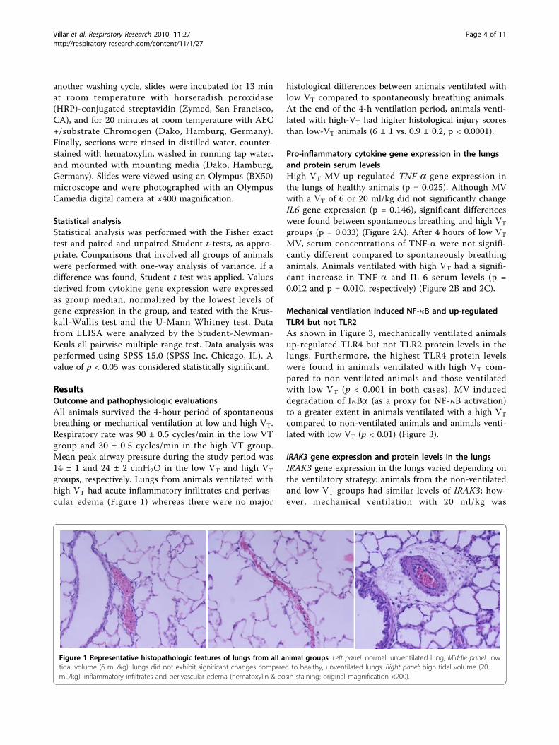
groups, respectively. Lungs from animals ventilated withhigh VT had acute inflammatory infiltrates and perivas-cular edema (Figure 1) whereas there were no major
histological differences between animals ventilated withlow VT compared to spontaneously breathing animals.At the end of the 4-h ventilation period, animals venti-lated with high-VT had higher histological injury scoresthan low-VT animals (6 ± 1 vs. 0.9 ± 0.2, p < 0.0001).
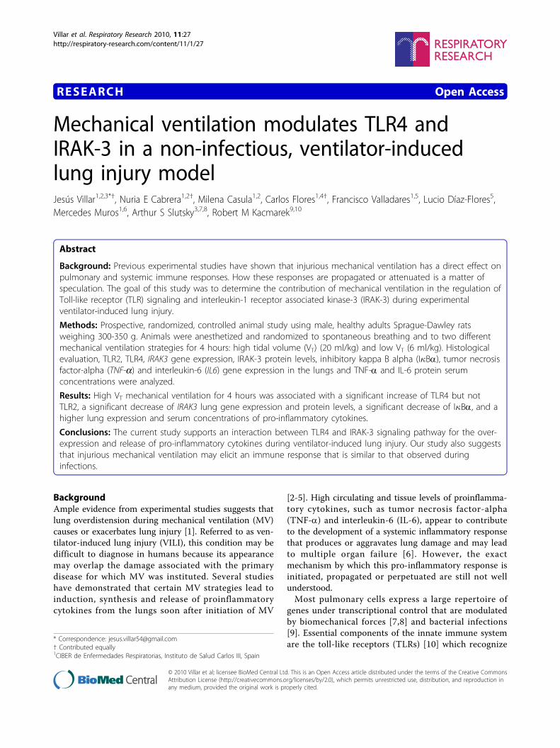
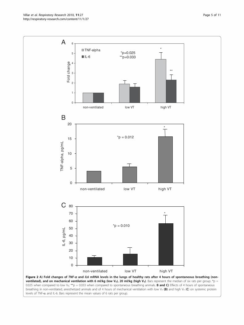
Pro-inflammatory cytokine gene expression in the lungsand protein serum levelsHigh VT MV up-regulated TNF-a gene expression inthe lungs of healthy animals (p = 0.025). Although MVwith a VT of 6 or 20 ml/kg did not significantly changeIL6 gene expression (p = 0.146), significant differenceswere found between spontaneous breathing and high VT
groups (p = 0.033) (Figure 2A). After 4 hours of low VT
MV, serum concentrations of TNF-a were not signifi-cantly different compared to spontaneously breathinganimals. Animals ventilated with high VT had a signifi-cant increase in TNF-a and IL-6 serum levels (p =0.012 and p = 0.010, respectively) (Figure 2B and 2C).
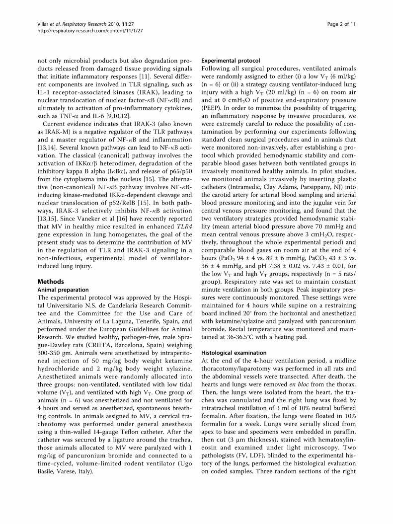
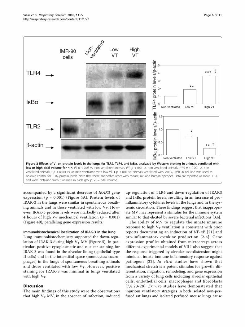
Mechanical ventilation induced NF-�B and up-regulatedTLR4 but not TLR2As shown in Figure 3, mechanically ventilated animalsup-regulated TLR4 but not TLR2 protein levels in thelungs. Furthermore, the highest TLR4 protein levelswere found in animals ventilated with high VT com-pared to non-ventilated animals and those ventilatedwith low VT (p < 0.001 in both cases). MV induceddegradation of I�Ba (as a proxy for NF-�B activation)to a greater extent in animals ventilated with a high VT
compared to non-ventilated animals and animals venti-lated with low VT (p < 0.01) (Figure 3).
IRAK3 gene expression and protein levels in the lungsIRAK3 gene expression in the lungs varied depending onthe ventilatory strategy: animals from the non-ventilatedand low VT groups had similar levels of IRAK3; how-ever, mechanical ventilation with 20 ml/kg was
Figure 1 Representative histopathologic features of lungs from all animal groups. Left panel: normal, unventilated lung; Middle panel: lowtidal volume (6 mL/kg): lungs did not exhibit significant changes compared to healthy, unventilated lungs. Right panel: high tidal volume (20mL/kg): inflammatory infiltrates and perivascular edema (hematoxylin & eosin staining; original magnification ×200).
Villar et al. Respiratory Research 2010, 11:27http://respiratory-research.com/content/11/1/27
Page 4 of 11
A
C
*p = 0.012*p = 0.010
*
0
10
20
30
40
50
60
70
80
non-ventilated low VT high VT
IL-6
, pg
/mL
B*
*p = 0.012
0
5
10
15
20
non-ventilated low VT high VT
TN
F-a
lph
a, p
g/m
L
0
1
2
3
4
5
6
non-ventilated low VT high VT
TNF-alpha
IL-6
Fo
ld c
han
ge
*p=0.025**p=0.033
*
**
Figure 2 A) Fold changes of TNF-a and IL6 mRNA levels in the lungs of healthy rats after 4 hours of spontaneous breathing (non-ventilated), and on mechanical ventilation with 6 ml/kg (low VT), 20 ml/kg (high VT). Bars represent the median of six rats per group. *p =0.025 when compared to low VT; **p = 0.033 when compared to spontaneous breathing animals. B and C) Effects of 4 hours of spontaneousbreathing in non-ventilated, anesthetized animals and of 4 hours of mechanical ventilation with low VT (B) and high VT (C) on systemic proteinlevels of TNF-a and IL-6. Bars represent the mean values of 6 rats per group.
Villar et al. Respiratory Research 2010, 11:27http://respiratory-research.com/content/11/1/27
Page 5 of 11
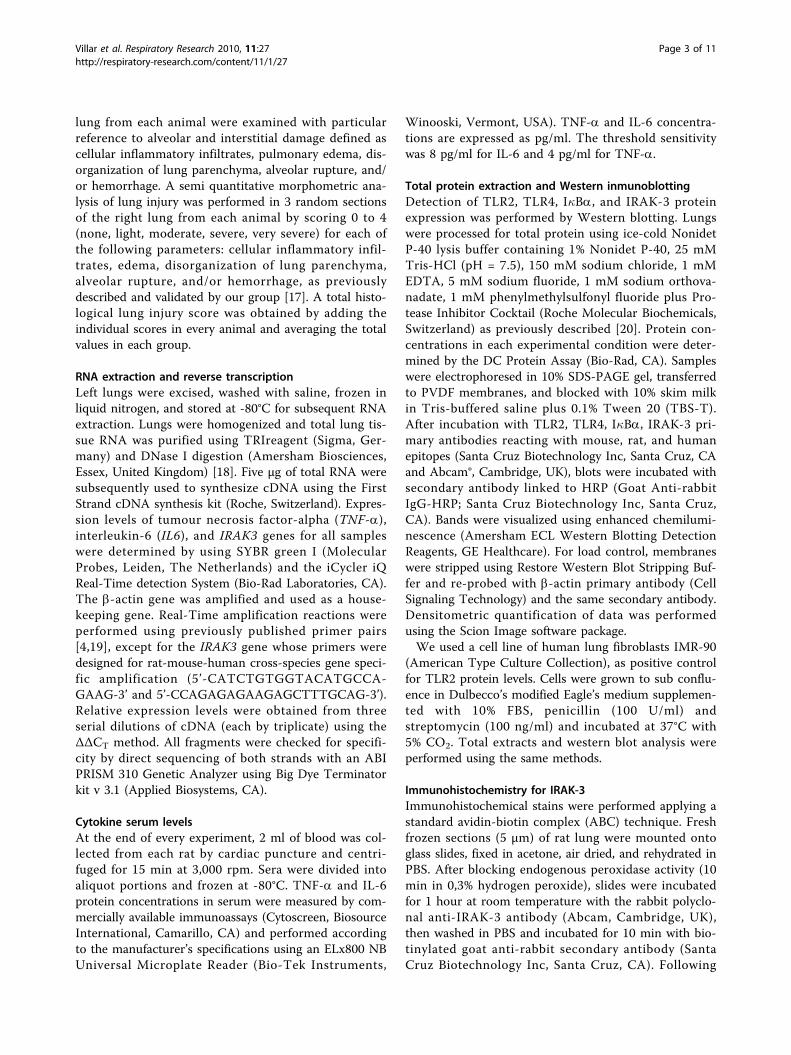
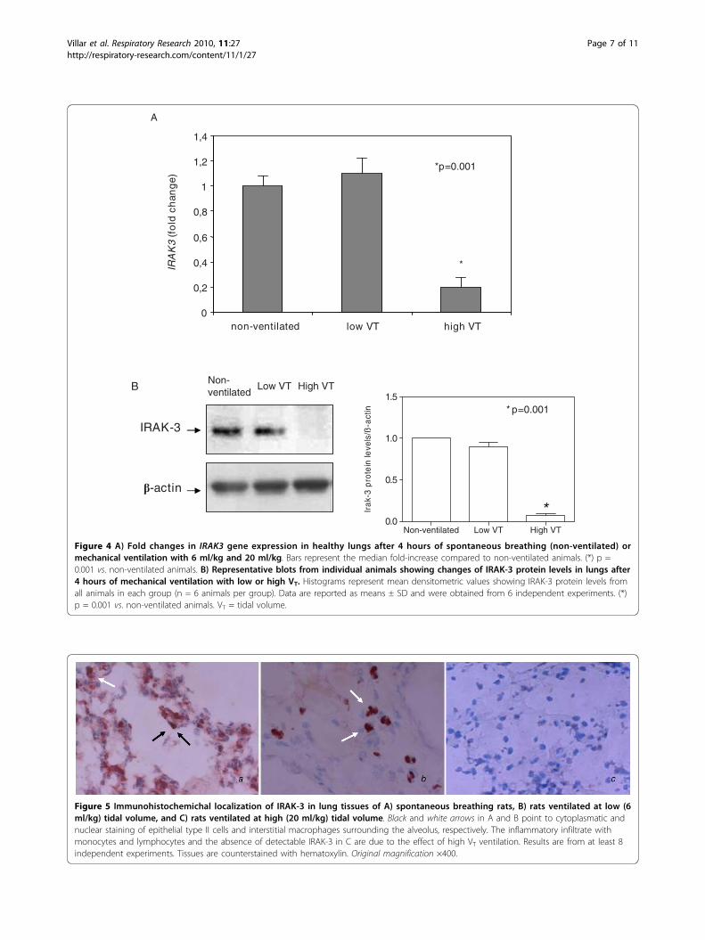
accompanied by a significant decrease of IRAK3 geneexpression (p = 0.001) (Figure 4A). Protein levels ofIRAK-3 in the lungs were similar in spontaneous breath-ing animals and in those ventilated with low VT. How-ever, IRAK-3 protein levels were markedly reduced after4 hours of high VT mechanical ventilation (p = 0.001)(Figure 4B), paralleling gene expression results.
Immunohistochemical localization of IRAK-3 in the lungLung immunohistochemistry supported the down-regu-lation of IRAK-3 during high VT MV (Figure 5). In par-ticular, positive cytoplasmatic and nuclear staining forIRAK-3 was found in the alveolar lining (epithelial typeII cells) and in the interstitial space (monocytes/macro-phages) in the lungs of spontaneous breathing animalsand those ventilated with low VT. However, positivestaining for IRAK-3 was minimal in lungs ventilatedwith high VT.
DiscussionThe main findings of this study were the observationsthat high VT MV, in the absence of infection, induced
up-regulation of TLR4 and down-regulation of IRAK3and I�Ba protein levels, resulting in an increase of pro-inflammatory cytokines levels in the lungs and in the sys-temic circulation. These findings suggest that inappropri-ate MV may represent a stimulus for the immune systemsimilar to that elicited by severe bacterial infections [3,4].The ability of MV to regulate the innate immune
response to high VT ventilation is consistent with priorreports documenting an induction of NF-�B [21] andpro-inflammatory cytokine production [2-4]. Geneexpression profiles obtained from microarrays acrossdifferent experimental models of VILI also suggest thatthe response triggered by alveolar overdistension mightmimic an innate immune inflammatory response againstpathogens [22]. In vitro studies have shown thatmechanical stretch is a potent stimulus for growth, dif-ferentiation, migration, remodeling, and gene expressionfrom a variety of lung cells including alveolar epithelialcells, endothelial cells, macrophages and fibroblasts[7,8,23-28]. Ex vivo studies have demonstrated thatinjurious ventilatory strategies in both isolated non-per-fused rat lungs and isolated perfused mouse lungs cause
Non
-ve
ntila
ted
TLR2
-actin
HighVT
TLR4
I B
IMR-90 cells
LowVT
Non-ventilated Low VT High VT0
1
2
3
4
5
TLR
4 pr
otei
n le
vels
/-a
ctin
*** t
**
Non-ventilated Low VT High VT0.0
0.5
1.0
1.5
IkB
pro
tein
leve
ls/
-act
in
**
*
Figure 3 Effects of VT on protein levels in the lungs for TLR2, TLR4, and I�Ba, analyzed by Western blotting in animals ventilated withlow or high tidal volume for 4 h. (*) p < 0.05 vs. non-ventilated animals, (**) p < 0.01 vs. non-ventilated animals, (***) p < 0.001 vs. non-ventilated animals, t p < 0.001 vs. animals ventilated with low VT, τ p < 0.01 vs. animals ventilated with low VT. IMR-90 cell line was used aspositive control for TLR2 protein levels. Note that these antibodies react with mouse, rat, and human epitopes. Data are reported as mean ± SDand were obtained from 6 animals in each group. VT = tidal volume.
Villar et al. Respiratory Research 2010, 11:27http://respiratory-research.com/content/11/1/27
Page 6 of 11
0
0,2
0,4
0,6
0,8
1
1,2
1,4
non-ventilated low VT high VT
IRA
K3
(fo
ld c
han
ge)
*p=0.001
*
B
IRAK-3
-actin
Non- ventilated
Low VT High VT
*Non-ventilated Low VT High VT
0.0
0.5
1.0
1.5
Ira
k-3
pro
tein
leve
ls/ß
-act
in * p=0.001
A
Figure 4 A) Fold changes in IRAK3 gene expression in healthy lungs after 4 hours of spontaneous breathing (non-ventilated) ormechanical ventilation with 6 ml/kg and 20 ml/kg. Bars represent the median fold-increase compared to non-ventilated animals. (*) p =0.001 vs. non-ventilated animals. B) Representative blots from individual animals showing changes of IRAK-3 protein levels in lungs after4 hours of mechanical ventilation with low or high VT. Histograms represent mean densitometric values showing IRAK-3 protein levels fromall animals in each group (n = 6 animals per group). Data are reported as means ± SD and were obtained from 6 independent experiments. (*)p = 0.001 vs. non-ventilated animals. VT = tidal volume.
Figure 5 Immunohistochemichal localization of IRAK-3 in lung tissues of A) spontaneous breathing rats, B) rats ventilated at low (6ml/kg) tidal volume, and C) rats ventilated at high (20 ml/kg) tidal volume. Black and white arrows in A and B point to cytoplasmatic andnuclear staining of epithelial type II cells and interstitial macrophages surrounding the alveolus, respectively. The inflammatory infiltrate withmonocytes and lymphocytes and the absence of detectable IRAK-3 in C are due to the effect of high VT ventilation. Results are from at least 8independent experiments. Tissues are counterstained with hematoxylin. Original magnification ×400.
Villar et al. Respiratory Research 2010, 11:27http://respiratory-research.com/content/11/1/27
Page 7 of 11
an increase in the induction and release of inflammatorymediators [2,3,29]. In vivo, injurious mechanical ventila-tion can cause an increase in pulmonary and systemicinflammatory cytokines [4,30]. Tremblay et al [3] venti-lated isolated lungs during 2 hours with a VT of 15 ml/kg and zero PEEP and found that average peak pres-sures increased 2.5-fold in the first 30 min (from 9 to23 cmH2O) and 2-fold (from 13 to 28 cmH2O) by theend of 2-hour period compared to control lungs. Wefound that average peak pressures in healthy lungs ven-tilated with 20 ml/kg increased almost 2-fold (from 14to 24 cmH2O) by the end of 4-hour period when com-pared to those animals ventilated with 6 ml/kg. Ventila-tory strategy also modulates alveolar and plasma levelsof pro-inflammatory cytokines in patients with acutelung injury [5,31].The ability of MV to induce inflammation may be in
part explained by its known ability to modulate theinduction of NF-�B in response to injurious ventilationalone or in combination with bacterial products [32]. Inan isolated perfused mouse lung model, Held et al [21]found that both overinflation of the lung (VT of 32 mL/kg) for 150 min and LPS treatment caused activation ofNF-�B in lung tissue and resulted in the release of asimilar cytokine profile. These experiments were per-formed during the same period in which IRAK-3 wasoriginally identified by Wesche et al in 1999 [33], andtherefore did not explore the possibility that deregula-tion of genes participating in the endogenous TLR-sig-naling cascades could be involved in the activation ofNF-�B. Increased TLR expression and/or signaling maycontribute to the pathophysiology of several importantdisease states since blunting the up-regulation of TLRexpression with an immunomodulator was correlatedwith improved outcome [34]. We observed that high VT
MV for 4 h induced up-regulation of TLR4 (and notTLR2) protein levels, a receptor related to LPS signaltransduction and the classical NF-�B pathway [9].Although our study was performed in rats with healthylungs, it may be possible that in addition to overdisten-sion by high VT, the lack of application of a low level ofPEEP (2-5 cmH2O) could contribute to ventilator-induced lung damage by causing the opening and clos-ing of lung units (volutrauma and atelectrauma) withevery respiratory cycle. Recently, Vaneker et al [16]reported the effects of ventilation for 4 hours with 8mL/kg VT, 4 cmH2O of PEEP and 40% oxygen inhealthy and knockout TLR2 and TLR4 mice. Theyfound that MV of healthy mice resulted in increasedexpression of endogenous TLR4 ligands in the bronch-oalveolar lavage fluid and enhanced TLR4 lung geneexpression in lung homogenates that was associatedwith increased levels of TNF-a and IL-6 in lung andplasma. However, in TLR4 knockout mice, MV did not
increase plasma levels of those cytokines. Therefore,their study also suggests that TLRs play a role in theinflammatory response initiated by MV in healthy lungs.Our study is complementary to a study by Moriyama etal [35] who found that animals ventilated with high VT
(20 ml/kg) for 4 hours had increased expression ofreceptor CD14 mRNA and protein in the absence ofLPS stimulation.IRAK-3 is a well described repressor of NF-�B signal-
ling and successful induction of pro-inflammatory sig-nals requires loss of IRAK-3 from the NF-�B pathway[13]. Although IRAK-3 was originally described inmonocytes and macrophages [33], and it is primarilypresent in the peripheral blood leukocytes and monocy-tic cell lines [36], subsequent studies have reported thatit is also expressed in other cell types. Balaci et al [37]found that IRAK3 is highly expressed in alveolar epithe-lial cells, congruent with our results (see Figure 5). Ourfindings suggest that MV functions as a modulator ofthe inflammatory response in the lung via the IRAK-3immune effects in alveolar macrophages and type IIcells. Although totally speculative, we think that thispivotal role of IRAK-3 in preventing excessive activationof NF-�B and subsequent inflammatory response mayalso be exploited by other cells. In this study, we haveshown that, in non-infected animals, MV for 4 hinduced IRAK-3 down-regulation, TLR4 up-regulationand that different patterns of MV caused different pat-terns of IRAK-3 expression. This is congruent with anenhanced NF-�B activation directly caused by IRAK-3deficiency [38]. Kobayashi et al [13] also showed thatmacrophages from IRAK-3-deficient mice producedmarkedly enhanced levels of inflammatory cytokines inresponse to TLR stimulation. This loss of IRAK-3 pro-tein levels could be responsible for the increased cyto-kine production. However, the mechanism for loss ofIRAK-3 expression remains incompletely understood.Components of the extracellular tissue matrix (includingproteoglycan, collagen and elastin) could play a key rolein the unremitting inflammation during ventilator-induced lung injury [34,39,40]. Moriondo et al [39]examined the effects of stretching lung tissue during 4hours of MV at various VT with zero PEEP in the lungsof healthy animals and found that significant fragmenta-tion and degradation of the components of the extracel-lular tissue matrix were observed after ventilatinghealthy rats with VT ≥ 16 ml/kg. Jiang et al [41] demon-strated that extracellular matrix fragments isolated fromserum of patients with acute lung injury stimulatedmacrophage chemokine and cytokine productionthrough a TLR-dependent activation of NF-�B. In addi-tion, down-regulation of IRAK3 expression by specificsmall interfering RNAs have been shown to reinstatethe production of TNF-a after re-stimulation of
Villar et al. Respiratory Research 2010, 11:27http://respiratory-research.com/content/11/1/27
Page 8 of 11
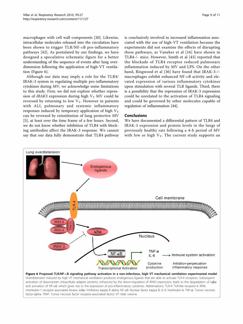
macrophages with cell wall components [20]. Likewise,intracellular molecules released into the circulation havebeen shown to trigger TLR/NF-�B pro-inflammatorypathways [42]. As postulated by our findings, we havedesigned a speculative schematic figure for a betterunderstanding of the sequence of events after lung over-distension following the application of high-VT ventila-tion (Figure 6).Although our data may imply a role for the TLR4/
IRAK-3 system in regulating multiple pro-inflammatorycytokines during MV, we acknowledge some limitationsto this study. First, we did not explore whether repres-sion of IRAK3 expression during high VT MV could bereversed by returning to low VT. However in patientswith ALI, pulmonary and systemic inflammatoryresponses induced by temporary application of high VT
can be reversed by reinstitution of lung protective MV[5], at least over the time frame of a few hours. Second,we do not know whether inhibition of TLR4 with block-ing antibodies affect the IRAK-3 response. We cannotsay that our data fully demonstrate that TLR4 pathway
is conclusively involved in increased inflammation asso-ciated with the use of high-VT ventilation because theexperiments did not examine the effects of disruptingthese pathways, as Vaneker et al [16] have shown inTLR4-/- mice. However, Smith et al [43] reported thatthe blockade of TLR4 receptor reduced pulmonaryinflammation induced by MV and LPS. On the otherhand, Ringwood et al [36] have found that IRAK-3-/-macrophages exhibit enhanced NF-�B activity and ele-vated expression of various inflammatory cytokinesupon stimulation with several TLR ligands. Third, thereis a possibility that the repression of IRAK-3 expressioncould be unrelated to the activation of TLR4 signalingand could be governed by other molecules capable ofregulation of inflammation [44].
ConclusionsWe have documented a differential pattern of TLR4 andIRAK-3 expression and protein levels in the lungs ofpreviously healthy rats following a 4-h period of MVwith low or high VT. The current study supports an
Figure 6 Proposed TLR/NF-�B signaling pathway activation in a non-infectious, high VT mechanical ventilation experimental model.Overdistension induced by high VT mechanical ventilation produces endogenous ligands that are able to activate TLR-4 receptors. Subsequentactivation of downstream intracellular adapter proteins, enhanced by the down-regulation of IRAK3 expression, leads to the degradation of I�baand activation of NF-�B, which gives rise to the expression of pro-inflammatory cytokines. Abbreviations: TLR-4: Toll-like receptor-4; IRAK:Interleukin-1 receptor-associated kinase; I�Ba: Inhibitory kappa B alpha; NF-�B: Nuclear factor kappa B; IL-6: Interleukin-6; TNF-a: Tumor necrosisfactor-alpha. TRAF: Tumor necrosis factor receptor-associated factor; VT: tidal volume.
Villar et al. Respiratory Research 2010, 11:27http://respiratory-research.com/content/11/1/27
Page 9 of 11
interaction between TLR4 and NF-�B signaling pathwayfor the over-expression and release of pro-inflammatorycytokines during ventilator-induced lung injury. Ourstudy also suggests that injurious MV may elicit animmune response that is similar to that observed duringsevere infections. Further studies are needed to fullyaddress these questions.
AbbreviationsELISA: enzyme-linked immunosorbent assay; I�Ba: inhibitory kappa B alpha;IL-6: interleukin-6; MV: mechanical ventilation; NF-�B: nuclear factor kappa B;PEEP: positive end-expiratory pressure; TLR2: Toll-like receptor-2; TLR-4: Toll-like receptor-4; TNF-a: tumor necrosis factor-alpha; VT: tidal volume.
AcknowledgementsThe study has been supported by grants from Ministerio de Ciencia of Spain(SAF 2004-06833), FUNCIS (53/04), and by a specific agreement betweenInstituto de Salud Carlos III and FUNCIS (EMER07/001) under the ENCYT 2015framework.
Author details1CIBER de Enfermedades Respiratorias, Instituto de Salud Carlos III, Spain.2Multidisciplinary Organ Dysfunction Evaluation Research Network(MODERN), Research Unit, Hospital Universitario Dr. Negrin, Las Palmas deGran Canaria, Spain. 3Keenan Research Center at the Li Ka Shing KnowledgeInstitute of St. Michael’s Hospital, Toronto, Canada. 4Research Unit, HospitalUniversitario N.S. de Candelaria, Tenerife, Spain. 5Department of Anatomy,Pathology & Histology, University of La Laguna, Tenerife, Spain. 6Departmentof Clinical Biochemistry, Hospital Universitario NS de Candelaria, Tenerife,Spain. 7Interdepartmental Division of Critical Care Medicine, University ofToronto, Toronto, Canada. 8King Saud University, Riyadh, Saudi Arabia.9Department of Respiratory Care, Massachusetts General Hospital, Boston,Massachusetts, USA. 10Department of Anesthesia, Harvard Medical School,Boston, MA, USA.
Authors’ contributionsJV, CF, RK and AS conceived and designed the study. JV obtained fundingfor the study. JV, NC, MC, FV, LDF, CF, MM performed the experiments. JV,CF, and FV coordinated data collection and data quality. CF, NC, LDF andMM performed statistical analysis. JV, NC, MC, CF, FV, RK, and AS participatedin the first draft of the manuscript. All authors participated in the writingprocess of the manuscript and read and approved the final manuscript.
Authors’ informationArthur S Slutsky is Adjunct Professor at King Saud University, Riyadh, SaudiArabia
Competing interestsThe authors declare that they have no competing interests.
Received: 13 December 2009Accepted: 3 March 2010 Published: 3 March 2010
References1. Dreyfuss D, Soler P, Basset G, Saumon G: High inflation pressure
pulmonary edema: respective effects of high airway pressure, high tidalvolume and positive end-expiratory pressure. Am Rev Respir Dis 1988,137:1159-1164.
2. von Bethmann AN, Brasch F, Nüsing R, Vogt K, Volk HD, Müller KM,Wendel A, Uhlig S: Hyperventilation induces release of cytokines fromperfused mouse lung. Am J Respir Crit Care Med 1998, 157:263-272.
3. Tremblay L, Valenza F, Ribeiro SP, Li J, Slutsky AS: Injurious ventilatorystrategies increase cytokines and c-fos m-RNA expression in an isolatedrat lung model. J Clin Invest 1997, 99:944-952.
4. Herrera MT, Toledo C, Valladares F, Muros M, Díaz-Flores L, Flores C, Villar J:Positive end-expiratory pressure modulates local and systemicinflammatory responses in a sepsis-induced lung injury model. IntensiveCare Med 2003, 29:1345-1353.
5. Stüber F, Wrigge H, Schroeder S, Wetegrove S, Zinserling J, Hoeft A,Putensen C: Kinetic and reversibility of mechanical ventilation-associatedpulmonary and systemic inflammatory response in patients with acutelung injury. Intensive Care Med 2002, 28:834-841.
6. Slutsky AS, Tremblay LN: Multiple system organ failure. Is mechanicalventilation a contributing factor?. Am J Respir Crit Care Med 1998,157:1721-1725.
7. Dekker RJ, van Soest S, Fontijn RD, Salamanca S, de Groot PG, VanBavel E,Pannekoek H, Horrevoets AJ: Prolonged fluid shear stress induces adistinct set of endothelial cell genes, most specifically lung Kruppel-likefactor (KLF2). Blood 2002, 100:1689-1698.
8. Liu M, Tanswell AK, Post M: Mechanical force-induced signal transductionin lung cells. Am J Physiol (Lung Cell Mol Physiol) 1999, 21:L667-L683.
9. Cohen J: The immunopathogenesis of sepsis. Nature 2002, 420:885-891.10. Aderem A, Ulevitch RJ: Toll-like receptors in the induction of the innate
immune response. Nature 2000, 406:782-787.11. Jiang D, Liang J, Li Y, Noble PW: The role of Toll-like receptors in non-
infectious lung injury. Cell Res 2006, 16:693-701.12. Liu SF, Malik AB: NF-�B activation as a pathological mechanism of septic
shock and inflammation. Am J Physiol 2006, 290:L622-L645.13. Kobayashi K, Hernandez LD, Galán JE, Janeway CA, Medzhitov R, Flavell RA:
IRAK-M is a negative regulator of Toll-like receptor signaling. Cell 2002,110:191-202.
14. Liew FY, Xu D, Brint EK, O’Neill LAJ: Negative regulation of Toll-likereceptor-mediated immune responses. Nature Rev Immunol 2005,5:446-458.
15. Su J, Zhang J, Tyson J, Li L: The Interleukin-1 Receptor-Associated KinaseM selectively inhibits the alternative, instead of the classical NF�Bpathway. J Innate Immun 2009, 1:164-174.
16. Vaneker M, Joosten LA, Heunks LMA, Snijdelaar DG, Halbertsma FJ, vanEgmond J, Netea MG, Hoeven van der JG, Scheffer GJ: Low tidal volumemechanical ventilation induces a Toll-like receptor 4-dependentinflammatory response in healthy mice. Anesthesiology 2008, 109:465-472.
17. Villar J, Herrera-Abreu MT, Valladares F, Muros M, Pérez-Méndez L, Flores C,Kacmarek RM: Experimental ventilator-induced lung injury. Exacerbationby positive end-expiratory pressure. Anesthesiology 2009, 110:1341-1347.
18. Chomczynski P, Sacchi N: Single-step method of RNA isolation by acidguanidium thiocyanate-phenol-chloroform extraction. Anal Biochem 1987,162:156-159.
19. Patak E, Candenas ML, Pennefather JN, Ziccone S, Lilley A, Martín JD,Flores C, Mantecón AG, Story ME, Pinto FM: Tachykinins and tachykininreceptors in human uterus. Br J Pharmacol 2003, 139:523-532.
20. Nakayama K, Okugawa S, Yanagimoto S, Kitazawa T, Tsukada K, Kawada M,Kimura S, Hirai K, Takagaki Y, Ota Y: Involvement of IRAK-M inpeptidoglycan-induced tolerance in macrophages. J Biol Chem 2004,279:6629-6634.
21. Held HD, Boettcher S, Hamann L, Uhlig S: Ventilation-induced chemokineand cytokine release is associated with activation of nuclear factor-�Band is blocked by steroids. Am J Respir Crit Care Med 2001, 163:711-716.
22. Wurfel MM: Microarray-based analysis of ventilator-induced lung injury.Proc Am Thorac Soc 2007, 4:77-84.
23. Wirtz HR, Dobbs LG: Calcium mobilization and exocytosis after onemechanical stretch of lung epithelial cells. Science 1990, 250:1266-1269.
24. Mourgeon E, Isowa N, Keshavjee S, Zhang X, Slutsky AS, Liu M: Mechanicalstretch stimulates macrophage inflammatory protein-2 secretion fromfetal rat lung cells. Am J Physiol Lung Cell Mol Physiol 2000, 279:L699-L706.
25. Quinn D, Tager A, Joseph PM, Bonventre JV, Force T, Hales CA: Stretch-induced mitogen-activated protein kinase activation and interleukin-8production in type II alveolar cells. Chest 1999, 116:S89-S90.
26. Vogel V, Sheetz M: Local force and geometry sensing regulate cellfunctions. Nature Rev Mol Cell Biol 2006, 7:265-275.
27. Pugin J, Dunn I, Jolliet P, Tassaux D, Magnenat JL, Nicod LP, Chevrolet JC:Activation of human macrophages by mechanical ventilation in vitro.Am J Physiol 1998, 275:L1040-1050.
28. Vlahakis NE, Schroeder MA, Limper AH, Hubmayr RD: Stretch inducescytokine release by alveolar epithelial cells in vitro. Am J Physiol 1999,277:L167-173.
29. Tremblay LN, Miatto D, Hamid Q, Govindarajan A, Slutsky AS: Injuriousventilation induces widespread pulmonary epithelial expression oftumor necrosis factor-alpha and interleukin-6 messenger RNA. Crit CareMed 2002, 30:1693-1700.
Villar et al. Respiratory Research 2010, 11:27http://respiratory-research.com/content/11/1/27
Page 10 of 11
30. Chiumello D, Pristine G, Slutsky AS: Mechanical ventilation affects localand systemic cytokines in an animal model of acute respiratory distresssyndrome. Am J Respir Crit Care Med 1999, 160:109-116.
31. Ranieri VM, Suter PM, Tortorella C, De Tullio R, Dayer JM, Brienza A, Bruno F,Slutsky AS: Effect of mechanical ventilation on inflammatory mediators inpatients with acute respiratory distress syndrome: a randomizedcontrolled trial. JAMA 1999, 282:54-61.
32. Martin TR: Interactions between mechanical and biological processes inacute lung injury. Proc Am Thorac Soc 2008, 5:291-296.
33. Wesche H, Gao X, Li X, Kirschning CJ, Stark GR, Cao Z: IRAK-M is a novelmember of the Pelle/interleukin-1 receptor-associated kinase (IRAK)family. J Biol Chem 1999, 274:19403-19410.
34. Williams DL, Ha T, Li C, Kalbfleisch JH, Schweitzer J, Vogt W, Browder W:Modulation of tissue Toll-like receptor 2 and 4 during the early phasesof polymicrobial sepsis correlates with mortality. Crit Care Med 2003,31:1808-1818.
35. Moriyama K, Ishizaka A, Nakamura M, Kubo H, Kotani T, Yamamoto S,Ogawa EN, Kajikawa O, Frevert CW, Kotake Y, Morisaki H, Koh H, Tasaka S,Martin TR, Takeda J: Enhancement of the endotoxin recognition pathwayby ventilation with a large tidal volume in rabbits. Am J Physiol Lung CellMol Physiol 2004, 286:L1114-L1121.
36. Ringwood L, Liwu L: The involvement of the interleukin-1 receptor-associated kinases (IRAKs) in cellular signaling networks controllinginflammation. Cytokine 2008, 42:1-7.
37. Balaci L, Spada MC, Olla N, Sole G, Loddo L, Anedda F, Naitza S,Zuncheddu MA, Maschio A, Altea D, et al: IRAK-M is involved in thepathogenesis of early-onset persistent asthma. Am J Hum Genet 2007,80:1103-1114.
38. Xie Q, Gan L, Wang J, Wilson I, Li L: Loss of the innate immunity negativeregulator IRAK-M leads to enhanced host immune defense againsttumor growth. Mol Immunol 2007, 44:3453-3461.
39. Moriondo A, Pelosi P, Passi A, Viola M, Marcozzi C, Severgnini P, Ottani V,Quaranta M, Negrini D: Proteoglycan fragmentation and respiratorymechanics in mechanically ventilated healthy rats. J Appl Physiol 2007,103:747-756.
40. Pelosi P, Rocco PR: Effects of mechanical ventilation on the extracellularmatrix. Intensive Care Med 2008, 34:631-639.
41. Jiang D, Liang J, Fan J, Yu S, Chen S, Luo Y, Prestwich GD,Mascarenhas MM, Garg HG, Quinn DA, Homer RJ, Goldstein DR, Bucala R,Lee PJ, Medzhitov R, Noble PW: Regulation of lung injury and repair byToll-like receptors and hyaluronan. Nat Med 2005, 11:1173-1179.
42. Pereira C, Schaer DJ, Bachli EB, Kurrer MO, Schoedon G: Wnt5A/CaMKIIsignaling contributes to the inflammatory response of macrophages andis a target for the anti-inflammatory action of activated protein C andinterleukin-10. Arterioscler Thromb Vasc Biol 2008, 28:504-510.
43. Smith LS, Kajikawa O, Elson G, Wick M, Mongovin S, Kosco-Vilbois M,Martin TR, Frevert CW: Effect of Toll-like receptor 4 blockage onpulmonary inflammation caused by mechanical ventilation and bacterialendotoxin. Exp Lung Res 2008, 34:225-243.
44. Matsuyama H, Amaya F, Hashimoto S, Ueno H, Beppu S, Mizuta M,Shime N, Ishizaka A, Hashimoto S: Acute lung inflammation andventilator-induced lung injury caused by ATP via the P2Y receptors: anexperimental study. Respir Res 2008, 9:79.
doi:10.1186/1465-9921-11-27Cite this article as: Villar et al.: Mechanical ventilation modulates TLR4and IRAK-3 in a non-infectious, ventilator-induced lung injury model.Respiratory Research 2010 11:27.
Submit your next manuscript to BioMed Centraland take full advantage of:
• Convenient online submission
• Thorough peer review
• No space constraints or color figure charges
• Immediate publication on acceptance
• Inclusion in PubMed, CAS, Scopus and Google Scholar
• Research which is freely available for redistribution
Submit your manuscript at www.biomedcentral.com/submit
Villar et al. Respiratory Research 2010, 11:27http://respiratory-research.com/content/11/1/27
Page 11 of 11