Embed Size (px)
Citation preview
Mechanism of action of a flavin-containing monooxygenaseSubramaniam Eswaramoorthy*, Jeffrey B. Bonanno†, Stephen K. Burley‡, and Subramanyam Swaminathan*§
*Biology Department, Brookhaven National Laboratory, Upton, NY 11973; †New York Structural Biology Center, New York, NY 10027; and ‡SGXPharmaceuticals, Inc., San Diego, CA 92121
Edited by Brian W. Matthews, University of Oregon, Eugene, OR, and approved May 11, 2006 (received for review March 24, 2006)
Elimination of nonnutritional and insoluble compounds is a criticaltask for any living organism. Flavin-containing monooxygenases(FMOs) attach an oxygen atom to the insoluble nucleophilic com-pounds to increase solubility and thereby increase excretion. Herewe analyze the functional mechanism of FMO from Schizosaccha-romyces pombe using the crystal structures of the wild type andprotein–cofactor and protein–substrate complexes. The structureof the wild-type FMO revealed that the prosthetic group FAD is anintegral part of the protein. FMO needs NADPH as a cofactor inaddition to the prosthetic group for its catalytic activity. Structuresof the protein–cofactor and protein–substrate complexes provideinsights into mechanism of action. We propose that FMOs exist inthe cell as a complex with a reduced form of the prosthetic groupand NADPH cofactor, readying them to act on substrates. The4�-hydroperoxyflavin form of the prosthetic group represents atransient intermediate of the monooxygenation process. The ox-ygenated and reduced forms of the prosthetic group help stabilizeinteractions with cofactor and substrate alternately to permitcontinuous enzyme turnover.
three-dimensional structure � xenobiotics � methimazole
F lavin-containing monooxygenases (FMOs) and cytochromesP450 are two important microsomal proteins involved in the
process of nonnutritional foreign compounds metabolism known asxenobiotics. Their main function is to add molecular oxygen tolipophilic compounds, making them soluble to ensure rapid excre-tion. FMOs oxygenate nucleophilic O, N, S, and Se atoms of a widerange of substrates, such as amines, amides, thiols, and sulfides (1).FMOs of liver microsomes are known as microsomal FMOs (2).They are divided into three classes, namely FMOs, N-hydroxylatingmonooxygenases, and Baeyer–Villigar monooxygenases (BVMOs).The signature sequence, FXGXXXHXXXW(P�D), of BVMOdiffers from FXGXXXHXXX(Y�F) of FMO, distinguishing thesetwo enzymes (3). The mechanism of action of FMOs is distinctlydifferent from that of other monooxygenases. FMOs do not requiresubstrate for dioxygen reduction of the prosthetic group FAD byNADPH. Instead, the protein plus the prosthetic group and thecofactor in its 4�-hydroperoxyflavin form stand ready to performchemistry on available nucleophiles (4).
The mammalian FMO gene family contains five similar genes(FMO1 through FMO5). FMO1 and FMO3 are prominentisoforms expressed mainly in liver microsomes and in othertissues. FMO1 expression is higher in fetal liver, whereas FMO3is more abundant in adult human. FMO2 is expressed in thelungs of nonhuman primates; FMO4 and FMO5 represent scarceisoforms (5, 6). Individuals with defective FMOs exhibit ‘‘fishodor syndrome’’ caused by excretion of trimethylamine insteadof its oxygenated form, trimethylamine N-oxide in urine, sweat,and breath (7).
Here we present three crystal structures, (i) FMO from Schizo-saccharomyces pombe with a bound FAD, (ii) its complex withNADPH, and (iii) an enzyme–substrate [N-methyl-2-mercaptoimi-dazole (methimazole)] complex. Hydride ion reduction of FAD toFADH2 and the mechanism of oxygenation of the substrate me-thimazole are discussed.
ResultsFMO Activity. The protein solution appeared dark yellow, possiblyarising from the prosthetic group FAD. On adding NADPH itturned pale yellow, apparently because of the FAD reduction toFADH2. The protein and NADPH complex was added to theaerated tricine reaction buffer. The protein was in the active format this stage and ready to oxygenate a suitable substrate (methim-azole in this case). The 5,5�-dithiobis(2-nitrobenzoate) (DTNB)added to the reaction mixture was reduced to nitro-5-thiobenzoateby DTT. OD was measured at 412 nm every 5 seconds to monitorthe DTNB concentration. OD values decreased with time for �5min and stabilized. Although the DTNB was reduced by DTT, therewas residual OD observed after 5 min. Methimazole was added tothe reaction mixture at this point, and the OD measurement wasstarted. The OD increased steadily for �10 min and stabilized.
Dixit and Roche (1) have demonstrated that oxygenated me-thimazole reacts with nitro-5-thiobenzoate to produce DTNB. Theincrease in OD corresponds to the increment of DTNB in thereaction mixture, confirming oxygenation of substrate. This reac-tion also demonstrated that the maximum DTNB production wasachieved within 10 min. In the control experiment without me-thimazole, the OD increased very slowly. It took �30 min to reachthe maximum OD of 1.12, which is considerably less than that of theexperiment with methimazole (1.32). The difference in OD readingbetween these two experiments (with and without methimazole)over 10 min is plotted in Fig. 1.
Structure of FMO. The crystal structure of the enzyme–FAD com-plex was first determined as part of the structural genomics effortby the New York SGX Research Consortium and has been depos-ited in the Protein Data Bank (PDB ID code 1VQW). Thestructural models of enzyme–FAD, enzyme–FAD–NADPH, andenzyme–FAD–methimazole complexes contain residues Leu-3 toGlu-444. No electron density was observed for the two N-terminalresidues, the three C-terminal residues, and the C-terminal hexa-histidine affinity tag. The electron density maps were of high qualityfor the entire model, and the protein exists as dimer (Fig. 5, whichis published as supporting information on the PNAS web site).Whereas the enzyme–FAD and enzyme–FAD–NADPH complexstructures have one dimer per unit cell of the P1 symmetry, theenzyme–FAD–methimazole complex has two. No conformationalchanges were evident when the three structures were compared indetail, permitting refinement with noncrystallographic symmetryrestraints.
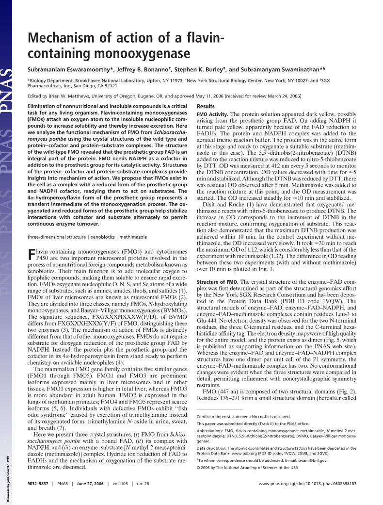
FMO (447 aa) is composed of two structural domains (Fig. 2).Residues 176–291 form a small structural domain (hereafter called
Conflict of interest statement: No conflicts declared.
This paper was submitted directly (Track II) to the PNAS office.
Abbreviations: FMO, flavin-containing monooxygenase; methimazole, N-methyl-2-mer-captoimidazole; DTNB, 5,5�-dithiobis(2-nitrobenzoate); BVMO, Baeyer–Villigar monooxy-genase.
Data deposition: The atomic coordinates and structure factors have been deposited in theProtein Data Bank, www.pdb.org (PDB ID codes 1VQW, 2GV8, and 2GVC).
§To whom correspondence should be addressed. E-mail: [email protected].
© 2006 by The National Academy of Sciences of the USA
9832–9837 � PNAS � June 27, 2006 � vol. 103 � no. 26 www.pnas.org�cgi�doi�10.1073�pnas.0602398103
Dow
nloa
ded
by g
uest
on
Mar
ch 1
, 202
0 D
ownl
oade
d by
gue
st o
n M
arch
1, 2
020
Dow
nloa
ded
by g
uest
on
Mar
ch 1
, 202
0
the insertion domain), with the remainder of the polypeptide chainforming a larger single domain. A channel is present between thesetwo domains. A 60-residue-long polypeptide chain segment ina predominantly random coil configuration with some minor sec-ondary structure elements occurs in the interface between thetwo domains, where it appears to stabilize the overall domainorganization.
The FMO large domain consists of a four-stranded parallel�-sheet flanked by a three-stranded antiparallel �-sheet on one sideand six �-helices on the other (Fig. 6, which is published assupporting information on the PNAS web site). The insertiondomain consists of a five-stranded parallel �-sheet flanked by a
three-stranded antiparallel �-sheet and a helix on one side, and an�-helix on the other. The C terminus of the polypeptide chain formsa bent helix (�81 and �82) that extends from the large domain to theinsertion domain. The C termini of the parallel �-sheets of the twodomains point toward each other. This structure also contains threestrand–turn–helix motifs. The first motif is formed by �1 and �1.This motif has a nucleotide binding sequence, GAGPSG(GXGXXG), which stabilizes binding of FAD in all three structurespresented here. The second motif, formed by �8 and �4, is locatedin the insertion domain, where it stabilizes binding of NADPH inthe enzyme–FAD–NADPH complex (Fig. 7, which is published assupporting information on the PNAS web site). In the enzyme–FAD–substrate complex, methimazole replaces NADPH and oc-cupies the nicotinamide site. The third strand–turn–helix motif,formed by �15 and �5, occurs where the C terminus of the insertiondomain connects to large domain. The second half of the largedomain contains the FMO identifying sequence FXGXXX-HXXXF and interacts with the flavin part of FAD.
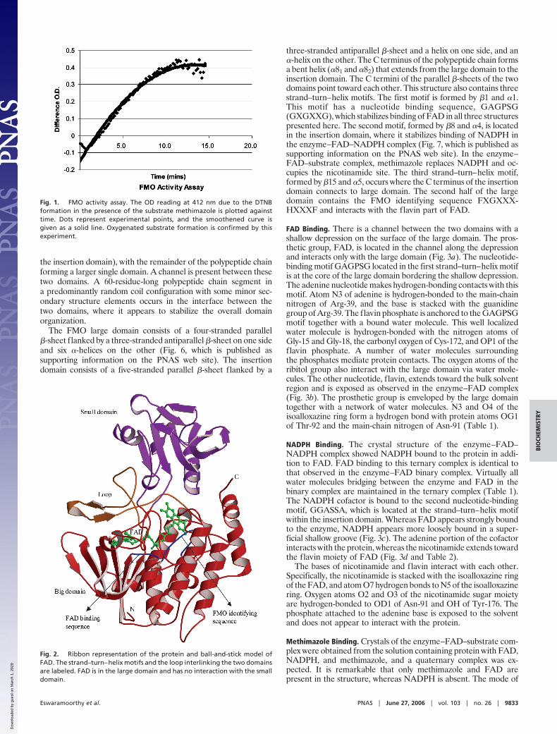
FAD Binding. There is a channel between the two domains with ashallow depression on the surface of the large domain. The pros-thetic group, FAD, is located in the channel along the depressionand interacts only with the large domain (Fig. 3a). The nucleotide-binding motif GAGPSG located in the first strand–turn–helix motifis at the core of the large domain bordering the shallow depression.The adenine nucleotide makes hydrogen-bonding contacts with thismotif. Atom N3 of adenine is hydrogen-bonded to the main-chainnitrogen of Arg-39, and the base is stacked with the guanidinegroup of Arg-39. The flavin phosphate is anchored to the GAGPSGmotif together with a bound water molecule. This well localizedwater molecule is hydrogen-bonded with the nitrogen atoms ofGly-15 and Gly-18, the carbonyl oxygen of Cys-172, and OP1 of theflavin phosphate. A number of water molecules surroundingthe phosphates mediate protein contacts. The oxygen atoms of theribitol group also interact with the large domain via water mole-cules. The other nucleotide, flavin, extends toward the bulk solventregion and is exposed as observed in the enzyme–FAD complex(Fig. 3b). The prosthetic group is enveloped by the large domaintogether with a network of water molecules. N3 and O4 of theisoalloxazine ring form a hydrogen bond with protein atoms OG1of Thr-92 and the main-chain nitrogen of Asn-91 (Table 1).
NADPH Binding. The crystal structure of the enzyme–FAD–NADPH complex showed NADPH bound to the protein in addi-tion to FAD. FAD binding to this ternary complex is identical tothat observed in the enzyme–FAD binary complex. Virtually allwater molecules bridging between the enzyme and FAD in thebinary complex are maintained in the ternary complex (Table 1).The NADPH cofactor is bound to the second nucleotide-bindingmotif, GGASSA, which is located at the strand–turn–helix motifwithin the insertion domain. Whereas FAD appears strongly boundto the enzyme, NADPH appears more loosely bound in a super-ficial shallow groove (Fig. 3c). The adenine portion of the cofactorinteracts with the protein, whereas the nicotinamide extends towardthe flavin moiety of FAD (Fig. 3d and Table 2).
The bases of nicotinamide and flavin interact with each other.Specifically, the nicotinamide is stacked with the isoalloxazine ringof the FAD, and atom O7 hydrogen bonds to N5 of the isoalloxazinering. Oxygen atoms O2 and O3 of the nicotinamide sugar moietyare hydrogen-bonded to OD1 of Asn-91 and OH of Tyr-176. Thephosphate attached to the adenine base is exposed to the solventand does not appear to interact with the protein.
Methimazole Binding. Crystals of the enzyme–FAD–substrate com-plex were obtained from the solution containing protein with FAD,NADPH, and methimazole, and a quaternary complex was ex-pected. It is remarkable that only methimazole and FAD arepresent in the structure, whereas NADPH is absent. The mode of
Fig. 1. FMO activity assay. The OD reading at 412 nm due to the DTNBformation in the presence of the substrate methimazole is plotted againsttime. Dots represent experimental points, and the smoothened curve isgiven as a solid line. Oxygenated substrate formation is confirmed by thisexperiment.
Fig. 2. Ribbon representation of the protein and ball-and-stick model ofFAD. The strand–turn–helix motifs and the loop interlinking the two domainsare labeled. FAD is in the large domain and has no interaction with the smalldomain.
Eswaramoorthy et al. PNAS � June 27, 2006 � vol. 103 � no. 26 � 9833
BIO
CHEM
ISTR
Y
Dow
nloa
ded
by g
uest
on
Mar
ch 1
, 202
0
FAD binding is the same as that observed for the wild-type protein.We believe that methimazole competes with the NADPH cofactorand replaces it. Methimazole occupies the nicotinamide bindingposition seen in the enzyme–FAD–NADPH complex and stackswith the isoalloxazine ring. It was previously thought that FMOforms a functional quaternary complex. Our results clearly showthat the cofactor and substrate occupy overlapping binding sites bystacking on the FAD, thereby precluding quaternary complexformation. Methimazole is surrounded by water molecules and doesnot make any direct contacts with the protein.
In our initial refinement models, an elongated electron densityfeature positioned 3.7 Å from the S of methimazole was inter-preted as a water molecule hydrogen-bonded to OD1 of Asn-91.However, the elongated electron density feature suggested thepresence of a dioxygen molecule. When O2 was modeled into thisfeature, the refinement converged well (Fig. 3e). It is, however,possible that this feature can be explained by one or moredisordered water molecules.
DiscussionThe biochemical assay of methimazole oxygenation unambigu-ously showed that this protein SPBP16F5.08c of S. pombe (GenBank accession no. GI: 19112574) is an FMO (8).
The structure of the wild-type protein is the first and the only onedetermined from the FMO protein family and will be useful as arepresentative model of this family. Cytochrome P450s contain
heme as a prosthetic group, whereas FMOs use FAD. Theseproteins need a cofactor NADPH in addition to the prostheticgroup to accomplish their functional goal. The structure includestwo similar structural domains with the C-terminal portion of theparallel �-sheets facing each other. These �-sheets hold the pros-thetic group (FAD) and the cofactor (NADPH), allowing them tointeract for FAD reduction. The helical bundle found within thelarge domain may be responsible for substrate capture, although therole played by the FMO identifying sequence (FXGXXXHXXXF)is not clear.
Mechanism of FMO Activity. The catalytic activity of this enzyme iscarried out mainly through the prosthetic group FAD and cofactorNADPH. Asn-91 is the only protein residue directly involved in thecatalytic mechanism as substantiated from the crystal structures. Tothe best of our knowledge, this is the first study to elucidate themechanism of action of FMO via crystallographic studies of distinctsteps in the reaction pathway (Fig. 4).
FAD is a major electron carrier in the oxidation–reductionprocesses catalyzed by enzymes. The electron donor in most ofreductive biosyntheses is NADPH, a reducing species that is readilyavailable in the cells. In the process of monooxygenation by theenzyme FMO, the prosthetic group FAD is first reduced to FADH2through a hydride ion transfer from NADPH. In the second stepFADH2 accepts a molecular oxygen at the C4A (or C10) positionof the isoalloxazine ring and becomes 4�-hydroperoxyflavin, FAD-
Fig. 3. Protein–cofactor and protein–substrate interactions. (a) Electrostatic potential of the large domain of FMO. The insertion domain was excluded to clarifythe view of the cavity formed along the large domain. FAD is depicted as a stick model. This cavity accommodates the prosthetic group, with adenine completelyburied in the protein, and the flavin is more exposed to the solvent. (b) Hydrogen bonding interactions of the prosthetic group. FAD is represented as aball-and-stick model, and the protein residues are shown as sticks. Water molecules are shown as red spheres. (c) Electrostatic potential of the insertion domainof FMO. The NADPH cofactor is shown as a stick model. NADPH is bound to the protein in a shallow cavity. (d) Hydrogen bonding interactions of the cofactor.NADPH is rendered as a ball-and-stick model, and the protein residues are shown as sticks. Water molecules are shown as red spheres. (e) Stereo diagram ofmethimazole and the isoalloxazine ring stacking along with a nearby water molecule and Asn-91. The hydrogen bonding interactions are shown by dashed lines.Long dashed lines represent the possible interaction routes involved in the oxygen transfer to the substrate. Two water molecules bonded with a solid linerepresent the molecular oxygen.
9834 � www.pnas.org�cgi�doi�10.1073�pnas.0602398103 Eswaramoorthy et al.
Dow
nloa
ded
by g
uest
on
Mar
ch 1
, 202
0
OOH. When a suitable substrate with a nucleophilic atom, such asthe S in methimazole, binds productively to the protein�FAD-OOHcomplex, it is oxygenated to SO through the OOH moiety. A watermolecule is released during this reaction, and the substrate becomesoxygenated as depicted in Fig. 4 (4, 9).
All three structures presented here contain the prosthetic group(FAD) bound to the first nucleotide binding motif and a modeleddioxygen molecule in dihydrogen peroxide form. The enzyme–FAD–NADPH complex structure showed the cofactor bound tothe second nucleotide-binding motif located within the insertiondomain. Interactions between FAD and NADPH are well definedin the electron density maps. The nicotinamide portion of NADPHstacks with the flavin of FAD in a novel fashion. Atom C2 of thenicotinamide base is 3.35 Å from N5 of the isoalloxazine ring. Wepropose that the hydride ion transfer takes place through these twoatoms. The NH group (N5) of the reduced flavin moiety makes ahydrogen bond with O7 (3.08 Å) of the nicotinamide. In theenzyme–FAD–NADPH complex the prosthetic group exists in thereduced form (FADH2) and the cofactor in a protonated form(NADP�) as given in step 3 of Fig. 4. We suggest that the enzymeexists with FADH2 and NADP� bound state in the cell.
At this stage the prosthetic group FADH2 is ready to acceptmolecular oxygen. An electron density feature consistent with abound oxygen molecule hydrogen-bonded to Asn-91 was observedin all three structures presented here. This molecular oxygen islocated near the isoalloxazine ring and would be readily availablefor 4�-hydroperoxyflavin formation. When an appropriate sub-strate, such as methimazole, approaches the protein active site itreplaces NADP� and stacks with the isoalloxazine ring similarly
positioned as compared with the sugar moiety of the nicotinamide(Fig. 3e). Both NADPH and methimazole interact with the sameside of the isoalloxazine ring, and the substrate is able to competeand replace NADP� as seen in the ternary complex structures. Themethimazole complex structure has methimazole bound to theprotein in the vicinity of the modeled molecular oxygen hydrogen-bonded to Asn-91. This molecular oxygen could bind to C4A (orC10) of the isoalloxazine ring and then undergo rapid transfer tosubstrate. Our crystal structure did not show molecular oxygencovalently bound to the isoalloxazine ring. Instead, the structureshows simultaneous binding of molecular oxygen and methimazole.This observation suggests that a molecular oxygen-bound prostheticgroup represents an intermediate step in the oxygenation reactionpathway (step 4 of Fig. 4).
The dioxygen molecule was refined as a dihydrogen peroxidewith the distance between the two oxygen atoms of 1.46 Å. Thedioxygen could have been present in the enzyme. Atom O1 of thisdioxygen molecule is at a distance of 3.7, 3.5, and 3.0 Å from C4Aand C10 of isoalloxazine and ND2 of Asn-91, respectively. Hence,residue Asn-91 could play a critical role by supplying molecularoxygen to the isoalloxazine ring. The observation of dioxygenmolecules near enzyme active sites is precedented in other struc-tures (10, 11). Dioxygen is associated with metal atoms in naph-thalene dioxygenase and cytochromes P450 (10, 12). The proposeddioxygen in this FMO structure is bound to Asn-91. The refinedtemperature factor of the dioxygen is 49.4 Å2, whereas the averagevalue for the protein model is 25.9 Å2. The S atom of themethimazole is 4.8 Å from C4A and 4.5 Å from C10 atoms of theisoalloxazine ring. There is no indication that the isoalloxazine ringis present in the hydroperoxy form.
Once the substrate is oxygenated and a water molecule isreleased, FAD is regenerated from FADH2 and is ready for anothercatalytic cycle. Our structures suggest that the cofactor and thesubstrate interact with the prosthetic group alternately, renderingthe enzyme capable of oxygenating substrates continuously. Ourfinding suggests that the FAD reduction takes place before sub-strate binding but that the dioxygen acceptance occurs only in thepresence of the substrate.
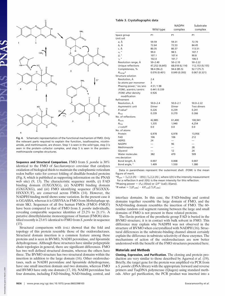
Table 2. Hydrogen bonding interactions of NADPH in the NADPHcomplex structure
NADPH Protein Distance, Å
AdenineN1� O Wat 2.99N3� O Wat 2.89N6� O Wat 3.10
PhosphatesO1� O Wat 2.55
O Wat 2.77O2� O Wat 2.81O1 O Wat 2.69
N Ser-223 2.89O2 O Wat 2.72
OG Ser-223 2.82Nicotinamide
O2* O Wat 3.08O Wat 2.61OD1 Asn-91 2.97
O3* OH Tyr-176 3.17N7 O Wat 3.03
O Wat 3.19O Gln-89 3.07OD2 Asp-226 2.87
O7 N5 FAD 3.05O4 FAD 3.20O Gln-89 2.99
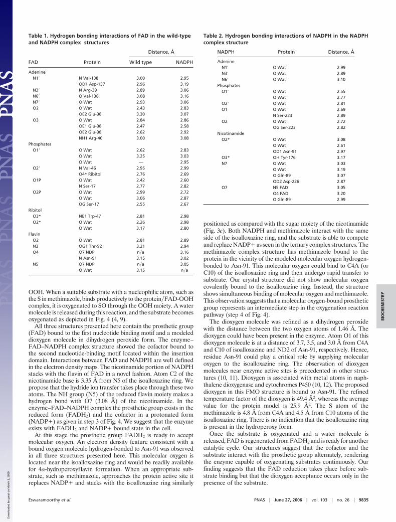
Table 1. Hydrogen bonding interactions of FAD in the wild-typeand NADPH complex structures
FAD Protein
Distance, Å
Wild type NADPH
AdenineN1� N Val-138 3.00 2.95
OD1 Asp-137 2.96 3.19N3� N Arg-39 2.89 3.06N6� O Val-138 3.08 3.16N7� O Wat 2.93 3.06O2 O Wat 2.43 2.83
OE2 Glu-38 3.30 3.07O3 O Wat 2.84 2.86
OE1 Glu-38 2.47 2.58OE2 Glu-38 2.62 2.92NH1 Arg-40 3.00 3.08
PhosphatesO1� O Wat 2.62 2.83
O Wat 3.25 3.03O Wat — 2.95
O2� N Val-46 2.95 2.99O4* Ribitol 2.76 2.69
O1P O Wat 2.42 2.60N Ser-17 2.77 2.82
O2P O Wat 2.99 2.72O Wat 3.06 2.87OG Ser-17 2.55 2.67
RibitolO3* NE1 Trp-47 2.81 2.98O2* O Wat 2.26 2.98
O Wat 3.17 2.80Flavin
O2 O Wat 2.81 2.89N3 OG1 Thr-92 3.21 2.94O4 O7 NDP n�a 3.16
N Asn-91 3.15 3.02N5 O7 NDP n�a 3.05
O Wat 3.15 n�a
Eswaramoorthy et al. PNAS � June 27, 2006 � vol. 103 � no. 26 � 9835
BIO
CHEM
ISTR
Y
Dow
nloa
ded
by g
uest
on
Mar
ch 1
, 202
0
Sequence and Structural Comparison. FMO from S. pombe is 30%identical to the FMO of Saccharomyces cerevisiae that catalyzesoxidation of biological thiols to maintain the endoplasmic reticulumredox buffer ratio for correct folding of disulfide-bonded proteins(Fig. 8, which is published as supporting information on the PNASweb site) (9, 13). The characteristic sequence motifs, (i) FADbinding domain (GXGXXG), (ii) NADPH binding domain(GXGXXG), and (iii) FMO identifying sequence (FXGXXX-HXXXY�F), are conserved across FMOs (14). However, theNADPH binding motif shows some variation. In the present case itis GGASSA, whereas it is GSSYSA in FMO from Methylophaga sp.strain SK1. Sequences of all five human FMOs (FMO1–FMO5)have been compared to that of FMO from S. pombe individually,revealing comparable sequence identities of 23.2% to 21.1%. Aputative dimethylalanine monooxygenase of human (FMO6) iden-tified recently is 23.6% identical to FMO from S. pombe in sequence(15).
Structural comparisons with DALI showed that the fold andtopology of this protein resemble those of the oxidoreductases.Structural domain insertion is a common feature among theseproteins and is seen in BVMO, NADH peroxidase, and lipoamidedehydrogenase. Although these structures have similar polypeptidechain topologies in general, there are significant differences. FMOhas two well defined structural domains, whereas the others havethree. The BVMO structure has two structural domains within theinsertion in addition to the large domain (16). Other oxidoreduc-tases, such as NADH peroxidase and lipoamide dehydrogenase,have one small insertion domain and two domains, whereas FMOand BVMO have only one domain (17, 18). NADH peroxidase hasfour domains, including FAD-binding, NAD-binding, central, and
interface domains. In this case, the FAD-binding and centraldomains together resemble the large domain of FMO, and theNAD-binding domain resembles the insertion of FMO. The 60-residue random coil segment running between the large and smalldomains of FMO is not present in these related proteins.
The flavin portion of the prosthetic group FAD is buried in theBVMO structure; it is in contact with bulk solvent in FMO. Thisdifference may explain why NADPH was not observed in thestructure of BVMO when cocrystallized with NADPH (16). Struc-tural differences in the substrate-binding channel almost certainlyexplain the difference in substrate selectivity of these enzymes. Themechanisms of action of the oxidoreductases are now betterunderstood with the benefit of the FMO structures presented here.
Materials and MethodsCloning, Expression, and Purification. The cloning and protein pro-duction are very similar to those described by Agarwal et al. (19).Briefly, the target gene for the protein was amplified via PCR froma S. pombe cDNA library with the appropriate forward and reverseprimers and TaqDNA polymerase (Qiagen) using standard meth-ods. After gel purification, the PCR product was inserted into a
Fig. 4. Schematic representation of the functional mechanism of FMO. Onlythe relevant parts required to explain the function, isoalloxazine, nicotin-amide, and methimazole, are shown. Step 1 is seen in the wild type, step 3 isseen in the protein–cofactor complex, and step 5 is seen in the protein–methimazole complex structures.
Table 3. Crystallographic data
Wild typeNADPHcomplex
Substratecomplex
Space group P1 P1 P1Unit cell
a, Å 59.59 59.31 72.70b, Å 72.64 72.53 84.45c, Å 80.35 80.37 113.51�, ° 99.0 98.5 107.7�, ° 107.1 107.4 90.8�, ° 102.0 101.7 106.5Resolution range, Å 50–2.40 50–2.10 50–2.22Unique reflections 47,252 (4,645) 68,018 (6,114) 112,153 (9,155)Completeness, % 98.4 (96.2) 94.6 (85.5) 92.7 (75.7)Rmerge* 0.076 (0.401) 0.049 (0.393) 0.067 (0.321)
Structure solutionResolution, Å 2.4Se atoms per monomer 3Phasing power,† iso�ano 4.51�1.98�FOM�, acentric�centric 0.441�0.339�FOM� after density
modification0.926
RefinementResolution, Å 50.0–2.4 50.0–2.1 50.0–2.22Asymmetric unit Dimer Dimer Two dimersR value‡ 0.226 0.239 0.241Rfree 0.239 0.270 0.266
No. of reflectionsRwork 42,885 61,400 100,941Rfree 1,335 1,940 4,254� cutoff 0.0 0.0 0.0
No. of atomsProtein 6,978 6,978 13,956FAD 106 106 212HEPES 24 — —NADPH — 96 —Methimazole — — 28Glycerol — 12 24Water molecules 382 470 509
rms deviationBond length, Å 0.007 0.008 0.007Bond angle, ° 1.409 1.550 1.380
Data in parentheses represent the outermost shell. �FOM� is the meanfigure of merit.*Rsym � �h�i�Ii (h) � �I(h)����h�i�Ii (h) �, where Ii(h) is the intensity measurementfor a reflection h and �I(h)� is the mean intensity for this reflection.
†Phasing power � �FH��E(iso) or �2F’’ (cal)��E(ano).‡R value � �i�Fi,obs� � k�Fi,cal���i�Fi,obs�.
9836 � www.pnas.org�cgi�doi�10.1073�pnas.0602398103 Eswaramoorthy et al.
Dow
nloa
ded
by g
uest
on
Mar
ch 1
, 202
0
modified pET26b vector for topoisomerase-directed cloning (In-vitrogen) designed to express the protein with a C-terminal hexa-histidine tag and transformed into BL21 (DE3) cells. The clone wasconfirmed for correct sequence. The expression and solubility weretested by standard methods. The protein yield was 75.1 mg, whichwas concentrated to 45.1 mg�ml. Selenomethionine-labeled proteinwas produced and purified in a similar manner.
FMO Activity. The enzymatic assay for measuring FMO activity ofthe wild-type protein was performed by using the most commonsubstrate, methimazole (1). A final volume of assay mixture (100 �l)contained 100 mM tricine buffer, 1 mM EDTA, 0.4 mg of proteinsample, 0.1 mM NADPH, 0.02 mM DTT, and 0.06 mM DTNB in6.0 mM phosphate buffer. FMO activity was evaluated by spectro-photometric measurement of OD at 412 nm after addition ofmethimazole.
Structure Determination of Enzyme–FAD Complex. Purified protein in10 mM Hepes (pH 7.0) plus 150 mM NaCl was used for crystalli-zation. Diffraction-quality crystals were obtained by sitting drop-vapor diffusion against a reservoir containing 20% PEG 4000 and0.1 M sodium citrate buffer (pH 5.8) with 1,6-diaminohexane asadditive. The same crystallization condition was used for the SeMetprotein. Cryoprotection was achieved by addition of glycerol to afinal concentration of 10% (vol�vol). X-ray diffraction data werecollected under standard cryogenic conditions at beamline X25 ofthe National Synchrotron Light Source at Brookhaven NationalLaboratory. Crystals belonged to the space group P1 with twomolecules in the unit cell and diffracted to 2.4-Å resolution. Thestructure was determined by the three-wavelength multi-wavelength anomalous dispersion method. Se positions were de-termined by using SOLVE (20). Refinement of the Se positions,phase refinement and extension, and density modification wereperformed by using SHARP and DM (21, 22). The experimentalelectron density map calculated with phases from SHARP showedclear secondary structure elements. Automatic model building wasattempted with ARP�WARP (23), which built 84% of the polypeptidechain without manual intervention. Model building was completedmanually by using O (24).
The structure was refined by using CNS 1.1 (25). Interpretableelectron density features were observed for the prosthetic groupFAD, water molecules, and a buffer component Hepes, which wereincluded in the final refinements. Data collection and refinementparameters are provided in Table 3.
Structure Determination of Enzyme–FAD–NADPH Complex. Purifiedprotein was incubated with NADPH in a 1:5 molar ratio for 30 minat room temperature before crystallization trials. Crystals wereobtained under the same conditions as for the enzyme–FADcomplex and belong to the same crystal form. The isomorphousstructure was determined by difference Fourier synthesis using theenzyme–FAD complex as a phasing model and refined to conver-gence with CNS 1.1. A composite omit map showed unambiguouslyinterpretable electron density for the cofactor NADPH. Furtherrefinement was carried out after addition of NADPH, watermolecules, and a glycerol molecule (located at the binding site forthe Hepes molecule seen in the enzyme–FAD complex structure).Refinement statistics are presented in Table 3.
Structure Determination of Enzyme–FAD–Methimazole Complex. Theenzyme–FAD–NADPH complex was prepared as before and in-cubated for 1 h at room temperature with 5-fold molar excess ofmethimazole. Crystals grew under the same conditions as before,and x-ray diffraction data were collected under identical conditions.These crystals belong to the same space group (P1), but the unit celldimensions differ from those obtained with the enzyme–FADcomplex crystals, giving four molecules per unit cell. The structurewas determined by molecular replacement using the enzyme–FADcomplex as a search model (26). A composite omit map showedresidual electron density for FAD and methimazole only. NADPHwas not observed in the crystal structure. Water molecules and oneglycerol per monomer were added, and the refinement was com-pleted (Table 3).
Sequence and Structural Comparisons. Primary structure compar-isons were performed initially by BLAST, and sequentially similarproteins were identified (27). One-to-one comparison of se-lected proteins was performed by CLUSTALW (28). Multiplesequence alignment of a set of selected proteins was done byT-COFFEE (29). Three-dimensional structure of the FMO wascompared with other structures using the DALI server to detectprotein fold similarity (30).
We thank Dr. Kumaran for helpful discussions. Research was supportedby National Institutes of Health Grant GM62529 to the New York SGXResearch Consortium under Department of Energy Prime ContractDEAC02-98CH10886 to Brookhaven National Laboratory.
1. Dixit, A. & Roche, T. E. (1984) Arch. Biochem. Biophys. 233, 50–63.2. Poulsen, L. L. & Ziegler, D. (1979) J. Biol. Chem. 254, 6449–6455.3. Fraaije, M. W., Kamerbeek, N. M., van-Berkel, W. J. J. & Janssen, D. B. (2002)
FEBS Lett. 518, 43–47.4. Ziegler, D. M. (1993) Annu. Rev. Pharmocol. Toxicol. 33, 179–199.5. Cashman, J. R. (2000) Curr. Drug Metab. 1, 181–191.6. Cashman, J. R. (2004) Drug Discovery Today 9, 574–581.7. Treacy, E. P., Akerman, B. R., Chow, L. M. L., Youil, R., Bibeau, C., Lin, J.,
Bruce, A. G., Knight, M., Danks, D. M., Cashman, J. R., et al. (1998) Hum. Mol.Genet. 7, 839–845.
8. Wood, V., Gwilliam, R., Rajandream, M. A., Lyne, M., Lyne, R., Stewart, A.,Sgouros, J., Peat, N., Hayles, J., Baker, S., et al. (2002) Nature 415, 871–880.
9. Suh, J. K., Poulsen, L. L., Ziegler, D. M. & Robertus, J. D. (1996) Arch.Biochem. Biophys. 336, 268–274.
10. Karlsson, A., Parales, J. V., Parales, R. E., Gibson, D. T., Eklund, H. &Ramaswamy, S. (2003) Science 299, 1039–1042.
11. Wilmot, C. M., Hajdu, J., McPherson, M. J., Knowles, P. F. & Phillips, S. E.(1999) Science 286, 1724–1728.
12. Schlichting, I., Berendzen, J., Chu, K., Stock, A. M., Maves, S. A., Benson,D. E., Sweet, R. M., Ringe, D., Petsko, G. A. & Sligar, S. G. (2000) Science 287,1615–1622.
13. Goffeau, A., Barrell, B. G., Bussey, H., Davis, R. W., Dujon, B., Feldmann, H.,Galibert, F., Hoheisel, J. D., Jacq, C., Johnston, M., et al. (1996) Science 274,546, 563–567.
14. Choi, H. S., Kim, J. K., Vho, E. H., Kim, Y. C., Kim, J. I. & Kim, S. W. (2003)Biochem. Biophys. Res. Commun. 306, 930–936.
15. Furnes, B., Feng, J., Sommer, S. S. & Schlenk, D. (2003) Drug Metab. Dispos.31, 187–193.
16. Malito, E., Alfieri, A., Fraaije, M. W. & Mattevi, A. (2004) Proc. Natl. Acad.Sci. USA 101, 13157–13162.
17. Mattevi, A., Obmolova, G., Sokatch, J. R., Betzel, C. & Hol, W. G. J. (1992)Proteins Struct. Funct. Genet. 13, 336–351.
18. Stehle, T., Ahmed, S. A., Claiborne, A. & Schulz, G. E. (1991) J. Mol. Biol. 221,1325–1344.
19. Agarwal, R., Bonanno, J. B., Burley, S. K. & Swaminathan, S. (2006) ActaCrystallogr. D 62, 383–391.
20. Terwilliger, T. C. & Berendzen, J. (1997) Acta Crystallogr. D 55, 849–861.21. Cowtan, K. D. & Main, P. (1996) Acta Crystallogr. D 52, 43–48.22. de La Fortelle, E. & Bricogne, G. (1997) Methods Enzymol. 276, 472–493.23. Perrakis, A., Morris, R. & Lamzin, V. S. (1999) Nat. Struct. Biol. 6, 458–463.24. Jones, T. A., Zou, J., Cowtan, S. & Kjeldgaard, M. (1991) Acta Crystallogr. A
47, 110–119.25. Brunger, A. T., Adams, P. D., Clore, G. M., Delano, W. L., Gros, P.,
Grosse-Kunstleve, R. W., Jiang, J. S., Kuszwewski, J., Nilges, M., Pannu, N. S.,et al. (1998) Acta Crystallogr. D 54, 905–921.
26. Vagin, A. & Teplyakov, A. (2000) Acta Crystallogr. D 56, 1622–1624.27. Altschul, S. F., Madden, T. L., Schaffer, A. A., Zhang, J., Zhang, Z., Miller, W.
& Lipman, D. J. (1997) Nucleic Acids Res. 25, 3389–3402.28. Thompson, J. D., Higgins, D. G. & Gibson, T. J. (1994) Nucleic Acids Res.
22, 4673–4680.29. Notredame, C., Higgins, D. G. & Heringa, J. (2000) J. Mol. Biol. 302, 205–217.30. Holm, L. & Sander, C. (1993) J. Mol. Biol. 233, 123–138.
Eswaramoorthy et al. PNAS � June 27, 2006 � vol. 103 � no. 26 � 9837
BIO
CHEM
ISTR
Y
Dow
nloa
ded
by g
uest
on
Mar
ch 1
, 202
0
Corrections and Retraction
CORRECTIONS
BIOCHEMISTRY. For the article ‘‘Mechanism of action of a flavin-containing monooxygenase,’’ by Subramaniam Eswaramoorthy,Jeffrey B. Bonanno, Stephen K. Burley, and SubramanyamSwaminathan, which appeared in issue 26, June 27, 2006, of ProcNatl Acad Sci USA (103:9832–9837; first published June 15, 2006;10.1073�pnas.0602398103), the authors note the following: ‘‘Areader of the published article pointed out that the mechanismof Fig. 4 showing hydride transfer from the C2 position is notcompatible with known NADH chemistry, which entails hydridetransfer to and from C4. Our explanation and Fig. 4 were basedon our crystal structure, in which C2 of NADH was close to N5of the isoalloxazine moiety of FMN. Accordingly, we suggest thatthe crystal structure possibly represents a transient stage beforeor after the proton transfer. The remainder of the article is notaffected by this change.’’
www.pnas.org�cgi�doi�10.1073�pnas.0707147104
MEDICAL SCIENCES. For the article ‘‘Isolation of anti-CD22 Fv withhigh affinity by Fv display on human cells,’’ by Mitchell Ho,Satoshi Nagata, and Ira Pastan, which appeared in issue 25, June20, 2006, of Proc Natl Acad Sci USA (103:9637–9642; firstpublished June 8, 2006; 10.1073�pnas.0603653103), the authorsnote that on page 9637, right column, in Results, beginning online 8, the phrase ‘‘the murine Ig � chain signal peptide (MET-DTLLLWVLLLWVPGSTGDJ) and the transmembrane do-main (amino acids 514–562) of PDGFR’’ should instead read:‘‘the murine Ig � chain signal peptide (METDTLLLWVLLL-WVPGSTGD) and the transmembrane domain (amino acidsAla513–Arg561) of PDGFR.’’
www.pnas.org�cgi�doi�10.1073�pnas.0707408104
RETRACTION
BIOCHEMISTRY. For the article ‘‘Vitamin C conjugates of genotoxiclipid peroxidation products: Structural characterization anddetection in human plasma,’’ by John Sowell, Balz Frei, and JanF. Stevens, which appeared in issue 52, December 28, 2004, ofProc Natl Acad Sci USA (101:17964–17969; first publishedDecember 17, 2004; 10.1073�pnas.0408433102), the authorsregret that the plasma analyte described in Figs. 3 and 5 wasincorrectly identified as ascorbylated 4-hydroxy-2-nonenal. Theauthors do not claim to have detected ascorbylated 4-hydroxy-2-nonenal in human plasma and, therefore, retract the paper.The reported in vitro chemistry is correct and is not affected bythis retraction.
John SowellBalz FreiJan F. Stevens
www.pnas.org�cgi�doi�10.1073�pnas.0706514104
www.pnas.org PNAS � September 4, 2007 � vol. 104 � no. 36 � 14543
CORR
ECTI
ON
SA
ND
RETR
ACT
ION


![by Joseph Bruchac illustrated by Teresa Flavin · illustrated by Teresa Flavin by Joseph Bruchac illustrated by Teresa Flavin Pushing Up)&(the Sky FjZhi^dc d[ i]Z LZZ` =dl Yd eZdeaZ](https://img.pdfslide.net/doc/110x75/5f8e7f4dad596368cb63a9b9/by-joseph-bruchac-illustrated-by-teresa-flavin-illustrated-by-teresa-flavin-by-joseph.jpg)