Embed Size (px)
Citation preview

ECOLE POLYTECHNIQUE FEDERALE DE LAUSANNE
SCHOOL OF LIFE SCIENCES
Master's project in Life Sciences and Technology
Mechanism of DegU-dependent activation of flagellar gene transcription in Listeria monocytogenes
Carried out in the laboratory of Prof. Darren E. Higgins at Harvard Medical School
Under the supervision of Prof. Darren E. Higgins
Done by
Diane Baer
Under the direction of
Prof. John D. McKinney In the laboratory of Bacteriology
EPFL
2
Table of Contents
ACKNOWLEDGEMENTS 3 1.SUMMARY 4 2.INTRODUCTION 5 3.MATERIALS AND METHODS 11 4.RESULTS 19
4.1 pfliN-gmaR activation by DegU in B. subtilis 19
4.2 A transposon mutagenesis screen for L. monocytogenes genes 22
involved in transcriptional activation of pfliN-gmaR at 30°
4.3 A transposon mutagenesis screen for L. monocytogenes genes 35
involved in the repression of pfliN-gmaR at 37°
5.DISCUSSION 38
6.CONCLUSION 45
7.REFERENCES 46

3
ACKNOWLEDGEMENTS I would like to thank Prof. Darren Higgins for welcoming me in his lab and making this year so
scientifically exciting and enriching. I am immensely grateful for the opportunity he has provided me and I would like to thank him for always taking the time for support and advice and for the many scientific discussions that have been incredibly helpful in teaching me how to better think as a scientist. I would like to thank Prof. John McKinney for taking the time on the afternoon of February 20th 2009 to explain me what research he was interested in and to make me realize that bacteria are the most interesting and fascinating organisms to work on. I would like to thank him for giving me the opportunity to work in his lab and learn as much as I could. I also would like to thank him for recommending me to Darren and for his incredible support throughout this year. I would like to thank the Higgins lab for making my stay so enjoyable and for their scientific help and advice throughout this year. I would also like to thank the EPFL WISH Foundation, the EPFL Excellence Scholarship program and Prof. Darren Higgins for financial support.

4
1. SUMMARY Flagellar motility is an important process for bacterial survival. It allows bacteria to move to
aquire nutrients or to retreat from unfavorable environments. In the Gram-positive bacterium Listeria monocytogenes, flagellar motility is a temperature-dependent process, being restricted to temperatures below 37°C. This regulation is mediated by the activities of the DegU response regulator, the MogR transcriptional repressor and GmaR, the MogR anti-repressor. Transcriptional regulation of the fliN-gmaR promoter is central to flagellar motility regulation as pfliN-gmaR is transcriptionally activated by DegU and is also MogR repressed. However, the mechanism by which DegU mediates transcriptional activation of the fliN-gmaR promoter is not well understood. In this study, we further investigated the mechanisms controlling transcription initiating at the fliN-gmaR promoter by performing two comprehensive transposon mutagenesis screens to identify any additional factors involved in the activation and repression of pfliN-gmaR. The mutagenesis screen performed at 30°C to identify factors involved in pfliN-gmaR transcriptional activation led to the identification of a novel factor. We demonstrated that Lmo0866, a protein from the DEAD-box protein family, is required for transcriptional activation of pfliN-gmaR and therefore flagellar motility. Moreover, we identified CodY as being involved in the repression of pfliN-gmaR at 37°C.

5
2. INTRODUCTION
Listeria monocytogenes is a Gram-positive bacterium and a facultative intracellular
pathogen. L. monocytogenes is the causative organism of the foodborne disease listeriosis, characterized by gastroenteritis, septicemia, meningitis and encephalitis, that can lead to fatal outcomes in immunocompromised individuals, pregnant women, newborns and the elderly (1).
L. monocytogenes can survive and multiply both inside and outside the host,
including in soil, unpasteurized dairy products and processed food. L. monocytogenes has therefore evolved mechanisms that enable rapid adaptation and survival in a wide range of environments and temperatures ranging from 3°C to 43°C. Once inside the host, environmental signals sensed by L. monocytogenes lead to the upregulation of virulence factors and downregulation of other genes, such as flagellar genes, that are unnecessary or impede survival in the host environment.
Flagellar motility is the primary mechanism enabling bacteria to move in the
extracellular environment and is widely distributed throughout bacterial species. It enables bacteria to move towards or away from environmental cues monitored by a chemotactic signal-transduction system controlling the direction of flagellar rotation. The bacterial flagellum is a complex self-assembling organelle consisting of three substructures: a membrane-bound basal body which anchors the structure in the cell
envelope and contains the ion-powered rotary motor, an extracellular helical filament measuring approximately 20 nm in diameter that acts as the propeller and extends many cell lengths from the cell and a flexible hook connecting the basal body and the filament (2). Flagella from Gram-positive and Gram-negative bacteria are essentially identical, except that flagella from Gram-negative bacteria extend through the outer-membrane absent in Gram-positive bacteria. Counter-clockwise rotation of the flagella

6
propels the cell forward at speeds of 15-100 μm per second (3). A quick reversal to clockwise rotation induces tumbling which allows reorientation of the cell and swimming in a new direction when the motor returns to counter-clockwise rotation. Change in
direction occurs approximately once a second in homogeneous environments resulting in random movement. In non-homogeneous environments the chemotactic signal-transduction system biases the overall direction of movement by decreasing the tumbling frequency.
Flagellar motility is important for bacterial survival as it enables bacteria to move to
acquire nutrients or retreat from unfavorable environments (4), to colonize surfaces, and is important to establish infection. In L. monocytogenes, flagellar motility has been shown to increase abiotic and cellular adherence (5), is essential for biofilm formation (6) and enhances cellular invasion (7). However, in response to temperature (37°C and above) and other environmental signals, such as the host cell cytosol, flagellar motility genes are repressed (8). Indeed, flagella recognition leads to activation of host innate immune responses (9).
Flagella production is a very energy-demanding process that requires the
coordinate activation of more than 40 genes and assembly of thousands of interacting proteins (10). The regulation of bacterial flagella production has been studied in different bacterial species and appears to be regulated at multiple levels in a hierarchal manner (10). Interestingly, regulation of flagellar motility in L. monocytogenes differs significantly
from other bacterial species as conserved master regulators involved in the transcriptional hierarchal regulation of flagellar production are absent in L. monocytogenes (11).
The temperature-dependent regulation of flagellar motility in L. monocytogenes
involves three key players, MogR (12), a transcriptional repressor, GmaR (13), the

7
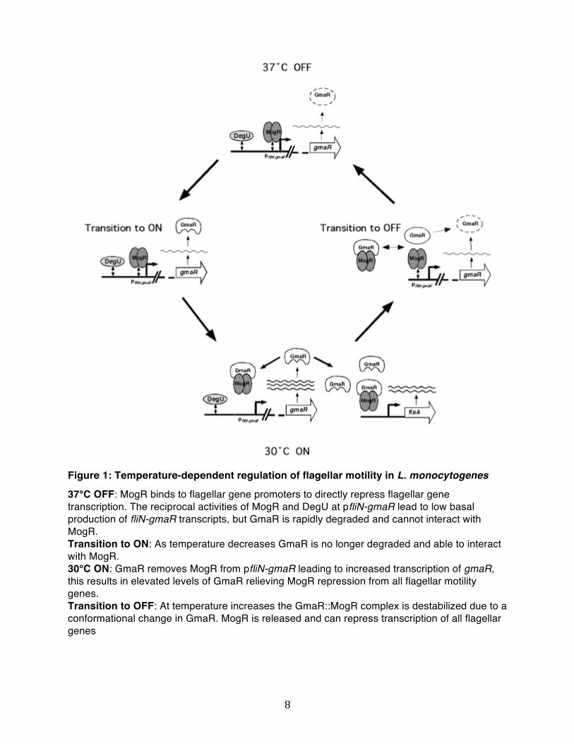
MogR anti-repressor, and DegU (14), a response regulator (Fig. 1). At temperatures of 37°C and above, MogR binds to flagellar gene promoters to directly repress flagellar gene transcription (11). Transcription of the first operon of the flagellar motility gene
cluster (fliN-gmaR) is activated by the DegU response regulator and is also repressed by MogR. The fliN-gmaR operon is of principal importance for flagellar gene regulation as it encodes the MogR anti-repressor GmaR (13). At non-permissive temperatures (37°C and above) the reciprocal activities of MogR and DegU lead to low basal production of fliN-gmaR transcripts, but a secondary post-transcriptional mechanism facilitates degradation of GmaR (15). As temperature decreases, GmaR undergoes a conformational change preventing its degradation (Kamp and Higgins, unpublished data). Therefore, low levels of GmaR can now bind MogR removing MogR from pfliN-gmaR leading to increased transcription of gmaR. This results in elevated levels of GmaR relieving MogR repression from all flagellar motility genes allowing production of flagella. Therefore, transcriptional and post-transcriptional regulation of the GmaR anti-repressor governs temperature-dependent control of flagellar motility in L. monocytogenes. However, the mechanism by which DegU mediates transcriptional activation of the fliN-gmaR promoter is not well understood.
DegU is an orphan two-component system response regulator as no cognate
histidine kinase has been identified in the L. monocytogenes genome. DegU is composed of two distinct domains, a phosphorylation receiver domain and a LuxR family helix-turn-helix DNA binding domain. It shares significant homology with Bacillus
subtilis (Bs) DegU as they are 63% identical and 78% similar (15). Interestingly, BsDegU is also required for flagellar motility gene activation in B. subtilis, but activation or repression of flagellar genes is dependent on the state of phosphorylation of the BsDegU receiver domain, which is phosphorylated by the cognate histidine sensor kinase BsDegS (16).
8
Figure 1: Temperature-dependent regulation of flagellar motility in L. monocytogenes
37°C OFF: MogR binds to flagellar gene promoters to directly repress flagellar gene transcription. The reciprocal activities of MogR and DegU at pfliN-gmaR lead to low basal production of fliN-gmaR transcripts, but GmaR is rapidly degraded and cannot interact with MogR. Transition to ON: As temperature decreases GmaR is no longer degraded and able to interact with MogR. 30°C ON: GmaR removes MogR from pfliN-gmaR leading to increased transcription of gmaR, this results in elevated levels of GmaR relieving MogR repression from all flagellar motility genes. Transition to OFF: At temperature increases the GmaR::MogR complex is destabilized due to a conformational change in GmaR. MogR is released and can repress transcription of all flagellar genes

9
DegU in L. monocytogenes has been shown to activate the fliN-gmaR promoter (15) and to bind and repress its own promoter (17). Protein levels of DegU have been shown to be temperature-independent (13). In addition, activation of the fliN-gmaR promoter by
DegU is also temperature-independent, as quantitative RT-PCR experiments in a ΔmogR strain indicate that fliN-gmaR transcripts are produced at both 30°C and 37°C (15). It has not been determined if DegU is phosphorylated in L. monocytogenes, but in vitro experiments have shown that BsDegS can phosphorylate DegU with even higher efficiency than BsDegU. However, DegU containing a mutation in the phosphorylation acceptor site is still able to activate fliN-gmaR transcription in L. monocytogenes, indicating that DegU does not require phosphorylation to activate transcription of the fliN-gmaR promoter (15). DegU binds the fliN-gmaR promoter region independently of other factors and DNase I footprinting analysis has identified a unique binding region at position -154 to -184 nt upstream of the fliN-gmaR transcription start site (15). Furthermore, electrophoretic mobility shift analysis suggests that DegU may bind as a multimer, as a super-shifted complex at higher concentration of DegU was observed (15). Interestingly, the N-terminal portion of DegU containing the phosphorylation receiver domain was shown to be dispensable for activation of the fliN-gmaR promoter (15). Despite sharing significant homology with DegU, BsDegU was not able to complement activation of fliN-gmaR in a L. monocytogenes degU mutant (15), underlying the differences existing between the two DegU proteins.
DegU is necessary for transcriptional activation at the fliN-gmaR promoter (pfliN-gmaR) however it is not known if it is sufficient. The DegU binding site within the fliN-gmaR promoter region is located far upstream of the transcriptional start site, leading to the question of whether DegU directly activates transcription of pfliN-gmaR. Activators typically bind close to the -35 region, but binding sites located at approximately -150 nt have also been described in bacteria (18). DegU binding may change the conformation

10
of promoter region DNA or DegU could induce DNA bending and directly contact RNA polymerase. Alternatively, DegU may be required for recruitment of a coactivator.
The goal of this project was to further characterize transcriptional regulation occurring at the fliN-gmaR promoter and to determine if DegU is sufficient for transcriptional activation of pfliN-gmaR or if additional bacterial factors are required. In this study, we first intended to determine if DegU was sufficient to activate transcription of pfliN-gmaR in B. subtilis, therefore using B. subtilis as a surrogate host bacterium. Furthermore, to fully characterize regulation of the fliN-gmaR promoter, we generated a mariner transposon mutant library in a pfliN-gmaR::gusA reporter strain and performed comprehensive library screens at 30°C and 37°C to identify any additional factors involved in regulation of the fliN-gmaR promoter. Here, we report the identification of a novel factor required for pfliN-gmaR transcriptional activation.

11
3. MATERIALS AND METHODS Bacterial strains and culture conditions Listeria monocytogenes, Escherichia coli and Bacillus Subtilis strains used in this study are listed in Table 1, Table 2 and Table 3, respectively. Oligonucleotides used in this study are listed in Table 4. All L. monocytogenes strains are in the EGDe background and were grown in Brain Heart Infusion (BHI) broth. E. coli strains were grown in Luria-Bertani (LB) media for plasmid isolation. B. subtilis strains were grown in LB media. All bacterial stocks were stored at -80°C in LB or BHI supplemented with 33% glycerol. Antibiotics were used in the following concentrations: chloramphenicol at 20 μg/ml for selection of pPL3 derivatives in E. coli, 7.5 μg/ml for selection of integrated pPL3 derivatives and pAM401 vectors in L. monocytogenes. 30 μg/ml kanamycin for selection pIMK vectors in E. coli and for selection of integrated pIMK derivatives in L. monocytogenes. 100 μg/ml carbenicillin for selection of pDG1663 derivatives and pDR110a amyE-pSpank derivatives in E. coli. 100 μg/ml spectinomycin for selection of integrated pDR110a amyE-pSpank derivatives in B. subtilis, and 5 μg/ml erythromycin for selection of integrated mariner transposon in L. monocytogenes and pDG1663 derivatives in B. subtilis. All plasmid constructs were confirmed by DNA sequencing. Plasmids were isolated from strains XL1-Blue and electroporated directly into L. monocytogenes or transformed into SM10 for conjugative transfer in L. monocytogenes. Table 1 Listeria monocytogenes strains used in this study
Strain Genotype and relevant features Strain designation Reference DH-L478 wild-type strain EGDe Wild-type
DH-L1056 gmaR in-frame deletion in EGDe ΔgmaR (13)
DH-L1156 mogR in-frame deletion in EGDe ΔmogR (12)
DH-L1273 degU in-frame deletion in EGDe ΔdegU (11)

12
DH-L1339 D55N point mutation in degU in EGDe LmDegU D55N (15)
DH-L1867 pPL3-pfliNgusA-278 in ΔmogR ΔmogR/pfliNgusA-278 (15)
LM1 pIMK-pfliNgusA-278 in EGDe pIMK-pfliNgusA-278 This study
LM2 pPL3-pfliNgusA-278 in ΔdegU ΔdegU/pfliNgusA-278 This study
LM3 Himar1 transposon generated library in LM1 This study
LM4 Transposon insertion into lmo0866 LM1 lmo0866/pIMK-pfliNgusA-278 This study
LM5 Transposon insertion into lmo0866 in DH-L478 lmo0866/ wt EGDe This study
LM6 Transposon insertion into lmo1280 in LM1 codY/ pIMK-pfliNgusA-278 This study
LM7 pAM401-lmo0866 in LM4 lmo0866/pAM401-lmo0866 This study
LM8 pAM401 empty in LM4 lmo0866/pAM401empty This study
LM9 pPL3-lmo0866 in LM5 lmo0866/pPL3-lmo0866 This study
Table 2 Escherichia coli strains used in this study Strains Genotype and relevant features Reference DH-E182 XL1-Blue {Fʼ proAB lacIq ∆(lacZ)M15 Tn10}
RecA1 endA1 gyrA96 thi-1 hsdR17 supE relA1 lac Stratagene
DH-E474 SM10
DH-E898 pPL3 in XL1-Blue (12)
DH-E476 pAM401 in 1855
DH-E1934 pIMK in DH10B (21)
pJZ037 in DH5 alpha (19)
DH-E1829 pPL3-pfliNgusA-278 in XL1-Blue (15)
DH-E1833 pPL3-pfliNgusA-278 in SM10 (15)
DH-E1564 pDG1663 in DH5 alpha (32)
DH-E1565 pDR110a amyE-Pspank in DH5 alpha (32)
EC1 pIMK-pfliNgusA-278 in XL1-Blue This study
EC2 pAM401-lmo0866 in XL1-Blue This study
EC3 pPL3-lmo0866 in XL1-Blue This study
EC4 pDG1663-pfliNlacZ in XL1-Blue This study
EC5 pDR110a-degU in XL1-Blue This study

13
EC6 pDR110a-degU D55N in XL1-Blue This study
Table 3 Bacillus subtilis strains used in this study
Strains Genotype and relevant features Reference
bDR124 thrC::cat in B. subtilis PY79 (32)
bDR993 amyE::pHyper-spac-lacZ in B. subtilis PY79
BS1 thrC::pfliNlacZ in bDR124 This study
BS2 amyE::pSpank-degU in BS1 This study
BS3 amyE::pSpank empty in BS1 This study
BS4 amyE::pSpank-degUD55N in BS1 This study
Table 4 Oligonucleotides used in this study Number Sequence Sitea
P1 AGATACAAGCTTTGAAGGAGGAGTAGTCATTATGGCAC HindIII
P2 AGATACGCATGCTAGTCCCCCTGAGATTTCTTTAGCG SphI
P3 AGATACGAATTCGGCATACAATCACATACCTCTCTATC EcoRI
P4 AGATACGGATCCCTTTCACTCCCTTCATCAGATTAC BamHI
P5 AGATACGGATCCTGTATGGGACTAATAAAAAGCTGG BamHI
P6 AGATACGTCGACAACTAGCACCACACTCCCGTATC SalI
P7 GGCCACGCGTCGACTAGTACNNNNNNNNNNGTAAT
P8 GCAATGAAACACGCCAAAGTAAAC
P9 GGCCACGCGTCGACTAGTAC
P10 CGCCTACGGGGAATTTGTATC
a. The indicated restriction endonuclease site is underlined within the oligonucleotide sequence
Strain construction A pIMK-pfliNgusA-278 L. monocytogenes strain was generated and used as strain background for the mariner transposon mutant library. To create the pIMK-pfliNgusA-278

14
L. monocytogenes strain plasmid pPL3-pfliNgusA-278 (DH-E1829) was digested with EagI and SalI and gel purified. The pfliNgusA-278 insert was ligated to plasmid pIMK digested with the same restriction enzymes and transformed into XL1-Blue to create
strain EC1. The resulting plasmid was introduced into wild-type EGDe (DH-L478) by electroporation. Strain LM2 was obtained by mating SM10 strain DH-E1833 containing the pPL3-pfliNgusA-278 plasmid with L. monocytogenes ΔdegU strain (DH-L1273). To generate the complementation constructs for the transposon insertion into lmo0866 primer pair P5 and P6 were used with EGDe genomic DNA to amplify the 2 kb lmo0866 gene region. It comprises the lmo0866 open reading frame and 500 bp of intergenic sequence upstream of the lmo0866 start codon containing the putative lmo0866 native promoter. The 2 kb PCR product was gel purified and digested with BamHI and SalI and ligated to plasmids pAM401 and pPL3 digested with the same restriction enzymes and transformed to XL1-Blue to generate strain EC2 and EC3, respectively. The resulting plasmids were sequenced verified and electroporated into strains LM4 and LM5 to create LM7 and LM9, respectively. B. subtilis thrC::pfliNlacZ strain was constructed by PCR amplification of the fliN promoter sequence from EGDe genomic DNA with primer pair P3 and P4. The resulting PCR product was gel purified and digested with BamHI and EcoRI, ligated into pDG1663 digested with the same restriction enzymes and transformed into XL1-Blue yielding strain EC4. The resulting plasmid was sequenced and transformed into bDR124
generating strain BS1. Transformants were restreaked onto chloramphenicol LB plates to select for double cross-over recombination events at the thrC locus. To construct pDR110a-pSpank-degU and pDR110a-pSpank-degUD55N, the degU coding sequence was amplified with primer pair P1 and P2 from EGDe genomic DNA or from genomic DNA isolated from strain DH-L1339 containing the D55N L. monocytogenes DegU point mutant. The resulting PCR products were digested with HindIII and SphI and ligated to
15
plasmid pDR110a digested with the same restriction enzymes. The ligation reactions were transformed into XL1-Blue generating strain EC5 and EC6, respectively. The resulting plasmids were sequenced and transformed into strain BS1 yielding strain BS2
and BS4. Double cross-over events were selected by screening for α-amylase deficient clones grown on 1% starch-containing plates and flooded with iodine solution. X-gal/IPTG plate assay Single colonies were inoculated in 3 ml of LB medium and incubated at 37 °C for 6 hours. Dilutions were plated on either 1 mM IPTG/X-gal LB plates or X-gal LB plates and incubated 16 hours at 37 °C. X-gal was used in plates at a concentration of 40 μg/ml. Color of colonies was then visually assessed. Beta-glucuronidase assay Single colonies of L. monocytogenes strains were inoculated in 3 ml of BHI medium plus appropriate antibiotics and incubated for 20 hr without shaking at the indicated temperatures. OD600 readings were taken to normalize β-glucuronidase activity for bacterial density. Bacteria were pelleted in 1.5-ml aliquots at 13,000 rpm for 10 min and resuspended in 150 μl ABT buffer (60 mM K2HPO4/40 mM KH2PO4/100 mM NaCl/0.1% Triton X-100, pH 7.0, filter sterilized). Reactions were set up in a 96-well plate format and done in triplicate by mixing 10 μl of GUS substrate (0.4 mg/ml 4-methyl-umbelliferyl-β-D-glucuronide in DMSO) and 50 μl of sample using ABT buffer as control. After incubation for 60 minutes at room temperature in the dark, 20 μl from each reaction
were removed and diluted into 180 μl of ABT buffer in a black 96-well plate and fluorescence values were determined by using a SpectraMAX GeminiXS instrument (Molecular Devices) at excitation and emission wavelengths of 366 and 445 nm, respectively. Known concentrations of the fluorescent 4-methylumbelliferone (MU) product ranging from 25 to 4000 pmol were used to obtain a standard curve. Units were calculated as (picomoles of substrate hydrolyzed × dilution factor)/(ml culture in final

16
sample × OD600 x minute) where pmol of substrate hydrolyzed is calculated from the standard curve as (emission reading – y intercept)/(slope). A student t-test was performed for statistical analysis.
Western blot analysis Colonies from B. subtilis strain BS2 were scraped from the agar plates, resuspended in 100 ml TE buffer with 5 mg/ml lysozyme and incubated at 37°C for 1 hour. An equal volume of 2X FSB was added to the supernatant and samples were boiled for 5 min at 95°C and centrifuged at 13,000 rpm for 1 min. 15 μl was loaded on a 12% SDS-PAGE gel and a Western blot was performed as previously described (11) using a polyclonal antibody for DegU. BHI medium supplemented with appropriate antibiotics was inoculated with L. monocytogenes overnight culture and incubated for 6 h at the indicated temperature or until cultures reach mid-exponential phase. A culture volume equivalent of 1 ml of OD600= 1.5 was pelleted and resuspended in 75 μl of TE/lysozyme (10 mM Tris-HCL pH 8.0, 1 mM EDTA, 5mg/ml lysozyme) and incubated for 1h at 37°C. 5X FSB was added to yield a 1X final concentration, samples were boiled for 5 min at 95°C and then centrifuged for 1 min at 13,000 rpm. 15 μl was loaded on a 12% SDS-PAGE gel for analysis of DegU and MogR and western blots were performed as previously described (11) using polyclonal antibodies specific for DegU or MogR.
Generation of a mariner transposon mutant library Electrocompetent L. monocytogenes LM1 cells were prepared following repeated treatments with 1mM Hepes/0.5 M sucrose. Cells were grown in vegetable peptone broth instead of BHI to increase electroporation efficiency. Approximately 1 μg of pJZ037 was used to electroporate each 50-μl aliquot of electrocompetent cells. After electroporation 1ml of BHI-0.5 M sucrose was added to cells to allow recovery for 1 hr at

17
30°C. Cells were then plated over approximately 19 100-mm BHI 5 μg/ml erythromycin agar plates. Plates were incubated for 60 hr at 30°C the permissive temperature and then replica-plated onto BHI 5 μg/ml erythromycin plates and incubated at 42°C, the
non-permissive temperature, to cure the plasmid. Colonies were counted, scraped and resuspended in BHI 5 ug/ml erythromycin 40% glycerol for storage at -80°C. To test for plasmid curing efficiency, 10-fold serial dilutions were prepared from a frozen aliquot of the library and plated onto BHI 5 μg/ml erythromycin (the resistance marker carried by the transposon) and onto BHI 7.5 μg/ml chloramphenicol plates (the resistance marker carried by the delivery vector). Screening Screening was performed on BHI kanamycin 30 μg/ml, erythromycin 5 μg/ml, X-gluc (5-
bromo-4-chloro-3-indolyl-beta-D-glucuronic acid) 50 μg/ml 245 mm X 245 mm plates (Corning). Dilutions from two different frozen aliquots of the library were made to reach a concentration of approximately 1500 bacteria per ml and 3 ml were plated on each plate. Plates were incubated at the indicated temperatures for 60 h and putative mutants visually looking white or less blue were picked and restreaked on BHI kanamycin 30 μg/ml, erythromycin 5 μg/ml, X-gluc 50 μg/ml to confirm phenotype. Motility phenotype was then assessed for mutants (see below) and frozen stocks of each mutant were stored at -80°C. Identification of transposon insertion sites Identification of transposon insertion sites was performed through semi-arbitrary PCR of a mutant colony. The first PCR from a mutant colony uses primer pair P7 and P8. Primer pair P9 and P10 is used in the second round of PCR with1 μl from the first PCR reaction used as template. The PCR products were purified and sequenced with primer P10.

18
Phage Transduction L. monocytogenes bacteriophage P35 was used to transduce the mariner transposon. Bacteriophages (107 and 108 pfu) grown on the appropriate donor strains were mixed
with 108 of mid-exponential phase recipient cells and incubated at RT for 40 min. Cells were centrifuged for 5 min at 13,000 rpm and resuspended in 1 ml of BHI medium containing 10 mM sodium citrate pH 7.5 and 0.04 μg/ml erythromycin and incubated for 2 hrs at 37°C. Cells were pelleted and resuspended in 100 μl supernatant and plated on BHI 10 mM sodium citrate 5 μg/ml erythromycin and incubated at 37 °C for 48 h. Motility assay analysis A single colony was inoculated with a small pipet tip into a low agar content BHI plate (0.375% agar). Plates were inoculated at 30°C for 48 h and then photographed or scanned to evaluate size of swimming area. Transmission electron microscope A single colony was inoculated in 2 ml BHI and grown for 14 hr at 30°C without shaking. 5µl of the culture was adsorbed for 1 minute to a carbon coated grid that had been made hydrophilic by a 30 second exposure to a glow discharge. Excess liquid was removed with a filterpaper (Whatman #1) and the samples were stained with 0.75% uranyl formate for 30 seconds. After removing the excess uranyl formate with a filterpaper the grids were examined in a TecnaiG² Spirit BioTWIN transmission electron microscope and images were recorded with an AMT 2k CCD camera.
Growth curve A single colony was inoculated in 2 ml BHI and grown for 16 hr at 30°C without shaking. It was back-diluted 1:40 in 30 ml of BHI medium and grown shaking at the indicated temperature. OD600 readings from three independent cultures were taken at regular intervals until cultures reached stationary phase.

19
4.RESULTS 4.1 pfliN-gmaR activation by DegU in B. subtilis
To determine if activation of pfliN-gmaR can occur in B. subtilis, the L. monocytogenes degU gene was cloned under the control of the pSpank inducible promoter in the pDR110a plasmid, which also contains a constitutively expressed lacI repressor gene. The fliN-gmaR promoter region DNA was cloned upstream of a lacZ reporter gene in the pDG1663 plasmid, and both vectors were integrated into the B. subtilis genome. Upon induction with IPTG, DegU is produced and can bind the fliN-gmaR promoter region. Transcriptional activation was monitored by beta-galactosidase activity using X-gal plates. The use of B. subtilis to analyze activation of pfliN-gmaR in vivo has several advantages. It allows the use of an in vivo system closely related to L. monocytogenes. Moreover, transcriptional activation can easily be monitored in an in vivo environment by utilizing the B. subtilis transcription and translation machinery.
B. subtilis strain BS2, containing the pSpank::degU and pfliN-gmaR::lacZ
constructs, was plated on LB agar plates containing 1 mM IPTG/X-gal or on LB plates
with X-gal alone and incubated 16 hours at 37°C. B. subtilis colonies on the 1 mM
IPTG/X-gal plates should be blue if pfliN-gmaR is activated by DegU, and colonies on
the X-gal LB plates should remain white as no DegU is produced. A B. subtilis strain
containing a strong IPTG inducible promoter upstream of the lacZ gene (pHyper-
spac::lacZ) was used as a positive control for the assay. The results obtained are
summarized in Table 5. The presence of DegU in strain BS2 plated on 1 mM IPTG/X-
gal plates was confirmed by Western blot (Fig. 2). We can see a band corresponding to
DegU in the well loaded with sample from BS2 strain colonies scraped from 1 mM
IPTG/X-gal plates. The upper band present in both BS2 samples is a non-specific band.

20
Table 5: X-gal/IPTG plate assay
Strain number Strain 1mM IPTG
/ X-gal X-gal
BS1 pfliN-gmaR::lacZ blue blue
BS2 pSpank::LmdegU + pfliN-gmaR::lacZ white blue
BS4 pSpank::LmdegUD55N + pfliN-gmaR::lacZ white blue
BS3 pDR110a empty + pfliN-gmaR::lacZ blue blue
bdr993 pHyper-spac::lacZ blue white
X-gal plate
BS2
X-gal/1mM IPTG
BS2
DegU purification elution
Figure 2: DegU is produced in BS2 strain plated on 1 mM IPTG/X-gal plates. Western blot for DegU from BS2 strain colonies scrapped from X-gal and 1mM IPTG/ X-gal plates. DegU purification elution is a His-tagged DegU purified using a Ni-NTA (QIAGEN) column.
The results indicate that the fliN-gmaR promoter is active in B. subtilis in the absence of
DegU. pfliN-gmaR has been shown to have basal activity in a L. monocytogenes ΔdegU
ΔmogR strain. Alternatively, BsRNA polymerase may recognize another initiating
sequence within the 300 bp fliN-gmaR promoter region DNA or further upstream.
Following induction of DegU, transcription of lacZ was repressed (Table 5, BS2 strain),
suggesting that DegU may bind the fliN-gmaR promoter region in B. subtilis and inhibit
transcription. We know that BsDegS can very efficiently phosphorylate DegU in vitro

21
(15). It is possible that phosphorylation of DegU by BsDegS in vivo could inhibit the
ability of DegU to activate pfliN-gmaR. Alternatively, DegU may not function with the
BsRNA polymerase, or other B. subtilis factors could associate with DegU on the fliN-
gmaR promoter region to prevent transcriptional activation. Lastly, DegU may require
additional L. monocytogenes factors to activate transcription. To address the possibility
of DegU phosphorylation by BsDegS affecting pfliN-gmaR activation, L. monocytogenes
degU D55N, the L. monocytogenes degU gene containing a mutation in the
phosphoacceptor site, therefore preventing phosphorylation by BsDegS (15), was
cloned behind the Spank promoter in the pDR110a plasmid. B. subtilis strain BS4,
containing the pSpank::degUD55N and pfliN-gmaR::lacZ constructs did not show any
phenotypic difference from B. subtilis strain BS2 (Table 5) indicating that possible
phosphorylation of DegU by BsDegS is not responsible for the transcriptional repression
of pfliN-gmaR following IPTG induction. B. subtilis strain BS3, containing pfliN-
gmaR::lacZ and the empty pDR110a vector has the same phenotype as BS1 (Table 5)
proving that the pDR110a vector is not responsible for transcriptional repression of
pfliN-gmaR following IPTG induction.
These results are inconclusive, but suggest that DegU binds pfliN-gmaR and is unable to activate transcription in B. subtilis. This could indicate that another factor is required for pfliN-gmaR activation. In light of these results, we initiated a transposon mutagenesis screen to identify L. monocytogenes genes involved in transcriptional activation of pfliN-gmaR at 30°C.

22
4.2 A transposon mutagenesis screen for L. monocytogenes genes involved in transcriptional activation of pfliN-gmaR at 30°C Construction of a mariner transposon mutant library
Recently, a new Himar1-based transposon system was developed for use in L. monocytogenes (19). This new mariner transposon system provides a better alternative to the prominently used Tn917 transposon system, which has low transposition efficiency and a non-random pattern of insertion leading to insertional “hot spots” and poor genomic coverage (20). In comparison, the Himar1-based transposon system has been shown to be effective in multiple bacterial species and has a low site specificity, the dinucleotide TA, very common in the low G+C L. monocytogenes genome.
pfliN-gmaR fused to a gusA reporter gene was cloned into the pIMK vector (21).
This vector was integrated into the genome of wild-type L. monocytogenes strain EGDe, resulting in strain LM1, and a mariner transposon library of approximately 90,000 independent insertion mutants was generated using pJZ307 (19). A curing efficiency of approximately 97% was determined based on the number of colonies that were chloramphenicol resistant (drug marker carried by delivery plasmid) compared to the number of colonies that were erythromycin resistant (resistance marker carried by the transposon). Transposon mutagenesis screen for white mutants on X-gluc plates at 30°C
The transposon screen aims to identify additional factors required for transcriptional activation of pfliN-gmaR by screening the generated mariner transposon mutant library for white colonies on X-gluc plates after incubation at 30°C, indicating that pfliN-gmaR can no longer be activated. The pfliN-gmaR::gusA construct integrated into a L. monocytogenes ΔdegU strain (LM2) was used as a control and produced white colonies on X-gluc plates incubated at 30°C for 48 hours verifying the ability of the
23
screen to identify degU mutants. The screen should identify mariner insertions in degU, gmaR, pfliN-gmaR and any additional factor(s) required for activation of pfliN-gmaR. A partial library screen of approximately 60,000 transposon insertions was performed on
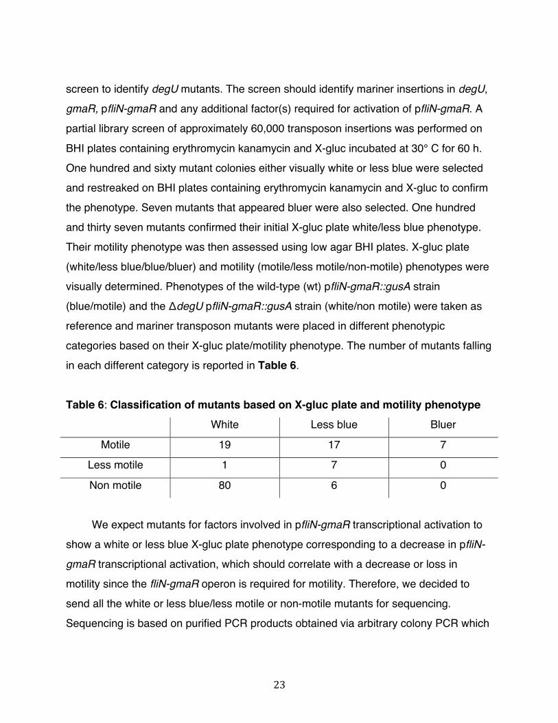
BHI plates containing erythromycin kanamycin and X-gluc incubated at 30° C for 60 h. One hundred and sixty mutant colonies either visually white or less blue were selected and restreaked on BHI plates containing erythromycin kanamycin and X-gluc to confirm the phenotype. Seven mutants that appeared bluer were also selected. One hundred and thirty seven mutants confirmed their initial X-gluc plate white/less blue phenotype. Their motility phenotype was then assessed using low agar BHI plates. X-gluc plate (white/less blue/blue/bluer) and motility (motile/less motile/non-motile) phenotypes were visually determined. Phenotypes of the wild-type (wt) pfliN-gmaR::gusA strain (blue/motile) and the ΔdegU pfliN-gmaR::gusA strain (white/non motile) were taken as reference and mariner transposon mutants were placed in different phenotypic categories based on their X-gluc plate/motility phenotype. The number of mutants falling in each different category is reported in Table 6. Table 6: Classification of mutants based on X-gluc plate and motility phenotype
White Less blue Bluer
Motile 19 17 7
Less motile 1 7 0
Non motile 80 6 0
We expect mutants for factors involved in pfliN-gmaR transcriptional activation to show a white or less blue X-gluc plate phenotype corresponding to a decrease in pfliN-gmaR transcriptional activation, which should correlate with a decrease or loss in motility since the fliN-gmaR operon is required for motility. Therefore, we decided to send all the white or less blue/less motile or non-motile mutants for sequencing. Sequencing is based on purified PCR products obtained via arbitrary colony PCR which
24
enables us to determine the precise transposon insertion locus, to the exact base pair. The sequencing results are provided in Table 7. Multiple degU mutants, representing 10 distinct transposon insertion sites were isolated in the category white/non-motile as well
as two independent insertions in pfliN-gmaR and 3 independent insertions in the fliN-gmaR operon, genes lmo0676 and lmo0677. We were therefore able to retrieve all the expected controls suggesting that we were approaching screening saturation and no additional screening was necessary.
Table 7: Transposon insertions from sequenced mutants
X-gluc plate /motility phenotype
Transposon insertion
# of independent
insertions
Gene name
Annotation
White/motile gusA 1 gusA pfliN-gmaR reporter gene
White/ less motile no curing 1 Mariner delivery vector
lmo2515 10 degU Two-component DegU response
regulator
pfliN-gmaR 2 Promoter region of the fliN-gmaR
operon White/non-motile
lmo0676 3 fliP similar to flagellar biosynthesic protein
FliP
lmo0889 1 rsbR highly similar to positive regulator of
sigma-B activity
lmo0896 1 rsbX
Indirect negative regulation of sigma B
dependant gene expression (serine
phosphatase)
lmo0894 1 rsbW sigma-B activity negative regulator
RsbW
Less blue/ less
motile
lmo0539 1 similar to tagatose-1,6-diphosphate
aldolase

25
no curing 2 Mariner delivery vector
lmo2515 2 degU Two-component DegU response
regulator
lmo0676 1 fliP similar to flagellar biosynthesic protein
FliP
lmo0677 1 fliQ similar to flagellar biosynthesis protein
FliQ
Less blue/ non-
motile
lmo0866 2 similar to ATP-dependent RNA helicase
lmo0027 1 similar to PTS system, beta-glucosides
specific enzyme IIABC
lmo0734 2 similar to LacI transcriptional regulator Bluer/ motile
lmo0785 1 similar to transcriptional regulator
NifA/NtrC
The less blue/less motile category led to the identification of 3 distinct insertions in
the sigma B operon. SigB is an alternative sigma factor involved in stress response and virulence. SigB has been proposed to play a role in regulation of motility as ΔsigB mutants in L. monocytogenes strain 10430S have an increased motility at 30°C (22). Those results could not however be reproduced in our lab strain L. monocytogenes
EGDe. In our screen we found that mutants with distinct transposon insertions in the sigB operon have a decreased motility at 30°C. It is difficult to identify which gene is responsible for this phenotype as we identified three different insertions, which may have polar effects on other genes in the operon. Due to the fact that we did not see any effect on motility in a ΔsigB mutant in our lab strain, the difficulty to identify which gene is directly responsible for the observed phenotype, and the fact that these mutants are still motile we decided not to further characterize these mutants. Another gene, lmo0539, was identified in the less blue/less motile category and encodes an enzyme
similar to a tagatose-1,6-diphosphate aldolase which catalyzes the reversible condensation of dihydroxyacetone phosphate with glyceraldehyde 3-phosphate to

26
produce tagatose 1,6-bisphosphate. The less blue/less motile phenotype could be the result of decreased metabolism and is unlikely to be directly involved in the regulation of pfliN-gmaR, we therefore decided not to further characterize this mutant. The
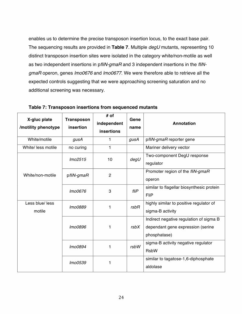
bluer/motile category retrieved mutations that seem to be involved in sugar metabolism and may be involved in X-gluc uptake. Interestingly, we also identified degU, lmo0676 and lmo0677 mutants in the less blue/non-motile category suggesting that the phenotypic difference between white and less blue was difficult to determine. Furthermore, we identified two independent insertions in the gene lmo0866, which encodes a protein similar to an ATP-dependent RNA helicase. We decided to further investigate the lmo0866::mariner mutant, which displays a phenotype very similar to a degU mutant, a known transcriptional activator of pfliN-gmaR. Lmo0866 is required for pfliN-gmaR activation and motility
Both transposon insertions into lmo0866 were transduced into both pfliN-gmaR::gusA and wt EGDe backgrounds using bacteriophage P35 to confirm linkage between the phenotype and transposon insertion. The X-gluc plate phenotype of lmo0866::mariner is presented in Figure 3. The initial streak appears less blue than the initial streak of wt pfliN-gmaR::gusA strain and single colonies are white. A ΔdegU pfliN-gmaR::gusA mutant has a white initial streak and white single colonies. To characterize in a more quantitative manner the level of transcriptional activation of pfliN-gmaR in the lmo0866::mariner mutant, a β-glucuronidase assay was performed (Fig. 4).
lmo0866::mariner showed a β-glucuronidase activity very similar to a ΔdegU mutant and the difference from the pfliN-gmaR::gusA strain is statistically significant with p-value < 0.001.

27
Figure 3: X-gluc plate phenotype of wt pfliN-gmaR::gusA strain, ΔdegU and lmo0866 mutant. Wild-type pfliN-gmaR::gusA strain, ΔdegU pfliN-gmaR::gusA and lmo0866::mariner were streaked on X-gluc plate and incubated 60 hr at 30°C. The decrease in pfliN-gmaR transcriptional activation directly correlated with a loss of flagellar motility. Indeed, similar to a ΔdegU mutant, lmo0866::mariner did not swim at
30°C in a motility plate assay (Fig. 5). We therefore hypothesized that lmo0866::mariner does not express flagella due to a decrease in transcription of the fliN-gmaR operon resulting in the non-motile phenotype. We further analyzed the lmo0866 mutant strain by transmission electron microscopy. Wild-type EGDe typically expresses between 4 to 6 flagella. As expected, both the lmo0866 and ΔdegU mutant did not express flagella
28
(Fig. 6). Taken together, these data indicate that lmo0866 is required for pfliN-gmaR transcriptional activation and flagellar motility.
Figure 4: Analysis of the fliN-gmaR promoter activity determined by β-glucuronidase assay. L. monocytogenes strains were grown 18 to 20 hr at 30°C in BHI broth. β-glucuronidase activity represents the mean and standard deviation of three independent cultures. Miller units were calculated per 109 bacteria. Experiment was repeated once in triplicate and similar results were obtained.
****

29
Figure 5: lmo0866 mutant is not motile at 30°C. The swimming phenotype of wt EGDe, ΔdegU and lmo0866::mariner when analyzed by motility plate assay (BHI with 0.375% agar) at 30°C.
Figure 6: Transmission electron microscopy of L. monocytogenes strains. Five microliters of bacterial cultures grown for 16 hr at 30°C not shaking were used for negative staining procedure. Bacteria were imaged at 11,000 X.
wt EGDe ΔdegU lmo0866 mutant

30
Complementation of the lmo0866 transposon insertion To verify that the observed phenotypes were specifically due to the absence of
Lmo0866, the lmo0866 mutant was complemented by the introduction of a wild-type
copy of the lmo0866 locus on the multicopy plasmid pAM401 and on the single integration vector pPL3 resulting in strains LM6 and LM8, respectively. The introduced lmo0866 locus comprises the upstream intergenic sequence, containing the putative native lmo0866 promoter, the lmo0866 open reading frame and its terminator sequence. Flagellar motility was restored in both complemented strains (Fig. 7), however not to a wild-type level. Figure 7: Motility phenotype of lmo0866 complemented strains. The swimming phenotype of wt EGDe pPL3 pfliN-gmaR::gusA strain, lmo0866/pPL3-lmo0866, lmo0866/pAM401-lmo0866 and lmo0866/pAM401-empty when analyzed by motility plate assay (BHI with 0.375% agar) at 30°C.

31
To investigate whether there was a difference in the number of flagella expressed in the complemented strains, which could account for the difference in flagellar motility, we further analyzed the complemented strains by transmission electron microscopy (Fig. 8).
Figure 8: Transmission electron microscopy of lmo0866 complemented strains. Five microliters of each respective bacterial culture grown for 16 hr at 30°C were used for negative staining procedure. Wild-type EGDe and lmo0866/pPL3-lmo0866 strains were imaged at 11,000X. lmo0866/pAM401-lmo0866 strain was imaged at 6,800X. In the lmo0866 /pAM401-lmo0866 complemented strain we observed the same number of flagella as in wild-type EGDe. Bacteria of the complemented strain lmo0866/ pPL3 lmo0866 expressed only one flagellum. The integration of a single copy of the lmo0866 gene at an exogeneous locus could result in lower level of Lmo0866 expression. These complementation data demonstrate that the abrogation of functional Lmo0866 is responsible for the loss of flagellar motility in the lmo0866 mutant.
wt EGDe lmo0866/pAM401-lmo0866 lmo0866/pPL3-lmo0866
32
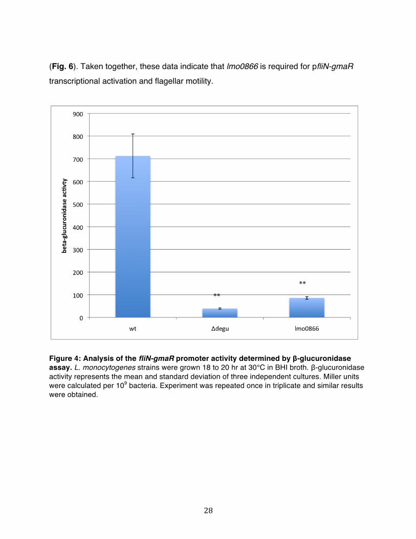
Lmo0866 is similar to an ATP-dependent RNA helicase Lmo0866 is predicted to be a single gene, 1563 base pairs long with an intergenic
region of 451 base pairs upstream of the start codon. lmo0866 encodes a hypothetical
protein similar to an ATP-dependent RNA helicase belonging to the DEAD-box protein family. It is composed of the 9 conserved motifs characteristic of DEAD-box proteins (reviewed in 23) as well as a 177 amino acid long non-conserved C-terminal domain (Fig. 6).
Figure 6: Schematic representation of the Lmo0866 protein. Lmo0866 is composed of a DEAD-box core containing the 9 conserved motifs, represented here as black stripes. The non-conserved C-terminal domain is shown in red.
DEAD-box proteins are a family of proteins highly conserved from bacteria through humans composed of a core of 350-400 amino acids containing 9 conserved sequence motifs. Motif II has the amino acids D-E-A-D giving the name to the family. Motifs I, II, VI and Q are required for ATP binding and hydrolysis. Motifs Ia, Ib, III, IV and V are less well characterized but may be involved in interaction with RNA. DEAD-box proteins N- or C- terminal domains are highly divergent and may provide specificity for interacting with specific RNA substrates or other factors. In vitro all DEAD-box proteins tested show RNA-dependent ATPase activity and many of them are ATP-dependent RNA helicases, meaning that they are able to dissociate short RNA duplexes in an ATP-dependent manner. DEAD-box proteins play important roles in many processes of RNA metabolism. In eukaryotes, they are involved in RNA degradation, pre-mRNA splicing and mRNA export as well as ribosome biogenesis and translation initiation. Interestingly, several DEAD-box proteins have been shown to be involved in

33
transcriptional regulation, which does not alwas require their RNA helicase activity. They are thought to act as adaptors or bridging factors between coactivators or corepressors and components of the transcription machinery. In prokaryotes, DEAD-box proteins
have been involved in mRNA processing and decay, ribosome biogenesis and translation initiation. To our knowledge, no prokaryotic DEAD-box protein has been found to be involved in transcriptional regulation.
lmo0866 was identified by microarray analysis as having higher transcript levels in
both log- and stationary- phase at 4°C compared to 37°C (24). Interestingly, lmo0866::mariner displays a growth defect when grown in BHI medium shaking at 30°C (Fig. 9) and 37°C (data not shown).
Figure 9: Growth curve of lmo0866 mutant. L. monocytogenes cultures were grown at 30 °C in BHI shaking. OD600 readings for each strain were taken from three independent cultures at regular time intervals.

34
As the DEAD-box proteins shown to act as transcriptional regulators in eukaryotes are also involved in other cellular processes, the growth defect observed suggests that
Lmo0866 also participates in processes other than pfliN-gmaR transcriptional activation. A ΔdegU mutant did not have a growth defect under these conditions, suggesting that the growth defect is not a DegU-dependent pathway. Lmo0866 does not affect DegU protein levels
As we identified lmo0866 as being required for pfliN-gmaR transcriptional activation Lmo0866 may directly act at the fliN-gmaR promoter in conjunction with DegU or could alternatively be involved in DegU regulation. To test if DegU levels are affected in the lmo0866 mutant we performed a western blot for DegU at 30°C (Fig. 10). Western blot analysis showed that DegU protein is still produced in the lmo0866 mutant and protein levels appear similar or maybe slightly lower than wild-type EGDe DegU protein levels. The faint DegU band in the ΔdegU sample is the result of some spillage from adjacent wells. We therefore hypothesize that Lmo0866 has a direct effect on transcriptional activation of the fliN-gmaR promoter.
lmo0866 ΔdegU wt
Figure 10: Western blot analysis of DegU protein levels in the lmo0866 mutant. Cultures were grown until mid exponential phase at 30°C and a western blot with a DegU polyclonal antibody was performed.

35
4.3 A transposon mutagenesis screen for L. monocytogenes genes involved in the repression of pfliN-gmaR at 37°C
The screen aims to identify additional factors required for transcriptional repression of pfliN-gmaR by screening the pfliN-gmaR::gusA transposon mutant library for blue colonies on X-gluc plates after incubation at 37°C, indicating that pfliN-gmaR is derepressed. The pfliN-gmaR::gusA construct integrated into a L. monocytogenes ΔmogR strain was used as a control and produced blue colonies on X-gluc plates incubated at 37°C for 48 hours (data not shown), verifying the ability of the screen to identify mogR mutants. The screen should identify mariner insertions in mogR and any additional factor(s) involved in the transcriptional repression of pfliN-gmaR. A partial library screen of approximately 50,000 transposon insertions was performed on BHI plates containing erythromycin kanamycin and X-gluc incubated at 37° C for 60 h. Ninety-seven blue colonies were selected and restreaked on BHI plates containing erythromycin kanamycin and X-gluc to confirm the phenotype. Ninety-six mutants confirmed their initial blue X-gluc plate phenotype. We performed a β-glucuronidase assay to better quantify the transcriptional activation level of pfliN-gmaR to select interesting mutants to send for sequencing. From the 96 mutants, 41 mutants showed a β-glucuronidase activity very similar to the wild-type pfliN-gmaR::gusA strain, 9 showed an intermediate phenotype and 46 showed an activity similar to a ΔmogR pfliN-gmaR::gusA strain. We decided to send the intermediate and high β-glucuronidase activity mutants for sequencing. We only identified insertions in mogR in the high β-
glucuronidase activity mutants with 6 independent transposon insertions in mogR. The intermediate β-glucuronidase activity category led to the identification of four independent transposon insertions in the codY gene and one insertion in mogR, inserted 12 bp upstream of the stop codon. It is possible that this mutant is expressing a shorter MogR protein that still retains some activity.
36
As we were able to isolate multiple mogR mutants, the expected control, representing 7 independent insertions it suggested that we were approaching saturation and we did not perform any additional screening.
Interestingly, codY was identified in a previous screen performed in our lab looking
at the regulation of pflaA, the promoter of the flagellin gene, at 37°C. Furthermore, a microarray of ΔcodY EGDe mutant and EGDe strain showed that genes involved in flagellar biosynthesis were derepressed in the ΔcodY mutant (24). We therefore decided to further investigate the role of CodY in the transcriptional repression of pfliN-gmaR at 37°C. MogR protein levels are decreased in a codY mutant
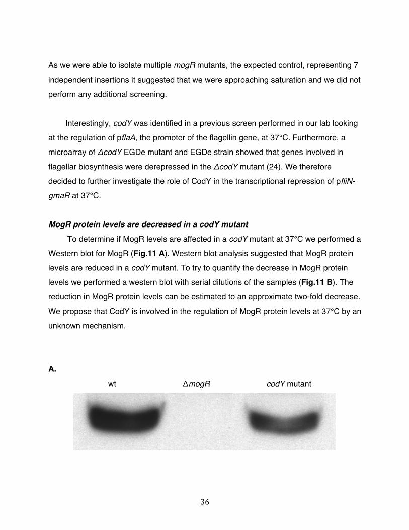
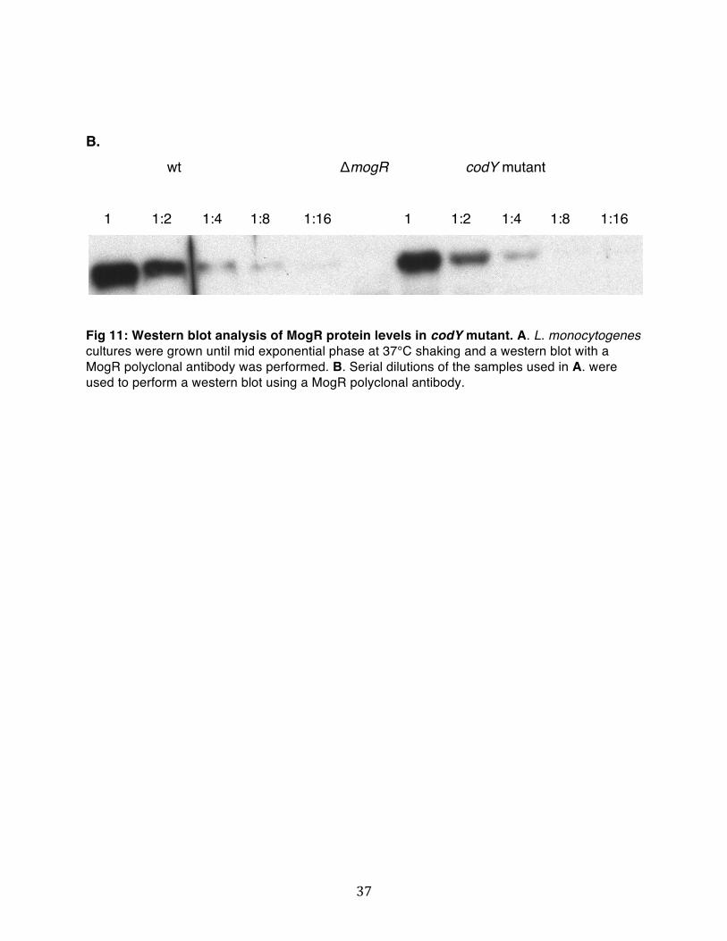
To determine if MogR levels are affected in a codY mutant at 37°C we performed a Western blot for MogR (Fig.11 A). Western blot analysis suggested that MogR protein levels are reduced in a codY mutant. To try to quantify the decrease in MogR protein levels we performed a western blot with serial dilutions of the samples (Fig.11 B). The reduction in MogR protein levels can be estimated to an approximate two-fold decrease. We propose that CodY is involved in the regulation of MogR protein levels at 37°C by an unknown mechanism.
A.
wt ΔmogR codY mutant
37
B.
wt ΔmogR codY mutant
1 1:2 1:4 1:8 1:16 1 1:2 1:4 1:8 1:16
Fig 11: Western blot analysis of MogR protein levels in codY mutant. A. L. monocytogenes cultures were grown until mid exponential phase at 37°C shaking and a western blot with a MogR polyclonal antibody was performed. B. Serial dilutions of the samples used in A. were used to perform a western blot using a MogR polyclonal antibody.

38
5. DISCUSSION
Transcriptional regulation of the fliN-gmaR promoter, the promoter of the first operon of the flagellar gene cluster, is central to the temperature-dependent regulation of flagellar motility in L. monocytogenes and is mediated by the activities of the DegU response regulator, the MogR transcriptional repressor, and GmaR, the MogR anti-repressor. In this study, we further investigated the mechanisms controlling transcription initiating at the fliN-gmaR promoter by performing two comprehensive transposon mutagenesis screens to identify factors involved in the activation and repression of pfliN-gmaR. The mutagenesis screen performed at 30°C to identify factors involved in pfliN-gmaR transcriptional activation led to the identification of a novel regulatory factor in L. monocytogenes. We demonstrated that Lmo0866, a protein from the DEAD-box protein family, is required for transcriptional activation of pfliN-gmaR and therefore flagellar motility. Moreover, we identified codY as being involved in the repression of pfliN-gmaR at 37°C.
Comprehensive transposon mutagenesis screens for factors involved in the
transcriptional regulation of pfliN-gmaR
To further investigate the regulation of transcription initiating at the fliN-gmaR promoter, we generated a mariner transposon library in a pfliN-gmaR::gusA reporter strain and performed two screens at 30°C and 37°C to, respectively, identify additional factors involved in the activation and repression of pfliN-gmaR. Comprehensive transposon mutagenesis in L. monocytogenes was made possible by the development of a new Himar1-based transposon system (19). This transposon system provides increased genomic coverage and is far superior to the well-established Tn917 transposon system. Indeed, due to the poor genomic coverage of Tn917, a previous

39
screen performed in our lab using the Tn917 transposon was unable to identify degU when screening for activating factors of pflaA, the promoter for the flagellin gene. We were able to isolate 75 degU mutants representing 10 distinct transposon insertions, in
our mariner transposon screen for additional factors involved in the transcriptional activation of pfliN-gmaR. We also isolated transposon insertions in pfliN-gmaR and the fliN-gmaR operon. In the mutagenesis screen to identify factors involved in repression of pfliN-gmaR at 37°C, we isolated 46 mogR mutants representing 7 independent insertions. We therefore conclude that the newly generated transposon mutant library allowed for comprehensive screening for factors controlling regulation of transcription occurring at pfliN-gmaR.
Identification of a novel factor involved in transcription of pfliN-gmaR and flagellar motility
We identified two independent transposon insertions in lmo0866 in the 30°C screen to identify factors involved in the activation of pfliN-gmaR. Both mutants were less blue on X-gluc plates (Fig. 3) and non-motile (Fig. 4). Furthermore, complementation with the cloned lmo0866 locus was able to restore flagellar motility (Fig. 7) confirming that Lmo0866 is required for transcription of pfliN-gmaR and therefore flagellar motility. As we preferentially screened for white colonies rather than less blue colonies on X-gluc plates, it is possible that we could have identified additional independent lmo0866 mutants by screening more extensively for less blue colonies.
Western blot analysis showed that DegU is still produced in the lmo0866 mutant (Fig. 10). DegU protein levels were similar to those in wild-type bacteria suggesting that Lmo0866 does not regulate DegU protein levels, but may act directly at the fliN-gmaR promoter in conjunction with DegU. Overexpression of DegU in the lmo0866 mutant and

40
testing for suppression of the non-motile phenotype may provide insights into the function of Lmo0866.
Lmo0866 is highly similar to an ATP-dependent RNA helicase from the DEAD-box protein family
DEAD-box proteins are highly conserved from bacteria through humans and play important roles in many cellular processes including mRNA export from the nucleus to the cytoplasm, ribosome biogenesis and RNA degradation. The L. monocytogenes genome encodes four ATP-dependent RNA helicases from the DExD-box protein family (where x can be any amino acid), two of them being DEAD-box proteins. Interestingly, Lmo0866 shares significant homology with the B. subtilis cold-shock protein A, CshA. They are 59 % identical and 74% similar (Fig. 13). cshA transcript levels are upregulated in response to cold-shock (26) and CshA was proposed to function in conjunction with cold-shock proteins to help rescue misfolded mRNA molecules to maintain proper initiation of translation at low temperatures in B. subtilis (27). Similarly, lmo0866 transcript levels were found by microarray analysis to be upregulated at 4°C compared to 37°C (24). It is therefore also possible that Lmo0866 is involved in cold adaptation in L. monocytogenes.
Recently, CshA has been identified as the major RNA helicase of the RNA degradosome complex in B. subtilis (28). CshA was shown to be able to interact with several proteins of the degradosome complex, including RNase Y and the polynucleotide phosphorylase. Interestingly, the non-conserved C-terminal domain of CshA was shown to be required for dimerization of CshA and for interactions with proteins of the RNA degradosome. Based on the significant homology with CshA and the growth defect of the lmo0866 mutant, suggesting that Lmo0866 is involved in

41
important cellular processes, it is tempting to speculate that Lmo0866 may also be involved in RNA degradation in L. monocytogenes.
Figure 13: Alignment of amino acid residues of L. monocytogenes Lmo0866 and B. subtilis CshA proteins. Conserved identical residues are blocked in blue and similar residues are blocked in purple. Alignment was obtained through the Needleman-Wunsch algorithm using matrix BLOSUM62.
Nonetheless, our studies indicate that Lmo0866 is required for transcription of pfliN-gmaR and therefore flagellar motility. Since DegU protein levels do not appear to be affected in the lmo0866 mutant, we hypothesize that Lmo0866 has a direct effect on
transcriptional activation of pfliN-gmaR. In recent years, there has been increasing evidence showing that some members of the DEAD-box protein family play important roles in transcriptional regulation in eukaryotes (reviewed in 29). DEAD-box proteins can function through interaction with transcriptional coactivators or corepressors, suggesting they may function as bridging factors between other regulators and components of the

42
transcription machinery, stabilizing the transcription initiation complex by interacting directly with proteins in the complex, or helping recruit transcriptional regulators to the initiation complex. For example, the human DEAD-box protein p68 interacts with the
transcriptional coactivators CBP/p300 and RNA pol II, stimulating transcriptional activation mediated by CBP/p300. p68 also acts as a transcriptional coactivator for the nuclear estrogen receptor alpha (Erα). While no DEAD-box protein has been reported to act as transcriptional regulator in prokaryotes, it is possible that Lmo0866 is directly involved in transcriptional activation of pfliN-gmaR.
The DegU binding site in the pfliN-gmaR region is located -184 to -154 bp
upstream of the transcriptional start site, however it is unclear how the binding of DegU to the pfliN-gmaR region is transmitted downstream to RNA polymerase to allow activation of transcription. In this instance, Lmo0866 could act as a bridging factor between the DegU response regulator and RNA polymerase. As the C-terminal domain of CshA allows for dimerization and interaction with specific proteins, it is tempting to speculate that the C-terminal domain of Lmo0866, which shares significant homology to the CshA C-terminal domain, may enable dimerization of Lmo0866 and interaction with factors such as DegU or RNA polymerase. Pull-down experiments to determine if Lmo0866 is able to interact with DegU would provide insights into a potential Lmo0866/DegU interaction. Alternatively, interaction between DegU and Lmo0866 may be dependent on DegU binding to the pfliN-gmaR region, therefore a gel mobility shift analysis with pfliN-gmaR DNA, and purified DegU and Lmo0866 proteins should be performed. These studies would also provide insights into the ability of Lmo0866 to bind to pfliN-gmaR DNA. However, it is less likely that Lmo0866 binds DNA since no DNA-binding domain was identified in the Lmo0866 protein sequence and no DEAD-box protein previously identified to be involved in transcriptional regulation has been shown to bind DNA.
43
Alternatively, another model for Lmo0866 regulation of pfliN-gmaR transcription could involve Lmo0866 helicase activity. Lmo0866 could bind to DegU and mediate unwinding of pfliN-gmaR DNA to favor transcriptional activation. It would therefore be
interesting to determine if the helicase activity of Lmo0866 is required for transcription of pfliN-gmaR activity by mutating the DEAD box motif to a helicase inactive NEAD sequence (30). This would provide additional insights into the mechanism of Lmo0866-mediated transcriptional activation of pfliN-gmaR. Nonetheless, we cannot exclude the remote possibility that because of the potential role of Lmo0866 in the RNA degradosome complex, a mutation in lmo0866 may cause a more general metabolic defect that results in poor flagellar production. Therefore, further analysis is needed to determine how Lmo0866 is involved in regulating transcription initiating at pfliN-gmaR and as a result flagellar motility.
CodY is involved in the regulation of MogR protein levels at 37°C
We were able to isolate multiple mogR mutants from the mutagenesis screen for factors involved in repression of pfliN-gmaR at 37°C. Furthemore, only mogR mutants demonstrated high β-glucuronidase activity, suggesting that MogR is the primary
repressor of pfliN-gmaR and does not require any additional factor(s) to function as a repressor, this is in accordance with previously published data. We were however able to identify CodY as being involved in the repression of pfliN-gmaR, but a codY mutant had an intermediate β-glucuronidase activity suggesting that the CodY effect on transcription of pfliN-gmaR may be indirect. CodY is a transcriptional regulator controlling genes involved in carbon and nitrogen assimilation. CodY is able to monitor the energetic and nutritional status of the cell by sensing the levels of GTP and branched chain amino acids (BCAA). Interestingly, the codY regulon, as defined by microarray analysis, identified the flagellar motility gene cluster as being derepressed in a ΔcodY mutant (25). Moreover, in B. subtilis CodY was shown to directly repress the flagellin gene in a nutrient rich environment by binding to the flagellin gene promoter

44
(31). Since, we hypothesized that the role of CodY for repression of pfliN-gmaR was indirect, we investigated MogR protein levels in a codY mutant and found that MogR levels are indeed decreased. To further investigate if CodY regulates mogR
transcription or MogR protein levels, a q-RT PCR for mogR should be performed. As mogR was not identified in the codY regulon, it is tempting to speculate that CodY is involved in the regulation of MogR protein levels by the action of another CodY regulated protein, such as a protease.

45
6. CONCLUSION
Flagellar motility is an essential mechanism allowing bacteria to move and to
survive in very diverse environments. L. monocytogenes is a facultative intracellular pathogen that represses transcription of flagellar motility genes at physiological temperatures (37°C and above). Interestingly, regulation of flagellar motility genes in L. monocytogenes differs significantly from other bacterial species as homologs to the master regulators are lacking. In L. monocytogenes, the MogR transcriptional repressor represses transcription of all flagellar motility genes in a non-hierarchal manner at temperatures of 37°C and above. GmaR, the MogR anti-repressor binds to MogR preventing MogR repression of flagellar motility genes at temperatures below 37°C. The response regulator DegU is required for transcription of all flagellar motility genes and acts by transcriptionally activating fliN-gmaR, the first operon of the flagellar gene cluster. In this study, we identified a novel factor, Lmo0866, as being required for pfliN-gmaR transcriptional activation and therefore flagellar motility. Moreover, we identified CodY as being involved in pfliN-gmaR repression at 37°C. Given the multiple environments L. monocytogenes can encounter during its extracellular and intracellular existence and the importance of proper regulation of flagellar motility genes for bacterial survival, L. monocytogenes has evolved a complex regulatory system involving multiple factors that work together to fine tune flagellar motility gene expression in response to environmental conditions.

46
7. REFERENCES
1. Ramaswamy V, Cresence VM, Rejitha JS, Lekshmi MU, Dharsana KS, Prasad SP, Vijila HM. (2007) Listeria--review of epidemiology and pathogenesis. J Microbiol Immunol Infect 40:4-13.
2. Jarrell KF, McBride MJ. (2008) The surprisingly diverse ways that prokaryotes move, Nat Rev Microbiol. 6:466-76.
3. Wadhams GH, Armitage JP. (2004) Making sense of it all: bacterial chemotaxis.
Nat Rev Mol Cell Biol. 5:1024-37.
4. Armitage, J.P. (1999) Bacterial tactic responses. Adv Microb Physiol 41: 229-289.
5. Dons L, Eriksson E, Jin Y, Rottenberg ME, Kristensson K, Larsen CN, Bresciani
J, Olsen JE. (2004) Listeria monocytogenes flagella are used for motility, not as adhesins, to increase host cell invasion. Infect Immun. 72:3237-44.
6. Lemon KP, Higgins DE, Kolter R. (2007) Flagellar motility is critical for Listeria
monocytogenes biofilm formation. J Bacteriol. 189:4418-24.
7. O'Neil HS, Marquis H. (2006) Role of flagellin and the two-component CheA/CheY system of Listeria monocytogenes in host cell invasion and virulence. Infect Immun.74:6675-81.
8. Peel M, Donachie W, Shaw A. (1988) Temperature-dependent expression of
flagella of Listeria monocytogenes studied by electron microscopy, SDS-PAGE and western blotting. J Gen Microbiol. 134:2171-8.
9. Hayashi F, Smith KD, Ozinsky A, Hawn TR, Yi EC, Goodlett DR, Eng JK, Akira S, Underhill DM, Aderem A. (2001) The innate immune response to bacterial flagellin is mediated by Toll-like receptor 5. Nature. 410:1099-103.
10. McCarter LL. (2006) Regulation of flagella. Curr Opin Microbiol. 9:180-6.

47
. 11. Shen A, Higgins DE. (2006) The MogR transcriptional repressor regulates
nonhierarchal expression of flagellar motility genes and virulence in Listeria monocytogenes. PLoS Pathog.2:e30
12. Gründling A, Burrack LS, Bouwer HG, Higgins DE. (2004) Listeria
monocytogenes regulates flagellar motility gene expression through MogR, a transcriptional repressor required for virulence. Proc Natl Acad Sci USA.101:12318-23.
13. Shen A, Kamp HD, Gründling A, Higgins DE. (2006) A bifunctional O-GlcNAc
transferase governs flagellar motility through anti-repression. Genes Dev. 20:3283-95.
14. Knudsen GM, Olsen JE, Dons L. (2004) Characterization of DegU, a response
regulator in Listeria monocytogenes, involved in regulation of motility and contributes to virulence. FEMS Microbiol Lett. 240:171-9.
15. Kamp HD, Higgins DE. (2009) Transcriptional and post-transcriptional regulation
of the GmaR antirepressor governs temperature-dependent control of flagellar motility in Listeria monocytogenes Mol Microbiol. 74:421-35.
16. Kobayashi K. (2007) Gradual activation of the response regulator DegU controls
serial expression of genes for flagellum formation and biofilm formation in Bacillus subtilis Mol Microbiol. 66:395-409
17. Gueriri I, Cyncynatus C, Dubrac S, Arana AT, Dussurget O, Msadek T.(2008)
The DegU orphan response regulator of Listeria monocytogenes autorepresses its own synthesis and is required for bacterial motility, virulence and biofilm formation Microbiology. 154:2251-64.
18. Buck M, Bose D, Burrows P, Cannon W, Joly N, Pape T, Rappas M, Schumacher
J, Wigneshweraraj S, Zhang X.(2006) A second paradigm for gene activation in bacteria Biochem Soc Trans. 34:1067-71.

48
19. Zemansky J, Kline BC, Woodward JJ, Leber JH, Marquis H, Portnoy DA. (2009) Development of a mariner-based transposon and identification of Listeria monocytogenes determinants, including the peptidyl-prolyl isomerase PrsA2, that contribute to its hemolytic phenotype. J Bacteriol. 191:3950-64.
20. Garsin DA, Urbach J, Huguet-Tapia JC, Peters JE, Ausubel FM. (2004)
Construction of an Enterococcus faecalis Tn917-mediated-gene-disruption library offers insight into Tn917 insertion patterns J Bacteriol. 186:7280-9
21. Monk IR, Gahan CG, Hill C. (2008) Tools for functional postgenomic analysis of
listeria monocytogenes Appl Environ Microbiol 74:3921-34
22. Toledo-Arana et al. (2009), The Listeria transcriptional landscape from saprophytism to virulence. Nature 459:950-6
23. Linder P. (2006) Dead-box proteins: a family affair-active and passive players in
RNP-remodeling. Nucleic Acids Res 34:4168-80.
24. Chan YC, Raengpradub S, Boor KJ, Wiedmann M (2007) Microarray-based characterization of the Listeria monocytogenes cold regulon in log-and stationary-phase cells. Appl Environ Microbiol 73:6484-98
25. Bennett HJ et al. (2007) Characterization of relA and codY mutants of Listeria
monocytogenes: identification of the CodY regulon and its role in virulence. Mol Microbiol 63:1453-67
26. Beckering CL, Steil L, Weber MH, Völker U, Marahiel MA. (2002) Genomewide
transcriptional analysis of the cold shock respons in Bacillus subtilis. J Bacteriol 184:6395-402
27. Hunger K, Beckering CL, Wiegeshoff F, Graumann PL, Marahiel MA. (2006)
Cold-induced putative DEAD box RNA helicases CshA and CshB are essential for cold adaptation and interact with cold shock protein B in Bacillus subtilis. J Bacteriol 188:240-8

49
28. Lehnik-Habrink M, Pförtner H, Rempeters L, Pietack N, Herzberg C, Stülke J. (2010) The RNA degradosome in Bacillus subtilis: identification of CshA as the major RNA helicase in the multiprotein complex. Mol Microbiol [Epub ahead of print]
29. Fuller-Pace FV. (2006) DexD/H box RNA helicases: multifunctional proteins with
important roles in transcriptional regulation. Nucleic Acids Res. 34:4206-15.
30. Wortham et al (2009) The DEAD-box protein p72 regulates ERalpha-/oestrogen-dependent transcription and cell growth, and is associated with improved survival in ERalpha-positive breast cancer. Oncogene 28:4053-64
31. Bergara et al (2003) CodY is a nutritional repressor of flagellar gene expression
in Bacillus subtilis. J Bacteriol 185:3118-26
32. Guérout-Fleury et al. (1996) Plasmids for ectopic integration in Bacillus subtilis. Gene 180:57-61

50


![Torque Generated by Flagellar Motorof Escherichia - DAMTP · TorqueGenerated bythe Flagellar Motorof Escherichiacoil ... TES,N-tris[hydroxymethyl]methyl-2-aminoethanesulfonic acid](https://img.pdfslide.net/doc/110x75/5c90c4f509d3f2c8148bd888/torque-generated-by-flagellar-motorof-escherichia-torquegenerated-bythe-flagellar.jpg)