Embed Size (px)
Citation preview
ARTICLE IN PRESSAm J of Geriatric Psychiatry&&:&& (2019)&&−&&
Available online at www.sciencedirect.com
ScienceDirect
journal homepage: www.ajgponline.org
Regular Research Article
Mechanisms Linking White MatterLesions, Tract Integrity, andDepression in Alzheimer DiseaseChathuri Yatawara, Ph.D., Daryl Lee, B.S.Eng, Kok Pin Ng, M.B.B.S.,Russell Chander, B.A., Debby Ng, B.A., Fang Ji, M.Sc, Hee Youn Shim, B.S.Eng,Saima Hilal, Ph.D., Narayanaswamy Venketasubramanian, M.B.B.S.,Christopher Chen, B.M.B.Ch, Juan Zhou, Ph.D.,Nagaendran Kandiah, M.B.B.S., M.R.C.P., F.R.C.P.
AR T I C L E I N FO
Article history:
Received November, 22 2018
Revised April, 4 2019
Accepted April, 12 2019
From the Department of Neurology (CY, DL, KNeuroscience, Neuroscience and Behavioral DiHealth System (SH, CC), Memory Aging & Copore, Singapore. Send correspondence and repNational Neuroscience Institute, 11 Jalan Tan T
© 2019 American Association for Geriatric Phttps://doi.org/10.1016/j.jagp.2019.04.004
Am J Geriatr Psychiatry &&:&&, && 201
AB S TRA C T
Objective: Late-life depression involves the disconnection of white matter
tracts that regulate mood. A pathogenic link between poor tract integrity and
depressive symptoms is believed to be white matter lesions (WML), however
the mechanisms linking tract integrity, WML, and depression remains unex-
plored. The authors sought to identify whether the association between
reduced tract integrity and depressive symptoms is mediated by WML in
patients with Alzheimer disease (AD), and whether individual characteristics
moderate this effect. Methods: This was a cross-sectional study in a tertiary
memory clinic. A total of 91 patients with mild AD and 79 healthy elderly, com-
parable in depressive symptoms, white matter hyperintensities (WMH) volume,
cardiovascular risk, age, and sex were chosen. Tract integrity was assessed
using diffusion tensor imaging, WML were indexed as WMH, measured using
fluid-attenuation inversion recovery imaging, and depressive symptoms were
measured with the informant-based Geriatric Depression Scale. Results: In
patients with mild AD, reduced tract integrity in right hemispheric cortical-sub-
cortical tracts and the genu of the corpus callosum was moderately associated
with depressive symptoms. This association was fully mediated by WML. Modera-
tion analysis indicated that old age strengthened the association between all
tracts and depressive symptoms, as mediated by WML. In cognitively healthy
elderly, neither tracts nor WML were related to depressive symptoms.
Conclusion: Reduced tract integrity may be important but not sufficient for the
manifestation of depressive symptoms in mild AD. Instead, WML may drive the
Key Words:
Cerebrovascular disease
depression
Alzheimer disease
dementia
white matter tracts
structural imaging
PN, RC, DN, NK), National Neuroscience Institute, Singapore, Singapore ; Center for Cognitivesorders Program (FJ, HYS, JZ, NK), Duke-NUS Medical School, Singapore; National Universitygnition Centre, Singapore; and the Raffles Neuroscience Centre (NV), Raffles Hospital Singa-rint requests to Nagaendran Kandiah, M.B.B.S., M.R.C.P., F.R.C.P., Level 3, Clinical Staff Office,ock Seng, Singapore 308433. e-mail: [email protected]. Published by Elsevier Inc. All rights reserved.
9 1

ARTICLE IN PRESS
Mechanisms Linking White Matter Lesions, Tract Integrity, and Depression in
2
pathogenic link between reduced tract integrity and depressive symptoms. (Am J
Geriatr Psychiatry 2019;&&:&&−&&)
INTRODUCTION
L ate-life depression is one of the most commonmental illnesses in the aging population and its
prevalence is exacerbated in those with dementia.1
Onset of depressive symptoms in late life is precipi-tated by risk factors including cerebrovascular disease,old age, vulnerable genotypes, low socioeconomicstatus, and stressful life events.2−4 Of these risks, themost studied has been cerebrovascular disease, whichhas been proposed by the vascular depression hypoth-esis to predispose, precipitate, and perpetuate late-lifedepression by altering cerebral white matter.5
The link between cerebrovascular disease and late-life depression has been supported by a large body ofclinical and magnetic resonance imaging (MRI) stud-ies. Clinically, cerebrovascular disease risk factorssuch as diabetes, high blood pressure, and atrialfibrillation frequently co-occur with depression andindependently predict incidence of depressive symp-toms.6 However, this association between clinicalmarkers of cerebrovascular disease and depressionhas been inconsistent,7 and a more reliable link hasbeen observed with MRI markers, such as white mat-ter hyperintensities (WMH), chronic lacunes, andenlarged perivascular spaces.8,9 WMH, in particular,have been shown to quadruple the odds of develop-ing depressive symptoms in cognitively healthyelderly,8 and in patients with dementia.10
WMH are believed to contribute to depression bydisrupting white matter tracts that are central formood regulation.11 White matter tracts can be visual-ized using diffusion tensor imaging (DTI), which usesdiffusion to measure the restriction of water move-ment through tissue due to structural abnormalities.Restricted mobility of water molecules is an indicationof reduced tract integrity, caused by abnormal axonalmembranes, myelin sheaths, and neurofibrils.12
Reduced tract integrity plays an important role indepressive symptomology, discriminating betweenpatients with and without symptoms13 and predictingthe incidence of late-life depression.14 Tracts that playan important role in late-life depression involve thosethat link the frontal cortex with other mood-relatedcortical and subcortical areas, such as the amygdala
and hypothalamus.15 Alterations to these tracts dis-rupt the transfer of information between cortical andsubcortical regions, lending support to the idea thatdepression may be a disconnection syndromebetween cortical and subcortical regions.13
Tract integrity is influenced by individual charac-teristics that enhance or protect against structuralabnormalities. For instance, reduced tract integrity hasbeen associated with a greater accumulation of cardio-vascular risk factors,16 severe cognitive decline,17 oldage,18 and the APOE-e4 gene.19 Conversely, cognitivereserve, which is often characterized by high educa-tional attainment and vocabulary knowledge, hasbeen observed with healthier tract integrity.17
The direct relationships between depressive symp-toms, WMH, tract integrity, and individual character-istics have been well established, however, whatremains unclear is how WMH and tract integrityinteract in the process of depression and under whatconditions their relationship with depression is stron-gest. Based on the vascular depression hypothesis,we predict that the association between reduced tractintegrity and depressive symptoms may be mediatedby WMH and that this effect may be enhanced inindividuals with a high load of cardiovascular riskfactors, old age, lower educational attainment,impaired global cognition, and the APOE-e4 geno-type. We applied this hypothesis to subsyndromaldepressive symptoms given that most patients seenin memory clinics present with subsyndromal depres-sive symptoms.20
METHODS
Participants
Ninety-one patients with mild Alzheimer disease(AD) and 79 cognitively healthy elderly wererecruited into the study. Patients with mild AD wererecruited from a tertiary neurology center in Singa-pore (National Neuroscience Institute) between 2013and 2016. Diagnosis of mild probable AD was basedon the National Institute on Aging-Alzheimer’s Asso-ciation Criteria21 and included patients with a ClinicalDementia Rating Scale22 of 1. Diagnosis was made by
Am J Geriatr Psychiatry &&:&&, && 2019

ARTICLE IN PRESS
Yatawara et al.
cognitive neurologists using structural MRI as a sup-portive biomarker and a comprehensive neuropsy-chological evaluation. Healthy elderly were recruitedat the National Neuroscience Institute, Singapore andNational University Hospital, Singapore from 2010−2016 and included elderly who were “cognitivelynormal,” as determined by a comprehensive neuro-psychological assessment, including a Mini-MentalState Exam (MMSE) score greater than 28, and had aClinical Dementia Rating Scale of 0.
Exclusion criteria for all participants included 1) acurrent or history of alcohol or drug abuse; 2) comor-bid neurodegenerative diseases (e.g., Parkinson dis-ease); 3) history of acute clinical strokes; 4) cerebralamyloid angiopathy based on neuroimaging criteria;5) participants with MRI contradictions; and 6) a cur-rent or known history of major depression (definedusing Diagnostic and Statistical Manual of Mental Disor-ders, Fourth Edition, Text Revision) or other psychiatricconditions. Therefore, reported depressive symptomswere subsyndromal and unrelated to a premorbidpsychiatric disorder.
The study was approved by the SingHealth Cen-tralized Institutional Review Board and conducted inaccordance with Singhealth Centralized InstitutionalReview Board guidelines and the Declaration of Hel-sinki. Written informed consent was obtained fromall participants or their next of kin if they were men-tally incapable of giving consent.
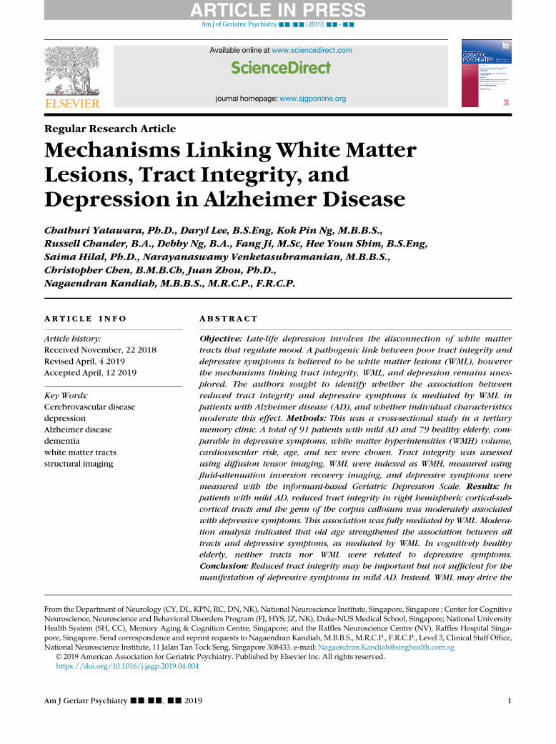
FIGURE 1. The cortical-subcortical and cortical-cortical white matteThe tract skeleton was chosen using TBSS and the JHU atlas which(red-orange) indicate right anatomic features, whereas cool colors(cingulate gyrus); COR: coronal; IFOF: inferior fronto-occipital fasciUniversity; PTR: posterior thalamic radiation; SAG: sagittal; SCR: sTBSS: Tract-Based Spatial Statistics; UF: uncinate fasciculus.
Am J Geriatr Psychiatry &&:&&, && 2019
Measures
Demographics were collected via clinical inter-view; cardiovascular risk profile was indexed usingthe Framingham office-based cardiovascular diseaserisk prediction model;23 global cognition was indexedusing the Montreal Cognitive Assessment24 and theMMSE;25 and APOE-e4 genotyping was performedusing TaqMan SNP genotyping assay and ABI7900HT PCR system (Applied Biosystems, FosterCity, CA). Depressive symptoms were measuredusing the Geriatric Depression Scale-Short Form,26
which is an informant-based questionnaire consistingof 15 yes/no items that has good validity (r =−0.73)and internal consistency (Cronbach a = 0.82) in theelderly with an MMSE score greater than 10.27 A cut-off of 5 points or more indicated significant depres-sive symptoms, as per locally validated norms.28
Target white matter tracts (Fig. 1) were chosen basedon a literature review of the most frequently reportedtracts from tract-based spatial statistics studies thatanalyzed the entire brain for group differences betweendepressed and nondepressed individuals, includingpatients with clinically diagnosed major depres-sion14,29,30 and subsyndromal depression,31 and meta-analyses investigating the association between whitematter tracts and late-life depression.13 Total WMHburden was used instead of regional WMH becausetarget tracts traverse across multiple cerebral regions.
r tracts proposed to be associated with depressive symptoms.was layered on top to identify individual tracts. Warm colors(blue) indicate left anatomic features. AX: axial; CC: cingulumculus; ILF: inferior longitudinal fasciculus; JHU: John Hopkinsuperior corona radiata; SLF: superior longitudinal fasciculus;
3

ARTICLE IN PRESS
Mechanisms Linking White Matter Lesions, Tract Integrity, and Depression in
Image Protocol
Participants underwent MRI in a whole body mag-netic resonance system. Participants undergoing studyprocedures before 2015 were scanned using a 3T Sie-mens Tim Trio system (Siemens, Erlangen, Germany),and participants recruited in 2015 and beyond werescanned using a 3T Siemens Prisma system (Siemens).Three-dimensional volumetric scans were obtainedusing a T1-weighted magnetization-prepared rapidgradient-echo sequence (repetition time 2300 ms, echotime 2.98 ms, matrix = 192£ 256£ 256, voxel = 1.0 mmisotropic) as per the AD Neuroimaging Initiativeprotocols (http://adni.loni.usc.edu/). Images weresegmented into gray matter, white matter, and cerebro-spinal fluid maps using a unified segmentationpipeline32 including affine regularization to the Interna-tional Consortium for Brain Mapping space templatefor East Asian brains. The generated gray matter andwhite matter maps were then used to generate globalvolumes of gray and white matter, respectively. Volu-metric analysis of WMHwas done in SPM8 (WellcomeTrust, London) using an existing workflow.33 Fluid-attenuated inversion recovery sequences were co-regis-tered with their corresponding Montreal NeurologicalInstitute-normalized magnetization-prepared rapidgradient-echo sequences, and voxels of WMH wereidentified based on having an intensity of at least1.4 times compared with the surrounding whitematters.34
DTI sequences were acquired using a single-shotspin echo echo-planar sequence (repetition time9600 ms, echo time 107 ms, matrix = 128£ 128, 54 sli-ces, voxel = 2.0 mm isotropic, 30 diffusion directions,b = 1000 s/mm2, and 6 b = 0 s/mm2). The DTI datawere preprocessed using FMRIB Software Library(University of Oxford, UK) (http://www.fmrib.ox.ac.uk/fsl). Following our previous approach,35 each dif-fusion-weighted volume was aligned to the b = 0image using EDDY current correction to correct for thedistortions caused by eddy currents and head move-ments. Data were discarded if the maximum displace-ment relative to the first b = 0 volume was more than3 mm. Diffusion gradients were rotated to improveconsistency with the motion parameters. TheDTI images were corrected for the geometric distortioncaused by magnetic field inhomogeneity usingFUGUE with gradient-echo field maps. Fractionalanisotropy (FA) images were created by fitting a
4
diffusion tensor model to the diffusion data at eachvoxel. We then applied tract-based spatial statistics36
to carry out a voxel-wise analysis of FA data withinmajor white matter pathways throughout the wholebrain following our previous approach.37 The meanFA of the predefined white matter tracts wereextracted from individual tracts of interest (Fig. 1)located according to the Johns Hopkins Universitywhite matter tractography atlas and the Johns HopkinsUniversity ICBM-DTI-81 white matter labels atlas.38
Statistical Analyses
Group differences in demographics, cognition, car-diovascular risk score, depressive symptoms, andMRI markers between patients with mild AD andhealthy elderly were determined using a t test for con-tinuous variables and the x2 test for categorical varia-bles, or the Fisher’s exact test for variables with smallcell counts. For unequal variances, the Welch adjust-ment was used.
Path analysis was used to examine the direct andindirect relationships between white matter anddepressive symptoms. First, the direct relationshipbetween predictor variables (tract integrity andWMH) and the outcome (depressive symptoms) wasdetermined using independent regression analysesfor each diagnostic group. Multicollinearity betweenall tracts and WMH was examined using varianceinflation factors,39 and normality was examined usingskew and kurtosis, with log transformation wherenecessary. All analyses controlled for age, educationalattainment, cardiovascular risk profile (Sex wasincluded in the cardiovascular risk score; therefore,we did not include it as a separate covariate. Age andeducation also make up the cardiovascular risk score;however, because of their variability, we includedthem as covariates.), global cognition, APOE-e4 geno-type, total gray matter volume, and MRI scanner typeto control for the use of different scanners. Second,mediation analysis40 determined whether the indirectrelationship between tract integrity (X in Fig. 2) anddepressive symptoms (Y in Fig. 2) could be explainedby a mediating variable, WMH (M in Fig. 2). If therelationship between X-Y reduced in significance afterincluding WMH, partial mediation was implied.Alternatively, if X-Y was no longer significant in thepresence of WMH, full mediation was implied. Medi-ation analysis was carried out independently for each
Am J Geriatr Psychiatry &&:&&, && 2019

FIGURE 2. The model implies that the relationship between reduced white matter tract integrity (X) and depressive symptoms (Y)may be driven by WMHs (M). The model additionally implies that individual risk factors (W) may moderate this indirect effectbetween tract integrity and depressive symptoms. Adapted from Hayes.40
ARTICLE IN PRESS
Yatawara et al.
diagnostic group, each white matter tract, and eachhemisphere of each tract due to possible structuraland functional hemispheric asymmetry.41 To com-pare the structure of the regression models betweendiagnostic groups, Steiger’s z test42 was applied. Firstthe predictive value for depressive symptoms wascalculated for each diagnostic group; next the correla-tion between each predicted value and the criterionwas determined to calculate Steiger’s z. A simulatedprocedure using bootstrap methods corrected formultiple comparisons, which has been empiricallyvalidated to derive robust parameter estimates basedon maximized power and limited type 1 error rates.43
As a subanalysis, the mediation analyses wereassessed in participants without significant depres-sive symptoms (Geriatric Depression Scale [GDS]score <5) to confirm consistency of results.
As a third step for significant mediation findings, amoderation analysis40 was conducted to quantifywhether the indirect relationship between tracts anddepressive symptoms, via WMH, was enhanced orattenuated by individual characteristics, such as age,educational attainment, cardiovascular risk profile,global cognition, or the APOE-e4 genotype. Modera-tion was calculated by first z-scoring all variablesthen multiplying each tract (X in Fig. 2) with each riskfactor (W in Fig. 2) to create the interaction variable(WX). If XW was significantly related to WMH (M inFig. 2) (WX-M), and indirectly related to the outcome(Y in Fig. 2) (WX-M-Y), we noted that moderatedmediation had occurred (for more detail see Supple-mentary Material).
Am J Geriatr Psychiatry &&:&&, && 2019
Path analyses for both the mediation and modera-tion effect were conducted using SPSS Amos version20 (SPSS, Inc, Chicago, IL). Model fit for each pathanalysis was determined using recommended crite-ria:44 1) x2 p value >0.05; 2) Bentler Comparative FitIndex (CFI >0.95); and 3) Root Mean Square Error ofApproximation (RMSEA <0.06). Model fit wasrevised using modification indices. A bias correctedbootstrap estimation with 1,000 resamples wasapplied to both the mediation and moderation analy-ses as a nonparametric approach for effect-size esti-mation. Here, a 95% confidence interval that did notcontain zero indicated a significant effect.45 Effectsizes for the direct effects were indexed using thestandardized coefficient of the slope (B), which isidentical to the benchmark set for Pearsons r, 0.10(small), 0.30 (moderate), and 0.50 (large).46 Effect sizefor indirect effects were indexed by squaringCohen’s46 estimations because indirect effects repre-sent a product of two effects,40 0.01 (small), 0.09(moderate), and 0.25 (large).
An exploratory analysis sought to verify the speci-ficity of our model to mood-related white mattertracts by testing whether tracts outside of the moodnetwork, such as motor tracts, were significant in ourmodel. Specifically, we tested one cortical-subcorticaltract, the cerebellar peduncle, which connects the cer-ebellum to the brainstem, and one interhemispherictract, the splenium, which connects the motor regionsof each hemisphere. We explored whether thesemotor-related tracts were directly and indirectlyrelated to depression, as mediated by WMH.
5

ARTICLE IN PRESS
Mechanisms Linking White Matter Lesions, Tract Integrity, and Depression in
RESULTS
Participants
The AD and healthy elderly groups did not dif-fer in age, sex, race, cardiovascular risk profile,WMH volume, or severity of depressive symptoms(Table 1). Compared with the healthy elderly,patients with mild AD had lower educationalattainment, greater global cognitive impairment,reduced gray matter volume, and higher preva-lence of the APOE-e4 genotype. Patients with mildAD also exhibited reduced tract integrity com-pared with controls for all tracts except the rightand left interior occipital fasciculus and left fornix(Supplementary Table S1).
TABLE 1. Participant Characteristics and Group Differences
Mean (SD)Healthy Elderly
(N = 79)
Age (years) 62.85 (7.12)Sex (male, %) 33 (41%)Years of education 13.33 (3.25)Race (%)Chinese 72 (91%)Malay 1 (1%)Indian 3 (4%)Euroasian 3 (4%)Other 0
Medication (frequency, %)Cognitive enhancers 0Antidepressants 2 (2%)Antipsychotics 0
APOE-e4 carrier (%) 7 (9%)MoCA(score range 0−30)
27.86 (1.83)
MMSE(score range 0−30)
28.24 (1.55)
Framingham score(score range 0−30)
12.84 (3.37)
GDS Total (mean, SD, score range)(score range 0−15)
2.01 (2.68)range: 0−12
GDS ≥5 (frequency, %) 9 (11%)WMH volume 4.96 (6.53)Gray matter volume 553.81 (60.69)
Notes: MoCA: Montreal Cognitive Assessment; SD: standard deviation;Donepezil.
a t test values with df = 168.bWelch adjusted t test adjusted df = 119.89.cWelch adjusted t test adjusted df = 118.59.d The x2 test values with df = 1.e Fisher’s exact test values.f p <0.01.g p <0.001.
6
Direct Relationships Between Tract Integrity,
WMH, and Depressive Symptoms
For patients with mild AD, path analysis modelstesting the direct relationship between white mattertract integrity and depressive symptoms (X-Y associa-tion in Fig. 2), while controlling for all covariates andWMH had good model fit (x2 [14] <10, p >0.05, CFI>0.99 and RMSEA <0.09). Depressive symptomswere significantly related to the right side of the pos-terior thalamic radiation, uncinate fasciculus, inferiorfronto-occipital fasciculus, fornix, superior coronaradiate, and the genu of corpus callosum (Table 2).All relationships were negative with a moderate effectsize, suggesting that as tract integrity decreased,severity of depressive symptoms increased. All leftside tracts were not related to depressive symptoms.
Mild AD(N = 91) Test Statistic
69.25 (8.56) 0.16a
47 (52%) 1.65d
9.72 (3.91) 3.23a,f
4.58e
85 (93%)2 (2%)3 (3%)
01 (2%)
12 (13%) 2.16d,f
1 (1%) 1.60d
0 -41 (45%) 21.09d,g
21.99 (5.27) 92.21b,g
24.92 (4.09) 51.24c,g
14.98 (3.94) 0.85a
3.08 (2.53)range: 0−10
0.72a
18 (20%) 1.49d
7.03 (9.10) −1.67a
520.40 (62.14) 3.53a,g
WMH: white matter hyperintensities, cognitive enhancers included
Am J Geriatr Psychiatry &&:&&, && 2019
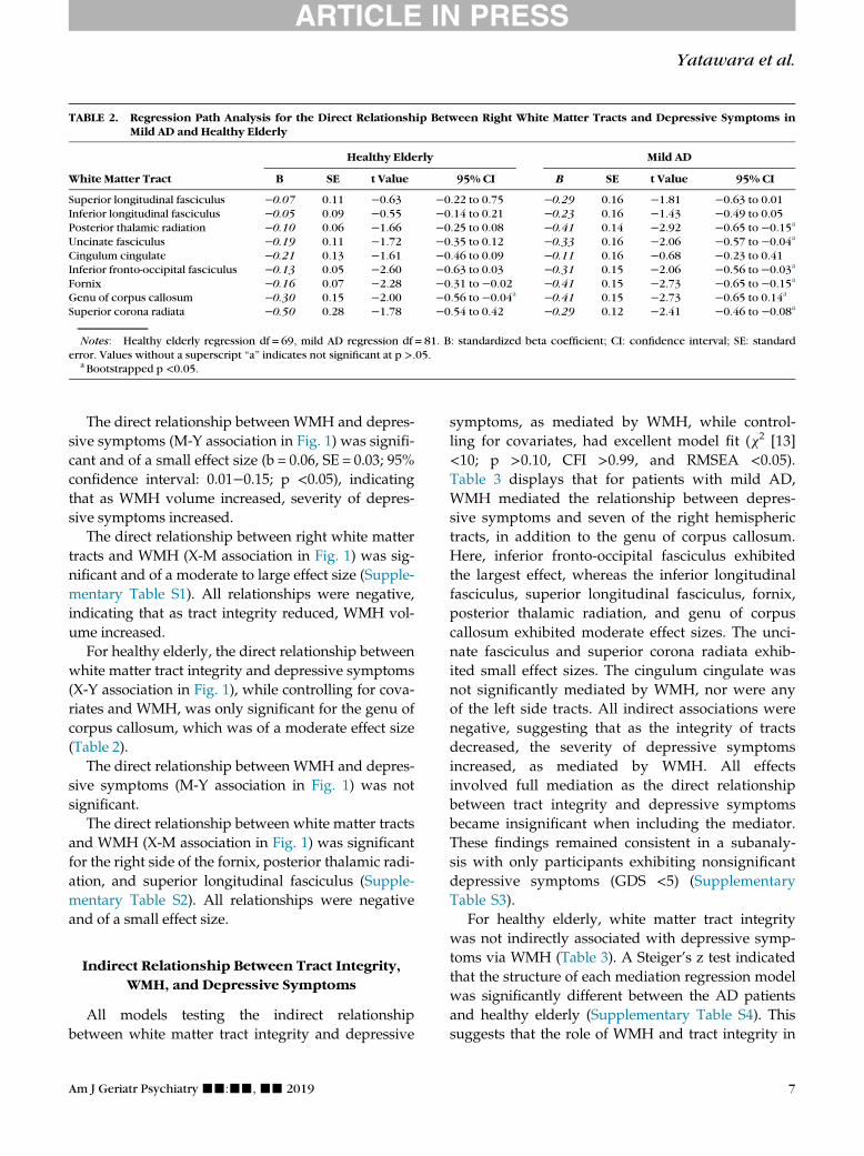
TABLE 2. Regression Path Analysis for the Direct Relationship Between Right White Matter Tracts and Depressive Symptoms inMild AD and Healthy Elderly
Healthy Elderly Mild AD
White Matter Tract B SE t Value 95% CI B SE t Value 95% CI
Superior longitudinal fasciculus −0.07 0.11 −0.63 −0.22 to 0.75 −0.29 0.16 −1.81 −0.63 to 0.01Inferior longitudinal fasciculus −0.05 0.09 −0.55 −0.14 to 0.21 −0.23 0.16 −1.43 −0.49 to 0.05Posterior thalamic radiation −0.10 0.06 −1.66 −0.25 to 0.08 −0.41 0.14 −2.92 −0.65 to −0.15a
Uncinate fasciculus −0.19 0.11 −1.72 −0.35 to 0.12 −0.33 0.16 −2.06 −0.57 to −0.04a
Cingulum cingulate −0.21 0.13 −1.61 −0.46 to 0.09 −0.11 0.16 −0.68 −0.23 to 0.41Inferior fronto-occipital fasciculus −0.13 0.05 −2.60 −0.63 to 0.03 −0.31 0.15 −2.06 −0.56 to −0.03a
Fornix −0.16 0.07 −2.28 −0.31 to −0.02 −0.41 0.15 −2.73 −0.65 to −0.15a
Genu of corpus callosum −0.30 0.15 −2.00 −0.56 to −0.04a −0.41 0.15 −2.73 −0.65 to 0.14a
Superior corona radiata −0.50 0.28 −1.78 −0.54 to 0.42 −0.29 0.12 −2.41 −0.46 to −0.08a
Notes: Healthy elderly regression df = 69, mild AD regression df = 81. B: standardized beta coefficient; CI: confidence interval; SE: standarderror. Values without a superscript “a” indicates not significant at p >.05.
a Bootstrapped p <0.05.
ARTICLE IN PRESS
Yatawara et al.
The direct relationship between WMH and depres-sive symptoms (M-Y association in Fig. 1) was signifi-cant and of a small effect size (b = 0.06, SE = 0.03; 95%confidence interval: 0.01−0.15; p <0.05), indicatingthat as WMH volume increased, severity of depres-sive symptoms increased.
The direct relationship between right white mattertracts and WMH (X-M association in Fig. 1) was sig-nificant and of a moderate to large effect size (Supple-mentary Table S1). All relationships were negative,indicating that as tract integrity reduced, WMH vol-ume increased.
For healthy elderly, the direct relationship betweenwhite matter tract integrity and depressive symptoms(X-Y association in Fig. 1), while controlling for cova-riates and WMH, was only significant for the genu ofcorpus callosum, which was of a moderate effect size(Table 2).
The direct relationship between WMH and depres-sive symptoms (M-Y association in Fig. 1) was notsignificant.
The direct relationship between white matter tractsand WMH (X-M association in Fig. 1) was significantfor the right side of the fornix, posterior thalamic radi-ation, and superior longitudinal fasciculus (Supple-mentary Table S2). All relationships were negativeand of a small effect size.
Indirect Relationship Between Tract Integrity,
WMH, and Depressive Symptoms
All models testing the indirect relationshipbetween white matter tract integrity and depressive
Am J Geriatr Psychiatry &&:&&, && 2019
symptoms, as mediated by WMH, while control-ling for covariates, had excellent model fit (x2 [13]<10; p >0.10, CFI >0.99, and RMSEA <0.05).Table 3 displays that for patients with mild AD,WMH mediated the relationship between depres-sive symptoms and seven of the right hemispherictracts, in addition to the genu of corpus callosum.Here, inferior fronto-occipital fasciculus exhibitedthe largest effect, whereas the inferior longitudinalfasciculus, superior longitudinal fasciculus, fornix,posterior thalamic radiation, and genu of corpuscallosum exhibited moderate effect sizes. The unci-nate fasciculus and superior corona radiata exhib-ited small effect sizes. The cingulum cingulate wasnot significantly mediated by WMH, nor were anyof the left side tracts. All indirect associations werenegative, suggesting that as the integrity of tractsdecreased, the severity of depressive symptomsincreased, as mediated by WMH. All effectsinvolved full mediation as the direct relationshipbetween tract integrity and depressive symptomsbecame insignificant when including the mediator.These findings remained consistent in a subanaly-sis with only participants exhibiting nonsignificantdepressive symptoms (GDS <5) (SupplementaryTable S3).
For healthy elderly, white matter tract integritywas not indirectly associated with depressive symp-toms via WMH (Table 3). A Steiger’s z test indicatedthat the structure of each mediation regression modelwas significantly different between the AD patientsand healthy elderly (Supplementary Table S4). Thissuggests that the role of WMH and tract integrity in
7

TABLE 3. Regression Path Analysis Models for the Indirect Relationship Between White Matter Tracts and Depressive Symptoms asMediated by WMH in Mild AD and Healthy Elderly
Mild AD Healthy Elderly
Right Side Tract B SE t Value BC 95% CI B SE t Value BC 95% CI
Superior longitudinal fasciculus −0.16 0.09 1.77 −0.38 to −0.03b −0.04 0.08 −0.50 −0.13 to 0.36Inferior longitudinal fasciculus −0.24 0.12 −2.00 −0.48 to −0.05a −0.01 0.02 −0.50 −0.11 to 0.23Fornix −0.22 0.12 −1.83 −0.42 to −0.04a −0.00 0.02 0.00 −0.07 to 0.31Posterior thalamic radiation −0.22 0.12 −1.83 −0.42 to −0.04a −0.01 0.09 −0.11 −0.05 to 0.04Uncinate fasciculus −0.06 0.04 −1.50 −0.13 to −0.01a −0.01 0.02 −0.50 −0.04 to 0.67Cingulum cingulate −0.03 0.04 −0.75 −0.19 to 0.00 −0.01 0.03 −0.33 −0.06 to 0.66Inferior fronto-occipital fasciculus −0.31 0.16 −1.93 −0.65 to −0.07a −0.00 0.02 0.00 −0.06 to 0.02Genu of corpus callosum −0.22 0.12 −1.83 −0.41 to −0.03a −0.01 0.02 −0.50 −0.06 to 0.37Superior corona radiata −0.04 0.05 −0.80 −0.10 to −0.01b −0.04 0.07 −0.57 −0.17 to 0.04
Notes: Healthy elderly regression df = 68, mild AD regression df = 80. B: standardized beta coefficient; BC: bias corrected; CI: confidence inter-val; SE: standard error.
a Bootstrapped p <0.05.b Bootstrapped p <0.01.
ARTICLE IN PRESS
Mechanisms Linking White Matter Lesions, Tract Integrity, and Depression in
depressive symptoms were significantly differentacross patients with AD and healthy elderly.
Moderators of the Relationship Between White
Matter and Depressive Symptoms
For patients with mild AD, all models testingwhether individual characteristics moderated theindirect effects between white matter tracts anddepressive symptoms, via WMH, had good model fit(x2 [47] <70; p >0.05, CFI >0.95, and RMSEA <0.04).The only significant moderator was old age for alltracts, except for the inferior longitudinal fasciculus(Table 4). The effect suggests that patients olderthan 65 years exhibited the strongest associationbetween low tract integrity and severity of depressivesymptoms.
For healthy elderly, no significant moderation wasobserved.
TABLE 4. Regression Path Analysis Models for Age (≥65 years) as aTracts and Depressive Symptoms via WMH for AD
Right Side Tract B
Superior longitudinal fasciculus −0.08Inferior longitudinal fasciculus −0.01Fornix −0.04Posterior thalamic radiation −0.05Uncinate fasciculus −0.05Inferior fronto-occipital fasciculus −0.04Genu body −0.69Superior corona radiata −0.54
Notes: Healthy elderly regression df = 63, mild AD regression df = 75. B:val; SE: standard error.
a Bootstrapped p <0.05.
8
Exploratory Analysis with Non-Mood Related
White Matter Tracts
Motor-related tracts, namely the cerebellar pedun-cle and the splenium, were not directly or indirectlyrelated to depressive symptoms, as mediated byWMH in patients with mild AD (SupplementaryTable S5), supporting the specificity of our model tomood-related tracts.
DISCUSSION
Main Findings
In patients with mild AD, we observed thatreduced integrity in mood-related white matter tractswas important but not sufficient for the manifestationof subsyndromal depressive symptoms. Instead,WMH were the catalyst that connected reduced tract
Moderator for the Indirect Relationship Between White Matter
SE t value BC 95% CI
0.02 −4.00 −0.17 to −0.01a
0.02 −0.50 −0.09 to 0.050.02 −2.00 −0.11 to −0.01a
0.03 −1.66 −0.12 to −0.01a
0.03 −1.66 −0.11 to −0.01a
0.03 −1.33 −0.12 to −0.01a
0.47 −1.46 −1.63 to −0.10a
0.29 −1.86 −1.36 to −0.20a
standardized beta coefficient; BC: bias corrected; CI: confidence inter-
Am J Geriatr Psychiatry &&:&&, && 2019
ARTICLE IN PRESS
Yatawara et al.
integrity to depressive symptoms. Patients with ADwere found to be most vulnerable to this effect,whereas reduced tract integrity and WMH played alimited role in subsyndromal depressive symptomsin cognitively healthy elderly comparable on age, sex,cardiovascular risk profile, WMH volume, and sever-ity of depressive symptoms. We further observed thatdepressive symptoms may not just involve a discon-nection between cortical-subcortical tracts, which waslocalized to the right hemisphere, but also a discon-nection between the interhemispheric tracts. Finally,we observed the association between white matterand depressive symptoms was strongest in elderlyolder than 65 years.
Theoretical and Clinical Implications
Consistent with previous findings in cognitivelynormal elderly with major depression,47 we observedthat reduced white matter tract integrity was directlyrelated to depressive symptoms; however after theinclusion of WMH, this association was no longer sig-nificant. WMH is a macrostructural representation ofneuronal loss, demyelination, and gliosis48 and repre-sents the most advanced stage of white matter degen-eration. Alternatively, DTI metrics provide amicrostructural representation of fiber density andaxon diameter and represent subtle early stages ofwhite matter degeneration. Therefore, our findingssuggest that subtle changes in mood-related tractsmay only be associated with depressive symptomswhen the overall severity of white matter damage isadvanced. Our findings are supported by previousresearch demonstrating that depression severity cor-relates strongest with white matter tracts intersectedby white matter lesions,29 and that a direct relation-ship between tract integrity and depressive symp-toms is commonly observed in elderly withcerebrovascular disease,49 but not in healthy elderlywithout cerebrovascular disease.50 Further research isrequired to determine whether severe white matterdamage located specifically in target tracts is drivingthis effect.
Consistent with previous perspectives,5 weobserved that mood-related cortical-subcortical tractswere implicated in subsyndromal depressive symp-toms, however, we further found that interhemi-spheric tracts may be of equal clinical relevance.Specifically, the genu of the corpus callosum
Am J Geriatr Psychiatry &&:&&, && 2019
exhibited the largest association with depressivesymptoms, and was the only tract significantlyrelated to depressive symptoms in cognitivelyhealthy elderly. The corpus callosum transfers infor-mation between the two hemispheres and the genuconnects the prefrontal with orbitofrontal regions.51
This significance of the genu of the corpus callosumin depressive symptomology converges with previ-ous findings13 and signifies the importance of dis-rupted information transfer between frontalhemispheres in depression. With regards to cortical-subcortical tracts, we observed that only the righthemisphere tracts were associated with depressivesymptoms in patients with mild AD, which is consis-tent with the growing evidence that the right side ofthe brain may have a primary role for depressivesymptoms in the elderly.52 Importantly, we verifiedthe specificity of our model to mood-related tracts bydemonstrating that motor-related cortical-subcorticaland interhemispheric tracts were not significant pre-dictors of depression in patients with mild AD.
In our study, cognitively healthy elderly and ADpatients had comparable WMH volumes and severityof depressive symptoms. Despite this, poor white mat-ter health was only associated with subsyndromaldepression in patients with AD. This implies the mani-festation of subsyndromal depressive symptoms incognitively healthy elderly may be independent ofwhite matter health and the vascular depressionhypothesis may have limited applicability for this pop-ulation. A significant finding only in the AD group sug-gests that AD pathology may have provided avulnerable context for poor white matter health tocause observable abnormalities. Amyloid has previ-ously been shown to compromise the integrity of whitematter tracts53 and greater AD pathology has beenassociated with greater WMH accumulation.54 There-fore, late-life subsyndromal depressive symptoms mayinvolve an interaction between the pathophysiologicalchanges of depression and changes initiated by otherdiseases, such as AD.55 We note that mechanisms otherthan cerebrovascular disease may degrade white mat-ter structures in AD, such as inflammatory processes,56
and further research is required to understand thesemechanisms.
We have demonstrated that the vascular depres-sion hypothesis, originally intended for understand-ing late-life major depression,55 may also begeneralized to understand subsyndromal depressive
9

ARTICLE IN PRESS
Mechanisms Linking White Matter Lesions, Tract Integrity, and Depression in
symptoms. This is particularly important given sub-syndromal symptoms are more prevalent in theelderly than major depression20 and are a risk for neu-rodegeneration of AD-related brain regions,57 greaterconversion to AD,58 and poorer functional out-comes.59 By excluding patients with major depres-sion, our findings suggest that subsyndromaldepressive symptoms in AD may have a comparableetiology to major depression in AD. Therefore, impli-cations from the vascular depression hypothesis55
may be useful to guide assessment and treatmentstrategies for subsyndromal depressive symptoms inmild AD. We note, however, that subsyndromaldepression may involve unique etiologies notobserved in major depression and the vasculardepression hypothesis may only describe one poten-tial mechanism. Pathologies other than WMH may becontributing to poor tract integrity as previousresearch has demonstrated that even after removingWMH, reduced white matter tract integrity was stillassociated with depressive symptoms in the elderly.60
Other pathologies such as inflammatory demyelinat-ing disease61 and amyloid deposition53 have beenassociated with poorer tract integrity and should beinvestigated in future research.
Our exploratory analysis identified that the linkbetween tract integrity and subsyndromal depressivesymptoms was strongest in AD patients over the ageof 65 years. The size of this modulatory effect wassmall for most tracts except for the inferior fronto-occipital fasciculus and genu of the corpus callosum,suggesting that not all tracts are equally affected byage-related changes. Interestingly, a high cardiovas-cular risk profile did not moderate the associationbetween white matter health and symptom severityin AD. Although cardiovascular risk factors contrib-ute to the development of poor white matter health,16
it appears to have limited effects on moderatingwhether poor white matter health manifests as sub-syndromal depression.
Limitations and Future Directions
We note that our cross-sectional data cannot provecausation, only association. Future research may ben-efit from exploring the causal links between tractintegrity, WMH, AD pathology, and depressivesymptoms. We note that we included total WMH inour model instead of regional WMH given that tracts
10
explored in this study transversed across multipleregions. Indeed, regional WMH plays a discrimina-tory role in late-life depression,62 however, mostWMH observed in patients with late-life depression8
and with mild AD63 are often found in the frontalregions. Furthermore, in patients with mild AD,WMH in all regions have been found to have conse-quences on frontal hypometabolism.63 As a result, wepropose that including regional WMH in our modelmay unlikely change the findings, however, this spec-ulation should be tested in future research with tract-based spatial statistics of each tract as it travels acrossvarious regions. We further note that three partici-pants (2%) were on antidepressants. These partici-pants did not present as outliers with their GDSscores (the GDS score range for these three partici-pants was 0−6), thus we did not exclude them, nordid we feel they would affect the results. Futureresearch may further benefit from investigating howdifferent DTI metrics are mediated by WMH to influ-ence symptom severity.
CONCLUSION
Reduced tract integrity may be important butnot sufficient for the manifestation of depressivesymptoms in mild AD. Instead, WMH may drivethe pathogenic link between reduced tract integrityand depressive symptoms, and Alzheimer pathol-ogy may provide a vulnerable context for poorwhite matter health to manifest symptomatically.We further found that depression in mild AD maynot just involve a disconnection between cortical-subcortical tracts, localized to the right hemi-sphere, but also between the interhemispheric tractgenu of corpus callosum. Finally, we demon-strated that after the age of 65 years, the associa-tion between white matter and depressivesymptoms may be strengthened. Implications ofthis study reinforce the need for vascular controlin the management of subsyndromal depressivesymptoms in AD.
This research was funded by the A*STAR BiomedicalResearch Council, Singapore (BMRC 04/1/36/372 to JZ andNK), an NMRC Centre Grant and Clinical IndividualResearch Grant (NMRC/CG/013/2013, NMRC/CG/NUHS/2010, NMRC/CIRG/1446/2016 to CC), and Duke-NUS
Am J Geriatr Psychiatry &&:&&, && 2019

ARTICLE IN PRESS
Yatawara et al.
Medical School Signature Research Program funded by Min-istry of Health (JZ), Singapore. The views expressed in thecurrent article are the authors and not the official position ofthe institution of funding agency.
Author Contributions: CY contributed to the studydesign, statistical analysis, and drafting the manuscript.DL contributed to study design, data processing, anddrafting of manuscript. KPN contributed to drafting ofmanuscript. RC contributed to data collection and manage-ment. DN contributed to study design and drafting ofmanuscript. FJ contributed to data processing and draftingof manuscript. HYS contributed to data collection andmanagement. SH contributed to data collection and
Am J Geriatr Psychiatry &&:&&, && 2019
revising the manuscript for intellectual content. NV, CC,and JZ contributed to revising the manuscript for intellec-tual content. NK contributed to the study design and revis-ing the manuscript for intellectual content.
JZ and NK are joint senior authors.
SUPPLEMENTARY MATERIALS
Supplementary material associated with this articlecan be found, in the online version, at https://doi:10.1016/j.jagp.2019.04.004.
References
1. Polyakova M, Sonnabend N, Sander C, et al: Prevalence of minor
depression in elderly persons with and without mild cognitive
impairment: a systematic review. J Affect Disord 2014; 152:
28–38
2. van Sloten TT, Sigurdsson S, van Buchem MA, et al: Cerebral
small vessel disease and association with higher incidence of
depressive symptoms in a general elderly population: the AGES-
Reykjavik Study. Am J Psychiatry 2015; 172:570–578
3. Fiske A, Gatz M, Pedersen NL: Depressive symptoms and aging:
the effects of illness and non-health-related events. J Gerontol B
Psychol Sci Soc Sci 2003; 58:P320–P328
4. Aziz R, Steffens DC: What are the causes of late-life depression?
Psychiatr Clin North Am 2013; 36:497–516
5. Alexopoulos GS, Meyers BS, Young RC, et al: ‘Vascular
depression’ hypothesis. Arch Gen Psychiatry 1997; 54:
915–922
6. Lyness JM, King DA, Conwell Y, et al: Cerebrovascular risk fac-
tors and 1-year depression outcome in older primary care
patients. Am J Psychiatry 2000; 157:1499–1501
7. Lyness JM, Caine ED, Cox C, et al: Cerebrovascular risk factors
and later-life major depression: testing a small-vessel brain dis-
ease model. Am J Geriatr Psychiatry 1998; 6:5–13
8. Herrmann LL, Le Masurier M, Ebmeier KP: White matter hyperin-
tensities in late life depression: a systematic review. J Neurol
Neurosurg Psychiatry 2008; 79:619–624
9. Staals J, Makin SD, Doubal FN, et al: Stroke subtype, vascular risk
factors, and total MRI brain small-vessel disease burden. Neurol-
ogy 2014; 83:1228–1234
10. Soennesyn H, Oppedal K, Greve OJ, et al: White matter hyperin-
tensities and the course of depressive symptoms in elderly peo-
ple with mild dementia. Dement Geriatr Cogn Dis Extra 2012;
2:97–111
11. Taylor WD, Payne ME, Krishnan KRR, et al: Evidence of white
matter tract disruption in MRI hyperintensities. Biol Psychiatry
2001; 50:179–183
12. Alexander DC, Hubbard PL, Hall MG, et al: Orientationally invari-
ant indices of axon diameter and density from diffusion MRI.
Neuroimage 2010; 52:1374–1389
13. Liao Y, Yang C, Kuang W, et al: Is depression a disconnection
syndrome? Meta-analysis of diffusion tensor imaging studies in
patients with MDD. J Psychiatry Neurosci 2013; 38:49
14. Reppermund S, Zhuang L, Wen W, et al: White matter integrity
and late-life depression in community-dwelling individuals:
diffusion tensor imaging study using tract-based spatial statistics.
Br J Psychiatry 2014; 205:315–320
15. Price JL, Drevets WC: Neurocircuitry of mood disorders. Neuro-
psychopharmacology 2010; 35:192–216
16. Wang R, Fratiglioni L, Laukka EJ, et al: Effects of vascular risk fac-
tors and APOE e4 on white matter integrity and cognitive
decline. Neurology 2015; 84:1128–1135
17. Teipel SJ, Meindl T, Wagner M, et al: White matter microstruc-
ture in relation to education in aging and Alzheimer’s disease.
J Alzheimers Dis 2009; 17:571–583
18. Salat DH, Kaye JA, Janowsky JS: Prefrontal gray and white matter
volumes in healthy aging and Alzheimer disease. Arch Neurol
1999; 56:338–344
19. Heise V, Filippini N, Ebmeier K, et al: The APOE e 4 allele modu-
lates brain white matter integrity in healthy adults. Mol Psychia-
try 2011; 16:908–916
20. Cuijpers P, Smit F: Subclinical depression: a clinically relevant
condition? Tijdschr Psychiatr 2008; 50:519–528
21. Albert MS, DeKosky ST, Dickson D, et al: The diagnosis of mild
cognitive impairment due to Alzheimer’s disease: recommenda-
tions from the National Institute on Aging-Alzheimer’s Associa-
tion workgroups on diagnostic guidelines for Alzheimer’s
disease. Alzheimers Dement 2011; 7:270–279
22. Morris JC: The Clinical Dementia Rating (CDR): current version
and scoring rules. Neurology 1993; 43:2412–2414
23. D’Agostino RB, Vasan RS, Pencina MJ, et al: General cardiovascu-
lar risk profile for use in primary care. Circulation 2008;
117:743–753
24. Nasreddine ZS, Phillips NA, B�edirian V, et al: The Montreal Cogni-
tive Assessment, MoCA: a brief screening tool for mild cognitive
impairment. J Am Geriatr Soc 2005; 53:695–699
25. Folstein MF, Folstein SE, McHugh PR: “Mini-mental state”: a prac-
tical method for grading the cognitive state of patients for the
clinician. J Psychiatr Res 1975; 12:189–198
26. Yesavage JA, Sheikh JI: 9/Geriatric Depression Scale (GDS)
recent evidence and development of a shorter violence. Clin Ger-
ontol 1986; 5:165–173
27. Conradsson M, Rosendahl E, Littbrand H, et al: Usefulness of the
Geriatric Depression Scale 15-item version among very old peo-
ple with and without cognitive impairment. Aging Ment Health
2013; 17:638–645
28. Schwingel A, Niti MM, Tang C, et al: Continued work employ-
ment and volunteerism and mental well-being of older adults:
11
ARTICLE IN PRESS
Mechanisms Linking White Matter Lesions, Tract Integrity, and Depression in
Singapore longitudinal ageing studies. Age Ageing 2009; 38:531–
537
29. Dalby RB, Frandsen J, Chakravarty MM, et al: Depression severity
is correlated to the integrity of white matter fiber tracts in late-
onset major depression. Psychiatry Res Neuroimaging 2010;
184:38–48
30. WenMC, Steffens DC, ChenMK, et al: Diffusion tensor imaging stud-
ies in late�life depression: systematic review and meta�analysis.
Int J Geriatr Psychiatry 2014; 29:1173–1184
31. Duffy SL, Hickie IB, Lewis SJ, et al: Cognitive impairment with
and without depression history: an analysis of white matter
microstructure. J Psychiatry Neurosci 2014; 39:135
32. Ashburner J, Friston KJ: Unified segmentation. Neuroimage
2005; 26:839–851
33. Vasudev A, Saxby BK, O’Brien JT, et al: Relationship between
cognition, magnetic resonance white matter hyperintensities,
and cardiovascular autonomic changes in late-life depression.
Am J Geriatr Psychiatry 2012; 20:691–699
34. Smart SD, Firbank MJ, O’Brien JT: Validation of automated
white matter hyperintensity segmentation. J Aging Res 2011;
2011:391783
35. Liu S, Ong Y-T, Hilal S, et al: The association between retinal neu-
ronal layer and brain structure is disrupted in patients with cog-
nitive impairment and Alzheimer’s disease. J Alzheimers Dis
2016; 54:585–595
36. Smith SM, Jenkinson M, Johansen-Berg H, et al: Tract-based spa-
tial statistics: voxelwise analysis of multi-subject diffusion data.
Neuroimage 2006; 31:1487–1505
37. Ji F, Pasternak O, Liu S, et al: Distinct white matter microstruc-
tural abnormalities and extracellular water increases relate to
cognitive impairment in Alzheimer’s disease with and without
cerebrovascular disease. Alzheimers Res Ther 2017; 9:63
38. Zhang Y, Zhang J, Oishi K, et al: Atlas-guided tract reconstruction
for automated and comprehensive examination of the white mat-
ter anatomy. Neuroimage 2010; 52:1289–1301
39. Seber GA, Lee AJ: Linear regression analysis. Hoboken: John
Wiley & Sons, 2012
40. Hayes AF: Introduction to mediation, moderation, and condi-
tional process analysis: a regression-based approach. New York:
Guilford Press, 2013
41. Wahl M, Li Y-O, Ng J, et al: Microstructural correlations of white
matter tracts in the human brain. Neuroimage 2010; 51:531–541
42. Cohen J, Cohen P, West SG, et al: Applied multiple regression/
correlation analysis for the behavioral sciences. New York: Rout-
ledge, 2013
43. Westfall PH: On using the bootstrap for multiple comparisons.
J Biopharm Stat 2011; 21:1187–1205
44. Schreiber JB, Nora A, Stage FK, et al: Reporting structural equa-
tion modeling and confirmatory factor analysis results: a review.
J Educ Res 2006; 99:323–338
45. Shrout PE, Bolger N: Mediation in experimental and nonexperi-
mental studies: new procedures and recommendations. Psychol
Methods 2002; 7:422
46. Cohen J: Statistical power analysis for the behavioral sciences.
2nd ed. New York: Academic Press, 1988
12
47. van Uden IW, Tuladhar AM, de Laat KF, et al: White matter integ-
rity and depressive symptoms in cerebral small vessel disease:
the RUN DMC study. Am J Geriatr Psychiatry 2015; 23:525–535
48. Maillard P, Carmichael O, Harvey D, et al: FLAIR and diffusion
MRI signals are independent predictors of white matter hyperin-
tensities. AJNR Am J Neuroradiol 2013; 34:54–61
49. Xiao J, He Y, McWhinnie CM, et al: Altered white matter integrity
in individuals with cognitive vulnerability to depression: a tract-
based spatial statistics study. Sci Rep 2015; 5:9738
50. Charlton RA, Lamar M, Zhang A, et al: White-matter tract integrity
in late-life depression: associations with severity and cognition.
Psychol Med 2014; 44:1427–1437
51. Doron KW, Gazzaniga MS: Neuroimaging techniques offer new
perspectives on callosal transfer and interhemispheric communi-
cation. Cortex 2008; 44:1023–1029
52. Taylor WD, Aizenstein HJ, Alexopoulos GS: The vascular depres-
sion hypothesis: mechanisms linking vascular disease with
depression. Mol Psychiatry 2013; 18:963–974
53. Chao LL, DeCarli C, Kriger S, et al: Associations between white
matter hyperintensities and b amyloid on integrity of projection,
association, and limbic fiber tracts measured with diffusion ten-
sor MRI. PLoS One 2013; 8:e65175
54. Erten-Lyons D, Woltjer R, Kaye J, et al: Neuropathologic basis of
white matter hyperintensity accumulation with advanced age.
Neurology 2013; 81:977–983
55. Alexopoulos GS: Depression in the elderly. Lancet 2005; 365:
1961–1970
56. Rubio-Perez JM, Morillas-Ruiz JM: A review: inflammatory pro-
cess in Alzheimer’s disease, role of cytokines. ScientificWorld-
Journal 2012; 2012:756357
57. Donovan NJ, Hsu DC, Dagley AS, et al: Depressive symptoms and
biomarkers of Alzheimer’s disease in cognitively normal older
adults. J Alzheimers Dis 2015; 46:63–73
58. Lee GJ, Lu PH, Hua X, et al: Depressive symptoms in mild cogni-
tive impairment predict greater atrophy in Alzheimer’s disease-
related regions. Biol Psychiatry 2012; 71:814–821
59. Mackin RS, Insel P, Tosun D, et al: The effect of subsyndromal
symptoms of depression and white matter lesions on disability
for individuals with mild cognitive impairment. Am J Geriatr Psy-
chiatry 2013; 21:906–914
60. Shimony JS, Sheline YI, D’Angelo G, et al: Diffuse microstructural
abnormalities of normal-appearing white matter in late life
depression: a diffusion tensor imaging study. Biol Psychiatry
2009; 66:245–252
61. Ciccarelli O, Werring DJ, Barker GJ, et al: A study of the mecha-
nisms of normal-appearing white matter damage in multiple
sclerosis using diffusion tensor imaging. J Neurol 2003; 250:287–
292
62. O’Brien JT, Firbank MJ, Krishnan MS, et al: White matter hyperin-
tensities rather than lacunar infarcts are associated with depres-
sive symptoms in older people: the LADIS study. Am J Geriatr
Psychiatry 2006; 14:834–841
63. Tullberg M, Fletcher E, DeCarli C, et al: White matter lesions
impair frontal lobe function regardless of their location. Neurol-
ogy 2004; 63:246–253
Am J Geriatr Psychiatry &&:&&, && 2019


![Imaging of the urinary tract - ssregypt.com · Normal urinary bladder before and after micturation----- Cystoureathrography [ CUG ] Urethral lesions Vesicoureteral reflux Stress incontinence](https://img.pdfslide.net/doc/110x75/61483727cee6357ef9253599/imaging-of-the-urinary-tract-normal-urinary-bladder-before-and-after-micturation-.jpg)