Embed Size (px)
Citation preview
Critical Reviews in Biochemistry and Molecular Biology, 43:347–370, 2008Copyright c© Informa UK Ltd.ISSN: 1040-9238 print / 1549-7798 onlineDOI: 10.1080/10409230802485358
Mechanisms of Recombination: Lessons from E. coli
Nicole S. Persky and Susan T. LovettDepartment of Biology and Rosenstiel Basic Medical Sciences Research Center MS029,Brandeis University, Waltham, MA, USA
The genetics and biochemistry of genetic recombination in E. coli has been studied for over fourdecades and provides a useful model system to understand recombination in other organisms.Here we provide an overview of the mechanisms of recombination and how such processescontribute to DNA repair. We describe the E. coli functions that are known to contribute tothese mechanisms, step by step, and summarize their biochemical properties in relation to therole these proteins play in vivo. We feature areas of investigation that are newly emerging, aswell as work that provides a historical perspective to the field. Finally, we highlight some of thequestions that remain unanswered.
Keywords genetic recombination, DNA repair, genetic rearrangements, replication fork repair
INTRODUCTIONGenetic recombination between homologous DNA se-
quences permits rescue of broken or incompletely replicatedchromosomes in all organisms. The core proteins in homologousrecombination are related in the three domains of life, RecA inbacteria, RadA in archaea and Rad51 in eukaryotes, suggest-ing that homologous recombination is an ancient process andis likely to be similar in all cells. Nowhere is the process ofgenetic recombination better understood than in the bacteriumEscherichia coli. This review is designed to present an intro-duction to the mechanisms of recombination and a portal tomore detailed information concerning the biochemical and ge-netic properties of E. coli’s recombination proteins. In particular,we will feature the biological consequences of these functions.Finally, we will address some of the persistent or emerging ques-tions that remain to be answered.
RECOMBINATION MECHANISMS ANDTHEIR ASSOCIATION WITH DNA REPAIRThe Idea of Recombination as a Form of DNA Repair
The connection between homologous recombination func-tions and DNA repair was evident from the first recombinationmutants isolated in E. coli in what would become known as therecA, recB, and recC genes (Clark and Margulies, 1965; Howard-Flanders and Theriot, 1966; Emmerson and Howard-Flanders,
Address correspondence to: Susan T. Lovett, Department ofBiology and Rosenstiel Basic Medical Sciences Research CenterMS029, Brandeis University, Waltham, MA 02454-9110, USA. E-mail:[email protected]
1967). Mutants which failed to yield genetic recombinants afterHfr crosses or P1 transduction were found to be uniformly sensi-tive to ultraviolet light, which produces replication-blocking le-sions, and to X-irradiation, which produces DNA strand breaks.Recombination mutants also poorly survive treatment with re-active oxygen species (including hydrogen peroxide), alkylatingagents (including MMS), cross-linking agents (including Mito-mycin C and cisplatin) and replication inhibitors (hydroxyureaand azidothymidine) (Sargentini and Smith, 1986; Linn and Im-lay, 1987; Beam et al., 2002; Nowosielska et al., 2004; 2006;Foti et al., 2005). This property, sensitivity to DNA damage,is seen for many different recombination mutants in all genet-ically tractable organisms. It can be argued that the emergenceof homologous recombination derives from its primary impor-tance for repair of damaged chromosomes, especially damagethat arises inevitably during replication.
We now appreciate that three types of recombination reac-tions can mediate the repair of damaged chromosomes: bro-ken fork repair, double-strand break repair and recombinationalgap-filling repair. The reader is directed to a number of recentreviews on this topic for more details (Kuzminov, 1999; Cox,2001; Cromie et al., 2001; Wyman et al., 2004; Kreuzer, 2005;Michel et al., 2007). It is useful to consider these mechanismsprior to discussion of specific recombination enzymes to under-stand how these functions may be specialized for one or moreof these repair reactions. Recent evidence suggests that recom-binational repair is particularly important during DNA repli-cation; in higher organisms the lack of recombination causesaccumulation of catastrophic DNA breaks during replication re-sulting in cell death (Lim and Hasty, 1996; Tsuzuki et al., 1996).In E. coli the inactivation of recombination proteins leads to
347
Cri
tical
Rev
iew
s in
Bio
chem
istr
y an
d M
olec
ular
Bio
logy
Dow
nloa
ded
from
info
rmah
ealth
care
.com
by
Fran
cis
A C
ount
way
Lib
rary
of
Med
icin
e Fo
r pe
rson
al u
se o
nly.

348 N. S. PERSKY AND S. T. LOVETT
reduced cell viability (Capaldo and Barbour, 1975) and, if theburden of DNA lesions is elevated, lethality (Kouzminova et al.,2004).
Common to all these recombinational repair mechanisms isthe pairing of homologous DNA strands from two differentmolecules. This involves DNA strand transfer to form “heterodu-plex” DNA, which, through base-pairing interactions, providesprecision to the joining processes that will lead to repair. TheRecA protein plays a central role in this synapsis reaction. In ad-dition, another common feature of these recombinational mech-anisms is the formation of branched DNA structures that mustultimately be resolved into duplex DNA molecules for the com-pletion of recombination.
Broken Fork RepairThe first reaction we will discuss allows a broken replication
fork (also known as a “collapsed” fork) to be repaired, restoringan intact fork upon which replication can be reinitiated. Suchbroken forks can arise by replication on a nicked template, ashas been demonstrated in vivo (Kouzminova and Kuzminov,2004). Alternatively, stalled replication forks may be severedby endonucleases (Michel et al., 2007). Breakage of the forkappears to be stimulated by difficulties in replication (such asthose afforded by mutations to the replisome complex.) In theabsence of recombination to repair a broken fork, the brokenchromosome would be degraded.
In this model (Figure 1), one strand of the broken arm is di-gested (“resected”) to reveal a 3′ single-stranded tail (Figure 1B).This end invades the homologous duplex, causing displacementof one of the two strands and formation of a branched interme-diate known as a D (displacement)-loop (Figure 1C). Extensionof the heteroduplex region (see below) between the invadingstrand and its partner, with concomitant displacement of theother strand, moves the junction in a process known as “branchmigration” (Figure 1C’). [Note that branch migration in the op-posite direction will decrease the region of heteroduplex andultimately dissolve the joint; see Figures 5B and 6B.] Branchmigration that extends the heteroduplex into the duplex regionof the invading DNA molecule produces a 4-strand branchedstructure known as a Holliday junction. Cleavage or “resolu-tion” of the appropriate strands of these branched molecules,either the D-loop (Figure 1D) or Holliday junction (Figure 1D’)and ligation of strands restores a fork structure.
Broken fork repair has been well studied in E. coli, wherethe process has been called “recombination-dependent replica-tion” (RDR) (Kogoma, 1996), or double-strand end repair (DSErepair) (Cromie et al., 2001). After the completion of replica-tion fork repair, the replisome must be reassembled to reinitiatereplication. In E. coli, special replication restart factors are re-quired to reload the replisome’s DNA helicase after such repairreactions (Marians, 2004). In eukaryotes, this type of mecha-nism constitutes a process known as “break-induced replication”(Symington, 2002; McEachern and Haber, 2006).
FIG. 1. Broken fork repair–a one-ended repair event. (A) Thebroken chromosome is resected to reveal 3’ single-strand DNAby RecBCD. (B) RecA is loaded onto the ssDNa and catalyzesstrand invasion at the region of homology in an intact sister chro-mosome. (C) This produces the structure known as the “D-loop”.(C’) Branch migration of the D-loop can extend the heterodu-plex region and generate a 4-crossed strand intermediate, theHolliday junction. The branched structures are resolved by D-loop cleavage (D) or Holliday junction cleavage (D’). (E) and(E’) Fork structures formed by resolution. PriA re-establishesa replication fork by loading of DnaB helicase on the laggingstrand template.
Double-Strand Break RepairIn another mechanism, frank double-strand breaks can be re-
paired by interactions with a homologous chromosome. Thisdiffers from the first in that two ends, rather than one, are re-cruited into the repair reaction, but is similar in other respects.Because E. coli lacks a pathway to join non-homologous ends,homologous recombination is the only means to salvage brokenchromosomes.
The double-strand break repair model (Szostak et al.,1983), proposed to explain yeast meiotic recombination, isa useful starting point (Figure 2). A broken end invades to
Cri
tical
Rev
iew
s in
Bio
chem
istr
y an
d M
olec
ular
Bio
logy
Dow
nloa
ded
from
info
rmah
ealth
care
.com
by
Fran
cis
A C
ount
way
Lib
rary
of
Med
icin
e Fo
r pe
rson
al u
se o
nly.

MECHANISMS OF RECOMBINATION 349
FIG. 2. Double-strand break repair – a two-ended repair event.(A) A broken chromosome. (B) Resection by RecBCD to re-veal 3′ single-strand ends. (C) RecA catalyzes strand invasion,potentially of both ends of the break. (D) DNA synthesis fromthe invading strands regenerates any missing information fromthe break, and produces a two-Holliday junction intermediate.(E) Holliday junctions are resolved by cleavage. Shown is acleavage pattern, in a different sense at each junction, that willgive rise to crossover products. Non-crossovers can be producedby cleavage in the same sense at each junction, not shown.(F) Mature crossover products, with exchange of DNA flank-ing the site of repair. (C’) In the SDSA (“synthesis depen-dent strand annealing) variation of the mechanism, one bro-ken end is engaged, and primes DNA synthesis. (D’) Branchmigration in the direction to destabilize the joint. (E’) Disso-lution of the joint permits the two broken strands to anneal.(F’) The second end primes synthesis from the annealed inter-mediate to seal to the two strands. Note that genetic informationcan be transfered to the broken chromosome from the synthe-sis in part C’, although non-crossover products are exclusivelyformed.
form a D-loop or Holliday junction; the displaced strand inthis intermediate can recruit and pair with the second resectedbroken end. Lost information from either broken end can berestored by DNA synthesis in this paired intermediate. TwoHolliday junctions are formed by this process (a feature clearlydemonstrated for yeast meiotic recombination intermediates;see Schwacha and Kleckner, 1995). Their resolution yields twointact chromosomes, which may or may not have exchangedthe flanking information, (“crossover” or “non-crossover”products). Because E. coli possesses a circular chromosome,crossovers between sister chromosomes will form a dimericcircular chromosome. A site-specific recombination system,reliant on protein XerCD at the dif site near the terminus,resolves such dimeric chromosomes into monomers for propersegregation to daughter cells (Blakely et al., 1993).
In a variant version of double-strand break repair, known as“synthesis-dependent strand annealing” (SDSA), capture of thetwo broken ends occurs sequentially. The synapsis of the secondend is dependent on successful synapsis of the first end. When thefirst end invades, DNA synthesis from this invading strand ex-tends it beyond the point of the original break. Dissolution of thisstrand exchange intermediate frees the invading strand so that itmay now anneal to the other processed broken end. This accom-plishes repair of the break, without the requirement for cleavageof any branched structure, and so does not yield crossover prod-ucts. The SDSA variation of double-strand break repair is be-lieved to underlie mating-type switching in yeast (Haber, 1998),repair of transposon-induced breaks in Drosophila (Nassif et al.,1994), and extreme resistance to radiation-induced breaks in thebacterium, Deinococcus radiodurans (Zahradka et al., 2006).
In E. coli, DSB repair has been studied by introduction ofdouble-strand breaks from sequence-specific endonucleases andexcision of transposable elements. Ionizing radiation also pro-duces DSBs, a particularly lethal lesion to bacteria, but it isimportant to remember that other lesions such as base and sugaroxidation and single-strand breaks are also produced by suchradiation. In most of these repair events, the partner for repair isthe intact sister chromosome, although recombination with otherchromosomes sharing homologous sequences can be detected.
In practice, two-ended double-strand break repair can be diffi-cult to distinguish from one-ended replication fork break repair.Double-strand break repair is not necessarily associated withthe replication fork, although many agents that produce DSBswill produce broken forks. (For example, nicks or gaps are con-verted to DSBs at the fork by replication). Transposon excision isalso targeted to newly replicated DNA and coordinated with thepassage of the replication fork (Roberts et al., 1985; Yin et al.,1988), presumably so that the breaks may be more efficiently re-paired. DSB repair also differs from replication fork break repairin that repair of two ends could be coordinated, so may requirespecific functions not necessary for single-end break repair. Al-ternatively, repair of the two broken ends could involve inde-pendent and uncoordinated one-ended repair, with each event
Cri
tical
Rev
iew
s in
Bio
chem
istr
y an
d M
olec
ular
Bio
logy
Dow
nloa
ded
from
info
rmah
ealth
care
.com
by
Fran
cis
A C
ount
way
Lib
rary
of
Med
icin
e Fo
r pe
rson
al u
se o
nly.
350 N. S. PERSKY AND S. T. LOVETT
establishing a replication fork. The “classical” recombinationmeasured after Hfr conjugation or P1 transduction involves ds-DNA ends and is thought to occur in the latter way, with eachend of the transferred DNA fragment independently repaired toestablish a new replication fork (Smith, 1991; Kogoma et al.,1996).
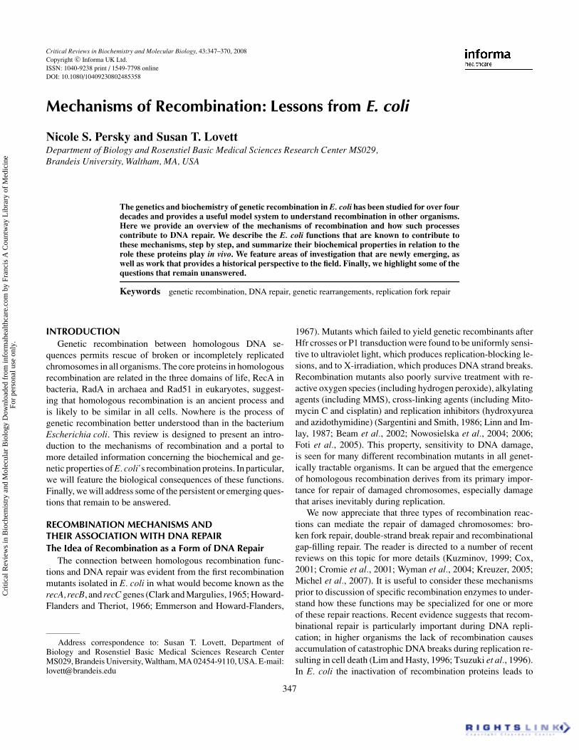
Gap-Filling Recombinational RepairIn the third type of DNA repair associated with homolo-
gous recombination, single-strand DNA gaps can be filled bystrand transfer reactions between sister or homologous chro-mosomes (Figure 3). A daughter-strand gap may be producedby incomplete replication, in which synthesis was blocked bytemplate or nascent strand damage, DNA secondary structureor tightly bound proteins. Although daughter strand gap repair(DSGR) was one of the first proposed mechanisms for recom-binational repair (Rupp et al., 1971), it is the most poorly un-derstood (Smith, 2004). The recombinational gap repair processhas been best studied in E. coli after UV irradiation, where ithas been called “post-replication repair.” Gap-filling was histor-ically assayed by conversion of low molecular weight newly-
replicated DNA into larger species by thymidine labeling anddenaturing sucrose gradient centrifugation (Rupp and Howard-Flanders, 1968; Rupp et al., 1971). Such studies demonstratedthat newly replicated DNA became associated with parentalDNA concomitant with repair, suggesting a recombinationalmechanism for gap-filling. Newer studies involve the transfor-mation of synthetically produced gapped plasmid molecules inwhich a replication-blocking lesion has been introduced into thegap (Berdichevsky et al., 2002). Although recombination clearlycontributes to gap-filling and in E. coli is arguably the predomi-nant mechanism of gap-filling (Berdichevsky et al., 2002), notethat other lesion tolerance mechanisms can also fill gaps, such astranslesion DNA synthesis (Goodman and Tippin, 2000). Thislatter mechanism is sometimes referred to as “error-prone post-replication repair” because of the involvement of low-fidelitypolymerases and its tendency to produce mutations.
A model for recombinational gap-filling is shown in Figure 3.The single-strand region of the gap pairs with its complementon an intact DNA molecule. This strand invasion may be aidedby displacement by one or both of the strands that flank thegap. Because this synapsis does not involve a free end, this
FIG. 3. Recombinational gap-filling repair. No dsDNA ends involved. (A) Gapped substrate molecule. (B) Strand exchange byRecA produces a joint molecule. Note that this pairing must be accompanied by topological changes to interwind the DNA strands.(C) Recruitment of ends flanking the joint produces Holliday junctions that can be resolved by cleavage. (C’) Alternatively, thecrossed strand of the half-Holliday junctions can be cleaved. (D) Recombinant product, with gap filled.
Cri
tical
Rev
iew
s in
Bio
chem
istr
y an
d M
olec
ular
Bio
logy
Dow
nloa
ded
from
info
rmah
ealth
care
.com
by
Fran
cis
A C
ount
way
Lib
rary
of
Med
icin
e Fo
r pe
rson
al u
se o
nly.
MECHANISMS OF RECOMBINATION 351
pairing presents topological problems that may be relieved bytopoisomerases. DNA synthesis primed from the ends replacesthe transferred strand, setting up a branched molecule, consistingof one or more crossed strands. Cleavage of these restores twoduplex molecules and the gap is filled. Like the double-strandbreak model, cleavage of these junctions can also yield crossoveror non-crossover products.
Recombination at gaps differs from the above double-strandbreak recombination mechanisms in the nature of the initiatingsubstrate. It is therefore not surprising to find that it depends ona different set of proteins from those that mediate DSB repair.Gap-filling may not necessarily require resection, although somesmall gaps may have to be widened to allow cooperative bindingof recombination proteins (see below). In addition, single-strandgaps left by incomplete replication are likely to be bound sta-bly by single-strand DNA binding protein, SSB, which must beremoved to allow access to recombination proteins.
Early genetic studies underestimated the contributions of thegap-filling mechanism of recombination. This is, in part, becausethe classical methods of measuring recombination in E. coli af-ter conjugation and transduction, involving dsDNA ends, reportDSB-mediated recombination and not that associated with gap-filling mechanisms. In repair of DNA damage, recombinationalgap repair can be difficult to distinguish in practice from repairof double-strand breaks. Many agents that produce gaps, suchas UV irradiation, will also produce double-strand breaks. In therepair of DNA damage, we likely underestimate the contributionof gap repair to survival: gaps that fail to be repaired are likelyconverted to double-strand breaks and repaired by DSB-specificrepair mechanisms.
ConclusionThese idealized mechanisms of recombinational repair pro-
vide us with a framework to understand the proteins involved indifferent recombination reactions and their influence on differenttypes of repair reactions and events involving genetic exchange.
RECOMBINATION PROTEINS AND THEIR FUNCTIONSIntroduction
In thinking about recombination mechanisms, it is useful tobreak down the process into three distinct steps in which theDNA substrates are processed prior to strand invasion, pairedto form heteroduplex DNA intermediates and are matured torecombinant products. The first step, “pre-synapsis” involvesthe production of recombinogenic single-strand DNA and thecooperative binding of the strand exchange protein, RecA. Thesecond-step, “synapsis” requires RecA-mediated homologouspairing, strand invasion and formation of heteroduplex interme-diates. These heteroduplexes are subject to processing whichmay stabilize or destabilize these recombination intermediates.The third step “post-synapsis” or “resolution” involves the dis-solution of intermediates, including any branched molecules in-volved in the process, removal of recombination proteins and for-
mation of intact duplex DNA recombinant products. Reassemblyof the replication fork may also be required post-synaptically. Inall the aforementioned recombination mechanisms, RecA pro-tein plays a central role. A number of these steps are fluid andcan be catalyzed in alternative ways. In addition, some proteinsappear to be specialized for particular recombination mecha-nisms: for instance, there are proteins involved in gap repair thatare not required for double-strand break repair and vice versa.
In this context, we will introduce the functions in E. colithat have been implicated in genetic recombination and brieflysummarize their biochemical properties. For more details of thebiochemical mechanisms, please consult recent reviews (Cox,2007b; Kowalczykowski, 2000). Additional discussion of thegenetic properties of E. coli recombination mutants can be founddispersed in several reviews (Clark, 1973; 1991; Smith, 1987;Smith and Wang, 1989; Clark and Sandler, 1994; West, 1997;Kuzminov and Stahl, 2005).
A. PresynapsisTo prepare for recombination, DNA substrates are processed
by nucleases and helicases to reveal single-strand DNA regions,which are bound by the central strand exchange protein RecA. Invivo, RecA is actively loaded by at least two distinct mechanismsinvolving RecBCD or RecFOR. This filament both protects theDNA and potentiates subsequent synapsis and strand exchange.A number of other proteins modulate the stability of the RecAhelical presynaptic filament. Special structural proteins may alsocoordinate or tether DNA ends.
Single-Strand DNA: the Signal for RecombinationPersistent single-strand DNA in the cell is recombinogenic.
Although ssDNA is produced transiently during replication, itspersistence signals the presence of gaps produced by incom-plete replication. Binding of RecA to this ssDNA (see below)will initiate the recombination process. Persistent ssDNA maybe bound with single-strand DNA binding protein, SSB, whichmust be removed and replaced with RecA.
Double-strand breaks in DNA, other lesions that require re-combination to be repaired, must be converted to partial singlestrands to initiate recombination. In this case, single-strand DNAis produced actively by exonuclease digestion of one of the twostrands, a process known as resection. Once the ends are single-stranded, they will bind RecA protein to initiate the homologysearch process (see below.) Because a 3′ end can prime newDNA synthesis, thereby aiding the recombination reaction andstabilizing the heteroduplex joint, (see Figure 5F below) resec-tion of DSBs appears to occur universally in the 5′ to 3′ direction,to reveal a 3′ ssDNA tail at the break site.
RecBCD and Resection of DSBsDouble strand breaks are processed into recombinogenic sin-
gle strands by the RecBCD nuclease. The RecBCD nuclease
Cri
tical
Rev
iew
s in
Bio
chem
istr
y an
d M
olec
ular
Bio
logy
Dow
nloa
ded
from
info
rmah
ealth
care
.com
by
Fran
cis
A C
ount
way
Lib
rary
of
Med
icin
e Fo
r pe
rson
al u
se o
nly.
352 N. S. PERSKY AND S. T. LOVETT
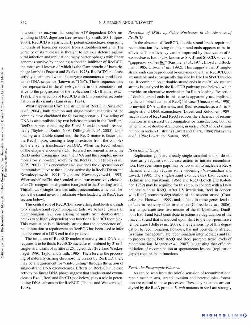
is a complex enzyme that couples ATP-dependent DNA un-winding to DNA digestion (see reviews by Smith, 2001; Spies,2005). RecBCD is a particularly potent exonuclease, degradinghundreds of bases per second from a double-strand end. Thevoracity of its nuclease is thought to act as a defense againstviral infection and replication; many bacteriophages with lineargenomes survive by encoding a specific inhibitor of RecBCD,the most well-known of which is the Gam protein of bacterio-phage lambda (Enquist and Skalka, 1973). RecBCD’s nucleaseactivity is tempered when the enzyme encounters a specific oc-tamer DNA sequence (known as “Chi”). These sequences areover-represented in the E. coli genome in one orientation rel-ative to the progression of the replication fork (Blattner et al.,1997). The interaction of RecBCD with Chi promotes recombi-nation in its vicinity (Lam et al., 1974).
What happens at Chi? The structure of RecBCD (Singletonet al., 2004), bulk reaction and single-molecule studies of thecomplex have elucidated the following scenario. Unwinding ofDNA is accomplished by two helicase motors in the RecB andRecD subunits, contacting the 5′ and 3′ ended strands, respec-tively (Taylor and Smith, 2003; Dillingham et al., 2005). Uponloading at a double-strand end, the RecD motor is faster thanthe RecB motor, causing a loop to extrude from the complexas the enzyme translocates on DNA. When the RecC subunitof the enzyme encounters Chi, forward movement arrests, theRecD motor disengages from the DNA and the complex movesmore slowly, powered solely by the RecB subunit (Spies et al.,2005; 2007). This encounter also switches the dispensation ofthe strands relative to the nuclease active site in RecB (Dixon andKowalczykowski, 1991; Dixon and Kowalczykowski, 1993).Whereas before Chi, the 3′ ended strand was extensively cleaved,after Chi recognition, digestion is targeted to the 5′ ending strand.This allows 3′ single-stranded tails to accumulate, which will be-come the strand invasion substrate when loaded with RecA (seesection below).
This central role of RecBCD in converting double-strand endsto 3′ single-strand recombinogenic tails, we believe, causes allrecombination in E. coli arising normally from double-strandbreaks to be highly dependent on a functional RecBCD complex.This correlation is sufficiently strong that the dependence of arecombination or repair event on RecBCD has been used to inferthe presence of a DSB end in the process.
The initiation of RecBCD nuclease activity on a DNA endrequires it to be flush; RecBCD nuclease is inhibited by 3′ or 5′
single-strand tails of as little as 25 nucleotides (Prell and Wacker-nagel, 1980; Taylor and Smith, 1985). Therefore, in the process-ing of naturally-arising chromosome breaks by RecBCD, theremay be a requirement for “end-blunting” through the action ofsingle-strand DNA exonucleases. Effects on RecBCD nucleaseactivity on linear DNA phage suggest that single-strand exonu-cleases Exo I, RecJ and SbcCD (see below) play a role in poten-tiating DNA substrates for RecBCD (Thoms and Wackernagel,1998).
Resection of DSBs by Other Nucleases in the Absence ofRecBCD
In the absence of RecBCD, double-strand break repair andrecombination involving double-strand ends appears to be in-efficient. This efficiency can be improved by inactivation of 3′
exonucleases Exo I (also known as SbcB) and SbcCD, so-called“suppressors of recBC” (Kushner et al., 1971; Lloyd and Buck-man, 1985; Gibson et al., 1992). This suggests that 3′ single-strand ends can be produced by enzymes other than RecBCD, butare unstable and subsequently digested by Exo I or SbcCD nucle-ase. Recombination at double-strand ends in recBC sbc mutantstrains is catalyzed by the RecFOR pathway (see below), whichprovides an alternative mechanism for RecA loading. Resectionof double-strand ends in this case is apparently accomplishedby the combined action of RecQ helicase (Umezu et al., 1990),to unwind DNA at the ends, and RecJ exonuclease, a 5′ to 3′
single-strand DNA exonuclease (Lovett and Kolodner, 1989).Inactivation of RecJ and RecQ reduces the efficiency of recom-bination as measured by conjugation or transduction, both ofwhich involve double-strand ends, in recBC sbcB sbcCD strainsbut not in recBCD+ strains (Lovett and Clark, 1984; Nakayamaet al., 1984; Lovett and Sutera, 1995).
Resection of Gaps?Replication gaps are already single-stranded and so do not
necessarily require exonuclease action to initiate recombina-tion. However, some gaps may be too small to nucleate a RecAfilament and may require some widening (Viswanathan andLovett, 1998). The single-strand exonucleases Exonuclease I(Lehman and Nussbaum, 1964) and RecJ (Lovett and Kolod-ner, 1989) may be required for this step, in concert with a DNAhelicase such as RecQ. After UV irradiation, RecJ in concertwith RecQ promotes degradation of the nascent strand (Cour-celle and Hanawalt, 1999) and defects in these genes lead todefects in recovery after irradiation (Courcelle et al., 2006).In a temperature-sensitive mutant of the fork helicase, DnaB,both Exo I and RecJ contribute to extensive degradation of thenascent strand that is induced upon shift to the non-permissivetemperature (Belle et al., 2007). The relationship of this degra-dation to recombination, however, has not been demonstrated.In strains that accumulate recombination intermediates and failto process them, both RecQ and RecJ promote toxic levels ofrecombination (Magner et al., 2007), suggesting that efficientinitiation of recombination at spontaneous lesions (replicationgaps?) requires both functions.
RecA: the Presynaptic FilamentAs can be seen from the brief discussion of recombinational
repair mechanisms, strand invasion and heteroduplex forma-tion are central to these processes. These key reactions are cat-alyzed by the RecA protein. E. coli mutants in recA are strongly
Cri
tical
Rev
iew
s in
Bio
chem
istr
y an
d M
olec
ular
Bio
logy
Dow
nloa
ded
from
info
rmah
ealth
care
.com
by
Fran
cis
A C
ount
way
Lib
rary
of
Med
icin
e Fo
r pe
rson
al u
se o
nly.
MECHANISMS OF RECOMBINATION 353
deficient for many types of recombination processes, includinga greater than 10,000-fold decrease in recombination after Hfrcrosses, and are exquisitely sensitive to many forms of DNAdamaging agents, including UV and ionizing-radiation (Clarkand Margulies, 1965; Howard-Flanders and Theriot, 1966).RecA (reviewed in Bianco et al., 1998; McGrew and Knight,2003; Bell, 2005; Cox, 2007a) is an ATPase that forms a right-handed helical filament on DNA, with a preference for single-strand DNA, which it binds at 3 nucleotides per RecA monomer.In vitro, RecA-ssDNA filaments can catalyze synapsis and strandexchange between homologous DNA molecules. The RecA fil-ament also promotes the self-cleavage of the LexA repressor, anactivity called “co-protease”, and thereby controls the onset ofthe SOS regulatory response to DNA damage (Walker, 1987).
A recent crystallographic study reveals its structures boundto single or double-strand DNA (Chen et al., 2008). One of theproperties of this RecA filament, conserved in its archaeal andeukaryotic orthologs, is the stretching of the DNA to a helicalpitch about 50% greater than that found in B-form double-strandDNA (Yu et al., 2004), exposing the bases in a way that aids inthe homology search process. Although initially, from RecA’sability to catalyze multiple important recombinational reactionsin vitro, much focus of the field was on the properties of RecAitself, we now appreciate that the formation, dissolution andactivity of the RecA filament is extensively regulated by otherproteins in the cell. Much of this information is newly emerging(reviewed recently by Cox (2007b)).
Conflict and Cooperation Between RecA and Single-Strand DNABinding Protein, SSB
Single-stranded DNA binding protein (SSB) is an abundantprotein that plays a ubiquitous role in many DNA processing ac-tivities, including replication and repair (see reviews by Lohmanand Ferrari, 1994; Meyer and Laine, 1990). SSB binds to singlestranded DNA with high affinity, removing its secondary struc-ture (Meyer and Laine, 1990). SSB exists as a tetramer in E. coliand wraps the DNA around itself in a fashion reminiscent of hi-stones and dsDNA (Chrysogelos and Griffith, 1982; Meyer andLaine, 1990; Raghunathan et al., 1997). Its C-terminal domain isdisordered in the crystal structure (Savvides et al., 2004) and isthe site of interaction with various replication and recombinationproteins.
SSB has both stimulatory and inhibitory actions on RecAstrand exchange in vitro. If single-strand DNA is pre-boundby SSB, it inhibits RecA filament formation (Kowalczykowskiet al., 1987). This inhibition appears to be at the initial nucle-ation step (Thresher et al., 1988; Joo et al., 2006). However, ifRecA is allowed to bind first, SSB is stimulatory to strand ex-change, presumably by its ability to melt secondary structuresthat would be inhibitory to RecA binding (Kowalczykowski andKrupp, 1987; Muniyappa et al., 1984). Once RecA has success-fully nucleated on ssDNA, the presence of SSB does not inhibitRecA filament extension (Joo et al., 2006).
In vivo, the presence of SSB on recombinogenic ssDNA maydirect its future processing. Mutants in ssb, including those inthe C-terminal interaction domain, are defective in genetic re-combination and highly sensitive to DNA damaging agents suchas UV light (Glassberg et al., 1979; Wang and Smith, 1982;Quinones and Piechocki, 1987; Carlini and Porter, 1997). SSBinteracts with a number of proteins, potentiating their activities(Molineux et al., 1974; Chase and Williams, 1986; Srivenugopaland Morris, 1986; Meyer and Laine, 1990; Umezu et al., 1993;Sandigursky and Franklin, 1994; Harmon and Kowalczykowski,2001; Han et al., 2006; Shereda et al., 2007), including somethat participate in recombination. A new concept is that, ratherthan acting merely as passive bystander, SSB may form a scaf-fold to assemble DNA processing enzymes. Although SSB hasbeen assumed to protect DNA from nuclease attack, it dramat-ically stimulates the digestion of ssDNA by exonucleases ExoI (Molineux and Gefter, 1975) and RecJ (Han et al., 2006).SSB also recruits and stimulates the RecQ helicase (Umezu andNakayama, 1993; Shereda et al., 2007). A genome-wide studyshows a whole system of DNA modification enzymes, includingtoposimerases, helicases and nucleases, that interact with SSB(although potentially indirectly) (Butland et al., 2005) and thereis much to be learned about how these interactions are managed.
Loading the RecA Filament by RecBCDAlthough RecBCD’s role in generating single-strand DNA
for initiation has been long appreciated, its involvement in es-tablishing the RecA presynaptic filament is a fairly recent dis-covery. The coordination of DNA digestion with RecA loadingis a regulated feature of the RecBCD complex. As discussed inthe previous section, the RecBCD nuclease degrades DNA untilthe octamer Chi site in DNA is encountered. Upon recognitionof Chi RecBCD’s nuclease activity is attenuated, its helicaseactivity is reduced two-fold, and preferential degradation of the5′ strand of the duplex produces a 3′ single stranded DNA tail(Spies, 2005). Recognition of Chi also triggers the RecBCDcomplex to direct loading of RecA onto the emergent 3′ singlestranded tail (Anderson and Kowalczykowski, 1997). This load-ing occurs through the RecB subunit C-terminal domain, whichby itself makes stable complexes with RecA (Spies and Kowal-czykowski, 2006). Through the active loading of RecA immedi-ately after the creation of ssDNA end, RecBCD is able to excludeSSB from binding, explaining why the RecBCD pathway is notinhibited by SSB overproduction (Moreau, 1988). Supporting arole in RecA filament formation, RecBCD is required for fullinduction of the SOS response following DSB-inducing treat-ments such as nalidixic acid (Chaudhury and Smith, 1985).
Loading the RecA Filament by RecFORAn alternative mechanism for RecA loading is provided by
the RecFOR proteins and they provide a solution for RecA togain access to SSB-coated single-stranded DNA. The recF gene
Cri
tical
Rev
iew
s in
Bio
chem
istr
y an
d M
olec
ular
Bio
logy
Dow
nloa
ded
from
info
rmah
ealth
care
.com
by
Fran
cis
A C
ount
way
Lib
rary
of
Med
icin
e Fo
r pe
rson
al u
se o
nly.
354 N. S. PERSKY AND S. T. LOVETT
was discovered as a component of a recombination pathway,initially called the “RecF pathway,” that functions independentof the RecBCD nuclease (Horii and Clark, 1973). Subsequentanalysis identified two other genes, recO and recR (Kolodneret al., 1985; Mahdi and Lloyd, 1989), with somewhat similareffects, now defining the “RecFOR pathway”. Genetic analy-sis suggested that RecF, RecO and RecR act in some commonfashion to promote RecA function. These mutants have similarphenotypes, and their recombination and repair deficiency couldbe suppressed by a lambda protein, known as “orf” (Sawitzkeand Stahl, 1992), or by mutations in RecA (known as “srf” forsuppressor of recF) (Volkert and Hartke, 1984; Wang and Smith,1986; Madiraju et al., 1988). One of these srf alleles, RecA803,altered strand exchange properties of RecA such that it becameless susceptible to inhibition by SSB, leading the authors to pro-pose that the RecF was involved in overcoming the inhibitoryeffects of SSB on RecA (Madiraju et al., 1988). Supporting thisidea, overproduction of SSB leads to phenotypes similar to RecFdeficiency (Moreau, 1988). A number of genetic studies sug-gested that the RecFOR pathway is specialized for single-strandgap repair (Wang and Smith, 1984; Galitski and Roth, 1997), incontrast to the RecBCD-promoted DSB repair, and one mightimagine unrepaired replication gaps would be pre-bound withSSB. Mutants in RecFOR also are defective in induction of theSOS response by UV irradiation (Sandler and Clark, 1994), con-sistent with a defect in RecA filament formation on replicationgaps caused by DNA polymerase arrest at UV lesions in thetemplate.
In vitro characterization of these proteins confirms geneticexpectations that RecFOR proteins act to control formation ofthe RecA filament and direct it to single-strand DNA gaps. Un-like RecBCD, these proteins appear not to work as a stoichio-metric machine. Instead, RecF, RecO, and RecR work eitherindependently or in pairs. Fulfilling the expectations from theirgenetic properties, the RecO and RecR proteins act as “media-tors” to allow RecA to replace SSB on ssDNA. The RecO proteincan bind directly to SSB-coated single stranded DNA as well asnaked single stranded DNA (Luisi-DeLuca and Kolodner, 1994;Umezuv, 1993; Umezu and Kolodner, 1994). The RecOR pairpromotes the dissociation of SSB and its replacement with RecAin the single-stranded DNA regions of the gap (Morimatsu andKowalczykowski, 2003; Umezu et al., 1993; Umezu and Kolod-ner, 1994). SSB lacking its C-terminal interaction domain blocksthe RecA-mediator function of RecOR, suggesting that specificinteractions between RecO and SSB are required (Hobbs et al.,2007). The role of RecF appears to direct and confine RecA fila-ment formation to the gapped region. The RecF protein binds tossDNA/dsDNA junctions preferentially and in conjunction withRecR prevents RecA loading onto the dsDNA regions (Webbet al., 1997; Morimatsu and Kowalczykowski, 2003). RecF mu-tant phenotypes can be partially suppressed by RecOR over-production (Sandler and Clark, 1994), supporting the idea thatRecOR provide the RecA-mediator function, which RecF directsto the right location.
Dynamics of the RecA/DNA Filament and its ControlLike other self-assembling structures such as actin and tubu-
lin, the RecA filament is a dynamic structure. RecA filamentson ssDNA are formed in two steps: first, the rate-limiting nu-cleation of 4–5 monomers (Galletto et al., 2006), then followedby filament extension. Net filament growth is 5′ to 3′ on ssDNA(Register and Griffith, 1985), although single-molecule studieshave shown that the filament can be extended in either direction(Galletto et al., 2006). ATP binding is required for RecA fila-ment nucleation (Menetski and Kowalczykowski, 1985; Gallettoet al., 2006) and ATP hydrolysis promotes dissociation of RecAfrom the filament, preferentially from the 5′ end (Lindsley andCox, 1990).
A number of proteins have been identified that modify thestability of the RecA:DNA filament (see review by Cox (2007)).These mechanisms may prevent RecA from initiating recombi-nation during the exposure of ssDNA during replication or dur-ing certain excision repair processes. The RecA filament mayalso be dismantled after an unsuccessful search for homology,to allow other gap-filling mechanisms to operate or for a brokenchromosome to be degraded. After the completion of recom-bination, destabilization of the RecA filament may also aid itsremoval from recombination products.
One of the most important modifiers of RecA function, as re-vealed by both in vitro and in vivo analysis, is the UvrD helicase,a superfamily-1 3′ to 5′ DNA helicase. UvrD can dismantle theRecA nucleoprotein filament (Veaute et al., 2005) and acts asan anti-recombinase when added to in vitro RecA DNA strandtransfer reactions (Morel et al., 1993). Accordingly, mutants inuvrD are hyper-recombinational in a number of assays (Zieget al., 1978; Arthur and Lloyd, 1980; Feinstein and Low, 1986;Bierne et al., 1997). These results are consistent with the ideathat UvrD may act as a “proofreader” of recombination so thatit cannot occur between short homologous sequences; in its ab-sence, chromosomal rearrangements are stimulated (Feschenkoet al., 2003; Kang and Blaser, 2006). When late stages of re-combination are blocked (such as RuvABC mutants, see below),loss of UvrD is lethal (Magner et al., 2007)–this results from in-creased accumulation of lethal intermediates in recombination,since blocks in early functions (such as RecA, RecF, RecQ andRecJ) relieve this toxicity.
The RecOR proteins, in addition to their role as RecA me-diator proteins, act to stabilize the RecA:ssDNA filament, es-pecially at its 5′ end, (Shan et al., 1997; Bork et al., 2001),either by inhibiting dissociation or encouraging reassociation ofRecA. In vivo, the importance of this filament stabilizing activityof RecOR versus their mediator activity in displacing SSB fromssDNA is unclear.
Recently, two other proteins have been identified that modu-late RecA filament formation: RecX and DinI. RecX was foundas an open reading frame, immediately downstream of the RecAgene in many organisms (Sano, 1993); DinI is a DNA damage in-ducible gene whose overexpression produced inhibitory effectson the SOS response (Yasuda et al., 1998). Visualization of RecA
Cri
tical
Rev
iew
s in
Bio
chem
istr
y an
d M
olec
ular
Bio
logy
Dow
nloa
ded
from
info
rmah
ealth
care
.com
by
Fran
cis
A C
ount
way
Lib
rary
of
Med
icin
e Fo
r pe
rson
al u
se o
nly.

MECHANISMS OF RECOMBINATION 355
cytologically suggest the two proteins have opposite effects onRecA:DNA structures: mutants in dinI had fewer spontaneousRecA-GFP foci whereas recX mutants exhibited more foci thanwild-type strains (Renzette et al., 2007). Both RecX and DinIhave been shown to directly bind to RecA (Voloshin et al., 2001;Stohl et al., 2003; Lusetti et al., 2004b) and have opposing effectson the stability of the RecA:DNA filament. RecX is able to in-hibit RecA strand exchange and RecA filament extension (Stohlet al., 2003; Drees et al., 2004). Interestingly, RecF and SSBcan suppress this inhibition of RecA strand extension by RecX(Baitin et al., 2008; Lusetti et al., 2006). DinI, at stoichiometricconcentrations, can stabilize already formed RecA filaments andprevent them from dissociating (Lusetti et al., 2004a). At theseconcentrations DinI does not inhibit RecA’s ability to catalyzestrand exchange nor its co-protease activity.
RecN and Organization of DNA EndsThe RecN protein is a predicted coiled coil protein of the SMC
(“structural maintenance of chromosomes”) family of proteinsfound in bacteria, archaea and eukaryotes, whose members playvarious roles in chromosome architecture, DNA repair and re-combination (Hirano, 2005; Strunnikov, 2006). SMC proteinsexhibit dumbbell structures, with two head domains connectedvia long coils. The recN gene was identified as a gene requiredfor optimal recombination via the RecFOR pathway, whose ex-pression is strongly induced by the SOS response to DNA dam-age (Picksley et al., 1984; Finch et al., 1985). In E. coli, RecN,a normally unstable protein and degraded by protease ClpXP,forms GFP-labeled foci after DNA damage. After completionof repair, aggregates of RecN must be removed by ClpXP torestore viability (Nagashima et al., 2006).
Further investigation has established RecN as particularlyimportant for repair of double-strand breaks. Mutants in recNare sensitive to ionizing radiation, bleomycin, EcoK restrictionfollowing 2-amino-purine incorporation and induction of theI-SceI nucleases (Picksley et al., 1984; Sargentini and Smith,1986; Cromie and Leach, 2001; Kosa et al., 2004; Meddowset al., 2005), all agents that produce double-strand breaks. Dur-ing conjugational recombination in E. coli, RecN may protectsingle-strand DNA or in some way promote single-strand an-nealing, since sectored colonies that presumably arise by anneal-ing of genetically distinct strands and formation of heteroduplexDNA are highly RecN-dependent (Lloyd and Buckman, 1995).
In analogy to the role of other SMC proteins, RecN may playa role in coordinating DNA ends during double-strand break re-pair and the subsequent recruitment of other repair factors. Al-though little direct evidence for this exists in E. coli, cytologicalstudies of RecN protein in Bacillus subtilis suggests that RecNis one of initiating factors in establishment of “repair centers”,visible assemblies of recombination proteins at sites of DNAdamage (Kidane et al., 2004). After treatment with agents thatinduce double-strand breaks, RecN-YFP can be seen to formdistinct foci; the later assembly of RecO and RecF fluorescent
foci requires RecN. During the process of natural competenceand DNA transformation, Bacillus subtilis RecN is seen to os-cillate dynamically throughout the cell; upon entry of ssDNARecN foci become fixed at the cell pole at the the DNA uptakemachinery is found (Kidane and Graumann, 2005). This stage islater followed by the appearance of RecA threads that emanatefrom this pole.
Although the biochemical properties of the E. coli RecN havebeen difficult to determine because of the insolubility of the pro-tein, a number of studies of the Bacillus subtilis protein haveelucidated RecN as a DNA binding protein. In vitro, Bsu RecNis an ATP-dependent ssDNA binding protein (Kidane and Grau-mann, 2005; Sanchezv, 2008). Among the SMC proteins, thispreference for ssDNA, rather than dsDNA, appears to be unique.As ascertained by atomic force microscopy, RecN binding to ss-DNA is insensitive to Bacillus SSB, SsbA, but reversed by RecA(Sanchezv, 2008). RecN forms aggregates (Kidane et al., 2004),and promotes rosette structures of protein and DNA (Sanchezet al., 2008). This activity could easily explain how RecN mayprotect single-stranded DNA and assist strand annealing duringdouble-strand break repair. One might imagine that this activ-ity would be quite useful in the annealing step of SDSA-modeof DSB repair, but this has yet to be confirmed by in vivo ex-periments. Specific interactions of other recombination proteinswith RecN have not been reported but appear likely, given itsfunction in the establishment of cytologically defined “repaircenters” in Bacillus.
SbcCD, an SMC NucleaseSbcCD is also an SMC-like protein and is arguably the struc-
tural and functional equivalent of the eukaryotic Mre11-Rad50-Xrs2/Nbs1 complex (reviewed in Stracker et al., 2004). TheSbcC subunit is a coiled coil protein with an ATPase domain;SbcD composes the nuclease activity of the protein. (Unlikethe complex in eukaryotes, there is no third subunit.) In vitro,SbcCD acts as an ATP-independent single-strand DNA endonu-clease and an ATP-dependent 3′ to 5′ exonuclease (Connellyand Leach, 1996; Connellyv, 1998; 1999). SbcCD also showspreference for hairpin secondary structures in DNA, which itcleaves close to the unpaired tip.
In vivo, SbcCD can initiate recombination between sisterchromosomes by the production of double-strand breaks, theresult of its cleavage at secondary structures formed by invertedrepeats, or palindromic DNA sequences (Cromie et al., 2000;Bzymek and Lovett, 2001; Eykelenboom et al., 2008). The ex-onuclease activity of SbcCD may also process ends of brokenchromosomes. This activity could be recombinogenic, poten-tially helpful in the removal of aberrant structures or tightlybound proteins at DNA ends. Indeed, in vitro, SbcCD can re-move a streptavidin/biotin moiety at a 5′ end by its endonucleaseactivity (Connelly et al., 2003). Conversely, SbcCD may also actto inhibit recombination by digestion of 3′ single-strand tailedrecombination substrates, an activity that may account for its
Cri
tical
Rev
iew
s in
Bio
chem
istr
y an
d M
olec
ular
Bio
logy
Dow
nloa
ded
from
info
rmah
ealth
care
.com
by
Fran
cis
A C
ount
way
Lib
rary
of
Med
icin
e Fo
r pe
rson
al u
se o
nly.
356 N. S. PERSKY AND S. T. LOVETT
discovery as a function inhibitory to the RecFOR-mediated re-combination of double-strand ends (Gibson et al., 1992; Lloydand Buckman, 1985). In a clever assay designed to assay therepair of double-strand chromosome breaks with its sister chro-mosome (Eykelenboom et al., 2008), SbcCD was found to benecessary for repair, leading to the suggestion that the SMCcharacter of the molecule may assist the coordination of twoDNA ends to allow healing specifically by DSB repair recom-bination. This coordination may channel the substrates into thetwo-ended DSB repair mechanism involving limited DNA syn-thesis (Figure 2) rather than one-ended recombination (brokenfork repair) that would be accompanied by initiation of newreplication forks (Figure 1).
B. Synapsis and Strand ExchangeOnce the RecA filament has been formed on ssDNA, it cat-
alyzes three important processes to accomplish strand pairingand synapsis: (1) a search for homology by transient interac-tions; (2) once homologous sequences have been located, strandinvasion and synapsis; and (3) extension of heteroduplex re-gions by branch migration. The recent crystal structure of RecAbound to single and double-strand DNA (Chen et al., 2008),and a single-molecule analysis of the strand transfer reaction(van der Heijden et al., 2008) offer some new insights.
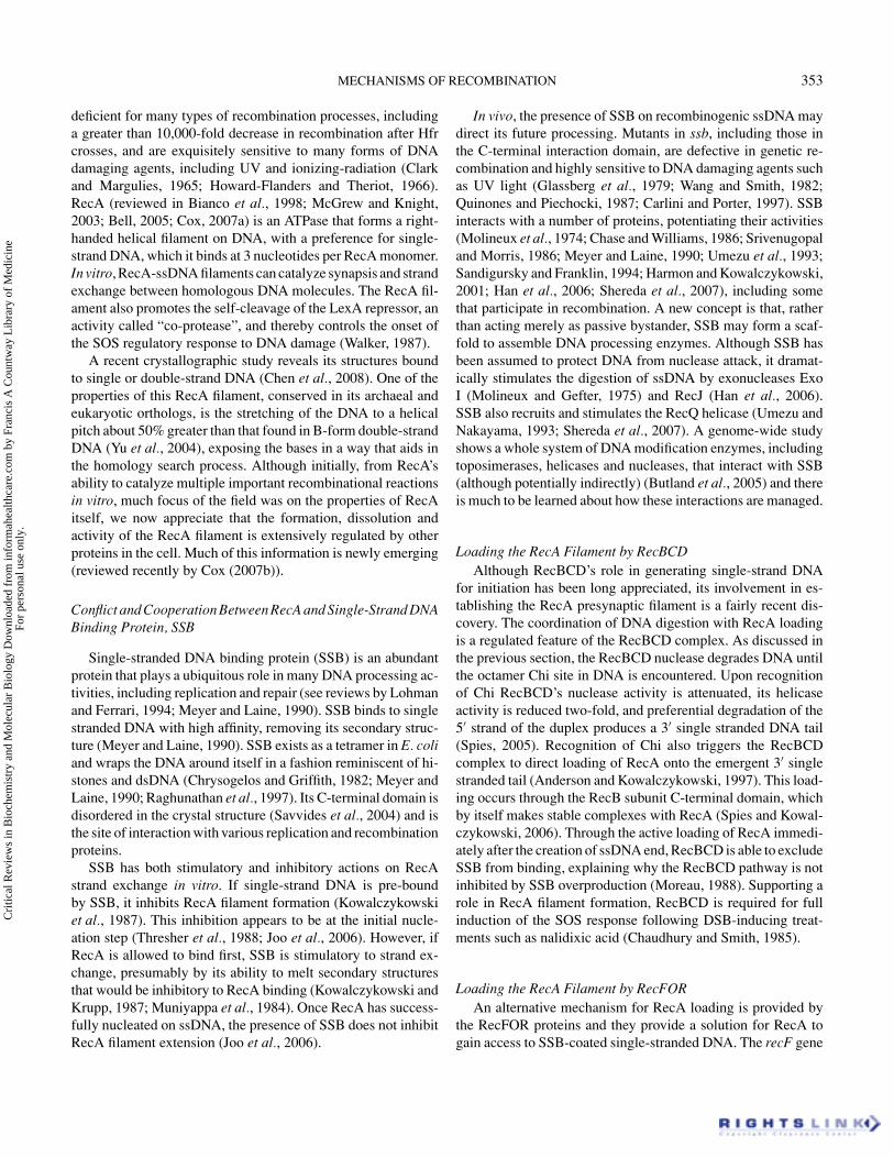
Homology Assessment and Strand ExchangeEvidence suggests that RecA filament has two sites for DNA
binding: a high affinity, primary site that is occupied when thefilament forms on ssDNA and a lower affinity, secondary site inwhich the donor dsDNA will be sampled for pairing-potential(Mazin and Kowalczykowski, 1998). Once homology is sensed,pairing of the donor duplex is destabilized and a strand from theduplex is transferred to the recipient single-strand for pairing(see diagram in Figure 4). In the crystal structure (Chen et al.,2008), both dsDNA and ssDNA when bound by RecA in theprimary site are stretched overall and underwound; however lo-cal areas resemble B-form DNA, explaining how Watson-Crickbase-pairing can be assessed in the synaptic complex. This is inagreement with the idea that the stretching of the donor dsDNAbreaks base-pairing and stacking interactions; the structure re-veals that these bases can be sampled for pairing, via the localB-form conformations, with the recipient strand for pairing inthe primary site. (See the schematic illustration of this processin Figure 4.)
Immediately after strand exchange, the newly formed het-eroduplex remains in the primary DNA binding site of the RecAfilament, while a single strand is left behind in the secondary site(Chow et al., 1986). Single molecule studies of RecA in the actof strand transfer have revealed several critical features (van derHeijden et al., 2008). Surprisingly, in the presence of ATP, theregion of synapsis, with three strands being protein-bound, oc-curs only over about 80 nucleotides at any given time, even whenlarge regions of homology are present. ATP hydrolysis promotes
FIG. 4. Schematic of the RecA-catalyzed strand exchange reac-tion. RecA binds ssDNA in high-affinity site 1 (A strand in bold)and recruits partner duplex in low-affinity site two, top of fig-ure. Stretching of the DNA destabilizes the pairing of the donorduplex and allows the recipient strand to sample pairing by lo-cal Watson-Crick base-pairing, middle of figure. Stable pairing,bottom of figure, causes strand-transfer, with the heteroduplexproduct in site 1 and a displaced single-strand in site 2.
release of RecA from the DNA products — as a wave of strandtransfer proceeds, the complex releases its products and leavesbehind an unpaired displaced strand that is wrapped around theduplex. In the presence of poorly hydrolyzable ATPgammaS,strand exchange occurs at the same rate, but the DNA is notreleased from the filament and a long ternary complex is formedbetween RecA and the two DNA molecules.
Topological Issues in Strand ExchangeStrand exchange initiated internally on a gapped molecule
(as in Figure 3) presents potential topological problems, sincethe ends are not free to rotate to establish interwound jointmolecules, so-called plectonemic joints, via the RecA filament.RecA can pair DNA strands that cannot intertwine, and such“paranemic” joints are stable but disrupted by deproteinization(DasGupta et al., 1980). These paranemic joints can be con-verted to plectonemic joints with the addition of topoisomeraseI (Cunningham et al., 1981), suggesting that paranemic jointsare precursors of plectonemic joints. Experiments with oligonu-cleotides with non-homologies at the ends shows that strandexchange can indeed occur when axial rotation is obstructed(Adzuma, 1992). Type I topoisomerases (in E. coli, topoiso-merase I and III) could in theory function to allow interwindingduring strand exchange at a gap. This topological problem isnot unique to single-strand gaps: even recombination involv-ing dsDNA linear molecules could initiate pairing somewhat
Cri
tical
Rev
iew
s in
Bio
chem
istr
y an
d M
olec
ular
Bio
logy
Dow
nloa
ded
from
info
rmah
ealth
care
.com
by
Fran
cis
A C
ount
way
Lib
rary
of
Med
icin
e Fo
r pe
rson
al u
se o
nly.

MECHANISMS OF RECOMBINATION 357
removed from the dsDNA end. However, how topological prob-lems are solved during recombination has not been well investi-gated. In a plasmid recombination assay, presumably involvinggaps because of its dependence on RecF, a requirement for TopoIhas been reported (Fishel and Kolodner, 1984).
The RecO protein can also anneal single-strand DNA, eventhat pre-bound by SSB, and can catalyze strand exchange to formD-loops (Luisi-DeLuca and Kolodner, 1994; Luisi-DeLuca,1995; Kantake et al., 2002). This suggests that RecO could assistRecA in synapsis at gaps that may have topological constraintsand pair less strongly; alternatively, RecO could help recruit thestrands flanking the gap into pairing to form Holliday junctions(Figure 3C’). In either case, we await genetic data to supportthis role for RecO.
Making Strand Exchange IrreversibleAfter strand displacement, the resulting single strand can be
bound by SSB. In vitro, this sequestration drives the exchangeto completion and prevents the product from re-initiating the re-verse reaction (Lavery and Kowalczykowski, 1992; Mazin andKowalczykowski, 1998). In addition, the displaced strand canbe subject to exonuclease digestion, which stimulates branchmigration by removing the competitor strand for pairing and re-moves the possibility of reaction reversal (See Figure 5D). Invitro, both RecJ and Exo I single-strand exonucleases stimu-late RecA strand exchange between linear duplex and circularsingle-strand molecules and so may have a synaptic-stabilizationrole in vivo (Bedale et al., 1993; Corrette-Bennett and Lovett,1995; Konforti and Davis, 1992). In assays for conjugationalrecombination that proceeds by the RecBCD-dependent DSBmechanism, inactivation of both RecJ and Exo I (but neithersingly) causes a reduction of recombination by about 10-fold(Viswanathan and Lovett, 1998). This is best explained by apost-synaptic role in which digestion by either RecJ or Exo Ihelps promote strand exchange, even when recombination isinitiated by RecBCD. In vivo, RecJ and Exo I appear to act post-synaptically on heteroduplex joints, as determined by physicalanalysis of the recombinant products formed after linearizationof plasmid DNA with terminal repeats (Friedman-Ohana andCohen, 1998; Friedman-Ohana et al., 1999). RecJ also stimu-lates branch migration past regions of non-homologies in vitro(Corrette-Bennett and Lovett, 1995).
Branch Migration by RecAOnce strand exchange has been initiated by joint molecule
formation, RecA can extend the heteroduplex region by a re-action known as branch migration. Branch migration can alsobe driven between two duplex molecules, with formation ofthe crossed 4-strand structure, the Holliday junction. RecA cancatalyze this phase of the reaction but is relatively inefficientwhen compared to other branch migration helicases (Adams andWest, 1996; see below), such as RuvAB and RecG. RecA also
is poor at traversing regions of non-homology between the re-acting molecules (Morel et al., 1994) and a 22 nt non-homologycan be sufficient to block further exchange by RecA.
C. Postsynaptic DNA ProcessingAn Introduction to D-loop Processing and Holliday Junctions
The product of strand exchange between a resected DSB andan intact duplex is a D-loop (“Displacement loop”), where aheteroduplex region is formed between the single strand tail ofthe linear molecule and its complement on the recipient dsDNAmolecule (Figure 5A). Although all models of recombination in-volving double-strand ends show initial strand exchange througha D-loop intermediate, the ultimate processing of these interme-diates is not well understood and could be quite complicated. TheD-loop heteroduplex joint is intrinsically unstable — branch mi-gration (Figure 5B) can diminish the heteroduplex region andultimately disrupt the joint structure. In contrast, branch migra-tion to extend the region of heteroduplex will stabilize the joint(Figure 5C), and will eventually form a 4-stranded branched
FIG. 5. Stabilization and destabilization of D-loop recombina-tion intermediate. (A) The D-loop produced by strand exchangebetween a resected dsDNA end and an intact homologous du-plex molecule. (B) Branch migration rightward will destabi-lize the D-loop by reducing the length of heteroduplex and willultimately dissove the joint. (C) Conversely, branch migrationleftward stablizes the joint by extending the heteroduplex, con-comitant with formation of a Holliday junction (4-strand jointmolecule). Cleavage of this junction, not shown) will produce afork structure. (D) Exonuclease digestion associated with branchmigration will also stabilize the joint, without Holliday junctionformation. (E) Cleavage of the D-loop stabilizes the joint andproduces a fork structure. (F) Priming of DNA synthesis fromthe invading 3′ strand will also stabilize the joint.
Cri
tical
Rev
iew
s in
Bio
chem
istr
y an
d M
olec
ular
Bio
logy
Dow
nloa
ded
from
info
rmah
ealth
care
.com
by
Fran
cis
A C
ount
way
Lib
rary
of
Med
icin
e Fo
r pe
rson
al u
se o
nly.
358 N. S. PERSKY AND S. T. LOVETT
molecule (the Holliday junction). Not all recombination inter-mediates will include true Holliday junctions. If exonucleaseactivity accompanies the branch migration phase, no such Hol-liday junction is formed concomitant with heteroduplex exten-sion (Figure 5D) and the D-loop just grows larger. Cleavage ofthe displaced strand of the D-loop can also stabilize the inter-mediate and can allow the transferred strand to be ligated to itsrecipient, producing a covalent recombinant strand and a forkstructure (Figure 5E). Finally, the 3′ end of the invading strandcan be extended by DNA polymerase, extending and stabilizingthe heteroduplex joint (Figure 5F).
Intermediate Structures Posed by DSB Repair vs. Gap RepairRepair of double-strand breaks, the DSB repair model, in-
volves D-loop formation by the invading dsDNA end, followedby recruitment of the second end into the D-loop, by pairing withthe displaced strand. This mechanism can produce two Hollidayjunctions (Figure 2C), whose resolution can yield crossovers(Figure 2D–F) or non-crossover products. During yeast meioticrecombination, double Holliday junctions have been detected(Schwacha and Kleckner, 1995). In the SDSA variant of DSBrepair, only one end need invade to form a D-loop and initiateDNA synthesis from the 3′ end (Figure 2C’–D’). The synapticintermediate is then dissolved and the 3′ ends of each dsDNA endcan anneal to heal the break, without the production of crossoverproducts between the donor and recipient (Figure 2E’–F’). TheDNA synthesis in the annealed intermediate (dashed lines inFigure 2E’–F’) can, however, produce some genetic exchangefrom the intact donor DNA molecule to the recipient brokenstrand even without crossover at the flanks.
In gap repair, initial pairing forms synaptic structures involv-ing junctions with a single crossed strand (half-Holliday junc-tions) connecting donor and recipient DNA molecules (Figure3B). Engagement of one or both of the broken strands that flankthe gap and branch migration can convert one or both of theseto four-strand Holliday junctions (Figure 3C). Although eitherhalf-Holliday or Holliday junctions in gap repair, in theory, canbe resolved to create recombinant products (Figure 3D), it notclear what forms exist in vivo.
Reverse versus Forward Branch Migration/RuvAB versus RecGRuvABC is a coordinated protein machine that acts in the
late stages of replication to migrate and resolve Holliday junc-tions. RuvA is a specificity factor that targets RuvB to Hollidayjunctions, RuvB is the Holliday junction branch migration heli-case, and RuvC is an endonuclease that specifically cleaves four-way junctions (West, 1997). RuvA exists as one tetramer or twotetramers that sandwich the junction, holding the four strandsof DNA in an open square configuration. RuvA is able to pro-tect the junction from being modified by other DNA processingenzymes while recruiting RuvB (Kaplan and O’Donnell, 2006).RuvB itself exists as a hexamer, and two of these hexamers bind
to opposite arms of the Holliday junction, pumping the DNA topower migration by its ATPase activity. RuvC is finally recruitedto the complex to cleave the Holliday junction DNA. RuvC hasa preferred site for cleavage, and it is thought that one functionof branch migration is to identify and present these sequences toRuvC for cleavage (West, 1997). RuvC exists as a dimer, and themechanism for loading of RuvC remains unclear. The nicks cre-ated by RuvC can be re-ligated into two DNA duplexes by DNAligase, and the Holliday junction becomes completely resolved(West, 1997). In the absence of RuvAB or RuvC in vivo, Holl-iday junction recombination intermediates have been shown toaccumulate in the cell (Donaldson et al., 2006). Mutations inRuvAB or RuvC can be suppressed by expression of RusA, acryptic bacteriophage-encoded Holliday junction endonuclease,that is normally not expressed in E. coli (Mandal et al., 1993;Sharples et al., 1994; Mahdi et al., 1996).
RecG is a helicase implicated in recombination that canbind to Holliday junctions, D-loops and other branched struc-ture, from which it promotes ATP-dependent branch migration.RecG can not only migrate Holliday junctions but can also formHolliday junctions from by fork regression in vitro, a mecha-nism thought to aid in restarting replication at blocked repli-cation forks (McGlynn and Lloyd, 2002; Briggs et al., 2004;Baharoglu et al., 2008), but not discussed further here. RecGbinds well to D-loop structures and its helicase activity is di-rectional on such substrates (Whitby et al., 1993). In contrast toRuvAB, RecG’s helicase activity acts to disrupt D-loop struc-tures (Figures 5B and 6B) and can reverse RecA-mediatedstrand exchange in vitro (McGlynn and Lloyd, 2002; West,1997).
Genetic Effects of RuvABC and RecG: Genetic Independenceand Synergy
Both RuvABC and RecG contribute to recombination in vivo.Inactivation of either complex has little effect on recombination,as measured after P1 transduction and conjugation, which oc-curs by a RecBCD-dependent DSB-repair pathway. Inactivationof both has some synergistic effects, with reduction of recombi-nation approximately 100-fold (Lloyd, 1991). This suggests thatRecBCD mediated recombination requires the function of eitherRuvABC or RecG for maximal efficiency. Survival of cells to X-irradiation and to chromosomal cleavage by the meganucleaseI-SceI likewise requires either RecG or RuvABC, suggestingthat the two functions define separate and redundant mecha-nisms of resolution after double-strand break repair (Meddowset al., 2004).
Although the branch migration and cleavage activity ofRuvABC can act to stabilize and resolve heteroduplex jointsin vitro and in vivo, it is much less clear how RecG’s heli-case activity contributes to recombination, since its preferreddirectionality dissolves heteroduplex joints (Figure 6AB). Oneidea is that RecG promotes the dissolution step of the SDSAvariant of double-strand break repair (Meddows et al., 2004)
Cri
tical
Rev
iew
s in
Bio
chem
istr
y an
d M
olec
ular
Bio
logy
Dow
nloa
ded
from
info
rmah
ealth
care
.com
by
Fran
cis
A C
ount
way
Lib
rary
of
Med
icin
e Fo
r pe
rson
al u
se o
nly.

MECHANISMS OF RECOMBINATION 359
FIG. 6. How reverse branch migration by RecG can yield re-combinants. (A) RecG catalyzes reverse branch migration onD-loops. (B) Joint dissoved by RecG. (C) Extension of 3′ end bya DNA polymerase. (D) Bubble migration, D-loop translocatedby reverse migration coupled to DNA synthesis. (E) Appropriatecleavage of D-loop to generate recombinants. (F) Recombina-tion product, with donor DNA covalently joined to recipient.(G) Bubble migration converging on replication fork. (H) Res-olution of half Holliday junction formed by the collision. (I)Recombinant product, after healing.
(Figure 2D’–E’), where after DNA synthesis in the recombina-tion intermediate, synapsis is reversed to permit annealing of thebroken chromosomes. This mechanism could clearly contributeto recombinational DNA repair of breaks but, because it doesnot promote genetic crossovers, it is difficult to understand how
this mechanism would contribute to inheritance of markers afterconjugation or transduction. Another notion to explain RecG’srole is that RecG’s helicase activity is coupled to DNA synthesis(Meddows et al., 2004), such that the D-loop is not dissolved butmigrates with the polymerase in a manner termed “bubble migra-tion” (Figure 6CD). Appropriate resolution of these structure orconvergence of the D-loop bubble onto another replication forkcan yield crossover products (Figure 6E–I). Recombination andrepair defects in recG mutants can be corrected by inactivationof the helicase activity of the PriA replication restart protein (seebelow), and it has been suggested that both proteins compete foraccess to the 3′ strand of the D-loop (Al-Deib et al., 1996).
Migration and cleavage of Holliday junctions appear to bemore critical for the gap repair recombinational mechanism thanfor double-strand break repair. The RuvABC functions havemore pronounced effects on recombination promoted by theRecFOR pathway than on the RecBCD pathway and were iden-tified as a classic “RecF pathway” genes for conjugational re-combination (Lloyd et al., 1984). This may be because RecBCD-mediated strand exchange at ends through D-loops are more sta-bilized, for example by larger heteroduplex joints (Figure 5D–F) and do not obligately form Holliday junctions. In addition,RecG appears to exhibit more anti-recombinational action ongap-mediated recombination. In another assay for recombina-tion induced by DNA damage, the RecFOR pathway is stimu-lated four-fold by inactivation of RecG (Bichara et al., 2006),supporting the idea that RecG exerts anti-recombinational ef-fects by reverse junction migration.
Although RuvABC and RecG can be somewhat redundantfor effects on recombination in some assays, both functions arerequired for efficient recombination in other assays, especiallythose that appear to involve breaks induced after passage of thereplication fork. In an assay of repair of chromosomal breaksinduced by cleavage at palindrome sequences, efficient survivalrequired both RuvABC and RecG (Eykelenboom et al., 2008). Inanother assay for recombination between sister chromosomes atshort tandem repeats, elevated recombination caused by defectsin DnaB (the replication fork helicase), requires both RuvABand RecG (Lovett, 2006) in a mechanism involving RecBCD(Saveson and Lovett, 1999).
A Role for a RecA Paralog in Late Stages of RecombinationAll organisms appear to encode one or more proteins that are
paralogous to their strand exchange proteins (Lin et al., 2006). Abacterial RecA paralog is encoded by a gene known as Sms (forsensitivity to MMS) or RadA (named for its sensitivity to radi-ation), found in virtually every bacterial genome, as well as inplants. [This nomenclature is unfortunate, since bacterial RadAprotein is not orthologous to archaeal RadA strand exchangeprotein and is more similar, in fact, to the archaeal paralog pro-tein, RadB.] Genetic studies of radA/sms of E. coli show that thestrain is mildly sensitive to certain form of DNA damage (Beamet al., 2002; Felzenszwalb et al., 1984; Neuwald et al., 1992;
Cri
tical
Rev
iew
s in
Bio
chem
istr
y an
d M
olec
ular
Bio
logy
Dow
nloa
ded
from
info
rmah
ealth
care
.com
by
Fran
cis
A C
ount
way
Lib
rary
of
Med
icin
e Fo
r pe
rson
al u
se o
nly.
360 N. S. PERSKY AND S. T. LOVETT
Sargentini and Smith, 1986; Song and Sargentini, 1996). How-ever, it has strong synergistic effects on DNA damage survivalwhen combined with mutants in other recombination genes, par-ticularly recG (Beam et al., 2002).
In measurements of conjugational recombination, RadA/Smsappears to have a role somewhat redundant to RuvAB and RecG:single mutants are not substantially reduced in recombination,whereas double mutants are more strongly deficient. The tripleradA recG ruvABC mutant is severely recombination defective,comparable to mutants in recA (Beam et al., 2002). Because ofthe known role of RuvABC and RecG in late stages of recom-bination, this implicates RadA/Sms likewise in post-synapticevents. RadA/Sms is required, as are RecG and RuvAB, for re-combination at tandem repeats associated with replication forkhelicase defects (Lovett, 2006) and radA mutants are particu-larly sensitive to the replication chain-terminator, azidothymi-dine (Cooper and Lovett, unpublished results). E. coli RadA,by itself, has no effect on RecFOR-mediated recombination(Beam and Lovett, unpublished data) and affects only RecBCD-mediated events (Beam et al., 2002; Lovett, 2006). The effectsof RadA are most pronounced in assays that report recombina-tion associated with replication, leading to the speculation thatit, in some unknown way, stabilizes D-loops associated withreplication fork repair (Lovett, 2006).
Bacterial RadA possesses, in addition to a domain homolo-gous to RecA, an N-terminal putative Zn finger and a C-terminaldomain related to the Lon protease. As the catalytic triad of Lonis not present in bacterial RadA sequences, RadA is unlikelyto function as a protease, leaving the function of this domainmysterious. The putative Zn finger is clearly important for func-tion, as the founding mutation, radA100, affects a conservedcysteine residue in this site (Song and Sargentini, 1996). Thereis no published biochemical characterization of the protein: ourunpublished results (Cooper and Lovett, unpublished results)confirm a weak ATPase activity and binding to single-strandDNA.
An Alternative for Resolution Involving RecQand Topoisomerase III
In yeast, double-Holliday junctions appear to be resolved,without crossing over, by a mechanism involving Sgs1 (a RecQ-family helicase) and the type 1 topoisomerase, topoisomerase III(Topo III), which specifically interact with each other (Gangloffet al., 1994). RecQ helicase and Topo III have been indicatedas partners in the resolution of replication intermediates. Themammalian counterparts resolve double Holliday junction struc-tures in vitro (Wu and Hickson, 2003; Lopez et al., 2005). In E.coli, RecQ and Topo III have not been shown to interact di-rectly. However, both proteins interact directly with SSB, aswith other SSB interacting proteins, through the C-terminal tailof SSB (Shereda et al., 2007; Suski and Marians, 2008). RecQdirectly stimulates Topo III activity (Harmon et al., 1999), andTopo III stimulates RecQ activity (Suski and Marians, 2008).
RecQ, Topo III, and SSB together in solution can both cate-nate and decatenate plasmids in vitro (Harmon et al., 1999).These three proteins together also can resolve a plasmid witha structure that resembles converging replication forks in vitro(Suski and Marians, 2008). Topo III mutants show genetic syn-ergy with those of RuvABC for DNA damage survival; fur-thermore, Topo III mutants are synthetically lethal with defectsin topoisomerase IV (a type II decatenating topoisomerase)and such lethality can be rescued by mutations in RecQ orRecA (Lopez et al., 2005). These findings support a role forTopo III and RecQ in a recombination pathway alternative toRuvABC.
Polymerization of 3′ Invading Ends and the Necessityfor Replication Restart
Strand exchange provides 3′ DNA ends that can be elongatedby DNA synthesis. This synthesis can stabilize joint molecules(Figure 5F) and restore lost information from breaks (Figure 2Dand D’). DNA synthesis following recombination may involvesingle DNA polymerase molecules or be associated withre-establishment of new bona fide replication forks, with coor-dinated leading and lagging strand synthesis (Figure 1E and E’).
The coordination of late stages of recombination with replica-tion is likely to be subject to regulation, depending on the natureof the recombination intermediate (see Cromie et al. (2001),Lovett (2003) and Briggs et al. (2004) for discussion of theseissues.) For example, it makes sense for one-ended recombi-nation events, such as replication fork break repair (Figure 1),to be obligately associated with establishment of new forksto allow efficient resumption of replication after fork break-age. However, double-strand break repair events (Figure 2) andgap repair recombination (Figure 3) may need only limitedamounts of DNA synthesis in the recombination intermediatesto restore missing information — it might be unnecessary toestablish replication forks from these. The factors and mecha-nisms that might allow this discrimination are still incompletelyunderstood.
E. coli has five DNA polymerases: during chromosomalreplication, DNA III synthesizes the bulk of DNA, with DNApolymerase I (and its associated 5′ flap endonuclease activity)involved in the maturation of Okazaki fragments (Kornberg andBaker, 1991). DNA polymerases II, IV and V are induced bythe SOS response to DNA damage and function in translesionsynthesis reactions (Pham et al., 2001; Tippin et al., 2004). Theinvolvement of these latter repair polymerases in recombina-tion events is still not clear, potentially because of redundantroles. The properties of DNA polymerase II make it particu-larly well-suited to synthesis from DNA gaps (Tomer et al.,1996) as might be presented by recombination intermediates:it has high fidelity due to associated 3′ exonuclease activityand Pol II is specifically stimulated by SSB-coated ssDNA(Sigal et al., 1972). There is evidence that DNA polymerase IV(DinB) is required for certain recombination events triggered
Cri
tical
Rev
iew
s in
Bio
chem
istr
y an
d M
olec
ular
Bio
logy
Dow
nloa
ded
from
info
rmah
ealth
care
.com
by
Fran
cis
A C
ount
way
Lib
rary
of
Med
icin
e Fo
r pe
rson
al u
se o
nly.

MECHANISMS OF RECOMBINATION 361
by replication stalling (Lovett, 2006) and that DinB-dependentpolymerization accompanies dsDNA break repair (Ponder et al.,2005).
Reestablishment of replication forks requires certain specificfactors to load the replication fork helicase, DnaB onto repairedstructures (reviewed in Marians, 2004). Once DnaB has beenloaded, the DNA polymerase III replisome and DNA primaseare believed to reassemble automatically, via interactions withDnaB. E. coli uses two systems for replication restart, whichdepend on the nature of the substrates presented (Heller andMarians, 2005). For structures in which a 3′ end, equivalentto the newly synthesized leading strand, abuts the fork, a PriAsystem is used to recruit DnaB and its escort proteins DnaC andDnaT. PriA seems to prevent access of DNA polymerase to the3′ end until DnaB is loaded (Xu and Marians, 2003), providing amechanism by which polymerase extension of the invading endis disfavored and establishment of symmetric forks is promoted.For a structure in which a gap is present between the 3′ end andthe fork, a PriC-dependent mechanism reloads DnaB. In vivo,loss of both PriA and PriC is lethal (Sandler and Marians, 2000).
PriA and RecombinationIn E. coli, the PriA system appears to be important for certain
recombination reactions, particularly those involving double-strand ends. Mutants in priA are strongly reduced for recombi-nation after transduction and more modestly reduced for recom-bination after Hfr conjugation (Kogoma et al., 1996; Sandleret al., 1999). This finding and other considerations have led tothe idea that recombination as measured by these processes oc-curs by two independent one-end events (broken fork repair), re-quiring re-establishment of replication forks (Figure 7) (Smith,1991; Kogoma et al., 1996). Although one might imagine thatDSB break repair (Figure 2) would not necessarily require estab-lishment of new replication forks, in vivo double-strand breakrepair appears to be PriA-dependent. Recombinational repairof I-SceI induced breaks in the E. coli chromosome requirePriA, regardless of whether they are processed by RuvABCor RecG (Meddows et al., 2004). Likewise, breaks inducedby SbcCD-induced cleavage at palindromic sequences requiresPriA (Eykelenboom et al., 2008), contradictory to an early re-port from the same group (Cromie et al., 2000). The require-ment for PriA for these events may be because RecBCD exonu-clease processing causes the ends to be quite far apart by thetime they are made competent for recombination by loading ofRecA – the ends may have lost sight of each other and eachend recombines independently to re-establish a new fork. Basedon its proposed mechanism (Figure 3) we would not expectthat daughter-strand gap repair would require PriA-dependentreplication fork reassembly. Mutants in PriA require RecFORfor viability, suggesting that gap-repair is indeed intact in suchstrains and required to repair replication forks (Grompone et al.,2004).
FIG. 7. Replication fork model for conjugational and trans-ductional recombination. Two separate one-ended break repairevents give rise to D-loops each end. Holliday junction inter-mediate is resolved by cleavage. (Other resolutions possible,for instance by D-loop cleavage, as shown in Figure 1C.) Newreplication forks are established at each joint by PriA mediatedloading of DnaB (ovals), yielding recombination between in theincoming fragment and the recipient chromosome.
ISSUES REMAINING IN E. COLI RECOMBINATIONAlthough a great deal has been deduced about recombination
using E. coli, several issues still remain unsettled. In particular,the diversity and regulation of recombination reactions in vivoraises new questions.
How is Recombination Regulated, in Time and Place?Recent studies in E. coli suggest that multiple mechanisms
exist to regulate recombination, via effects on the RecA fila-ment. For many of these, the biological wherewithal is not yetclear. By these mechanisms, recombination, particularly at DNAreplication gaps, may be restrained until other repair mecha-nisms have failed to remove the problem. Single-strand DNAis exposed naturally and transiently during the course of DNAreplication where it is inappropriate to initiate recombinationreactions. The RecOR mechanism to remove SSB from single-strand DNA and replace it with RecA may be a deliberatelyslow reaction in vitro (Hobbs et al., 2007), reflecting the factthat the cell exercises caution in replacing SSB with RecAin vivo.
Cri
tical
Rev
iew
s in
Bio
chem
istr
y an
d M
olec
ular
Bio
logy
Dow
nloa
ded
from
info
rmah
ealth
care
.com
by
Fran
cis
A C
ount
way
Lib
rary
of
Med
icin
e Fo
r pe
rson
al u
se o
nly.
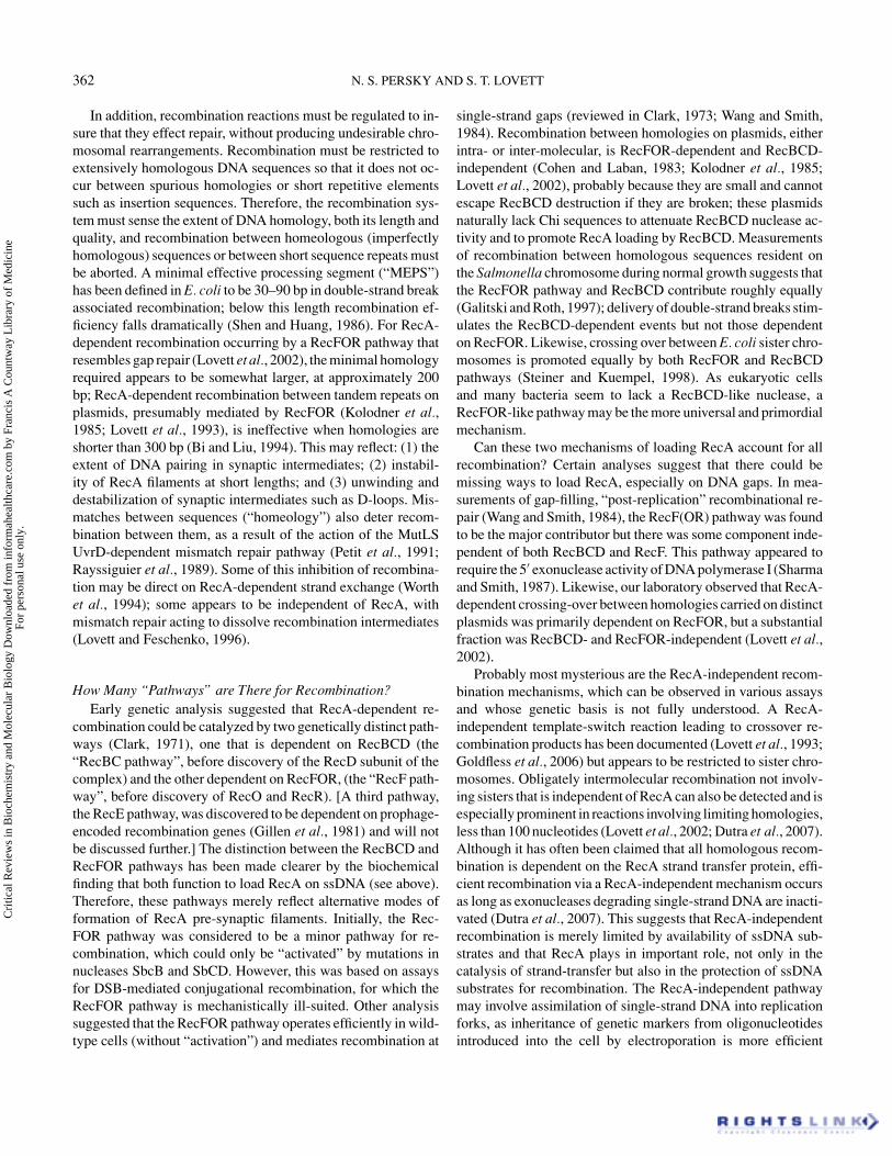
362 N. S. PERSKY AND S. T. LOVETT
In addition, recombination reactions must be regulated to in-sure that they effect repair, without producing undesirable chro-mosomal rearrangements. Recombination must be restricted toextensively homologous DNA sequences so that it does not oc-cur between spurious homologies or short repetitive elementssuch as insertion sequences. Therefore, the recombination sys-tem must sense the extent of DNA homology, both its length andquality, and recombination between homeologous (imperfectlyhomologous) sequences or between short sequence repeats mustbe aborted. A minimal effective processing segment (“MEPS”)has been defined in E. coli to be 30–90 bp in double-strand breakassociated recombination; below this length recombination ef-ficiency falls dramatically (Shen and Huang, 1986). For RecA-dependent recombination occurring by a RecFOR pathway thatresembles gap repair (Lovett et al., 2002), the minimal homologyrequired appears to be somewhat larger, at approximately 200bp; RecA-dependent recombination between tandem repeats onplasmids, presumably mediated by RecFOR (Kolodner et al.,1985; Lovett et al., 1993), is ineffective when homologies areshorter than 300 bp (Bi and Liu, 1994). This may reflect: (1) theextent of DNA pairing in synaptic intermediates; (2) instabil-ity of RecA filaments at short lengths; and (3) unwinding anddestabilization of synaptic intermediates such as D-loops. Mis-matches between sequences (“homeology”) also deter recom-bination between them, as a result of the action of the MutLSUvrD-dependent mismatch repair pathway (Petit et al., 1991;Rayssiguier et al., 1989). Some of this inhibition of recombina-tion may be direct on RecA-dependent strand exchange (Worthet al., 1994); some appears to be independent of RecA, withmismatch repair acting to dissolve recombination intermediates(Lovett and Feschenko, 1996).
How Many “Pathways” are There for Recombination?Early genetic analysis suggested that RecA-dependent re-
combination could be catalyzed by two genetically distinct path-ways (Clark, 1971), one that is dependent on RecBCD (the“RecBC pathway”, before discovery of the RecD subunit of thecomplex) and the other dependent on RecFOR, (the “RecF path-way”, before discovery of RecO and RecR). [A third pathway,the RecE pathway, was discovered to be dependent on prophage-encoded recombination genes (Gillen et al., 1981) and will notbe discussed further.] The distinction between the RecBCD andRecFOR pathways has been made clearer by the biochemicalfinding that both function to load RecA on ssDNA (see above).Therefore, these pathways merely reflect alternative modes offormation of RecA pre-synaptic filaments. Initially, the Rec-FOR pathway was considered to be a minor pathway for re-combination, which could only be “activated” by mutations innucleases SbcB and SbCD. However, this was based on assaysfor DSB-mediated conjugational recombination, for which theRecFOR pathway is mechanistically ill-suited. Other analysissuggested that the RecFOR pathway operates efficiently in wild-type cells (without “activation”) and mediates recombination at
single-strand gaps (reviewed in Clark, 1973; Wang and Smith,1984). Recombination between homologies on plasmids, eitherintra- or inter-molecular, is RecFOR-dependent and RecBCD-independent (Cohen and Laban, 1983; Kolodner et al., 1985;Lovett et al., 2002), probably because they are small and cannotescape RecBCD destruction if they are broken; these plasmidsnaturally lack Chi sequences to attenuate RecBCD nuclease ac-tivity and to promote RecA loading by RecBCD. Measurementsof recombination between homologous sequences resident onthe Salmonella chromosome during normal growth suggests thatthe RecFOR pathway and RecBCD contribute roughly equally(Galitski and Roth, 1997); delivery of double-strand breaks stim-ulates the RecBCD-dependent events but not those dependenton RecFOR. Likewise, crossing over between E. coli sister chro-mosomes is promoted equally by both RecFOR and RecBCDpathways (Steiner and Kuempel, 1998). As eukaryotic cellsand many bacteria seem to lack a RecBCD-like nuclease, aRecFOR-like pathway may be the more universal and primordialmechanism.
Can these two mechanisms of loading RecA account for allrecombination? Certain analyses suggest that there could bemissing ways to load RecA, especially on DNA gaps. In mea-surements of gap-filling, “post-replication” recombinational re-pair (Wang and Smith, 1984), the RecF(OR) pathway was foundto be the major contributor but there was some component inde-pendent of both RecBCD and RecF. This pathway appeared torequire the 5′ exonuclease activity of DNA polymerase I (Sharmaand Smith, 1987). Likewise, our laboratory observed that RecA-dependent crossing-over between homologies carried on distinctplasmids was primarily dependent on RecFOR, but a substantialfraction was RecBCD- and RecFOR-independent (Lovett et al.,2002).
Probably most mysterious are the RecA-independent recom-bination mechanisms, which can be observed in various assaysand whose genetic basis is not fully understood. A RecA-independent template-switch reaction leading to crossover re-combination products has been documented (Lovett et al., 1993;Goldfless et al., 2006) but appears to be restricted to sister chro-mosomes. Obligately intermolecular recombination not involv-ing sisters that is independent of RecA can also be detected and isespecially prominent in reactions involving limiting homologies,less than 100 nucleotides (Lovett et al., 2002; Dutra et al., 2007).Although it has often been claimed that all homologous recom-bination is dependent on the RecA strand transfer protein, effi-cient recombination via a RecA-independent mechanism occursas long as exonucleases degrading single-strand DNA are inacti-vated (Dutra et al., 2007). This suggests that RecA-independentrecombination is merely limited by availability of ssDNA sub-strates and that RecA plays in important role, not only in thecatalysis of strand-transfer but also in the protection of ssDNAsubstrates for recombination. The RecA-independent pathwaymay involve assimilation of single-strand DNA into replicationforks, as inheritance of genetic markers from oligonucleotidesintroduced into the cell by electroporation is more efficient
Cri
tical
Rev
iew
s in
Bio
chem
istr
y an
d M
olec
ular
Bio
logy
Dow
nloa
ded
from
info
rmah
ealth
care
.com
by
Fran
cis
A C
ount
way
Lib
rary
of
Med
icin
e Fo
r pe
rson
al u
se o
nly.

MECHANISMS OF RECOMBINATION 363
when they are single-stranded and when complementary tothe corresponding lagging strand DNA template (Dutra et al.,2007).
Are There Additional Ways to Resolve Branched RecombinationIntermediates?
As discussed above, RuvABC branch migration and cleavagecan resolve Holliday junction recombination intermediates andRecG can resolve structures such as D-loops by reverse branchmigration. Surprisingly, an assay designed to detect crossoverproducts between two plasmids did not detect any decrement ofrecombination in ruvC recG mutants (Lovett et al., 2002), sug-gesting that there could be alternative mechanisms for junctionresolution. (We rule out the RecQ Topo III alternative resolutionmechanism because it does not yield crossover products.)
In addition, although cleavage of D-loops can stabilize repli-cation fork break repair intermediates and models for DSB re-combination (Figure 1) invoke such processing, the existence ofsuch an activity in E. coli, or any other organism, remains un-known. A D-loop cleavage activity was identified and purified(Chiu et al., 1997) but its genetic basis has not been established.
How are Late Events in Recombination, Including DNA Synthe-sis, Coordinated?
Early steps in recombination involve “hand-off” mecha-nisms, such as the recruitment of RecA to single-strand gapsvia RecO bound to SSB. Our knowledge of the coordinationand regulation of the later steps of recombination is less clear.Does RecA eventually fall off post-synaptic structures or is it sta-bilized or removed by specific factors? Are resolution proteinsrecruited to recombination intermediates through the early re-combination proteins? What DNA polymerases are required forgap repair? How is the need for re-establishment of replicationforks after recombination assessed and fulfilled?
SUMMARY AND CONCLUSIONSA little over four decades of genetic and biochemical analysis
has provided key insights into the mechanisms of recombinationin E. coli that have relevance for other organisms. A number ofparadigms established for the function of E. coli recombinationproteins have held true in eukaryotic systems. The connectionbetween genetic recombination and DNA repair mechanismswas discovered in E. coli by early genetic analysis, and has beencodified into three mechanisms, “broken fork repair”, “DSB re-pair” and “recombinational gap-filling repair”. Genetics and bio-chemistry have established functions that operate in three stagesof recombination: prior to synapsis of two DNA molecules, dur-ing their synapsis and in the processing that occurs afterwards.Work in E. coli established the RecA protein as a key proteinmediating homologous pairing and strand exchange during re-combination. Two mechanisms or “pathways” of RecA loadinghave been deduced by the combined power of genetics and bio-
chemistry, one specific to double-strand ends, RecBCD, andthe other specific to replication gaps, involving RecA mediatorproteins, RecFOR. The latter proteins appear to be recruited tosubstrates through interaction with single-strand DNA bindingprotein, SSB. The pathway choice in vivo is dictated by the na-ture of the initiating DNA substrate: double-strand breaks forRecBCD and single-strand gaps for RecFOR. Exonucleolyticprocessing of recombination substrates involves either RecBCDat double-strand breaks or RecJ exonuclease and RecQ helicaseat replication gaps. At least two mechanisms for resolution ofrecombination intermediates have been deduced in late stagesof recombination: one involving migration and cleavage of Hol-liday junctions via RuvABC and the other involving RecG heli-case; still others may exist. Recombination intermediates suchas D-loops can be matured into replication forks via the action ofthe PriA replication restart protein. It is clear from E. coli genet-ics that recombinational repair of broken or damaged replicationforks plays a vital function for the cell.
As the proteins involved in recombination and replication inE. coli are better understood mechanistically and likely to be lesscomplex than their eukaryotic counterparts, it is our hope thatcontinued biochemical and genetic analysis in this system willprovide further lessons in the difficult questions that remain con-cerning coordination and regulation of genetic recombination,applicable to all cells.
ACKNOWLEDGMENTSWe thank Tom Silhavy for suggesting that a basic review of E.
coli recombination genetics and biochemistry would be useful.STL is grateful for early training in the joys and complexitiesofE. coli recombination genetics by A. J. Clark and for manyinteractions over the years with members of the recombinationfield. Unpublished work cited in this article was supported byNIH award GM51753.
REFERENCESAdams, D.E., and West, S.C. 1996. Bypass of DNA heterologies during
RuvAB-mediated three- and four-strand branch migration. J Mol Biol263:582–596.
Adzuma, K. 1992. Stable synapsis of homologous DNA molecules me-diated by the Escherichia coli RecA protein involves local exchangeof DNA strands. Genes Dev 6:1679–1694.
Al-Deib, A.A., Mahdi, A.A., and Lloyd, R.G. 1996. Modulation ofrecombination and DNA repair by the RecG and PriA helicases ofEscherichia coli K-12. J Bacteriol 178:6782–6789.
Anderson, D.G., and Kowalczykowski, S.C. 1997. The translocatingRecBCD enzyme stimulates recombination by directing RecA pro-tein onto ssDNA in a chi-regulated manner. Cell 90:77–86.
Arthur, H.M., and Lloyd, R.G. 1980. Hyper-recombination in uvrDmutants of Escherichia coli K-12. Mol Gen Genet 180:185–191.
Baharoglu, Z., Bradley, A.S., Le Masson, M., Tsaneva, I., and Michel,B. 2008. ruvA Mutants that resolve Holliday junctions but do notreverse replication forks. PLoS Genet 4:e1000012.
Baitin, D.M., Gruenig, M.C., and Cox, M.M. 2008. SSB antagonizesRecX-RecA interaction. J Biol Chem 283:14198–14204.
Cri
tical
Rev
iew
s in
Bio
chem
istr
y an
d M
olec
ular
Bio
logy
Dow
nloa
ded
from
info
rmah
ealth
care
.com
by
Fran
cis
A C
ount
way
Lib
rary
of
Med
icin
e Fo
r pe
rson
al u
se o
nly.
364 N. S. PERSKY AND S. T. LOVETT
Beam, C.E., Saveson, C.J., and Lovett, S.T. 2002. Role for radA/sms inrecombination intermediate processing in Escherichia coli. J Bacte-riol 184:6836–6844.
Bedale, W.A., Inman, R.B., and Cox, M.M. 1993. A reverse DNA strandexchange mediated by recA protein and exonuclease I. The genera-tion of apparent DNA strand breaks by recA protein is explained. JBiol Chem 268:15004–15016.
Bell, C.E. 2005. Structure and mechanism of Escherichia coli RecAATPase. Mol Microbiol 58:358–366.
Belle, J.J., Casey, A., Courcelle, C.T., and Courcelle, J. 2007. Inacti-vation of the DnaB helicase leads to the collapse and degradation ofthe replication fork: a comparison to UV-induced arrest. J Bacteriol189:5452–5462.
Berdichevsky, A., Izhar, L., and Livneh, Z. 2002. Error-free recombi-national repair predominates over mutagenic translesion replicationin E. coli. Mol Cell 10:917–924.
Bi, X., and Liu, L.F. 1994. recA-independent and recA-dependent in-tramolecular plasmid recombination. Differential homology require-ment and distance effect. J Mol Biol 235:414–423.
Bianco, P.R., Tracy, R.B., and Kowalczykowski, S.C. 1998. DNA strandexchange proteins: a biochemical and physical comparison. FrontBiosci 3:D570–603.
Bichara, M., Pinet, I., Origas, M., and Fuchs, R.P. 2006. Inactivationof recG stimulates the RecF pathway during lesion-induced recom-bination in E. coli. DNA Repair (Amst) 5:129–137.
Bierne, H., Seigneur, M., Ehrlich, S.D., and Michel, B. 1997. uvrDmutations enhance tandem repeat deletion in the Escherichia colichromosome via SOS induction of the RecF recombination pathway.Mol Microbiol 26:557–567.
Blakely, G., May, G., McCulloch, R., Arciszewska, L.K., Burke, M.,Lovett, S.T., and Sherratt, D.J. 1993. Two related recombinases arerequired for site-specific recombination at dif and cer in E. coli K12.Cell 75:351–361.
Blattner, F.R., Plunkett, G., 3rd, Bloch, C.A., Perna, N.T., Burland, V.,Riley, M., Collado-Vides, J., Glasner, J.D., Rode, C.K., Mayhew,G.F., Gregor, J., Davis, N.W., Kirkpatrick, H.A., Goeden, M.A.,Rose, D.J., Mau, B., and Shao, Y. 1997. The complete genome se-quence of Escherichia coli K-12. Science 277:1453–1474.
Bork, J.M., Cox, M.M., and Inman, R.B. 2001. The RecOR proteinsmodulate RecA protein function at 5′ ends of single-stranded DNA.EMBO J 20:7313–7322.
Briggs, G.S., Mahdi, A.A., Weller, G.R., Wen, Q., and Lloyd, R.G.2004. Interplay between DNA replication, recombination and repairbased on the structure of RecG helicase. Philos Trans R Soc Lond,B, Biol Sci 359:49–59.
Butland, G., Peregrin-Alvarez, J.M., Li, J., Yang, W., Yang, X.,Canadien, V., Starostine, A., Richards, D., Beattie, B., Krogan, N.,Davey, M., Parkinson, J., Greenblatt, J., and Emili, A. 2005. Interac-tion network containing conserved and essential protein complexesin Escherichia coli. Nature 433:531–537.
Bzymek, M., and Lovett, S.T. 2001. Evidence for two mechanismsof palindrome-stimulated deletion in Escherichia coli: single-strandannealing and replication slipped mispairing. Genetics 158:527–540.
Capaldo, F.N., and Barbour, S.D. 1975. The role of the rec genes in theviability of Escherichia coli K12. Basic Life Sci 5A:405–418.
Carlini, L.E., and Porter, R.D. 1997. Analysis of ssb mutations in vivoimplicates SSB protein in two distinct pathways of SOS inductionand in recombinational DNA repair. Mol Microbiol 24:129–139.
Chase, J.W., and Williams, K.R. 1986. Single-stranded DNA bindingproteins required for DNA replication. Annu Rev Biochem 55:103–136.
Chaudhury, A.M., and Smith, G.R. 1985. Role of Escherichia coliRecBC enzyme in SOS induction. Mol Gen Genet 201:525–528.
Chen, Z., Yang, H., and Pavletich, N. 2008. Mechanism of homolo-gous recombination from the RecA–ssDNA/dsDNA structures. Na-ture 453:489–484.
Chiu, S.K., Low, K.B., Yuan, A., and Radding, C.M. 1997. Resolutionof an early RecA-recombination intermediate by a junction-specificendonuclease. Proc Natl Acad Sci USA 94:6079–6083.
Chow, S.A., Honigberg, S.M., Bainton, R.J., and Radding, C.M. 1986.Patterns of nuclease protection during strand exchange. recA proteinforms heteroduplex DNA by binding to strands of the same polarity.J Biol Chem 261:6961–6971.
Chrysogelos, S., and Griffith, J. 1982. Escherichia coli single-strandbinding protein organizes single-stranded DNA in nucleosome-likeunits. Proc Natl Acad Sci USA 79:5803–5807.
Clark, A.J. 1971. Toward a metabolic interpretation of genetic recom-bination of E. coli and its phages. Annu Rev Microbiol 25:437–464.
Clark, A.J. 1973. Recombination deficient mutants of E. coli and otherbacteria. Annu Rev Genet 7:67–86.
Clark, A.J. 1991. rec genes and homologous recombination proteins inEscherichia coli. Biochimie 73:523–532.
Clark, A.J., and Margulies, A.D. 1965. Isolation and characterization ofrecombination-deficient nutants of Escherichia coli K12. Proc NatlAcad Sci USA 53:451–459.
Clark, A.J., and Sandler, S.J. 1994. Homologous genetic recombination:the pieces begin to fall into place. Crit Rev Microbiol 20:125–142.
Cohen, A., and Laban, A. 1983. Plasmidic recombination in Escherichiacoli K-12: the role of recF gene function. Mol Gen Genet 189:471–474.
Connelly, J.C., and Leach, D.R. 1996. The sbcC and sbcD genes ofEscherichia coli encode a nuclease involved in palindrome inviabilityand genetic recombination. Genes Cells 1:285–291.
Connelly, J.C., Kirkham, L.A., and Leach, D.R. 1998. The SbcCDnuclease of Escherichia coli is a structural maintenance of chromo-somes (SMC) family protein that cleaves hairpin DNA. Proc NatlAcad Sci USA 95:7969–7974.
Connelly, J.C., de Leau, E.S., and Leach, D.R. 1999. DNA cleavageand degradation by the SbcCD protein complex from Escherichiacoli. Nucleic Acids Res 27:1039–1046.
Connelly, J.C., de Leau, E.S., and Leach, D.R. 2003. Nucleolytic pro-cessing of a protein-bound DNA end by the E. coli SbcCD (MR)complex. DNA Repair (Amst) 2:795–807.
Corrette-Bennett, S.E., and Lovett, S.T. 1995. Enhancement of RecAstrand-transfer activity by the RecJ exonuclease of Escherichia coli.J Biol Chem 270:6881–6885.
Courcelle, C.T., Chow, K.H., Casey, A., and Courcelle, J. 2006. NascentDNA processing by RecJ favors lesion repair over translesion synthe-sis at arrested replication forks in Escherichia coli. Proc Natl AcadSci USA 103:9154–9159.
Courcelle, J., and Hanawalt, P.C. 1999. RecQ and RecJ process blockedreplication forks prior to the resumption of replication in UV-irradiated Escherichia coli. Mol Gen Genet 262:543–551.
Cox, M.M. 2001. Recombinational DNA repair of damaged replica-tion forks in Escherichia coli: questions. Annu Rev Genet 35:53–82.
Cri
tical
Rev
iew
s in
Bio
chem
istr
y an
d M
olec
ular
Bio
logy
Dow
nloa
ded
from
info
rmah
ealth
care
.com
by
Fran
cis
A C
ount
way
Lib
rary
of
Med
icin
e Fo
r pe
rson
al u
se o
nly.

MECHANISMS OF RECOMBINATION 365
Cox, M.M. 2007a. Motoring along with the bacterial RecA protein. NatRev Mol Cell Biol 8:127–138.
Cox, M.M. 2007b. Regulation of bacterial RecA protein function. CritRev Biochem Mol Biol 42:41–63.
Cromie, G.A., and Leach, D.R. 2001. Recombinational repair of chro-mosomal DNA double-strand breaks generated by a restriction en-donuclease. Mol Microbiol 41:873–883.
Cromie, G.A., Millar, C.B., Schmidt, K.H., and Leach, D.R. 2000.Palindromes as substrates for multiple pathways of recombination inEscherichia coli. Genetics 154:513–522.
Cromie, G.A., Connelly, J.C., and Leach, D.R. 2001. Recombinationat double-strand breaks and DNA ends: conserved mechanisms fromphage to humans. Mol Cell 8:1163–1174.
Cunningham, R.P., Wu, A.M., Shibata, T., DasGupta, C., and Radding,C.M. 1981. Homologous pairing and topological linkage of DNAmolecules by combined action of E. coli RecA protein and topoiso-merase I. Cell 24:213–223.
DasGupta, C., Shibata, T., Cunningham, R.P., and Radding, C.M. 1980.The topology of homologous pairing promoted by RecA protein. Cell22:437–446.
Dillingham, M.S., Webb, M.R., and Kowalczykowski, S.C. 2005. Bipo-lar DNA translocation contributes to highly processive DNA unwind-ing by RecBCD enzyme. J Biol Chem 280:37069–37077.
Dixon, D.A., and Kowalczykowski, S.C. 1991. Homologous pairingin vitro stimulated by the recombination hotspot, Chi. Cell 66:361–371.
Dixon, D.A., and Kowalczykowski, S.C. 1993. The recombinationhotspot chi is a regulatory sequence that acts by attenuating the nu-clease activity of the E. coli RecBCD enzyme. Cell 73:87–96.
Donaldson, J.R., Courcelle, C.T., and Courcelle, J. 2006. RuvABCis required to resolve holliday junctions that accumulate followingreplication on damaged templates in Escherichia coli. J Biol Chem281:28811–28821.
Drees, J.C., Lusetti, S.L., and Cox, M.M. 2004. Inhibition of RecA pro-tein by the Escherichia coli RecX protein: modulation by the RecAC terminus and filament functional state. J Biol Chem 279:52991–52997.
Dutra, B.E., Sutera, V.A., and Lovett, S.T. 2007. RecA-independentrecombination is efficient but limited by exonucleases. Proc NatlAcad Sci USA 104:216–221.
Emmerson, P.T., and Howard-Flanders, P. 1967. Cotransduction withthy of a gene required for genetic recombination in Escherichia coli.J Bacteriol 93:1729–1731.
Enquist, L.W., and Skalka, A. 1973. Replication of bacteriophagelambda DNA dependent on the function of host and viralgenes. I. Interaction of red, gam and rec. J Mol Biol 75:185–212.
Eykelenboom, J.K., Blackwood, J.K., Okely, E., and Leach, D.R. 2008.SbcCD causes a double-strand break at a DNA palindrome in theEscherichia coli chromosome. Mol Cell 29:644–651.
Feinstein, S.I., and Low, K.B. 1986. Hyper-recombining recipientstrains in bacterial conjugation. Genetics 113:13–33.
Felzenszwalb, I., Sargentini, N.J., and Smith, K.C. 1984. Character-ization of a new radiation-sensitive mutant, Escherichia coli K-12radC102. Radiat Res 97:615–625.
Feschenko, V.V., Rajman, L.A., and Lovett, S.T. 2003. Stabilization ofperfect and imperfect tandem repeats by single-strand DNA exonu-cleases. Proc Natl Acad Sci USA 100:1134–1139.
Finch, P.W., Chambers, P., and Emmerson, P.T. 1985. Identification ofthe Escherichia coli recN gene product as a major SOS protein. JBacteriol 164:653–658.
Fishel, R.A., and Kolodner, R. 1984. Escherichia coli strains containingmutations in the structural gene for topoisomerase I are recombina-tion deficient. J Bacteriol 160:1168–1170.
Foti, J.J., Schienda, J., Sutera, V.A., Jr., and Lovett, S.T. 2005. A bac-terial G protein-mediated response to replication arrest. Mol Cell17:549–560.
Friedman-Ohana, R., and Cohen, A. 1998. Heteroduplex joint forma-tion in Escherichia coli recombination is initiated by pairing of a3′-ending strand. Proc Natl Acad Sci USA, 95:6909–6914.
Friedman-Ohana, R., Karunker, I., and Cohen, A. 1999. A RecG-independent nonconservative branch migration mechanism in Es-cherichia coli recombination. J Bacteriol 181:7199–7205.
Galitski, T., and Roth, J.R. 1997. Pathways for homologous recom-bination between chromosomal direct repeats in Salmonella ty-phimurium. Genetics 146:751–767.
Galletto, R., Amitani, I., Baskin, R.J., and Kowalczykowski, S.C. 2006.Direct observation of individual RecA filaments assembling on singleDNA molecules. Nature 443:875–878.
Gangloff, S., McDonald, J.P., Bendixen, C., Arthur, L., and Rothstein,R. 1994. The yeast type I topoisomerase Top3 interacts with Sgs1, aDNA helicase homolog: a potential eukaryotic reverse gyrase. MolCell Biol 14:8391–8398.
Gibson, F.P., Leach, D.R., and Lloyd, R.G. 1992. Identification of sbcDmutations as cosuppressors of recBC that allow propagation of DNApalindromes in Escherichia coli K-12. J Bacteriol 174:1222–1228.
Gillen, J.R., Willis, D.K., and Clark, A.J. 1981. Genetic analysis of theRecE pathway of genetic recombination in Escherichia coli K-12. JBacteriol 145:521–532.
Glassberg, J., Meyer, R.R., and Kornberg, A. 1979. Mutant single-strand binding protein of Escherichia coli: genetic and physiologicalcharacterization. J Bacteriol 140:14–19.
Goldfless, S.J., Morag, A.S., Belisle, K.A., Sutera, V.A., and Lovett,S.T. 2006. DNA repeat rearrangements mediated by DnaK-dependent replication fork repair. Mol Cell 21:595–604.
Goodman, M.F., and Tippin, B. 2000. Sloppier copier DNA poly-merases involved in genome repair. Curr Opin Genet Dev 10:162–168.
Grompone, G., Sanchez, N., Dusko Ehrlich, S., and Michel, B. 2004.Requirement for RecFOR-mediated recombination in priA mutant.Mol Microbiol 52:551–562.
Haber, J.E. 1998. Mating-type gene switching in Saccharomyces cere-visiae. Annu Rev Genet 32:561–599.
Han, E.S., Cooper, D.L., Persky, N.S., Sutera, V.A., Jr., Whitaker, R.D.,Montello, M.L., and Lovett, S.T. 2006. RecJ exonuclease: substrates,products and interaction with SSB. Nucleic Acids Res 34:1084–1091.
Harmon, F.G., and Kowalczykowski, S.C. 2001. Biochemical charac-terization of the DNA helicase activity of the Escherichia coli RecQhelicase. J Biol Chem 276:232–243.
Harmon, F.G., DiGate, R.J., and Kowalczykowski, S.C. 1999. RecQhelicase and topoisomerase III comprise a novel DNA strand passagefunction: a conserved mechanism for control of DNA recombination.Mol Cell 3:611–620.
Heller, R.C., and Marians, K.J. 2005. The disposition of nascent strandsat stalled replication forks dictates the pathway of replisome loadingduring restart. Mol Cell 17:733–743.
Cri
tical
Rev
iew
s in
Bio
chem
istr
y an
d M
olec
ular
Bio
logy
Dow
nloa
ded
from
info
rmah
ealth
care
.com
by
Fran
cis
A C
ount
way
Lib
rary
of
Med
icin
e Fo
r pe
rson
al u
se o
nly.

366 N. S. PERSKY AND S. T. LOVETT
Hirano, T. 2005. SMC proteins and chromosome mechanics: from bac-teria to humans. Philos Trans R Soc Lond, B, Biol Sci 360:507–514.
Hobbs, M.D., Sakai, A., and Cox, M.M. 2007. SSB protein limitsRecOR binding onto single-stranded DNA. J Biol Chem 282:11058–11067.
Horii, Z., and Clark, A.J. 1973. Genetic analysis of the recF pathway togenetic recombination in Escherichia coli K12: isolation and char-acterization of mutants. J Mol Biol 80:327–344.
Howard-Flanders, P., and Theriot, L. 1966. Mutants of Escherichia coliK-12 defective in DNA repair and in genetic recombination. Genetics53:1137–1150.
Joo, C., McKinney, S.A., Nakamura, M., Rasnik, I., Myong, S., andHa, T. 2006. Real-time observation of RecA filament dynamics withsingle monomer resolution. Cell 126:515–527.
Kang, J., and Blaser, M.J. 2006. UvrD helicase suppresses recombi-nation and DNA damage-induced deletions. J Bacteriol 188:5450–5459.
Kantake, N., Madiraju, M.V., Sugiyama, T., and Kowalczykowski,S.C. 2002. Escherichia coli RecO protein anneals ssDNA com-plexed with its cognate ssDNA-binding protein: A common stepin genetic recombination. Proc Natl Acad Sci USA 99:15327–15332.
Kaplan, D.L., and O’Donnell, M. 2006. RuvA is a sliding collar thatprotects Holliday junctions from unwinding while promoting branchmigration. J Mol Biol 355:473–490.
Kidane, D., and Graumann, P.L. 2005. Intracellular protein and DNAdynamics in competent Bacillus subtilis cells. Cell 122:73–84.
Kidane, D., Sanchez, H., Alonso, J.C., and Graumann, P.L. 2004. Vi-sualization of DNA double-strand break repair in live bacteria re-veals dynamic recruitment of Bacillus subtilis RecF, RecO and RecNproteins to distinct sites on the nucleoids. Mol Microbiol 52:1627–1639.
Kogoma, T. 1996. Recombination by replication. Cell 85:625–627.Kogoma, T., Cadwell, G.W., Barnard, K.G., and Asai, T. 1996. The
DNA replication priming protein, PriA, is required for homologousrecombination and double-strand break repair. J Bacteriol 178:1258–1264.
Kolodner, R., Fishel, R.A., and Howard, M. 1985. Genetic recombina-tion of bacterial plasmid DNA: effect of RecF pathway mutations onplasmid recombination in Escherichia coli. J Bacteriol 163:1060–1066.
Konforti, B.B., and Davis, R.W. 1992. ATP hydrolysis and the displacedstrand are two factors that determine the polarity of RecA-promotedDNA strand exchange. J Mol Biol 227:38–53.
Kornberg, A., and Baker, T.A. 1991. DNA replication, 2nd edn W. H.Freeman, New York, NY.
Kosa, J.L., Zdraveski, Z.Z., Currier, S., Marinus, M.G., and Essigmann,J.M. 2004. RecN and RecG are required for Escherichia coli survivalof Bleomycin-induced damage. Mutat Res 554:149–157.
Kouzminova, E.A., and Kuzminov, A. 2004. Chromosomal fragmen-tation in dUTPase-deficient mutants of Escherichia coli and its re-combinational repair. Mol Microbiol 51:1279–1295.
Kouzminova, E.A., Rotman, E., Macomber, L., Zhang, J., and Kuzmi-nov, A. 2004. RecA-dependent mutants in Escherichia coli revealstrategies to avoid chromosomal fragmentation. Proc Natl Acad SciUSA 101:16262–16267.
Kowalczykowski, S.C. 2000. Initiation of genetic recombination andrecombination-dependent replication. Trends Biochem Sci 25:156–165.
Kowalczykowski, S.C., and Krupp, R.A. 1987. Effects of Escherichiacoli SSB protein on the single-stranded DNA-dependent ATPase ac-tivity of Escherichia coli RecA protein. Evidence that SSB proteinfacilitates the binding of RecA protein to regions of secondary struc-ture within single-stranded DNA. J Mol Biol 193:97–113.
Kowalczykowski, S.C., Clow, J., Somani, R., and Varghese, A. 1987.Effects of the Escherichia coli SSB protein on the binding of Es-cherichia coli RecA protein to single-stranded DNA. Demonstrationof competitive binding and the lack of a specific protein-protein in-teraction. J Mol Biol 193:81–95.
Kreuzer, K.N. 2005. Interplay between DNA replication and recombi-nation in prokaryotes. Annu Rev Microbiol 59:43–67.
Kushner, S.R., Nagaishi, H., Templin, A., and Clark, A.J. 1971. Geneticrecombination in Escherichia coli: the role of exonuclease I. ProcNatl Acad Sci USA 68:824–827.
Kuzminov, A. 1999. Recombinational repair of DNA damage in Es-cherichia coli and bacteriophage lambda. Microbiol Mol Biol Rev63:751–813.
Kuzminov, A., and Stahl, F.W., Editor N.P. Higgins. 2005. Overviewof homologous recombination and repair machines. In The BacterialChromosome, 2nd edition: 349–368. Washington, DC: American So-ciety of Microbiology Press.
Lam, S.T., Stahl, M.M., McMilin, K.D., and Stahl, F.W. 1974. Rec-mediated recombinational hot spot activity in bacteriophage lambda.II. A mutation which causes hot spot activity. Genetics 77:425–433.
Lavery, P.E., and Kowalczykowski, S.C. 1992. A postsynaptic rolefor single-stranded DNA-binding protein in recA protein-promotedDNA strand exchange. J Biol Chem 267:9315–9320.
Lehman, I.R., and Nussbaum, A.L. 1964. The deoxyribonucleases ofEscherichia coli. V. On the specificity of Exonuclease I (phosphodi-esterase). J Biol Chem 239:2628–2636.
Lim, D.S., and Hasty, P. 1996. A mutation in mouse rad51 results in anearly embryonic lethal that is suppressed by a mutation in p53. MolCell Biol 16:7133–7143.
Lin, Z., Kong, H., Nei, M., and Ma, H. 2006. Origins and evolution of therecA/RAD51 gene family: evidence for ancient gene duplication andendosymbiotic gene transfer. Proc Natl Acad Sci USA 103:10328–10333.
Lindsley, J.E., and Cox, M.M. 1990. Assembly and disassembly ofRecA protein filaments occur at opposite filament ends. Relationshipto DNA strand exchange. J Biol Chem 265:9043–9054.
Linn, S., and Imlay, J.A. 1987. Toxicity, mutagenesis and stress re-sponses induced in Escherichia coli by hydrogen peroxide. J CellSci Suppl 6:289–301.
Lloyd, R.G. 1991. Conjugational recombination in resolvase-deficientruvC mutants of Escherichia coli K-12 depends on recG. J Bacteriol173:5414–5418.
Lloyd, R.G., and Buckman, C. 1985. Identification and genetic anal-ysis of sbcC mutations in commonly used recBC sbcB strains ofEscherichia coli K-12. J Bacteriol 164:836–844.
Lloyd, R.G., and Buckman, C. 1995. Conjugational recombination inEscherichia coli: genetic analysis of recombinant formation in Hfr xF- crosses. Genetics 139:1123–1148.
Lloyd, R.G., Benson, F.E., and Shurvinton, C.E. 1984. Effect of ruvmutations on recombination and DNA repair in Escherichia coli K12.Mol Gen Genet 194:303–309.
Lohman, T.M., and Ferrari, M.E. 1994. Escherichia coli single-strandedDNA-binding protein: multiple DNA-binding modes and coopera-tivities. Annu Rev Biochem 63:527–570.
Cri
tical
Rev
iew
s in
Bio
chem
istr
y an
d M
olec
ular
Bio
logy
Dow
nloa
ded
from
info
rmah
ealth
care
.com
by
Fran
cis
A C
ount
way
Lib
rary
of
Med
icin
e Fo
r pe
rson
al u
se o
nly.
MECHANISMS OF RECOMBINATION 367
Lopez, C.R., Yang, S., Deibler, R.W., Ray, S.A., Pennington, J.M.,Digate, R.J., Hastings, P.J., Rosenberg, S.M., and Zechiedrich, E.L.2005. A role for topoisomerase III in a recombination pathway al-ternative to RuvABC. Mol Microbiol 58:80–101.
Lovett, S.T. 2003. Connecting replication and recombination. Mol Cell11:554–556.
Lovett, S.T. 2006. Replication arrest-stimulated recombination: Depen-dence on the RecA paralog, RadA/Sms and translesion polymerase,DinB. DNA Repair (Amst) 5:1421–1427.
Lovett, S.T., and Clark, A.J. 1984. Genetic analysis of the recJ gene ofEscherichia coli K-12. J Bacteriol 157:190–196.
Lovett, S.T., and Feschenko, V.V. 1996. Stabilization of diverged tan-dem repeats by mismatch repair: evidence for deletion formationvia a misaligned replication intermediate. Proc Natl Acad Sci USA93:7120–7124.
Lovett, S.T., and Kolodner, R.D. 1989. Identification and purifica-tion of a single-stranded-DNA-specific exonuclease encoded by therecJ gene of Escherichia coli. Proc Natl Acad Sci USA 86:2627–2631.
Lovett, S.T., and Sutera, V.A. 1995. Suppression of recJ exonucleasemutants of Escherichia coli by alterations in DNA helicases II (uvrD)and IV (helD). Genetics 140:27–45.
Lovett, S.T., Drapkin, P.T., Sutera, V.A., and Gluckman-Peskind, T.J.1993. A sister-strand exchange mechanism for recA-independentdeletion of repeated DNA sequences in Escherichia coli. Genetics135:631–642.
Lovett, S.T., Hurley, R.L., Sutera, V.A., Jr., Aubuchon, R.H., andLebedeva, M.A. 2002. Crossing over between regions of limited ho-mology in Escherichia coli. RecA-dependent and RecA-independentpathways. Genetics 160:851–859.
Luisi-DeLuca, C. 1995. Homologous pairing of single-stranded DNAand superhelical double-stranded DNA catalyzed by RecO proteinfrom Escherichia coli. J Bacteriol 177:566–572.
Luisi-DeLuca, C., and Kolodner, R. 1994. Purification and character-ization of the Escherichia coli RecO protein. Renaturation of com-plementary single-stranded DNA molecules catalyzed by the RecOprotein. J Mol Biol 236:124–138.
Lusetti, S.L., Voloshin, O.N., Inman, R.B., Camerini-Otero, R.D., andCox, M.M. 2004a. The DinI protein stabilizes RecA protein fila-ments. J Biol Chem 279:30037–30046.
Lusetti, S.L., Drees, J.C., Stohl, E.A., Seifert, H.S., and Cox, M.M.2004b. The DinI and RecX proteins are competing modulators ofRecA function. J Biol Chem 279:55073–55079.
Lusetti, S.L., Hobbs, M.D., Stohl, E.A., Chitteni-Pattu, S., Inman,R.B., Seifert, H.S., and Cox, M.M. 2006. The RecF protein an-tagonizes RecX function via direct interaction. Mol Cell 21:41–50.
Madiraju, M.V., Templin, A., and Clark, A.J. 1988. Properties of amutant recA-encoded protein reveal a possible role for Escherichiacoli recF-encoded protein in genetic recombination. Proc Natl AcadSci USA 85:6592–6596.
Magner, D.B., Blankschien, M.D., Lee, J.A., Pennington, J.M., Lupski,J.R., and Rosenberg, S.M. 2007. RecQ promotes toxic recombinationin cells lacking recombination intermediate-removal proteins. MolCell 26:273–286.
Mahdi, A.A., and Lloyd, R.G. 1989. Identification of the recR locus ofEscherichia coli K-12 and analysis of its role in recombination andDNA repair. Mol Gen Genet 216:503–510.
Mahdi, A.A., Sharples, G.J., Mandal, T.N., and Lloyd, R.G. 1996. Hol-liday junction resolvases encoded by homologous rusA genes in Es-cherichia coli K-12 and phage 82. J Mol Biol 257:561–573.
Mandal, T.N., Mahdi, A.A., Sharples, G.J., and Lloyd, R.G. 1993. Res-olution of Holliday intermediates in recombination and DNA repair:indirect suppression of ruvA, ruvB, and ruvC mutations. J Bacteriol175:4325–4334.
Marians, K.J. 2004. Mechanisms of replication fork restart in Es-cherichia coli. Philos Trans R Soc Lond, B, Biol Sci 359:71–77.
Mazin, A.V., and Kowalczykowski, S.C. 1998. The function of thesecondary DNA-binding site of RecA protein during DNA strandexchange. EMBO J 17:1161–1168.
McEachern, M.J., and Haber, J.E. 2006. Break-induced replication andrecombinational telomere elongation in yeast. Annu Rev Biochem75:111–135.
McGlynn, P., and Lloyd, R.G. 2002. Genome stability and the process-ing of damaged replication forks by RecG. Trends Genet 18:413–419.
McGrew, D.A., and Knight, K.L. 2003. Molecular design and functionalorganization of the RecA protein. Crit Rev Biochem Mol Biol 38:385–432.
Meddows, T.R., Savory, A.P., and Lloyd, R.G. 2004. RecG helicasepromotes DNA double-strand break repair. Mol Microbiol 52:119–132.
Meddows, T.R., Savory, A.P., Grove, J.I., Moore, T., and Lloyd, R.G.2005. RecN protein and transcription factor DksA combine to pro-mote faithful recombinational repair of DNA double-strand breaks.Mol Microbiol 57:97–110.
Menetski, J.P., and Kowalczykowski, S.C. 1985. Interaction of recAprotein with single-stranded DNA. Quantitative aspects of bindingaffinity modulation by nucleotide cofactors. J Mol Biol 181:281–295.
Meyer, R.R., and Laine, P.S. 1990. The single-stranded DNA-bindingprotein of Escherichia coli. Microbiol Rev 54:342–380.
Michel, B., Boubakri, H., Baharoglu, Z., LeMasson, M., and Lestini,R. 2007. Recombination proteins and rescue of arrested replicationforks. DNA Repair (Amst), 6:967–980.
Molineux, I.J., Friedman, S., and Gefter, M.L. 1974. Purification andproperties of the Escherichia coli deoxyribonucleic acid-unwindingprotein. Effects on deoxyribonucleic acid synthesis in vitro. J BiolChem 249:6090–6098.
Molineux, I.J., and Gefter, M.L. 1975. Properties of the Escherichiacoli DNA-binding (unwinding) protein interaction with nucleolyticenzymes and DNA. J Mol Biol 98:811–825.
Moreau, P.L. 1988. Overproduction of single-stranded-DNA-bindingprotein specifically inhibits recombination of UV-irradiated bacte-riophage DNA in Escherichia coli. J Bacteriol 170:2493–2500.
Morel, P., Hejna, J.A., Ehrlich, S.D., and Cassuto, E. 1993. Antipairingand strand transferase activities of E. coli helicase II (UvrD). NucleicAcids Res 21:3205–3209.
Morel, P., Stasiak, A., Ehrlich, S.D., and Cassuto, E. 1994. Effect oflength and location of heterologous sequences on RecA-mediatedstrand exchange. J Biol Chem 269:19830–19835.
Morimatsu, K., and Kowalczykowski, S.C. 2003. RecFOR proteins loadRecA protein onto gapped DNA to accelerate DNA strand exchange:a universal step of recombinational repair. Mol Cell 11:1337–1347.
Muniyappa, K., Shaner, S.L., Tsang, S.S., and Radding, C.M. 1984.Mechanism of the concerted action of recA protein and helix-destabilizing proteins in homologous recombination. Proc Natl AcadSci USA, 81:2757–2761.
Cri
tical
Rev
iew
s in
Bio
chem
istr
y an
d M
olec
ular
Bio
logy
Dow
nloa
ded
from
info
rmah
ealth
care
.com
by
Fran
cis
A C
ount
way
Lib
rary
of
Med
icin
e Fo
r pe
rson
al u
se o
nly.
368 N. S. PERSKY AND S. T. LOVETT
Nagashima, K., Kubota, Y., Shibata, T., Sakaguchi, C., Shinagawa,H., and Hishida, T. 2006. Degradation of Escherichia coli RecNaggregates by ClpXP protease and its implications for DNA damagetolerance. J Biol Chem 281:30941–30946.
Nakayama, H., Nakayama, K., Nakayama, R., Irino, N., Nakayama, Y.,and Hanawalt, P.C. 1984. Isolation and genetic characterization of athymineless death-resistant mutant of Escherichia coli K12: identifi-cation of a new mutation (recQ1) that blocks the RecF recombinationpathway. Mol Gen Genet 195:474–480.
Nassif, N., Penney, J., Pal, S., Engels, W.R., and Gloor, G.B. 1994.Efficient copying of nonhomologous sequences from ectopic sitesvia P-element-induced gap repair. Mol Cell Biol 14:1613–1625.
Neuwald, A.F., Berg, D.E., and Stauffer, G.V. 1992. Mutational analysisof the Escherichia coli serB promoter region reveals transcriptionallinkage to a downstream gene. Gene 120:1–9.
Nowosielska, A., Calmann, M.A., Zdraveski, Z., Essigmann, J.M., andMarinus, M.G. 2004. Spontaneous and cisplatin-induced recombi-nation in Escherichia coli. DNA Repair (Amst), 3:719–728.
Nowosielska, A., Smith, S.A., Engelward, B.P., and Marinus, M.G.2006. Homologous recombination prevents methylation-inducedtoxicity in Escherichia coli. Nucleic Acids Res, 34:2258–2268.
Petit, M.A., Dimpfl, J., Radman, M., and Echols, H. 1991. Control oflarge chromosomal duplications in Escherichia coli by the mismatchrepair system. Genetics 129:327–332.
Pham, P., Rangarajan, S., Woodgate, R., and Goodman, M.F. 2001.Roles of DNA polymerases V and II in SOS-induced error-proneand error-free repair in Escherichia coli. Proc Natl Acad Sci USA98:8350–8354.
Picksley, S.M., Attfield, P.V., and Lloyd, R.G. 1984. Repair of DNAdouble-strand breaks in Escherichia coli K12 requires a functionalrecN product. Mol Gen Genet 195:267–274.
Ponder, R.G., Fonville, N.C., and Rosenberg, S.M. 2005. Aswitch from high-fidelity to error-prone DNA double-strandbreak repair underlies stress-induced mutation. Mol Cell 19:791–804.
Prell, A., and Wackernagel, W. 1980. Degradation of linear and circularDNA with gaps by the recBC enzyme of Escherichia coli. Effectsof gap length and the presence of cell-free extracts. Eur J Biochem105:109–116.
Quinones, A., and Piechocki, R. 1987. Differential suppressor effectsof the ssb-1 and ssb-113 alleles on uvrD mutator of Escherichiacoli in DNA repair and mutagenesis. J Basic Microbiol 27:263–273.
Raghunathan, S., Ricard, C.S., Lohman, T.M., and Waksman, G. 1997.Crystal structure of the homo-tetrameric DNA binding domain ofEscherichia coli single-stranded DNA-binding protein determinedby multiwavelength x-ray diffraction on the selenomethionyl proteinat 2.9-A resolution. Proc Natl Acad Sci USA, 94:6652–6657.
Rayssiguier, C., Thaler, D.S., and Radman, M. 1989. The barrier to re-combination between Escherichia coli and Salmonella typhimuriumis disrupted in mismatch-repair mutants. Nature 342:396–401.
Register, J.C., and Griffith, J. 1985. The direction of RecA proteinassembly onto single strand DNA is the same as the direction ofstrand assimilation during strand exchange. J Biol Chem 260:12308–12312.
Renzette, N., Gumlaw, N., and Sandler, S.J. 2007. DinI and RecX mod-ulate RecA-DNA structures in Escherichia coli K-12. Mol Microbiol63:103–115.
Roberts, D., Hoopes, B.C., McClure, W.R., and Kleckner, N. 1985.IS10 transposition is regulated by DNA adenine methylation. Cell43:117–130.
Rupp, W.D., and Howard-Flanders, P. 1968. Discontinuities in the DNAsynthesized in an excision-defective strain of Escherichia coli fol-lowing ultraviolet irradiation. J Mol Biol 31:291–304.
Rupp, W.D., Wilde, C.E., Reno, D.L., and Howard-Flanders, P.1971. Exchanges between DNA strands in ultraviolet-irradiatedEscherichia coli. J Mol Biol 61:25–44.
Sanchez, H., Cardenas, P.P., Yoshimura, S.H., Takeyasu, K., andAlonso, J.C. 2008. Dynamic structures of Bacillus subtilis RecN-DNA complexes. Nucleic Acids Res 36:110–120.
Sandigursky, M., and Franklin, W.A. 1994. Escherichia coli single-stranded DNA binding protein stimulates the DNA deoxyribophos-phodiesterase activity of exonuclease I. Nucleic Acids Res 22:247–250.
Sandler, S.J., and Clark, A.J. 1994. RecOR suppression of recF mutantphenotypes in Escherichia coli K-12. J Bacteriol 176:3661–3672.
Sandler, S.J., and Marians, K.J. 2000. Role of PriA in replication forkreactivation in Escherichia coli. J Bacteriol 182:9–13.
Sandler, S.J., Marians, K.J., Zavitz, K.H., Coutu, J., Parent, M.A.,and Clark, A.J. 1999. dnaC mutations suppress defects in DNAreplication- and recombination-associated functions in priB and priCdouble mutants in Escherichia coli K-12. Mol Microbiol 34:91–101.
Sano, Y. 1993. Role of the recA-related gene adjacent to the recA genein Pseudomonas aeruginosa. J Bacteriol 175:2451–2454.
Sargentini, N.J., and Smith, K.C. 1986. Quantitation of the involvementof the recA, recB, recC, recF, recJ, recN, lexA, radA, radB, uvrD,and umuC genes in the repair of X-ray-induced DNA double-strandbreaks in Escherichia coli. Radiat Res 107:58–72.
Saveson, C.J., and Lovett, S.T. 1999. Tandem repeat recombinationinduced by replication fork defects in Escherichia coli requires anovel factor, RadC. Genetics 152:5–13.
Savvides, S.N., Raghunathan, S., Futterer, K., Kozlov, A.G., Lohman,T.M., and Waksman, G. 2004. The C-terminal domain of full-lengthE. coli SSB is disordered even when bound to DNA. Protein Sci13:1942–1947.
Sawitzke, J.A., and Stahl, F.W. 1992. Phage lambda has an analog ofEscherichia coli recO, recR and recF genes. Genetics 130:7–16.
Schwacha, A., and Kleckner, N. 1995. Identification of double Hollidayjunctions as intermediates in meiotic recombination. Cell 83:783–791.
Shan, Q., Bork, J.M., Webb, B.L., Inman, R.B., and Cox, M.M. 1997.RecA protein filaments: end-dependent dissociation from ssDNA andstabilization by RecO and RecR proteins. J Mol Biol 265:519–540.
Sharma, R.C., and Smith, K.C. 1987. Role of DNA polymerase I inpostreplication repair: a reexamination with Escherichia coli deltapolA. J Bacteriol 169:4559–4564.
Sharples, G.J., Chan, S.N., Mahdi, A.A., Whitby, M.C., and Lloyd,R.G. 1994. Processing of intermediates in recombination and DNArepair: identification of a new endonuclease that specifically cleavesHolliday junctions. EMBO J 13:6133–6142.
Shen, P., and Huang, H.V. 1986. Homologous recombination inEscherichia coli: dependence on substrate length and homology. Ge-netics 112:441–457.
Shereda, R.D., Bernstein, D.A., and Keck, J.L. 2007. A central role forSSB in Escherichia coli RecQ DNA helicase function. J Biol Chem282:19247–19258.
Cri
tical
Rev
iew
s in
Bio
chem
istr
y an
d M
olec
ular
Bio
logy
Dow
nloa
ded
from
info
rmah
ealth
care
.com
by
Fran
cis
A C
ount
way
Lib
rary
of
Med
icin
e Fo
r pe
rson
al u
se o
nly.
MECHANISMS OF RECOMBINATION 369
Sigal, N., Delius, H., Kornberg, T., Gefter, M.L., and Alberts, B. 1972.A DNA-unwinding protein isolated from Escherichia coli: its inter-action with DNA and with DNA polymerases. Proc Natl Acad SciUSA 69:3537–3541.
Singleton, M.R., Dillingham, M.S., Gaudier, M., Kowalczykowski,S.C., and Wigley, D.B. 2004. Crystal structure of RecBCD enzymereveals a machine for processing DNA breaks. Nature 432:187–193.
Smith, G.R. 1987. Mechanism and control of homologous recombina-tion in Escherichia coli. Annu Rev Genet 21:179–201.
Smith, G.R. 1991. Conjugational recombination in E. coli: myths andmechanisms. Cell 64:19–27.
Smith, G.R. 2001. Homologous recombination near and far from DNAbreaks: alternative roles and contrasting views. Annu Rev Genet35:243–274.
Smith, K.C. 2004. Recombinational DNA repair: the ignored repairsystems. Bioessays 26:1322–1326.
Smith, K.C., and Wang, T.C. 1989. recA-dependent DNA repair pro-cesses. Bioessays 10:12–16.
Song, Y., and Sargentini, N.J. 1996. Escherichia coli DNA repair genesradA and sms are the same gene. J Bacteriol 178:5045–5048.
Spies, M. and Kowalczykowski, S.C. 2005. Homologous recombinationby the RecBCD and RecF pathways, pp. 389–403. In Higgins, N.P.,ed. The Bacterial Chromosome, 2nd edition. Washington, DC: ASMPress.
Spies, M., and Kowalczykowski, S.C. 2006. The RecA binding locus ofRecBCD is a general domain for recruitment of DNA strand exchangeproteins. Mol Cell 21:573–580.
Spies, M., Dillingham, M.S., and Kowalczykowski, S.C. 2005. Translo-cation by the RecB motor is an absolute requirement for {chi}-recognition and RecA protein loading by RecBCD enzyme. J BiolChem 280:37078–37087.
Spies, M., Amitani, I., Baskin, R.J., and Kowalczykowski, S.C. 2007.RecBCD enzyme switches lead motor subunits in response to chirecognition. Cell 131:694–705.
Srivenugopal, K.S., and Morris, D.R. 1986. Modulation of the relaxingactivity of Escherichia coli topoisomerase I by single-stranded DNAbinding proteins. Biochem Biophys Res Commun 137:795–800.
Steiner, W.W., and Kuempel, P.L. 1998. Sister chromatid exchangefrequencies in Escherichia coli analyzed by recombination at the difresolvase site. J Bacteriol 180:6269–6275.
Stohl, E.A., Brockman, J.P., Burkle, K.L., Morimatsu, K., Kowal-czykowski, S.C., and Seifert, H.S. 2003. Escherichia coli RecX in-hibits RecA recombinase and coprotease activities in vitro and invivo. J Biol Chem 278:2278–2285.
Stracker, T.H., Theunissen, J.W., Morales, M., and Petrini, J.H. 2004.The Mre11 complex and the metabolism of chromosome breaks:the importance of communicating and holding things together. DNARepair (Amst) 3:845–854.
Strunnikov, A.V. 2006. SMC complexes in bacterial chromosome con-densation and segregation. Plasmid 55:135–144.
Suski, C., and Marians, K.J. 2008. Resolution of converging replicationforks by RecQ and topoisomerase III. Mol Cell 30:779–789.
Symington, L.S. 2002. Role of RAD52 epistasis group genes in homol-ogous recombination and double-strand break repair. Microbiol MolBiol Rev 66:630–670, table of contents.
Szostak, J.W., Orr-Weaver, T.L., Rothstein, R.J., and Stahl, F.W.1983. The double-strand-break repair model for recombination. Cell33:25–35.
Taylor, A.F., and Smith, G.R. 1985. Substrate specificity of the DNAunwinding activity of the RecBC enzyme of Escherichia coli. J MolBiol 185:431–443.
Taylor, A.F., and Smith, G.R. 2003. RecBCD enzyme is a DNA helicasewith fast and slow motors of opposite polarity. Nature 423:889–893.
Thoms, B., and Wackernagel, W. 1998. Interaction of RecBCD en-zyme with DNA at double-strand breaks produced in UV-irradiatedEscherichia coli: requirement for DNA end processing. J Bacteriol180:5639–5645.
Thresher, R.J., Christiansen, G., and Griffith, J.D. 1988. Assembly ofpresynaptic filaments. Factors affecting the assembly of RecA proteinonto single-stranded DNA. J Mol Biol 201:101–113.
Tippin, B., Pham, P., and Goodman, M.F. 2004. Error-prone replicationfor better or worse. Trends Microbiol 12:288–295.
Tomer, G., Cohen-Fix, O., O’Donnell, M., Goodman, M., and Livneh,Z. 1996. Reconstitution of repair-gap UV mutagenesis with purifiedproteins from Escherichia coli: a role for DNA polymerases III andII. Proc Natl Acad Sci USA 93:1376–1380.
Tsuzuki, T., Fujii, Y., Sakumi, K., Tominaga, Y., Nakao, K., Sekiguchi,M., Matsushiro, A., Yoshimura, Y., and MoritaT, 1996. Targeteddisruption of the Rad51 gene leads to lethality in embryonic mice.Proc Natl Acad Sci USA 93:6236–6240.
Umezu, K., and Kolodner, R.D. 1994. Protein interactions in geneticrecombination in Escherichia coli. Interactions involving RecO andRecR overcome the inhibition of RecA by single-stranded DNA-binding protein. J Biol Chem 269:30005–30013.
Umezu, K., and Nakayama, H. 1993. RecQ DNA helicase of Es-cherichia coli. Characterization of the helix-unwinding activity withemphasis on the effect of single-stranded DNA-binding protein. JMol Biol 230:1145–1150.
Umezu, K., Nakayama, K., and Nakayama, H. 1990. Escherichia coliRecQ protein is a DNA helicase. Proc Natl Acad Sci USA 87:5363–5367.
Umezu, K., Chi, N.W., and Kolodner, R.D. 1993. Biochemical inter-action of the Escherichia coli RecF, RecO, and RecR proteins withRecA protein and single-stranded DNA binding protein. Proc NatlAcad Sci USA, 90:3875–3879.
van der Heijden, T., Modesti, M., Hage, S., Kanaar, R., Wyman, C.,and Dekker, C. 2008. Homologous recombination in real time: DNAstrand exchange by RecA. Mol Cell 30:530–538.
Veaute, X., Delmas, S., Selva, M., Jeusset, J., Le Cam, E., Matic, I.,Fabre, F., and Petit, M.A. 2005. UvrD helicase, unlike Rep helicase,dismantles RecA nucleoprotein filaments in Escherichia coli. EMBOJ 24:180–189.
Viswanathan, M., and Lovett, S.T. 1998. Single-strand DNA-specificexonucleases in Escherichia coli. Roles in repair and mutation avoid-ance. Genetics 149:7–16.
Volkert, M.R., and Hartke, M.A. 1984. Suppression of Escherichia colirecF mutations by recA-linked srfA mutations. J Bacteriol 157:498–506.
Voloshin, O.N., Ramirez, B.E., Bax, A., and Camerini-Otero, R.D.2001. A model for the abrogation of the SOS response by an SOSprotein: a negatively charged helix in DinI mimics DNA in its inter-action with RecA. Genes Dev, 15:415–427.
Walker, G.C. 1987. The SOS response of Escherichia coli andSalmonella: Cellular and Molecular Biology. pp. 1346–1357. InNeidhardt, F.C., ed. Cellular and Molecular Biology, Vol. 2.Washington, DC: American Society for Microbiology.
Cri
tical
Rev
iew
s in
Bio
chem
istr
y an
d M
olec
ular
Bio
logy
Dow
nloa
ded
from
info
rmah
ealth
care
.com
by
Fran
cis
A C
ount
way
Lib
rary
of
Med
icin
e Fo
r pe
rson
al u
se o
nly.

370 N. S. PERSKY AND S. T. LOVETT
Wang, T.C., and Smith, K.C. 1982. Effects of the ssb-1 and ssb-113 mutations on survival and DNA repair in UV-irradiated deltauvrB strains of Escherichia coli K-12. J Bacteriol 151:186–192.
Wang, T.V., and Smith, K.C. 1984. recF-dependent and recF recB-independent DNA gap-filling repair processes transfer dimer-containing parental strands to daughter strands in Escherichia coliK-12 uvrB. J Bacteriol 158:727–729.
Wang, T.C., and Smith, K.C. 1986. recA (Srf) suppression of recFdeficiency in the postreplication repair of UV-irradiated Escherichiacoli K-12. J Bacteriol 168:940–946.
Webb, B.L., Cox, M.M., and Inman, R.B. 1997. RecombinationalDNA repair: the RecF and RecR proteins limit the extension ofRecA filaments beyond single-strand DNA gaps. Cell 91:347–356.
West, S.C. 1997. Processing of recombination intermediates by theRuvABC proteins. Annu Rev Genet 31:213–244.
Whitby, M.C., Ryder, L., and Lloyd, R.G. 1993. Reverse branch mi-gration of Holliday junctions by RecG protein: a new mechanism forresolution of intermediates in recombination and DNA repair. Cell75:341–350.
Worth, L., Clark, S., Radman, M., and Modrich, P. 1994. Mismatchrepair proteins MutS and MutL inhibit RecA-catalyzed strand trans-fer between diverged DNAs. Proc Natl Acad Sci USA 91:3238–3241.
Wu, L., and Hickson, I.D. 2003. The Bloom’s syndrome helicase sup-presses crossing over during homologous recombination. Nature426:870–874.
Wyman, C., Ristic, D., and Kanaar, R. 2004. Homologousrecombination-mediated double-strand break repair. DNA Repair(Amst) 3:827–833.
Xu, L., and Marians, K.J. 2003. PriA mediates DNA replication path-way choice at recombination intermediates. Mol Cell 11:817–826.
Yasuda, T., Morimatsu, K., Horii, T., Nagata, T., and Ohmori, H. 1998.Inhibition of Escherichia coli RecA coprotease activities by DinI.Embo J 17:3207–3216.
Yin, J.C., Krebs, M.P., and Reznikoff, W.S. 1988. Effect of dam methy-lation on Tn5 transposition. J Mol Biol 199:35–45.
Yu, X., VanLoock, M.S., Yang, S., Reese, J.T., and Egelman, E.H. 2004.What is the structure of the RecA-DNA filament? Curr Protein PeptSci 5:73–79.
Zahradka, K., Slade, D., Bailone, A., Sommer, S., Averbeck, D.,Petranovic, M., Lindner, A.B., and Radman, M. 2006. Reassem-bly of shattered chromosomes in Deinococcus radiodurans. Nature443:569–573.
Zieg, J., Maples, V.F., and Kushner, S.R. 1978. Recombinant levelsof Escherichia coli K-12 mutants deficient in various replication,recombination, or repair genes. J Bacteriol 134:958–966.
Editor: William S. Reznikoff
Cri
tical
Rev
iew
s in
Bio
chem
istr
y an
d M
olec
ular
Bio
logy
Dow
nloa
ded
from
info
rmah
ealth
care
.com
by
Fran
cis
A C
ount
way
Lib
rary
of
Med
icin
e Fo
r pe
rson
al u
se o
nly.