Embed Size (px)
Citation preview
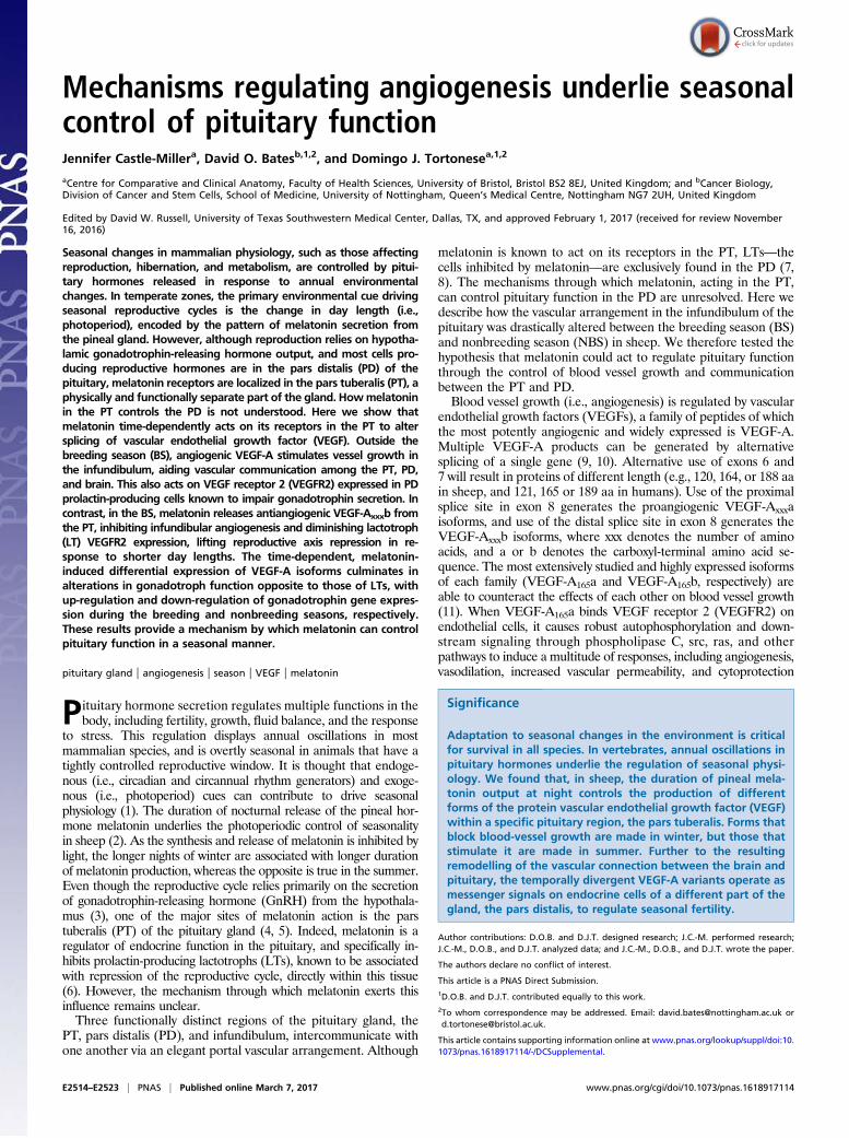
Mechanisms regulating angiogenesis underlie seasonalcontrol of pituitary functionJennifer Castle-Millera, David O. Batesb,1,2, and Domingo J. Tortonesea,1,2
aCentre for Comparative and Clinical Anatomy, Faculty of Health Sciences, University of Bristol, Bristol BS2 8EJ, United Kingdom; and bCancer Biology,Division of Cancer and Stem Cells, School of Medicine, University of Nottingham, Queen’s Medical Centre, Nottingham NG7 2UH, United Kingdom
Edited by David W. Russell, University of Texas Southwestern Medical Center, Dallas, TX, and approved February 1, 2017 (received for review November16, 2016)
Seasonal changes in mammalian physiology, such as those affectingreproduction, hibernation, and metabolism, are controlled by pitui-tary hormones released in response to annual environmentalchanges. In temperate zones, the primary environmental cue drivingseasonal reproductive cycles is the change in day length (i.e.,photoperiod), encoded by the pattern of melatonin secretion fromthe pineal gland. However, although reproduction relies on hypotha-lamic gonadotrophin-releasing hormone output, and most cells pro-ducing reproductive hormones are in the pars distalis (PD) of thepituitary, melatonin receptors are localized in the pars tuberalis (PT), aphysically and functionally separate part of the gland. Howmelatoninin the PT controls the PD is not understood. Here we show thatmelatonin time-dependently acts on its receptors in the PT to altersplicing of vascular endothelial growth factor (VEGF). Outside thebreeding season (BS), angiogenic VEGF-A stimulates vessel growth inthe infundibulum, aiding vascular communication among the PT, PD,and brain. This also acts on VEGF receptor 2 (VEGFR2) expressed in PDprolactin-producing cells known to impair gonadotrophin secretion. Incontrast, in the BS, melatonin releases antiangiogenic VEGF-Axxxb fromthe PT, inhibiting infundibular angiogenesis and diminishing lactotroph(LT) VEGFR2 expression, lifting reproductive axis repression in re-sponse to shorter day lengths. The time-dependent, melatonin-induced differential expression of VEGF-A isoforms culminates inalterations in gonadotroph function opposite to those of LTs, withup-regulation and down-regulation of gonadotrophin gene expres-sion during the breeding and nonbreeding seasons, respectively.These results provide a mechanism by which melatonin can controlpituitary function in a seasonal manner.
pituitary gland | angiogenesis | season | VEGF | melatonin
Pituitary hormone secretion regulates multiple functions in thebody, including fertility, growth, fluid balance, and the response
to stress. This regulation displays annual oscillations in mostmammalian species, and is overtly seasonal in animals that have atightly controlled reproductive window. It is thought that endoge-nous (i.e., circadian and circannual rhythm generators) and exoge-nous (i.e., photoperiod) cues can contribute to drive seasonalphysiology (1). The duration of nocturnal release of the pineal hor-mone melatonin underlies the photoperiodic control of seasonalityin sheep (2). As the synthesis and release of melatonin is inhibited bylight, the longer nights of winter are associated with longer durationof melatonin production, whereas the opposite is true in the summer.Even though the reproductive cycle relies primarily on the secretionof gonadotrophin-releasing hormone (GnRH) from the hypothala-mus (3), one of the major sites of melatonin action is the parstuberalis (PT) of the pituitary gland (4, 5). Indeed, melatonin is aregulator of endocrine function in the pituitary, and specifically in-hibits prolactin-producing lactotrophs (LTs), known to be associatedwith repression of the reproductive cycle, directly within this tissue(6). However, the mechanism through which melatonin exerts thisinfluence remains unclear.Three functionally distinct regions of the pituitary gland, the
PT, pars distalis (PD), and infundibulum, intercommunicate withone another via an elegant portal vascular arrangement. Although
melatonin is known to act on its receptors in the PT, LTs—thecells inhibited by melatonin—are exclusively found in the PD (7,8). The mechanisms through which melatonin, acting in the PT,can control pituitary function in the PD are unresolved. Here wedescribe how the vascular arrangement in the infundibulum of thepituitary was drastically altered between the breeding season (BS)and nonbreeding season (NBS) in sheep. We therefore tested thehypothesis that melatonin could act to regulate pituitary functionthrough the control of blood vessel growth and communicationbetween the PT and PD.Blood vessel growth (i.e., angiogenesis) is regulated by vascular
endothelial growth factors (VEGFs), a family of peptides of whichthe most potently angiogenic and widely expressed is VEGF-A.Multiple VEGF-A products can be generated by alternativesplicing of a single gene (9, 10). Alternative use of exons 6 and7 will result in proteins of different length (e.g., 120, 164, or 188 aain sheep, and 121, 165 or 189 aa in humans). Use of the proximalsplice site in exon 8 generates the proangiogenic VEGF-Axxxaisoforms, and use of the distal splice site in exon 8 generates theVEGF-Axxxb isoforms, where xxx denotes the number of aminoacids, and a or b denotes the carboxyl-terminal amino acid se-quence. The most extensively studied and highly expressed isoformsof each family (VEGF-A165a and VEGF-A165b, respectively) areable to counteract the effects of each other on blood vessel growth(11). When VEGF-A165a binds VEGF receptor 2 (VEGFR2) onendothelial cells, it causes robust autophosphorylation and down-stream signaling through phospholipase C, src, ras, and otherpathways to induce a multitude of responses, including angiogenesis,vasodilation, increased vascular permeability, and cytoprotection
Significance
Adaptation to seasonal changes in the environment is criticalfor survival in all species. In vertebrates, annual oscillations inpituitary hormones underlie the regulation of seasonal physi-ology. We found that, in sheep, the duration of pineal mela-tonin output at night controls the production of differentforms of the protein vascular endothelial growth factor (VEGF)within a specific pituitary region, the pars tuberalis. Forms thatblock blood-vessel growth are made in winter, but those thatstimulate it are made in summer. Further to the resultingremodelling of the vascular connection between the brain andpituitary, the temporally divergent VEGF-A variants operate asmessenger signals on endocrine cells of a different part of thegland, the pars distalis, to regulate seasonal fertility.
Author contributions: D.O.B. and D.J.T. designed research; J.C.-M. performed research;J.C.-M., D.O.B., and D.J.T. analyzed data; and J.C.-M., D.O.B., and D.J.T. wrote the paper.
The authors declare no conflict of interest.
This article is a PNAS Direct Submission.1D.O.B. and D.J.T. contributed equally to this work.2To whom correspondence may be addressed. Email: [email protected] [email protected].
This article contains supporting information online at www.pnas.org/lookup/suppl/doi:10.1073/pnas.1618917114/-/DCSupplemental.
E2514–E2523 | PNAS | Published online March 7, 2017 www.pnas.org/cgi/doi/10.1073/pnas.1618917114
(12). In contrast, although the binding affinity for VEGFR2 is thesame as that of VEGF-A165a, VEGF-A165b induces weak phos-phorylation (13), does not bind the coreceptor Neuropilin-1 (14),which is responsible for intracellular trafficking and recycling to themembrane (15), and does not activate the full signaling pathway.This means it does not induce angiogenesis or vasodilation (11) or asustained increase in vascular permeability (16), and results inVEGFR2 degradation, not recycling (15). It does, however, stimu-late cytoprotection of endothelial and epithelial cells (17) andneurons (18). These two isoform families therefore have very dif-ferent physiological consequences (19), but any differential role inseasonal pituitary angiogenesis is unknown.We found that, whereas total VEGF-A was not altered between
the BS and NBS, there was a dramatic switch in splicing in the BSfrom angiogenic VEGF-Axxxa isoforms to antiangiogenic VEGF-Axxxb isoforms in both the PT and PD. This was mirrored by areduction in the number of blood vessels in the infundibulum.
Melatonin receptor expression in the PT and the infundibulumcolocalized with cells expressing VEGF-A. Two potential mech-anisms for VEGF-A–mediated regulation of pituitary seasonalityare proposed here: (i) VEGF-A acting to regulate blood vesselfunction, which subsequently controls delivery of other hormonesto the PD; and (ii) VEGF-A acting directly on LTs to controlprolactin-associated down-regulation of the reproductive axis. Wefound that VEGFR2 colocalization with prolactin in the PD wasincreased outside the BS, consistent with a VEGF-Axxxa–mediatedrepression of fertility. In vitro culture of PT cells from BS sheepshowed that duration of melatonin exposure controlled VEGF-Aisoform secretion: long exposure induced VEGF-Axxxb produc-tion, whereas short exposure induced VEGF-Axxxa production.Culture of PT cells from the NBS revealed that melatonin given atfrequencies seen in the winter could switch the expression ofVEGF-A isoforms to BS levels. We then showed that PT cellstreated with NBS melatonin regimens release VEGF-Axxxa, which
VE
GF-
Axx
xb/to
tal
prot
ein
(pg/
mg)
0
200
400
600
800
1000
BS NBS 0%
20%
40%
60%
80%
BS NBS
BS NBS
0
5
10
15
BS NBS
***
0
0.5
1
1.5
BS NBS
p=0.056
BS NBS
CD31 CD31/PCNA
Pan
VE
GF-
A/to
tal
prot
ein
(pg/
mg)
PT
PD
VEGF-A Melatonin receptor Merge
Vascular Loops
No.
of v
ascu
lar
loop
s pe
r ani
mal
No.
of p
rolif
erat
ing
endo
thel
ial c
ells
pe
r vas
cula
r loo
p
PT/Stalk region PT/Stalk region
A B
C D **
F
PT/Stalk region
0
500
1000
1500
BS NBS
% o
f tot
al V
EG
F th
at is
V
EG
F-A
xxxb
***
E
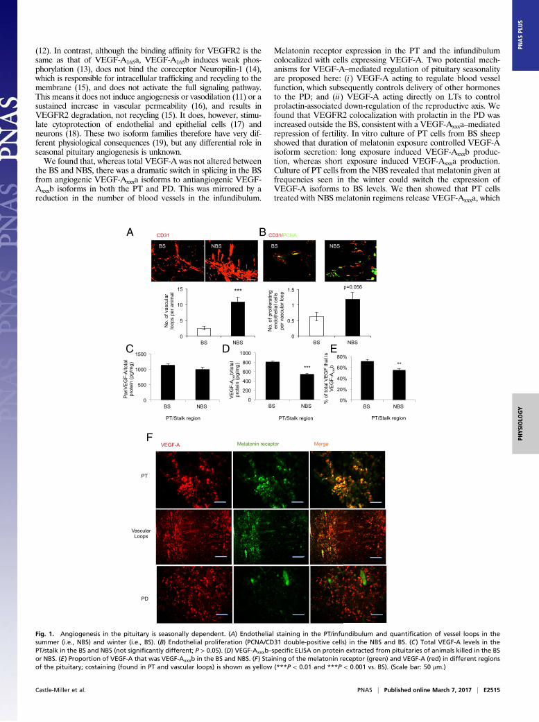
Fig. 1. Angiogenesis in the pituitary is seasonally dependent. (A) Endothelial staining in the PT/infundibulum and quantification of vessel loops in thesummer (i.e., NBS) and winter (i.e., BS). (B) Endothelial proliferation (PCNA/CD31 double-positive cells) in the NBS and BS. (C) Total VEGF-A levels in thePT/stalk in the BS and NBS (not significantly different; P > 0.05). (D) VEGF-Axxxb–specific ELISA on protein extracted from pituitaries of animals killed in the BSor NBS. (E) Proportion of VEGF-A that was VEGF-Axxxb in the BS and NBS. (F) Staining of the melatonin receptor (green) and VEGF-A (red) in different regionsof the pituitary; costaining (found in PT and vascular loops) is shown as yellow (***P < 0.01 and ***P < 0.001 vs. BS). (Scale bar: 50 μm.)
Castle-Miller et al. PNAS | Published online March 7, 2017 | E2515
PHYS
IOLO
GY
PNASPL
US
directly induced prolactin secretion from PD cells. Finally, thetime-dependent, melatonin-induced differential expression ofVEGF-A isoforms resulted in alterations of gonadotroph functionopposite to those of LTs in each season. Together, these resultsdemonstrate that melatonin-mediated control of VEGF splicingcould underlie intrapituitary regulation of seasonal fertility.
ResultsVascular Growth in the Pituitary Gland Is Seasonally Controlled. Toinvestigate the vascular architecture of the pituitary in a sea-sonally breeding mammal, we screened pituitary glands of sheepwith the endothelial marker CD31. Staining showed a significant(P < 0.001) increase in the number of vascular loops extendingfrom the PT into the infundibulum in the summer (i.e., NBS)compared with animals culled in the winter (i.e., BS; Fig. 1A). Todetermine whether this was a result of endothelial proliferationin the NBS, we costained for CD31 and proliferating cell nuclearantigen (PCNA). We detected proliferating endothelial cells inboth seasons, but a twofold increase in proliferating endothelialcells in the NBS (Fig. 1B). As angiogenesis is driven by VEGF,we measured VEGF-A in the pars tuberalis/stalk region of thepituitary. There was no difference in VEGF-A as measured byantibodies that detect all isoforms of VEGF-A (i.e., panVEGF;Fig. 1C). However, by using antibodies that specifically detectthe exon 8b splice variants (VEGF-Axxxb), the expression ofwhich has been shown to be antiangiogenic in vivo, a reductionin VEGF-Axxxb was measured in the NBS (Fig. 1D). Thisresulted in a change in the ratio of VEGF-A from 33% excessantiangiogenic isoforms in the BS to 60% excess angiogenicisoforms in the NBS (Fig. 1E). This indicates that the pituitaryis in an antiangiogenic state in the BS, and suggests a linkbetween day length and VEGF-A splicing.To determine whether VEGF-A was expressed in the pituitary
in cells that can respond to day length, we costained for mela-tonin receptor and VEGF. Fig. 1F shows that MT1 and VEGF-Awere colocalized in the PT and, interestingly, also in the vascularloops (Fig. 1F, arrows) that connect the PT with the infundibu-lum. In contrast, whereas VEGF-A was expressed in the PD,MT1 receptors were not. The antiangiogenic isoforms had notpreviously been cloned from sheep, so we examined RNA ex-pression by RT-PCR. Both isoforms were detected in pituitariesfrom sheep in both seasons (PD and PT; Fig. S1A). Cloning andsequencing of the PCR product confirmed that this was sheepVEGF-Axxxb (Fig. S1B).The sheep sequence has a single nucleotide substitution com-
pared with human DNA (a G in sheep, C in humans). This resultsin a single amino acid difference, with a sequence of SRTRKDinstead of SLTRKD in human. Thus, the sheep VEGF-Axxxbisoforms are 1 aa shorter than the human ones. The cell type inwhich VEGF-Axxxb was expressed in the PT was identified byimmunolocalization. Fig. S1C confirms that VEGF-Axxxb isexpressed in the MT1-positive cells, which, in the PT, are notendothelial or glial-type folliculostellate (S100+) cells. These re-sults suggested that melatonin could regulate expression of dif-ferent VEGF-A isoforms in the PT, regulating angiogenesis in thepituitary in a seasonally dependent manner.
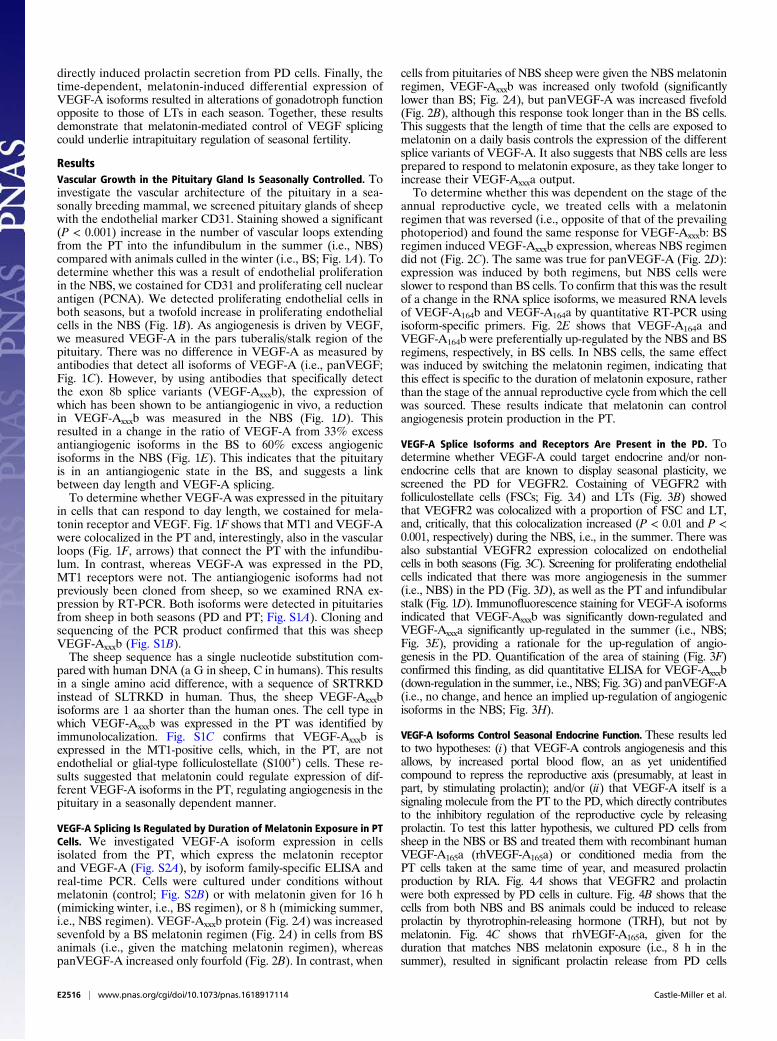
VEGF-A Splicing Is Regulated by Duration of Melatonin Exposure in PTCells. We investigated VEGF-A isoform expression in cellsisolated from the PT, which express the melatonin receptorand VEGF-A (Fig. S2A), by isoform family-specific ELISA andreal-time PCR. Cells were cultured under conditions withoutmelatonin (control; Fig. S2B) or with melatonin given for 16 h(mimicking winter, i.e., BS regimen), or 8 h (mimicking summer,i.e., NBS regimen). VEGF-Axxxb protein (Fig. 2A) was increasedsevenfold by a BS melatonin regimen (Fig. 2A) in cells from BSanimals (i.e., given the matching melatonin regimen), whereaspanVEGF-A increased only fourfold (Fig. 2B). In contrast, when
cells from pituitaries of NBS sheep were given the NBS melatoninregimen, VEGF-Axxxb was increased only twofold (significantlylower than BS; Fig. 2A), but panVEGF-A was increased fivefold(Fig. 2B), although this response took longer than in the BS cells.This suggests that the length of time that the cells are exposed tomelatonin on a daily basis controls the expression of the differentsplice variants of VEGF-A. It also suggests that NBS cells are lessprepared to respond to melatonin exposure, as they take longer toincrease their VEGF-Axxxa output.To determine whether this was dependent on the stage of the
annual reproductive cycle, we treated cells with a melatoninregimen that was reversed (i.e., opposite of that of the prevailingphotoperiod) and found the same response for VEGF-Axxxb: BSregimen induced VEGF-Axxxb expression, whereas NBS regimendid not (Fig. 2C). The same was true for panVEGF-A (Fig. 2D):expression was induced by both regimens, but NBS cells wereslower to respond than BS cells. To confirm that this was the resultof a change in the RNA splice isoforms, we measured RNA levelsof VEGF-A164b and VEGF-A164a by quantitative RT-PCR usingisoform-specific primers. Fig. 2E shows that VEGF-A164a andVEGF-A164b were preferentially up-regulated by the NBS and BSregimens, respectively, in BS cells. In NBS cells, the same effectwas induced by switching the melatonin regimen, indicating thatthis effect is specific to the duration of melatonin exposure, ratherthan the stage of the annual reproductive cycle from which the cellwas sourced. These results indicate that melatonin can controlangiogenesis protein production in the PT.
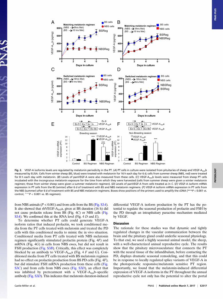
VEGF-A Splice Isoforms and Receptors Are Present in the PD. Todetermine whether VEGF-A could target endocrine and/or non-endocrine cells that are known to display seasonal plasticity, wescreened the PD for VEGFR2. Costaining of VEGFR2 withfolliculostellate cells (FSCs; Fig. 3A) and LTs (Fig. 3B) showedthat VEGFR2 was colocalized with a proportion of FSC and LT,and, critically, that this colocalization increased (P < 0.01 and P <0.001, respectively) during the NBS, i.e., in the summer. There wasalso substantial VEGFR2 expression colocalized on endothelialcells in both seasons (Fig. 3C). Screening for proliferating endothelialcells indicated that there was more angiogenesis in the summer(i.e., NBS) in the PD (Fig. 3D), as well as the PT and infundibularstalk (Fig. 1D). Immunofluorescence staining for VEGF-A isoformsindicated that VEGF-Axxxb was significantly down-regulated andVEGF-Axxxa significantly up-regulated in the summer (i.e., NBS;Fig. 3E), providing a rationale for the up-regulation of angio-genesis in the PD. Quantification of the area of staining (Fig. 3F)confirmed this finding, as did quantitative ELISA for VEGF-Axxxb(down-regulation in the summer, i.e., NBS; Fig. 3G) and panVEGF-A(i.e., no change, and hence an implied up-regulation of angiogenicisoforms in the NBS; Fig. 3H).
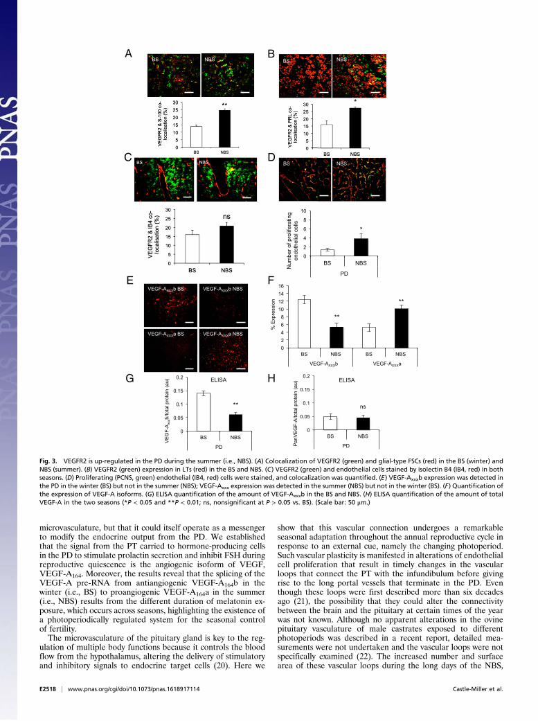
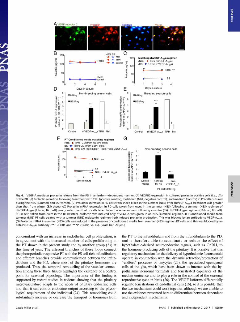
VEGF-A Isoforms Control Seasonal Endocrine Function. These results ledto two hypotheses: (i) that VEGF-A controls angiogenesis and thisallows, by increased portal blood flow, an as yet unidentifiedcompound to repress the reproductive axis (presumably, at least inpart, by stimulating prolactin); and/or (ii) that VEGF-A itself is asignaling molecule from the PT to the PD, which directly contributesto the inhibitory regulation of the reproductive cycle by releasingprolactin. To test this latter hypothesis, we cultured PD cells fromsheep in the NBS or BS and treated them with recombinant humanVEGF-A165a (rhVEGF-A165a) or conditioned media from thePT cells taken at the same time of year, and measured prolactinproduction by RIA. Fig. 4A shows that VEGFR2 and prolactinwere both expressed by PD cells in culture. Fig. 4B shows that thecells from both NBS and BS animals could be induced to releaseprolactin by thyrotrophin-releasing hormone (TRH), but not bymelatonin. Fig. 4C shows that rhVEGF-A165a, given for theduration that matches NBS melatonin exposure (i.e., 8 h in thesummer), resulted in significant prolactin release from PD cells
E2516 | www.pnas.org/cgi/doi/10.1073/pnas.1618917114 Castle-Miller et al.
fromNBS animals (P < 0.001) and from cells from the BS (Fig. S3A).It also showed that rhVEGF-A165a, given at BS duration (16 h) didnot cause prolactin release from BS (Fig. 4C) or NBS cells (Fig.S3A). We confirmed this at the RNA level (Fig. 4 D and E).To determine whether PT cells could generate VEGF-A
isoform ratios that induced prolactin, we took conditioned me-dia from the PT cells treated with melatonin and treated the PDcells with this conditioned media to mimic the in vivo situation.Conditioned media from PT cells treated with NBS melatoninregimen significantly stimulated prolactin protein (Fig. 4F) andmRNA (Fig. 4G) in cells from NBS ewes, but did not result inFSH production (Fig. S3B). Critically, this effect was completelyblocked by an antibody to VEGF-Axxxa (Fig. 4 F and G). Con-ditioned media from PT cells treated with BS melatonin regimenhad no effect on prolactin production from BS PD cells (Fig. 4F),but did stimulate FSH mRNA production from these cells (Fig.S3C) and from cells from NBS ewes (Fig. S3D), an effect thatwas inhibited by pretreatment with a VEGF-Axxxb–specificantibody (Fig. S3D). This indicates that melatonin duration-induced
differential VEGF-A isoform production by the PT has the po-tential to regulate the seasonal production of prolactin and FSH bythe PD through an intrapituitary paracrine mechanism mediatedby VEGF.
DiscussionThe rationale for these studies was that dynamic and tightlyregulated changes in the vascular communication between thebrain and the pituitary gland could underlie seasonal physiology.To that end, we used a highly seasonal animal model, the sheep,with a well-characterized annual reproductive cycle. The resultsshow that the pituitary microvasculature that connects the PTwith the neural tissue of the infundibulum, before contacting thePD, displays dramatic seasonal remodeling, and that this couldbe in response to locally regulated splice variants of VEGF-A inthe photoperiodic responsive/melatonin sensitive PT region.Importantly, we found that the melatonin-induced differentialexpression of VEGF-A isoforms in the PT throughout the annualreproductive cycle not only has the potential to alter the portal
A
C
E F
D
B
Fig. 2. VEGF-A isoforms levels are regulated by melatonin periodicity in the PT. (A) PT cells in culture were isolated from pituitaries of sheep and VEGF-Axxxbmeasured by ELISA. Cells from winter sheep (BS, blue) were treated with melatonin for 16 h each day for 6 d; cells from summer sheep (NBS, red) were treatedfor 8 h each day with melatonin. (B) Levels of panVEGF-A were also measured from these cells. (C) VEGF-Axxxb levels were measured from sheep PT cellsincubated with the incongruous melatonin exposure for the time from which they were harvested (cells from summer sheep were given a winter melatoninregimen; those from winter sheep were given a summer melatonin regimen). (D) Levels of panVEGF-A from cells treated as in C. (E) VEGF-A isoform mRNAexpression in PT cells from the BS (winter) after 6 d of treatment with BS and NBS melatonin regimens. (F) VEGF-A isoform mRNA expression in PT cells fromthe NBS (summer) after 6 d of treatment with BS and NBS melatonin regimens. Boxes show positions of the primers used to amplify the cDNA (***P < 0.001 vs.control, +++P < 0.001 vs. BS regimen).
Castle-Miller et al. PNAS | Published online March 7, 2017 | E2517
PHYS
IOLO
GY
PNASPL
US
microvasculature, but that it could itself operate as a messengerto modify the endocrine output from the PD. We establishedthat the signal from the PT carried to hormone-producing cellsin the PD to stimulate prolactin secretion and inhibit FSH duringreproductive quiescence is the angiogenic isoform of VEGF,VEGF-A164. Moreover, the results reveal that the splicing of theVEGF-A pre-RNA from antiangiogenic VEGF-A164b in thewinter (i.e., BS) to proangiogenic VEGF-A164a in the summer(i.e., NBS) results from the different duration of melatonin ex-posure, which occurs across seasons, highlighting the existence ofa photoperiodically regulated system for the seasonal controlof fertility.The microvasculature of the pituitary gland is key to the reg-
ulation of multiple body functions because it controls the bloodflow from the hypothalamus, altering the delivery of stimulatoryand inhibitory signals to endocrine target cells (20). Here we
show that this vascular connection undergoes a remarkableseasonal adaptation throughout the annual reproductive cycle inresponse to an external cue, namely the changing photoperiod.Such vascular plasticity is manifested in alterations of endothelialcell proliferation that result in timely changes in the vascularloops that connect the PT with the infundibulum before givingrise to the long portal vessels that terminate in the PD. Eventhough these loops were first described more than six decadesago (21), the possibility that they could alter the connectivitybetween the brain and the pituitary at certain times of the yearwas not known. Although no apparent alterations in the ovinepituitary vasculature of male castrates exposed to differentphotoperiods was described in a recent report, detailed mea-surements were not undertaken and the vascular loops were notspecifically examined (22). The increased number and surfacearea of these vascular loops during the long days of the NBS,
0
0.05
0.1
0.15
0.2
BS NBS
PD Pan
VE
GF-
A/to
tal p
rote
in (a
u)
0
0.05
0.1
0.15
0.2
BS NBS
PD
VE
GF-
Axx
xb/to
tal p
rote
in (a
u)
NBS BS
0
2
4
6
8
10
BS NBS
PD
Num
ber o
f pro
lifer
atin
g en
doth
elia
l cel
ls
BS
BS NBS
NBS
*
** ns
VEGF-AXXXb BS VEGF-AXXXb NBS
VEGF-AXXXa BS VEGF-AXXXa NBS
ELISA ELISA
A B
E
C D
F
G H
0 2 4 6 8
10 12 14 16
BS NBS BS NBS
VEGFxxxb VEGFxxx
% E
xpre
ssio
n
**
**
VEGF-AXXXb VEGF-AXXXa
Fig. 3. VEGFR2 is up-regulated in the PD during the summer (i.e., NBS). (A) Colocalization of VEGFR2 (green) and glial-type FSCs (red) in the BS (winter) andNBS (summer). (B) VEGFR2 (green) expression in LTs (red) in the BS and NBS. (C) VEGFR2 (green) and endothelial cells stained by isolectin B4 (IB4, red) in bothseasons. (D) Proliferating (PCNS, green) endothelial (IB4, red) cells were stained, and colocalization was quantified. (E) VEGF-Axxxb expression was detected inthe PD in the winter (BS) but not in the summer (NBS); VEGF-Axxx expression was detected in the summer (NBS) but not in the winter (BS). (F) Quantification ofthe expression of VEGF-A isoforms. (G) ELISA quantification of the amount of VEGF-Axxxb in the BS and NBS. (H) ELISA quantification of the amount of totalVEGF-A in the two seasons (*P < 0.05 and **P < 0.01; ns, nonsignificant at P > 0.05 vs. BS). (Scale bar: 50 μm.)
E2518 | www.pnas.org/cgi/doi/10.1073/pnas.1618917114 Castle-Miller et al.
concomitant with an increase in endothelial cell proliferation, isin agreement with the increased number of cells proliferating inthe PT shown in the present study and by another group (23) atthis time of year. The afferent branches of these loops connectthe photoperiodic-responsive PT with the FS cell-rich infundibulum,and efferent branches provide communication between the infun-dibulum and the PD, where most of the pituitary hormones areproduced. Thus, the temporal remodeling of the vascular connec-tion among these three tissues highlights the existence of a controlpoint for seasonal physiology. The importance of this finding issupported by recent studies in rodents showing that the pituitarymicrovasculature adapts to the needs of pituitary endocrine cellsand that it can control endocrine output according to the physio-logical requirement of the individual (24). This remodeling couldsubstantially increase or decrease the transport of hormones from
the PT to the infundibulum and from the infundibulum to the PD,and is therefore able to accentuate or reduce the effect ofhypothalamic-derived neuroendocrine signals, such as GnRH, tothe hormone-producing cells of the pituitary. It is possible that thisregulatory mechanism for the delivery of hypothalamic factors couldoperate in conjunction with the dynamic retraction/protraction of“endfeet” processes of tanycytes (25), the specialized ependymalcells of the glia, which have been shown to interact with the hy-pothalamic neuronal terminals and fenestrated capillaries of themedian eminence and to play a role in the control of the seasonalreproductive cycle in birds (26). The VEGF isoforms differentiallyregulate fenestrations of endothelial cells (16), so it is possible thatthe two mechanisms could work together, although we are unable touse the evidence presented here to differentiate between dependentand independent mechanisms.
0 1 2 3 40
50
100
150
Days in culture
Prol
actin
(ng/
ml) WCCM
SCCM + anti. 165
SCCM
0 1 2 3 40
50
100
150
Days in culture
Prol
actin
(ng/
ml)
Prolactin VEGF receptor 2 Merge Nucleus
Days in culture
NBS
BS
NBS
BS
PT Conditioned media matching regimen NBS 8hrs CM (from NBSPT cells) BS 16hrs CM (from BSPT cells) NBS 8hrs CM (from NBSPT cells)+anti-VEGF-Axxxa
0 2 4 6 80
50
100
150
Days in culture
Prol
actin
(ng/
ml)
MelTRHcontrol
+TRH
+Mel control
NBS BS
0
1
2
3
4
5
6 Breeding season cells BSReg
NBSReg
0
1
2
3
4
5
6
7
8 Non-breeding season cells BSReg
NBSReg
Control rhVEGF-A165a TRH Mel Control rhVEGF-A165a TRH Mel
Pro
lact
in m
RN
A ex
pres
sion
re
lativ
e to
GA
PD
H (2
^-C
t )
Pro
lact
in m
RN
A ex
pres
sion
re
lativ
e to
GA
PD
H (2
^-C
t )
0
1
2
3
4
5
6 Non-breeding season cells
PT CM NBSReg
Control media
+control for Ab
+anti-VEGF-Axxxa
Pro
lact
in m
RN
A ex
pres
sion
re
lativ
e to
GA
PD
H (2
^-C
t )
A
B C
D E
F G
NBS+ VEGF-Axxxa
*** ***
**
***
***
Matching rhVEGF-A165a regimen (NBS 8hrs rhVEGF-A165a) (BS 16 hrs rhVEGF-A165a)
Fig. 4. VEGF-A mediates prolactin release from the PD in an isoform-dependent manner. (A) VEGFR2 expression in cultured prolactin positive cells (i.e., LTs)of the PD. (B) Prolactin secretion following treatment with TRH (positive control), melatonin (Mel, negative control), and medium (control) in PD cells culturedduring the NBS (summer) and BS (winter). (C) Prolactin secretion in PD cells from sheep killed in the summer (NBS) after rhVEGF-A165a treatment was greaterthan that from winter (BS) sheep. (D) Prolactin mRNA expression in PD cells taken from ewes in the summer (NBS) following a summer (NBS) regimen ofrhVEGF-A165a (8 h on, 16 h off) was greater than that of cells taken from the same animals following a winter (BS) rhVEGF-A165a regimen (16 h on, 8 h off).(E) In cells taken from ewes in the BS (winter), prolactin was induced only if VEGF-A was given in an NBS (summer) regimen. (F) Conditioned media fromsummer (NBS) PT cells treated with a summer (NBS) melatonin regimen (red) induced prolactin production. This was blocked by an antibody to VEGF-Axxxa.(G) Prolactin mRNA in summer (NBS) cells was induced in the presence of conditioned media from summer (NBS) regimen PT cells, and this was blocked by ananti-VEGF-Axxxa antibody (**P < 0.01 and ***P < 0.001 vs. BS). (Scale bar: 20 μm.)
Castle-Miller et al. PNAS | Published online March 7, 2017 | E2519
PHYS
IOLO
GY
PNASPL
US
In other tissues, vascular remodeling and permeability is con-trolled by VEGF-A (19), so dynamic changes in the pituitarymicrovasculature were expected to correspond to alterations inVEGF-A expression. Indeed, VEGF-A–mediated changes in en-dothelial cell proliferation and angiogenesis were communicated inspecific regions of the songbird brain across seasons (27). However,VEGF-A had been previously reported to remain unchanged in thepituitary of sheep under different photoperiods (28), and, in thepresent study, panVEGF-A expression did not differ between BSand NBS animals. Critically, the use of specific antibodies to the pro-and antiangiogenic isoforms of VEGF-A (these distinguish betweenisoform families, but not between the different length isoforms,hence VEGF-Axxxa and VEGF-Axxxb) revealed differential isoformexpression between the long days of the NBS and the short days ofthe BS, with overexpression of antiangiogenic VEGF-Axxxb variantsin the BS and increased expression of proangiogenic VEGF-Axxxavariants in the NBS, providing an explanation for the observedchanges in the microvasculature. As the PT is the tissue with thehighest density of melatonin receptors (4, 5), and reliably translatesthe effects of photoperiod on circadian and circannual physiologywithin the pituitary (1, 29), our results showing the coexpression ofVEGF-A and melatonin receptors in PT-specific cells providecompelling evidence that the seasonal regulation of the vascularconnection between the brain and the pituitary gland is mediated bya melatonin-induced mechanism within the PT region that leads todifferential expression of pro- and antiangiogenic VEGF-A isoforms.Moreover, our results show that, in addition to the PT-specific
cells, melatonin could also act directly on the vascular loops,revealing a target for melatonin action to translate photoperiodiceffects on seasonal physiology. As the blood flows from the brainto the pituitary (11), alterations in the vascular loops of the in-fundibulum that will give rise to the long portal vessels (12) couldcontribute not only to regulate the transfer of PT products to thePD, but also to alter the delivery of hypothalamic factors; thus,the increased vascular connections during the long days ofsummer would be expected to favor increased supply of stimu-latory and inhibitory hypothalamic signals to the PD at this timeof year. Notwithstanding that, the reduction in vascularity duringthe short days of winter is likely to play a role in the modulationof the gonadotroph response to GnRH by means of preventingdesensitization of GnRH receptors (30) and fine-tuning thedifferential control of gonadotropin secretion (31, 32), which areessential processes to ensure normal fertility.Photoperiodic information is encoded by the duration of noc-
turnal melatonin secretion (2), so we used a paradigm wherebyovine PT cells were cultured and exposed daily to summer (i.e.,NBS) or winter (i.e., BS) durations of melatonin treatments (8 h vs.16 h, respectively) over a period of 6 d. PT cells from the sameanimals were exposed to the matching and nonmatching (i.e., op-posite season) melatonin regimens, so we were able to differentiatedirect effects of the melatonin signal and those resulting from itsinteraction with the circannual phase. We show that duration ofmelatonin exposure induced a striking differential expression ofVEGF-A isoforms, with up-regulation of the proangiogenic isoformVEGF-Axxxa by a short-duration regimen (i.e., 8 h, summer; NBS)and up-regulation of the antiangiogenic isoform VEGF-Axxxb by along-duration regimen (i.e., 16 h, winter; BS). This melatoninduration-dependent differential expression of VEGF-A isoformswas also recorded in cells obtained in the opposite season but at aslower rate, highlighting the requirement of PT cells to be entrainedto the new signal. Thus, the results are consistent with the findingsex vivo and demonstrate that pituitary microvascular remodeling islikely to be sensitive to the changing photoperiod and adapts to thephysiological requirements of the animal in response to time-dependent melatonin signals acting on VEGF-A. The mechanismthrough which melatonin switches splicing of the VEGF-A gene isnot yet known, but alternative splicing of VEGF has been shown to
be regulated by activation of the RNA binding proteins SRSF1,SRSF2, and SRSF6 by the kinases SRPK1 and Clk4 (33).We then investigated whether the seasonal regulation of
VEGF-A in the PT could affect the function of the PD. We showthat VEGF-A receptors are expressed in endocrine, endothelial,and FS cells in the PD, and that their colocalization is also underseasonal control, with up-regulation during the long days of theNBS. In addition, there was increased content of proangiogenicVEGF-A isoforms at this time of year and, conversely, increasedcontent of the antiangiogenic isoforms during the short days of theBS. As the seasonal regulation of VEGF-A isoform expressionwas shown to be melatonin-dependent and, in accordance withprevious studies (34), the PD was shown not to contain melatoninreceptors, the varying content of VEGF-A isoforms in the PD islikely to rely on a paracrine mechanism (35). The physiologicalsignificance of this was first revealed in the PD microvasculature,with increased endothelial cell proliferation demonstrated duringthe NBS. We show that this increase in angiogenesis at this timeof the annual reproductive cycle is concomitant with an increase inthe prevalence of FS cells containing VEGF receptors. FS cells areglial-like, nonendocrine cells that, via gap junctions, generate a 3Dnetwork throughout the pituitary to coordinate its function (36,37). These cells secrete an array of paracrine factors known toinfluence endocrine cells such as gonadotrophs and LTs, and are aprimary source of VEGF-A (35). In seasonal breeders, FS cells aredistributed throughout the PD and PT (38) and respond to pho-toperiodic changes with a high degree of plasticity (39, 40). Insheep, significant ultrastructural changes, together with enhancednumber of intercellular adherens junctions and increased numberof elongated processes surrounding endocrine cell clusters, werereported during the long days of the NBS (41). As FS cells do notcontain melatonin receptors (42), our findings revealing up-regulation of VEGF receptor content in these cells at this timeof year provide evidence for a role of VEGF-A in the dynamicchanges of the FS cell network to control vascular plasticity via theregulation of its own production during the annual reproductivecycle. The seasonally regulated differential expression of VEGF-Aisoforms in the pituitary gland of a short-day breeder unraveledhere could also operate in long-day breeders, such as hamsters andhorses, as part of the mechanisms controlling their annual physi-ology. Indeed, preliminary results have provided evidence that, inthoroughbred horses, VEGF-A isoform expression in the PT andPD regions of the pituitary is also seasonally regulated (43),suggesting that this is a conserved mechanism for seasonal ad-aptation in photoperiodic mammals.Notably, we show that, in addition to its actions on the pituitary
vasculature and FS cell population, VEGF-A has a potent prolactinreleasing effect, and that this stimulation depends on time of ex-posure of the ligand and density of VEGF receptors in LTs, whichis increased during the long days of the NBS. Moreover, thesestimulatory effects of VEGF-A on prolactin synthesis and releasewere accompanied by suppression of the gonadotrophic axis, asrevealed by inhibition of FSH gene expression. Melatonin wasshown to mediate the photoperiodic regulation of prolactin secre-tion through a direct action within the pituitary gland (6). BecauseMT1 melatonin receptors are selectively expressed in the PT, andthis region is deprived of LT cells (7, 8), a paracrine mechanism forthe control of prolactin secretion from the PD is warranted. Acti-vation of MT1 melatonin receptors in the PT is known to inhibitadenylyl cyclase, and pharmacological studies in sheep have shownthat melatonin impairs forskolin-induced hypersecretion of cAMP,with inhibition of prolactin from the PD through the reduction of aparacrine signal (44). However, although several compounds suchas tachykinins, substance P, and neurokinin A are produced by thePT and can stimulate prolactin release (45–47), characterization ofthe chemical identity of that signal has been elusive. Here we showthat the stimulatory effects of VEGF-A on prolactin were mimickedby conditioned media from PT cultures exposed to an NBS regimen
E2520 | www.pnas.org/cgi/doi/10.1073/pnas.1618917114 Castle-Miller et al.
of melatonin, and that these actions of PT media were blocked by aspecific VEGF-Axxxa antibody, demonstrating that VEGF-A is apotential paracrine signal, and that melatonin-induced differentialVEGF-A isoform production by the PT can regulate the seasonalproduction of prolactin and FSH.Because these effects were also recorded in PD cells obtained
in the opposite season (i.e., BS), albeit with a 3-d lag required foradaptation, our results show that the photoperiodically induced
paracrine mechanism mediated by VEGF-A can ultimately over-ride the circannual phase of the PD target cells, and entrain it tothe new photoperiod. The increased VEGF receptor content inthe PD during the NBS plays a major role in mediating this pro-cess, and thus in the biological adaptation to a summer physiology,because VEGF-A treatments mimicking an NBS melatonin regi-men showed a delayed response in BS cultures in which the VEGFreceptor content was reduced. Entrainment of the PD cells to a
A
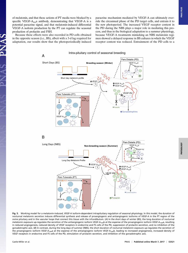
B
Fig. 5. Working model for a melatonin-induced, VEGF-A isoform-dependent intrapituitary regulation of seasonal physiology. In this model, the duration ofnocturnal melatonin secretion induces differential synthesis and release of proangiogenic and antiangiogenic isoforms of VEGF-A in the PT region of theovine pituitary and in the vascular loops that connect this tissue with the infundibulum. (A) In the short days of winter (BS), the long duration of nocturnalmelatonin exposure up-regulates the secretion of the antiangiogenic isoform VEGF-A164b at the expense of the proangiogenic isoform VEGF-A164a, resultingin reduced angiogenesis, reduced density of VEGF receptors in endocrine and FS cells of the PD, suppression of prolactin secretion, and no inhibition of thegonadotrophic axis. (B) In contrast, during the long days of summer (NBS), the short duration of nocturnal melatonin exposure up-regulates the secretion ofthe proangiogenic isoform VEGF-A164a at the expense of the antiangiogenic isoform VEGF-A164b, leading to increased angiogenesis, increased density ofVEGF receptors in endocrine and FS cells of the PD, stimulation of prolactin secretion, and inhibition of the gonadotrophic axis.
Castle-Miller et al. PNAS | Published online March 7, 2017 | E2521
PHYS
IOLO
GY
PNASPL
US
specific phase of the circannual cycle explains why NBS cells failedto secrete prolactin in response to the first 8 h of a BS (16 h)VEGF-A regimen or PT-conditioned media from the BS.In rodents, melatonin-induced suppression of cAMP is followed
by sensitization of adenosine A2b receptor signaling, leading tosubsequent increase in cAMP and cAMP response elementbinding protein (CREB) phosphorylation (28). Disruption of thissignaling pathway in MT1melatonin receptor-KOmice resulted inaltered prolactin secretion, implicating cAMP and adenosine inthis biological response to melatonin. Our results indicate thatVEGF-A is likely to be downstream of that pathway to bring aboutthe biological response. Indeed, cAMP signaling, CREB phos-phorylation, and adenosine are associated with angiogenesis (48,49) via stimulation of VEGF-A (50), and, whereas pharmacolog-ically induced cAMP up-regulation and treatment with adenosinestimulated VEGF-A expression in smooth muscle cells (50), theselective knockdown of all VEGF-A isoforms blocked the actionsof elevated cAMP on hippocampal neurons (51). The melatonin-induced VEGF-A regulation of prolactin secretion shown in thisstudy will have an impact on the gonadotrophic axis in addition toits direct inhibition of FSH, because, when combined with dopa-mine, prolactin impairs the gonadotroph response to GnRH ina seasonally dependent manner in long- and short-day breeders(52–54).Our results provide evidence for an intrapituitary mechanism
that responds to an external independent signal to regulate sea-sonal physiology. We propose a model whereby the duration ofnocturnal melatonin secretion promotes alternative splicing of theVEGF-A gene, leading to differential synthesis and release ofproangiogenic and antiangiogenic isoforms of VEGF-A within thePT region of the pituitary gland and in the vascular loops thatconnect this tissue with the infundibulum (Fig. 5). The resultingoutput of VEGF-A isoforms will have two complementary effects:(i) it alters the temporal vascular connection between the brain andthe pituitary gland and (ii) it can be used as a paracrine signal tomodify the seasonal activity of endocrine cells in the PD thatcontrol reproduction. In this model, the long duration of nocturnalmelatonin exposure during the winter up-regulates the secretion ofantiangiogenic isoforms VEGF-Axxxb at the expense of proangio-genic isoforms VEGF-Axxxa, resulting in reduced angiogenesis,reduced density of VEGF receptors in endocrine and FS cells,suppression of prolactin secretion, and no inhibition of the go-nadotrophic axis characteristic of the BS. Conversely, the shortduration of nocturnal melatonin exposure during the summer willup-regulate the secretion of proangiogenic isoforms VEGF-Axxxaat the expense of antiangiogenic isoforms VEGF-Axxxb, leading toincreased angiogenesis, increased density of VEGF receptors inendocrine and FS cells, stimulation of prolactin secretion, and in-hibition of the gonadotrophic axis, characteristic of the NBS. Thus,the model permits a physiological adaptation to the seasonal re-quirements of the species by means of an angiogenesis-dependentintercommunication between two regions of the pituitary.
Materials and MethodsDetails of standard protocols are given in SI Materials and Methods. Ovinepituitary glands were obtained from ovary-intact females during the BS (i.e.,December/January) and the NBS (i.e., June/July). Animals were killed forcommercial reasons at an abattoir, and pituitaries were removed immedi-ately after death. During the BS, ewes were confirmed to be sexually activeon the basis of a recently formed corpus luteum (CL) together with thepresence of a large follicle (>2 cm). By contrast, in the NBS, ewes wereconsidered to be anestrus when no CL but a corpus albicans was observed inthe gonad, and follicles present were < 2 mm in diameter.
Expression Studies. Pituitaries were stained and RNA was extracted (55) byusing standard procedures (antibodies are detailed in Tables S1 and S2 andprimers are shown in Table S3). The term “VEGF-Axxxb” is used because theantibodies do not distinguish between the different VEGF-Axxxb isoforms(e.g., VEGF-A121b, VEGF-A165b, VEGF-A189b). The term “VEGF-A164b” or “VEGF-A165b” is used when the methodology specifically describes the sheep 164-aaisoform (isoform-specific RT-PCR, as the forward primers cross exon 5 and exon 7,or the human 165-aa isoform when recombinant protein is used).
Primary Cell Cultures. Ovine primary pituitary cultures were produced bycareful dissection and dissociation of the PD and PT of three or four pituitariesas previously described (52). Previous studies have demonstrated the validityof this method for producing a reliable hormone output in response toexogenous hormone releasing secretagogues in vitro (52, 56).
Both ELISAmethods have beenpreviously described (11, 57, 58). A rhVEGF165b-positive control was included in triplicate for the human VEGF-A ELISA, allowingcalculation of VEGF-Atotal concentration to compensate for reduced VEGF-Axxxbaffinity of ∼42% as previously published (59). Prolactin was measured by RIAusing purified ovine prolactin for standards. A linear relationship was detectedwhen the measured hormone concentration (in nanograms per milliliter) wasplotted against the concentration of diluted serum samples.
Statistical Analysis. In the BS and NBS cultures, a total of five separate experi-mental treatments were applied to PT cells, and nine experimental treatmentswere applied to the PD cells. For each treatment, six wells were assigned, and theexperiments were repeated independently three times in both seasons with re-producible results. The reported values represent the mean ± SEM. The effects ofseason and experimental treatment and their interaction on the secretion ofVEGF-A and prolactin from ovine primary pituitary cell cultures were examined byusing ANOVA followed by Fisher’s post hoc test. Because a season by treatmentinteraction was observed for each compound, separate ANOVAs were then usedto examine the effects of experimental treatment within season. For all othervariables, one-way ANOVA was applied. All data were confirmed to be normallydistributed by D’Agostino and Pearson omnibus normality test. Data wereconsidered to be statistically significant at P < 0.05; however, whereverdetected, smaller log value (P < 0.01, P < 0.001) probabilities are reported.
ACKNOWLEDGMENTS. The authors thank the National Hormone and PeptideProgram and Dr. A. F. Parlow (University of California, Los Angeles) for prolactinradioimmunoassay standards, and Prof. A. S. McNeilly (Medical ResearchCouncil Human Reproductive Sciences Unit, Edinburgh, United Kingdom) forovine prolactin antibody. This work was supported by the Biotechnology andBiological Sciences Research Council (D.J.T., J.C.-M.), the British Society for Neu-roendocrinology (D.J.T.), and the British Heart Foundation (D.O.B.).
1. Lincoln GA, Clarke IJ, Hut RA, Hazlerigg DG (2006) Characterizing a mammalian cir-cannual pacemaker. Science 314(5807):1941–1944.
2. Bittman EL, Karsch FJ (1984) Nightly duration of pineal melatonin secretion deter-mines the reproductive response to inhibitory day length in the ewe. Biol Reprod30(3):585–593.
3. Clarke IJ, Campbell R, Smith JT, Prevot V, Wray S (2012) Neuroendocrine control ofreproduction. Handbook of Neuroendocrinology, eds Fink G, Pfaff DW, Levine JE(Elsevier, Amsterdam), pp 197–236.
4. de Reviers MM, Ravault JP, Tillet Y, Pelletier J (1989) Melatonin binding sites in thesheep pars tuberalis. Neurosci Lett 100(1-3):89–93.
5. Morgan PJ, Williams LM, Davidson G, Lawson W, Howell E (1989) Melatonin receptorson ovine pars tuberalis: Characterization and autoradiographicai localization.J Neuroendocrinol 1(1):1–4.
6. Lincoln GA, Clarke IJ (1994) Photoperiodically-induced cycles in the secretion of pro-lactin in hypothalamo-pituitary disconnected rams: Evidence for translation of themelatonin signal in the pituitary gland. J Neuroendocrinol 6(3):251–260.
7. Gross DS (1984) The mammalian hypophysial pars tuberalis: A comparative immu-nocytochemical study. Gen Comp Endocrinol 56(2):283–298.
8. Tortonese DJ, Brooks J, Ingleton PM, McNeilly AS (1998) Detection of prolactin re-
ceptor gene expression in the sheep pituitary gland and visualization of the specific
translation of the signal in gonadotrophs. Endocrinology 139(12):5215–5223.9. Bates DO, et al. (2002) VEGF165b, an inhibitory splice variant of vascular endothelial
growth factor, is down-regulated in renal cell carcinoma. Cancer Res 62(14):4123–4131.10. Houck KA, et al. (1991) The vascular endothelial growth factor family: Identification
of a fourth molecular species and characterization of alternative splicing of RNA. Mol
Endocrinol 5(12):1806–1814.11. Woolard J, et al. (2004) VEGF165b, an inhibitory vascular endothelial growth factor
splice variant: Mechanism of action, in vivo effect on angiogenesis and endogenous
protein expression. Cancer Res 64(21):7822–7835.12. Koch S, Tugues S, Li X, Gualandi L, Claesson-Welsh L (2011) Signal transduction by
vascular endothelial growth factor receptors. Biochem J 437(2):169–183.13. Kawamura H, Li X, Harper SJ, Bates DO, Claesson-Welsh L (2008) Vascular endothelial
growth factor (VEGF)-A165b is a weak in vitro agonist for VEGF receptor-2 due to lack
of coreceptor binding and deficient regulation of kinase activity. Cancer Res 68(12):
4683–4692.
E2522 | www.pnas.org/cgi/doi/10.1073/pnas.1618917114 Castle-Miller et al.
14. Cébe Suarez S, et al. (2006) A VEGF-A splice variant defective for heparan sulfate andneuropilin-1 binding shows attenuated signaling through VEGFR-2. Cell Mol Life Sci63(17):2067–2077.
15. Ballmer-Hofer K, Andersson AE, Ratcliffe LE, Berger P (2011) Neuropilin-1 promotesVEGFR-2 trafficking through Rab11 vesicles thereby specifying signal output. Blood118(3):816–826.
16. Oltean S, et al. (2015) Vascular endothelial growth factor-A165b is protective andrestores endothelial glycocalyx in diabetic nephropathy. J Am Soc Nephrol 26(8):1889–1904.
17. Magnussen AL, et al. (2010) VEGF-A165b is cytoprotective and antiangiogenic in theretina. Invest Ophthalmol Vis Sci 51(8):4273–4281.
18. Beazley-Long N, Gaston K, Harper SJ, Orlando A, Bates DO (2014) Novel mechanismsof resistance to vemurafenib in melanoma - V600E B-Raf reversion and switchingVEGF-A splice isoform expression. Am J Cancer Res 5(1):433–441.
19. Harper SJ, Bates DO (2008) VEGF-A splicing: The key to anti-angiogenic therapeutics?Nat Rev Cancer 8(11):880–887.
20. Houssay BA, Biasotti A, Sammartino R (1935) Modifications fonctionnelles del’hypophyse apres les lesions infundibulo-tuberiennes chez le crapaud. C R SeancesSoc Biol 120:725–727.
21. Daniel PM, Prichard MM (1957) The vascular arrangements of the pituitary gland. Q JExp Physiol Cogn Med Sci 42(3):237–248.
22. Wood SH, et al. (2015) Binary switching of calendar cells in the pituitary defines thephase of the circannual cycle in mammals. Curr Biol 25(20):2651–2662.
23. MigaudM, Batailler M, Pillon D, Franceschini I, Malpaux B (2011) Seasonal changes in cellproliferation in the adult sheep brain and pars tuberalis. J Biol Rhythms 26(6):486–496.
24. Mollard P, Hodson DJ, Lafont C, Rizzoti K, Drouin J (2012) A tridimensional view ofpituitary development and function. Trends Endocrinol Metab 23(6):261–269.
25. Prevot V, et al. (1999) Definitive evidence for the existence of morphological plasticityin the external zone of the median eminence during the rat estrous cycle: Implicationof neuro-glio-endothelial interactions in gonadotropin-releasing hormone release.Neuroscience 94(3):809–819.
26. Yamamura T, Hirunagi K, Ebihara S, Yoshimura T (2004) Seasonal morphologicalchanges in the neuro-glial interaction between gonadotropin-releasing hormonenerve terminals and glial endfeet in Japanese quail. Endocrinology 145(9):4264–4267.
27. Chen Z, Ye R, Goldman SA (2013) Testosterone modulation of angiogenesis andneurogenesis in the adult songbird brain. Neuroscience 239(3):139–148.
28. Jabbour HN, Boddy SC, Lincoln GA (1997) Pattern and localisation of expression ofvascular endothelial growth factor and its receptor flt-1 in the ovine pituitary gland:Expression is independent of hypothalamic control. Mol Cell Endocrinol 134(2):91–100.
29. von Gall C, et al. (2002) Rhythmic gene expression in pituitary depends on heterol-ogous sensitization by the neurohormone melatonin. Nat Neurosci 5(3):234–238.
30. McArdle CA, Franklin J, Green L, Hislop JN (2002) Signalling, cycling and desensiti-sation of gonadotrophin-releasing hormone receptors. J Endocrinol 173(1):1–11.
31. Clarke IJ, Burman KJ, Doughton BW, Cummins JT (1986) Effects of constant infusion ofgonadotrophin-releasing hormone in ovariectomized ewes with hypothalamo-pituitary disconnection: Further evidence for differential control of LH and FSH se-cretion and the lack of a priming effect. J Endocrinol 111(1):43–49.
32. McNeilly AS, Crawford JL, Taragnat C, Nicol L, McNeilly JR (2003) The differentialsecretion of FSH and LH: Regulation through genes, feedback and packaging. ReprodSuppl 61:463–476.
33. Nowak DG, et al. (2008) Expression of pro- and anti-angiogenic isoforms of VEGF isdifferentially regulated by splicing and growth factors. J Cell Sci 121(pt 20):3487–3495.
34. Williams LM (1989) Melatonin-binding sites in the rat brain and pituitary mapped byin-vitro autoradiography. J Mol Endocrinol 3(1):71–75.
35. Denef C (2008) Paracrinicity: The story of 30 years of cellular pituitary crosstalk.J Neuroendocrinol 20(1):1–70.
36. Fauquier T, Guérineau NC, McKinney RA, Bauer K, Mollard P (2001) Folliculostellatecell network: A route for long-distance communication in the anterior pituitary. ProcNatl Acad Sci USA 98(15):8891–8896.
37. Vila-Porcile E (1972) Le réseau des cellules folliculo-stellaireset les follicules de l’adé-nohypophyse du rat (Pars distalis). Cell Tissue Res 129(3):328–369.
38. Henderson HL, Hodson DJ, Gregory SJ, Townsend J, Tortonese DJ (2008)Gonadotropin-releasing hormone stimulates prolactin release from lactotrophs in
photoperiodic species through a gonadotropin-independent mechanism. Biol Reprod78(2):370–377.
39. Acosta M, Mohamed F (2011) Effect of the photoperiod and administration of mel-atonin on folliculostellate cells of the pituitary pars distalis of adult male viscacha(Lagostomus maximus maximus). Acta Histochem 113(6):640–646.
40. Vitale ML, Cardin J, Gilula NB, Carbajal ME, Pelletier RM (2001) Dynamics of connexin43 levels and distribution in the mink (Mustela vison) anterior pituitary are associatedwith seasonal changes in anterior pituitary prolactin content. Biol Reprod 64(2):625–633.
41. Christian HC, Imirtziadis L, Tortonese D (2015) Ultrastructural changes in lactotrophsand folliculo-stellate cells in the ovine pituitary during the annual reproductive cycle.J Neuroendocrinol 27(4):277–284.
42. Klosen P, et al. (2002) The mt1 melatonin receptor and RORbeta receptor are co-localized in specific TSH-immunoreactive cells in the pars tuberalis of the rat pitui-tary. J Histochem Cytochem 50(12):1647–1657.
43. Yeomans A, Thompson N, Castle-Miller J, Bates DO, Tortonese DJ (2014) Mechanismsunderlying pituitary microvasculature remodelling in Thoroughbred horses duringthe annual reproductive cycle. Reprod Abstracts 1:P342.
44. Morgan PJ, et al. (1996) The ovine pars tuberalis secretes a factor(s) that regulatesgene expression in both lactotropic and nonlactotropic pituitary cells. Endocrinology137(9):4018–4026.
45. Dupré SM, et al. (2010) Identification of Eya3 and TAC1 as long-day signals in thesheep pituitary. Curr Biol 20(9):829–835.
46. Eckstein N, et al. (1980) Effects of substance P on anterior pituitary secretion in thefemale rhesus monkey. Neuroendocrinology 31(5):338–342.
47. Skinner DC (2009) Rethinking the stalk effect: A new hypothesis explaining supra-sellar tumor-induced hyperprolactinemia. Med Hypotheses 72(3):309–310.
48. Grant S, Melan M, Latimer J, Witt-Enderby P (2009) Melatonin and breast cancer:Cellular mechanisms, clinical studies and future perspectives. Expert Rev Mol Med 11:e5.
49. Meininger CJ, Schelling ME, Granger HJ (1988) Adenosine and hypoxia stimulateproliferation and migration of endothelial cells. Am J Physiol 255(3 pt 2):H554–H562.
50. Pueyo ME, Chen Y, D’Angelo G, Michel JB (1998) Regulation of vascular endothelialgrowth factor expression by cAMP in rat aortic smooth muscle cells. Exp Cell Res238(2):354–358.
51. Lee JS, et al. (2009) Induction of neuronal vascular endothelial growth factor ex-pression by cAMP in the dentate gyrus of the hippocampus is required forantidepressant-like behaviors. J Neurosci 29(26):8493–8505.
52. Gregory SJ, Townsend J, McNeilly AS, Tortonese DJ (2004) Effects of prolactin on theluteinizing hormone response to gonadotropin- releasing hormone in primary pitu-itary cell cultures during the ovine annual reproductive cycle. Biol Reprod 70(5):1299–1305.
53. Hodson DJ, Henderson HL, Townsend J, Tortonese DJ (2012) Photoperiodic modula-tion of the suppressive actions of prolactin and dopamine on the pituitary gonado-tropin responses to gonadotropin-releasing hormone in sheep. Biol Reprod 86(4):122–130.
54. Hodson DJ, Townsend J, Gregory SJ, Walters C, Tortonese DJ (2010) Role of prolactinin the gonadotroph responsiveness to gonadotrophin-releasing hormone during theequine annual reproductive cycle. J Neuroendocrinol 22(6):509–517.
55. Chomczynski P, Sacchi N (1987) Single-step method of RNA isolation by acid guani-dinium thiocyanate-phenol-chloroform extraction. Anal Biochem 162(1):156–159.
56. Tortonese DJ, Lincoln GA (1995) Effects of melatonin in the mediobasal hypothalamuson the secretion of gonadotrophins in sheep: Role of dopaminergic pathways.J Endocrinol 146(3):543–552.
57. Bills VL, et al. (2009) Failure to up-regulate VEGF165b in maternal plasma is a firsttrimester predictive marker for pre-eclampsia. Clin Sci (Lond) 116(3):265–272.
58. Perrin RM, et al. (2005) Diabetic retinopathy is associated with a switch in splicingfrom anti- to pro-angiogenic isoforms of vascular endothelial growth factor.Diabetologia 48(11):2422–2427.
59. Varey AH, et al. (2008) VEGF 165 b, an antiangiogenic VEGF-A isoform, binds andinhibits bevacizumab treatment in experimental colorectal carcinoma: Balance of pro-and antiangiogenic VEGF-A isoforms has implications for therapy. Br J Cancer 98(8):1366–1379.
Castle-Miller et al. PNAS | Published online March 7, 2017 | E2523
PHYS
IOLO
GY
PNASPL
US