Embed Size (px)
Citation preview
Mediastinal staging in lung cancer
Tuncay GökselEge Üniversitesi Tıp Fakültesi
Göğüs Hastalıkları Anabilim Dalı
Clinical and radiological staging is enough
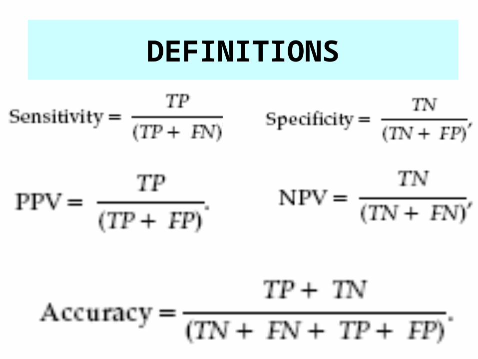
DEFINITIONS
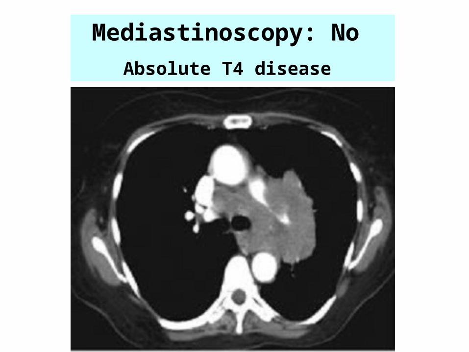
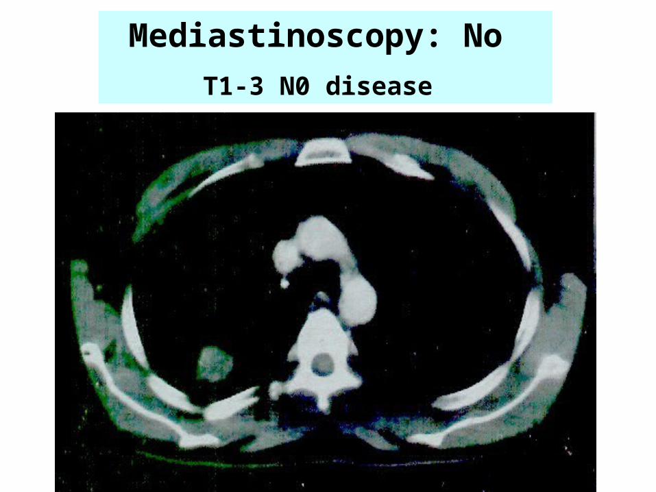
Mediastinoscopy: No
Absolute T4 disease
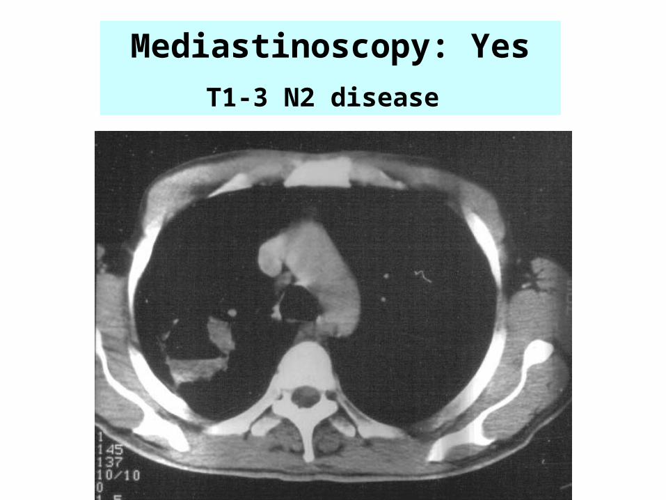
Mediastinoscopy: Yes
T1-3 N2 disease
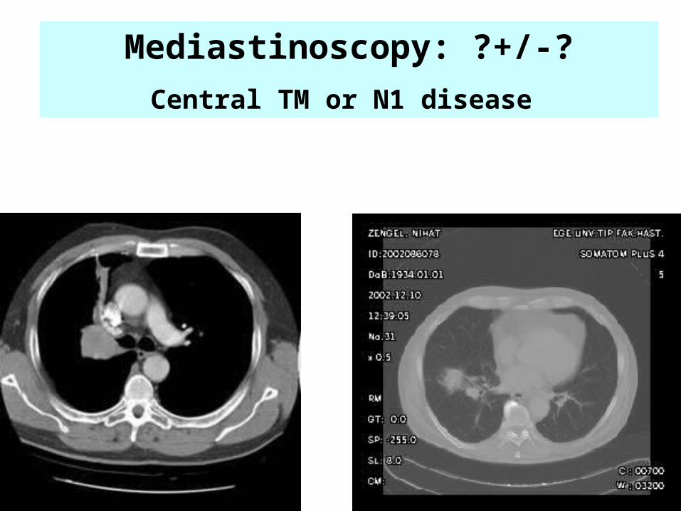
Mediastinoscopy: ?+/-?
Central TM or N1 disease
Mediastinoscopy: No
T1-3 N0 disease
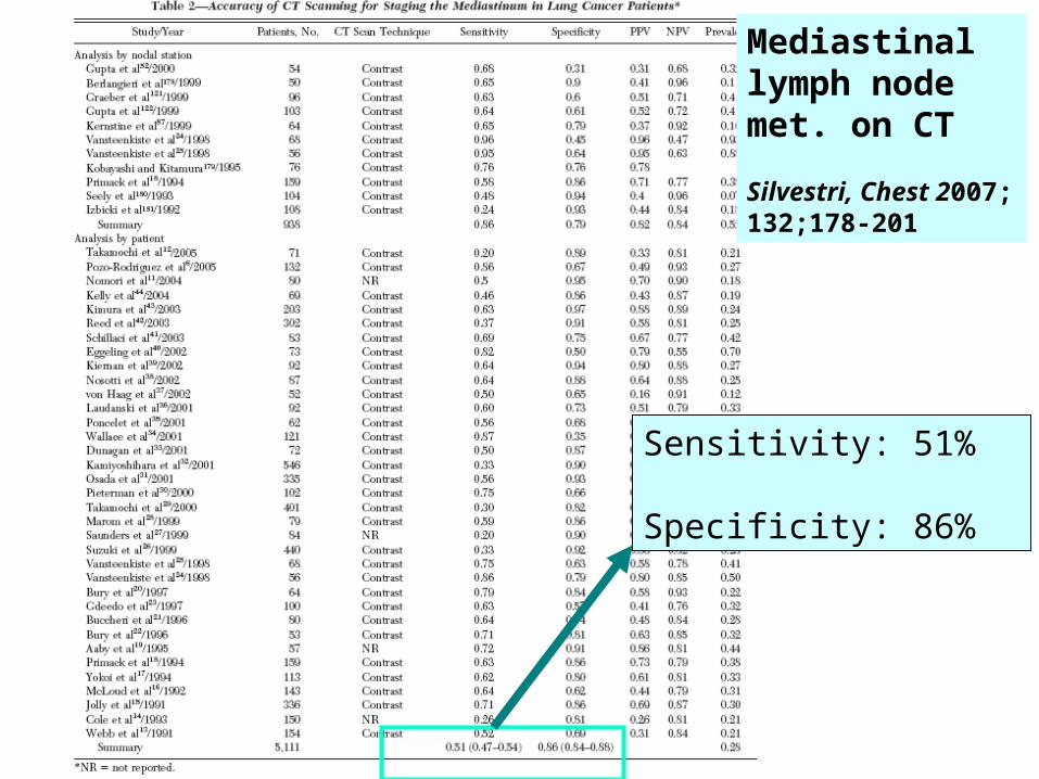
Sensitivity: 51%
Specificity: 86%
Mediastinal lymph node met. on CT
Silvestri, Chest 2007; 132;178-201
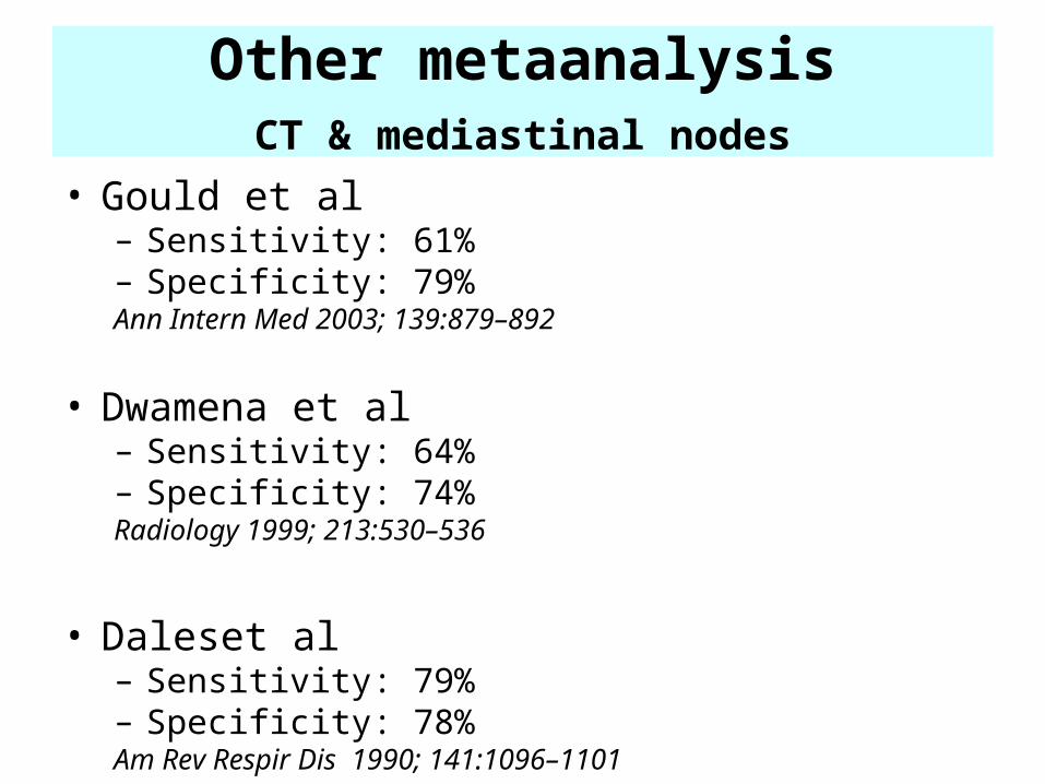
Other metaanalysis CT & mediastinal nodes
• Gould et al– Sensitivity: 61% – Specificity: 79%Ann Intern Med 2003; 139:879–892
• Dwamena et al – Sensitivity: 64% – Specificity: 74%Radiology 1999; 213:530–536
• Daleset al – Sensitivity: 79% – Specificity: 78%Am Rev Respir Dis 1990; 141:1096–1101
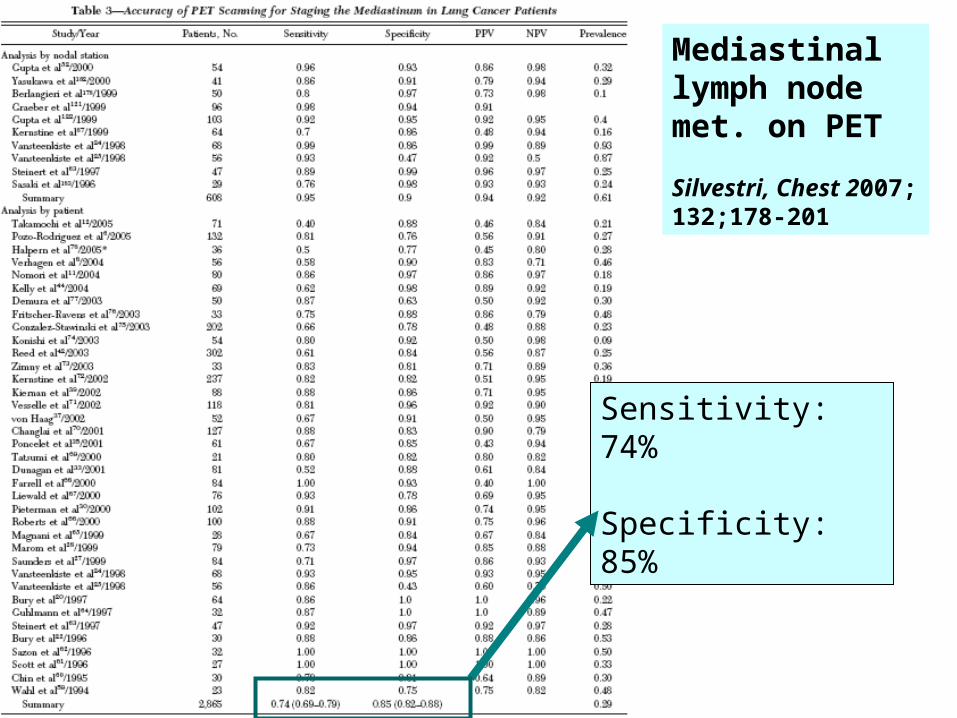
Sensitivity: 74%
Specificity: 85%
Mediastinal lymph node met. on PET
Silvestri, Chest 2007; 132;178-201
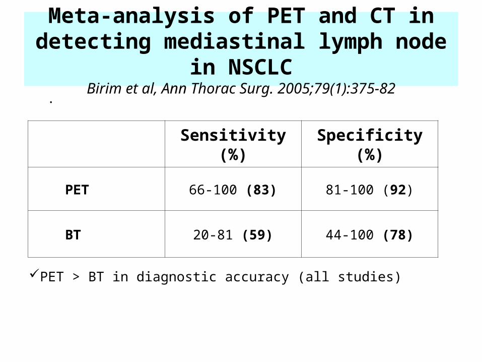
Meta-analysis of PET and CT in detecting mediastinal lymph node in NSCLC
Birim et al, Ann Thorac Surg. 2005;79(1):375-82
.
Sensitivity (%) Specificity (%)
PET 66-100 (83) 81-100 (92)
BT 20-81 (59) 44-100 (78)
PET > BT in diagnostic accuracy (all studies)
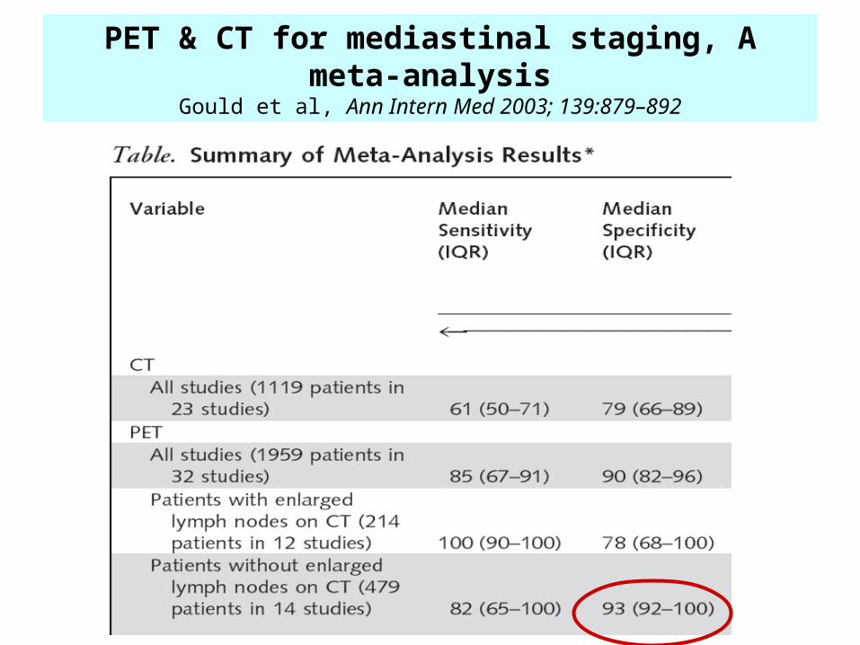
PET & CT for mediastinal staging, A meta-analysisGould et al, Ann Intern Med 2003; 139:879–892
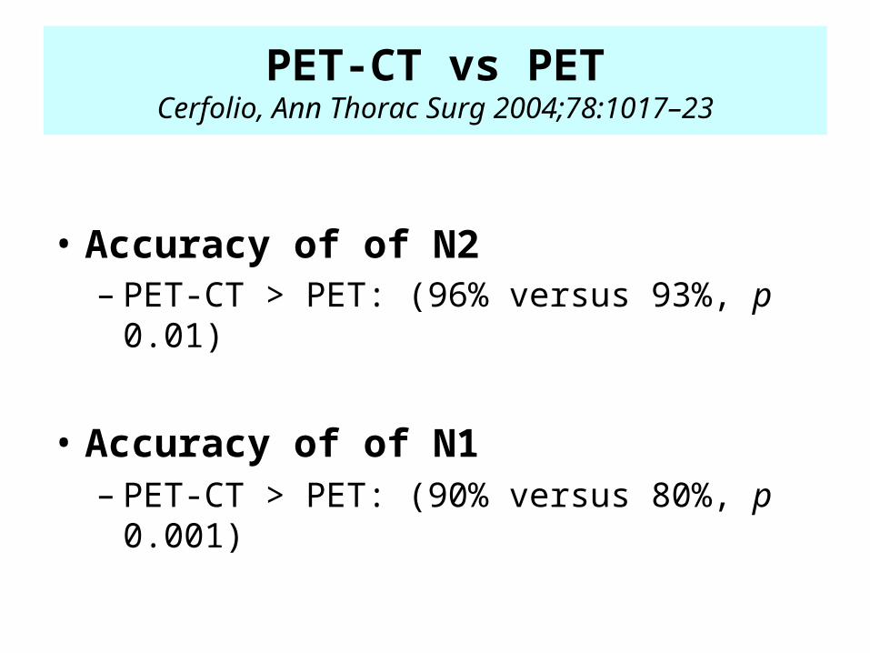
PET-CT vs PETCerfolio, Ann Thorac Surg 2004;78:1017–23
• Accuracy of of N2– PET-CT > PET: (96% versus 93%, p 0.01)
• Accuracy of of N1– PET-CT > PET: (90% versus 80%, p 0.001)
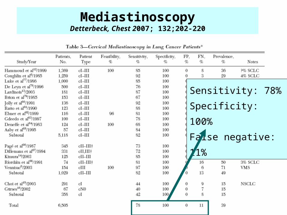
Sensitivity: 78%
Specificity: 100%
False negative: 11%
MediastinoscopyDetterbeck, Chest 2007; 132;202-220
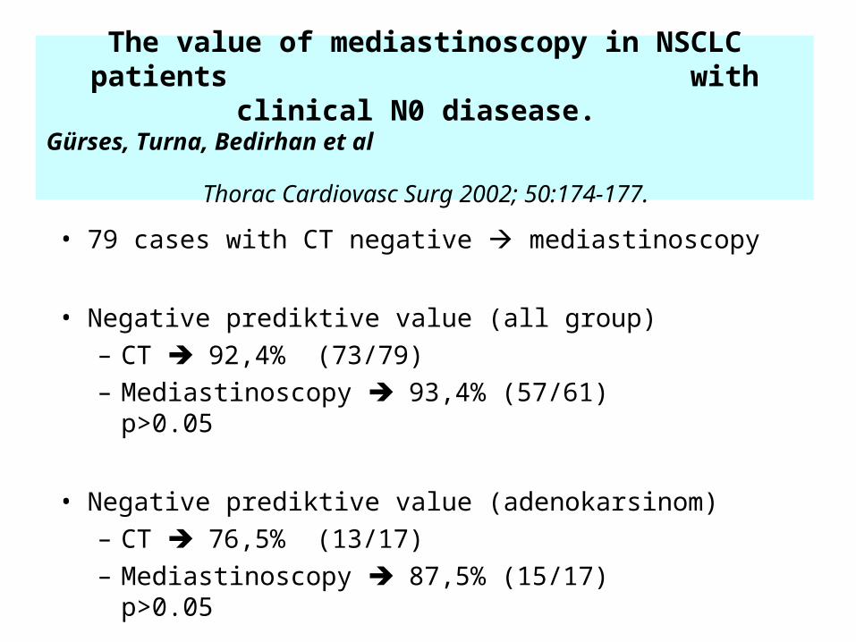
The value of mediastinoscopy in NSCLC patients with clinical N0 diasease.
Gürses, Turna, Bedirhan et al
Thorac Cardiovasc Surg 2002; 50:174-177. • 79 cases with CT negative mediastinoscopy
• Negative prediktive value (all group) – CT 92,4% (73/79)– Mediastinoscopy 93,4% (57/61) p>0.05
• Negative prediktive value (adenokarsinom) – CT 76,5% (13/17)– Mediastinoscopy 87,5% (15/17) p>0.05
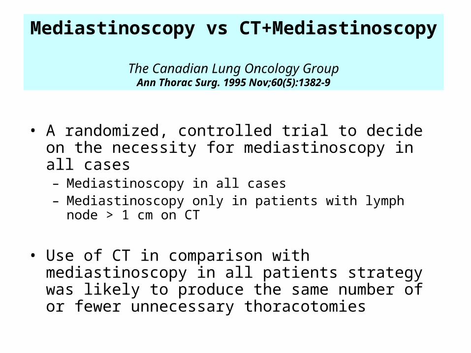
Mediastinoscopy vs CT+Mediastinoscopy The Canadian Lung Oncology Group
Ann Thorac Surg. 1995 Nov;60(5):1382-9
• A randomized, controlled trial to decide on the necessity for mediastinoscopy in all cases– Mediastinoscopy in all cases– Mediastinoscopy only in patients with lymph node > 1 cm on CT
• Use of CT in comparison with mediastinoscopy in all patients strategy was likely to produce the same number of or fewer unnecessary thoracotomies
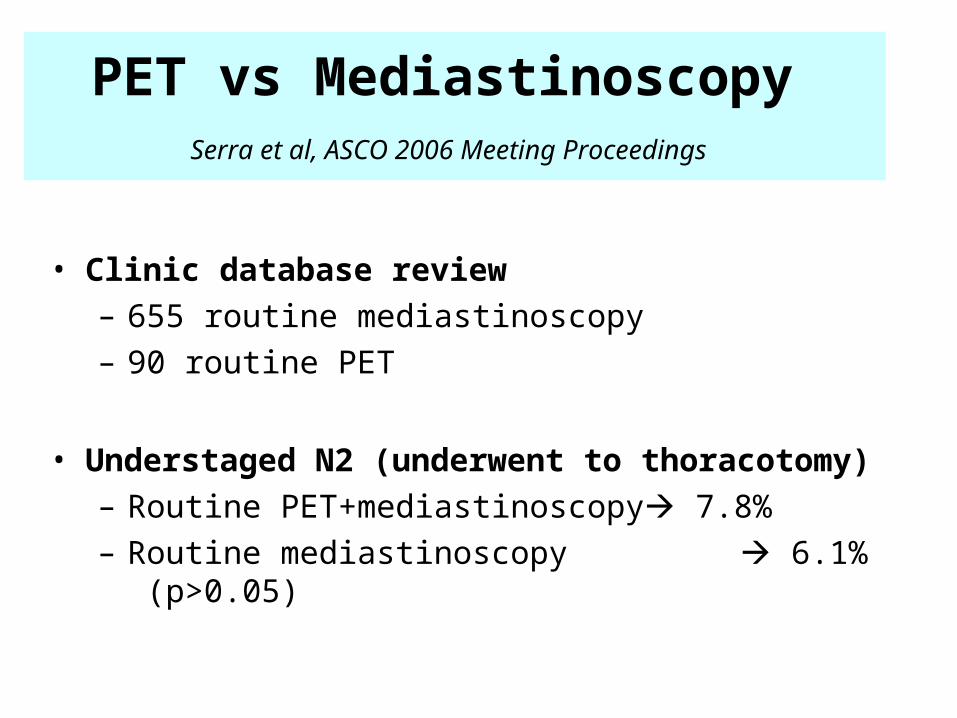
PET vs Mediastinoscopy
Serra et al, ASCO 2006 Meeting Proceedings
• Clinic database review– 655 routine mediastinoscopy– 90 routine PET
• Understaged N2 (underwent to thoracotomy)– Routine PET+mediastinoscopy 7.8% – Routine mediastinoscopy 6.1% (p>0.05)
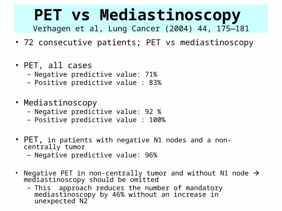
PET vs Mediastinoscopy Verhagen et al, Lung Cancer (2004) 44, 175—181
• 72 consecutive patients; PET vs mediastinoscopy
• PET, all cases– Negative predictive value: 71% – Positive predictive value : 83%
• Mediastinoscopy– Negative predictive value: 92 % – Positive predictive value : 100%
• PET, in patients with negative N1 nodes and a non-centrally tumor – Negative predictive value: 96%
• Negative PET in non-centrally tumor and without N1 node mediastinoscopy should be omitted– This approach reduces the number of mandatory
mediastinoscopy by 46% without an increase in unexpected N2
2 R L
4 R L
7
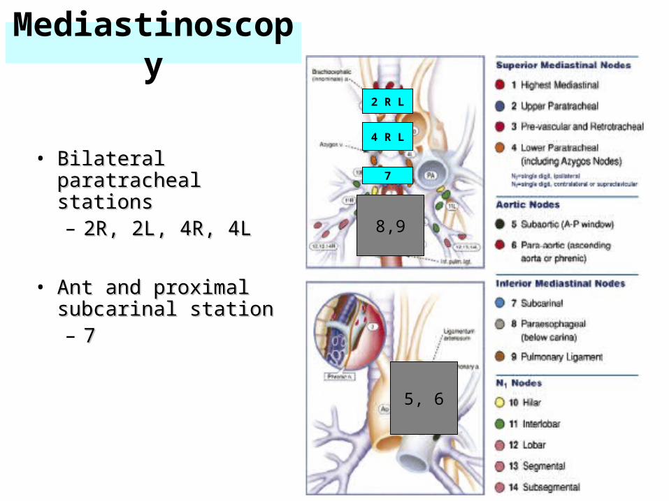
Mediastinoscopy
• Bilateral paratracheal Bilateral paratracheal stationsstations– 2R, 2L, 4R, 4L2R, 2L, 4R, 4L
• Ant and proximal Ant and proximal subcarinal stationsubcarinal station– 77
5, 6
8,9
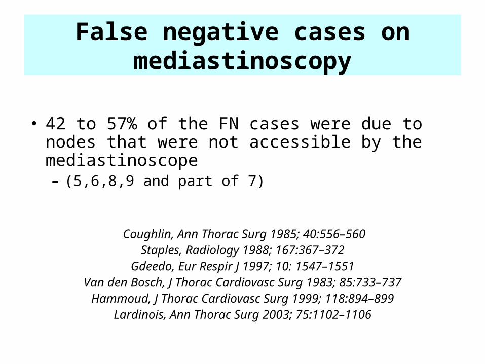
• 42 to 57% of the FN cases were due to nodes that were not accessible by the mediastinoscope– (5,6,8,9 and part of 7)
Coughlin, Ann Thorac Surg 1985; 40:556–560Staples, Radiology 1988; 167:367–372
Gdeedo, Eur Respir J 1997; 10: 1547–1551Van den Bosch, J Thorac Cardiovasc Surg 1983; 85:733–737
Hammoud, J Thorac Cardiovasc Surg 1999; 118:894–899Lardinois, Ann Thorac Surg 2003; 75:1102–1106
False negative cases on mediastinoscopy
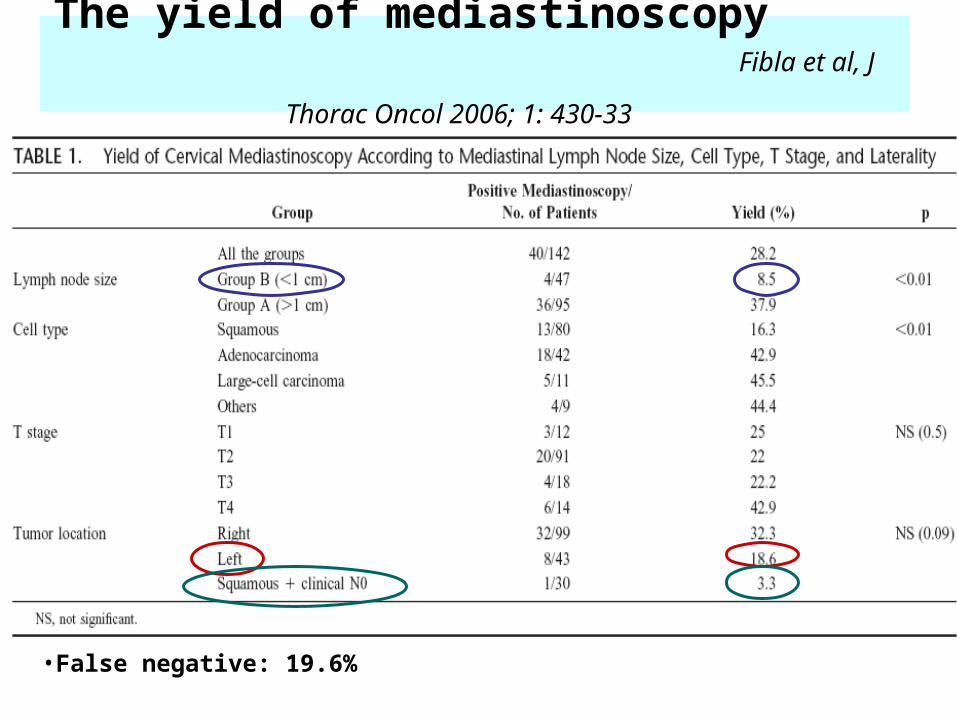
The yield of mediastinoscopy Fibla et al, J Thorac Oncol 2006; 1: 430-33
•False negative: 19.6%
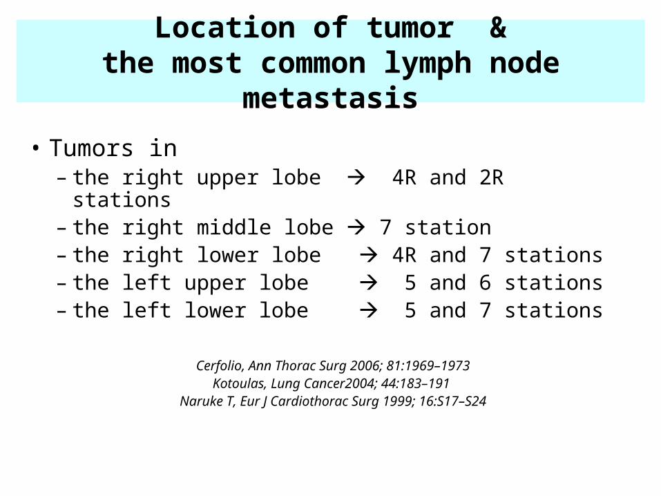
Location of tumor &the most common lymph node metastasis
• Tumors in– the right upper lobe 4R and 2R stations– the right middle lobe 7 station– the right lower lobe 4R and 7 stations– the left upper lobe 5 and 6 stations– the left lower lobe 5 and 7 stations
Cerfolio, Ann Thorac Surg 2006; 81:1969–1973Kotoulas, Lung Cancer2004; 44:183–191
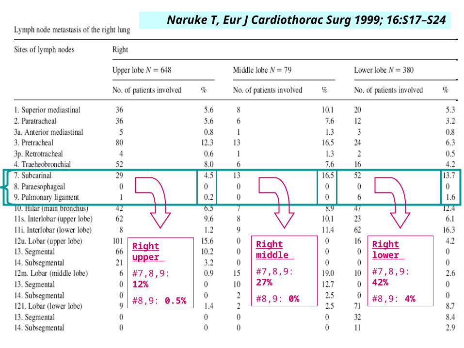
Naruke T, Eur J Cardiothorac Surg 1999; 16:S17–S24
Naruke T, Eur J Cardiothorac Surg 1999; 16:S17–S24
Right upper
#7,8,9: 12%
#8,9: 0.5%
Right middle
#7,8,9: 27%
#8,9: 0%
Right lower
#7,8,9: 42%
#8,9: 4%
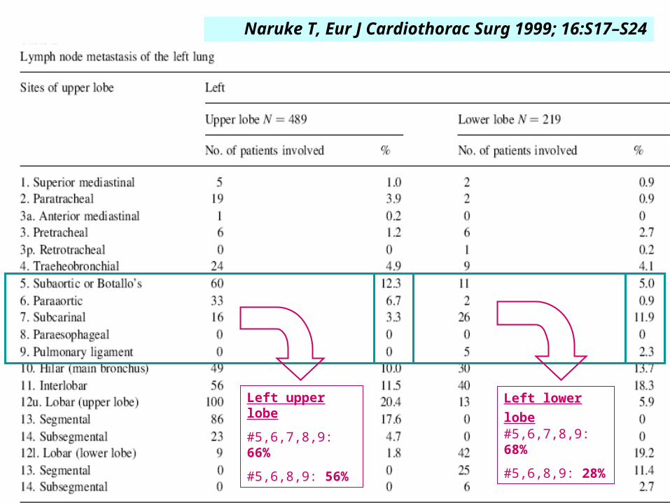
Naruke T, Eur J Cardiothorac Surg 1999; 16:S17–S24
Left upper lobe
#5,6,7,8,9: 66%
#5,6,8,9: 56%
Left lower lobe #5,6,7,8,9: 68%
#5,6,8,9: 28%
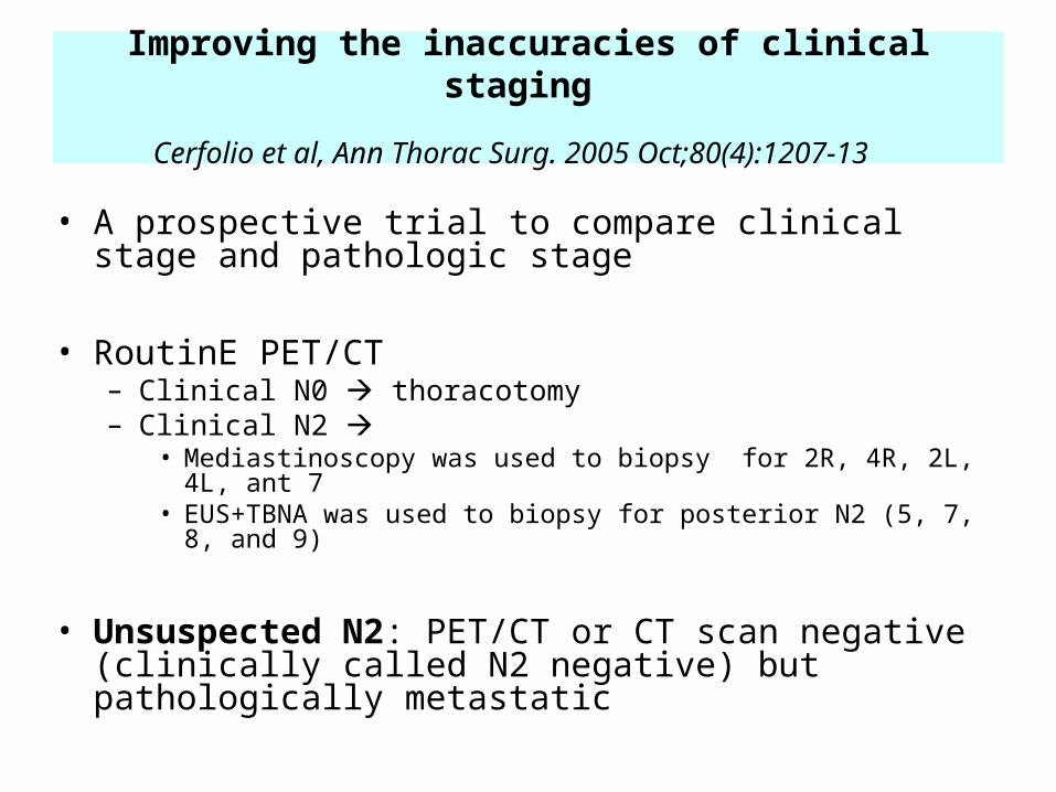
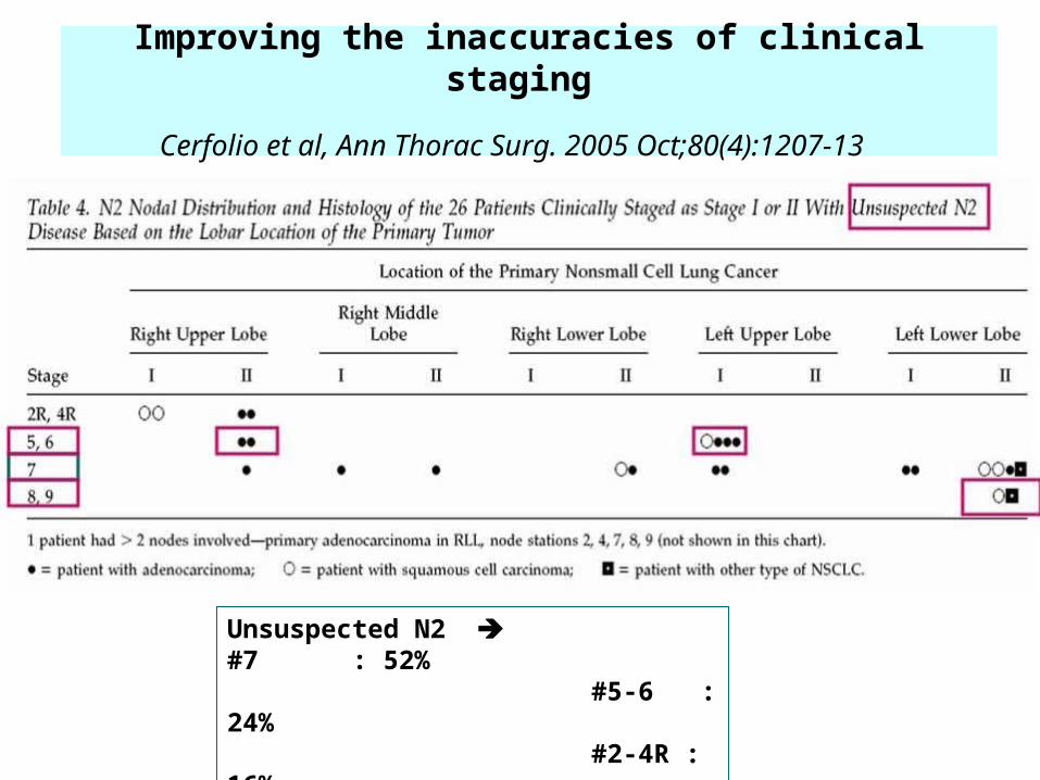
Improving the inaccuracies of clinical staging
Cerfolio et al, Ann Thorac Surg. 2005 Oct;80(4):1207-13
• A prospective trial to compare clinical stage and pathologic stage
• RoutinE PET/CT– Clinical N0 thoracotomy– Clinical N2
• Mediastinoscopy was used to biopsy for 2R, 4R, 2L, 4L, ant 7• EUS+TBNA was used to biopsy for posterior N2 (5, 7, 8, and 9)
• Unsuspected N2: PET/CT or CT scan negative (clinically called N2 negative) but pathologically metastatic
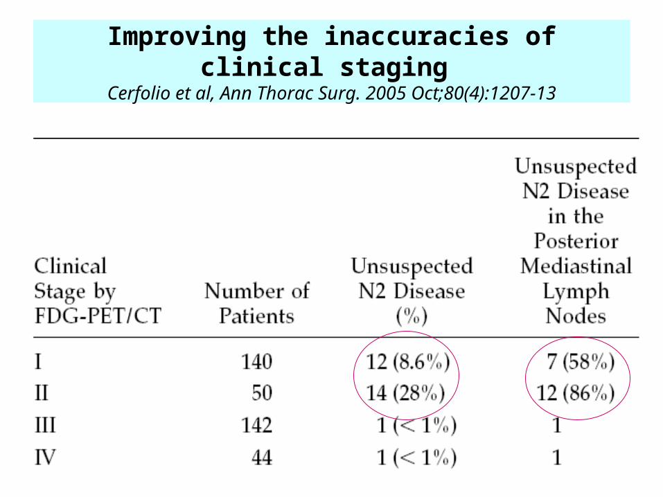
Improving the inaccuracies of clinical staging Cerfolio et al, Ann Thorac Surg. 2005 Oct;80(4):1207-13
Improving the inaccuracies of clinical staging
Cerfolio et al, Ann Thorac Surg. 2005 Oct;80(4):1207-13
Unsuspected N2 #7 : 52% #5-6 : 24% #2-4R : 16% #8-9 : 8%
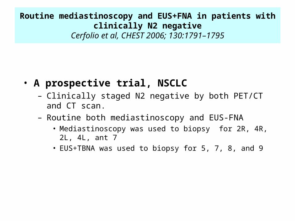
Routine mediastinoscopy and EUS+FNA in patients with clinically N2 negative
Cerfolio et al, CHEST 2006; 130:1791–1795
• A prospective trial, NSCLC – Clinically staged N2 negative by both PET/CT and CT scan. – Routine both mediastinoscopy and EUS-FNA
• Mediastinoscopy was used to biopsy for 2R, 4R, 2L, 4L, ant 7
• EUS+TBNA was used to biopsy for 5, 7, 8, and 9
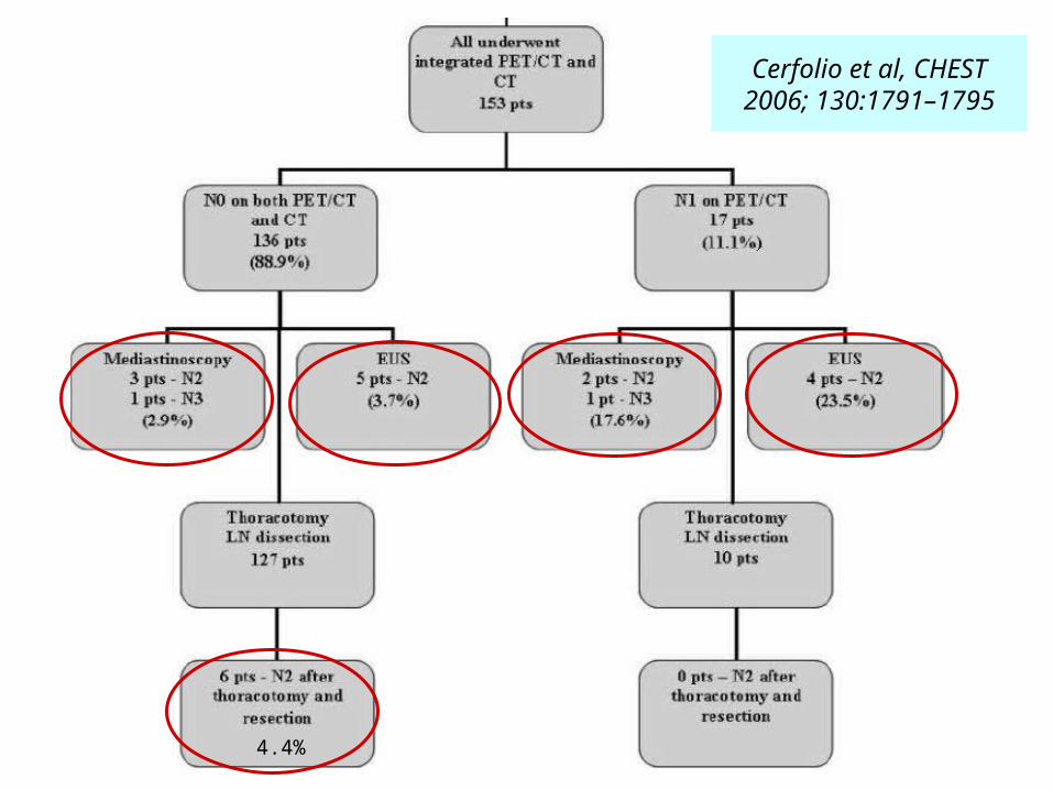
Cerfolio et al, CHEST 2006; 130:1791–1795
4.4%
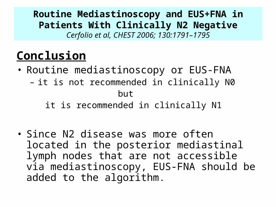
Conclusion• Routine mediastinoscopy or EUS-FNA
– it is not recommended in clinically N0 but it is recommended in clinically N1
• Since N2 disease was more often located in the posterior mediastinal lymph nodes that are not accessible via mediastinoscopy, EUS-FNA should be added to the algorithm.
Routine Mediastinoscopy and EUS+FNA in Patients With Clinically N2 Negative
Cerfolio et al, CHEST 2006; 130:1791–1795
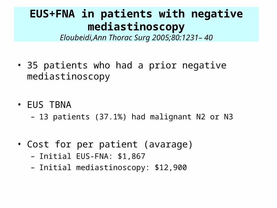
EUS+FNA in patients with negative mediastinoscopy
Eloubeidi,Ann Thorac Surg 2005;80:1231– 40
• 35 patients who had a prior negative mediastinoscopy
• EUS TBNA– 13 patients (37.1%) had malignant N2 or N3
• Cost for per patient (avarage) – Initial EUS-FNA: $1,867 – Initial mediastinoscopy: $12,900
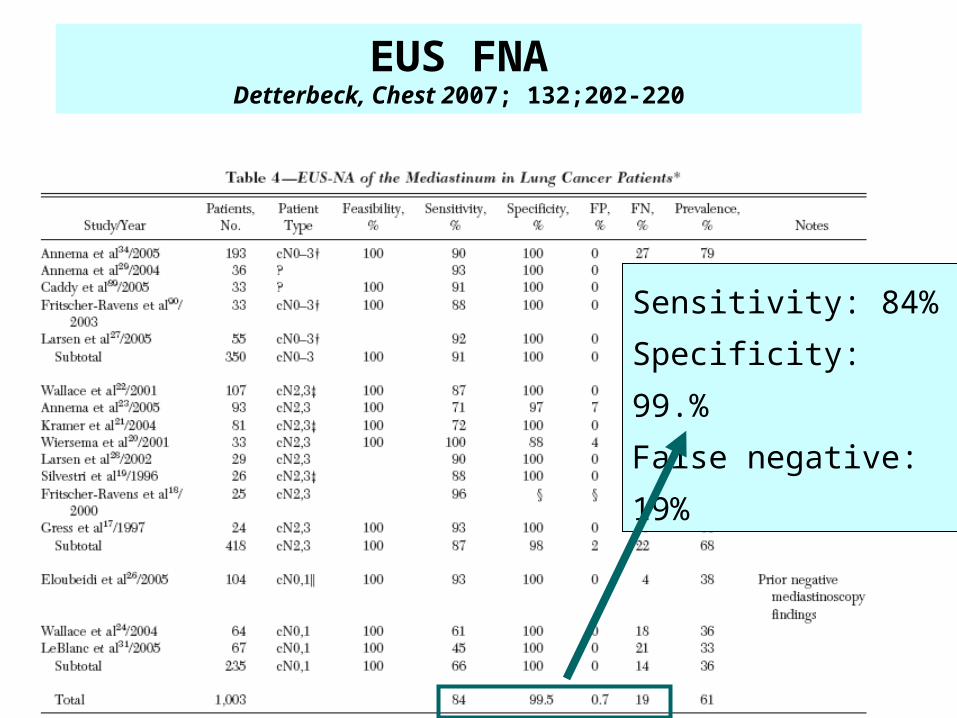
EUS FNADetterbeck, Chest 2007; 132;202-220
Sensitivity: 84%
Specificity: 99.%
False negative: 19%
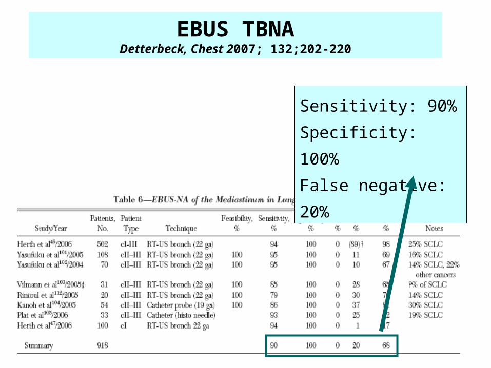
Sensitivity: 90%
Specificity: 100%
False negative: 20%
EBUS TBNADetterbeck, Chest 2007; 132;202-220
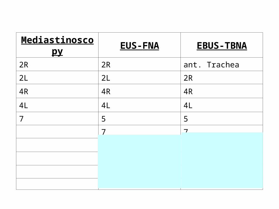
Mediastinoscopy EUS-FNA EBUS-TBNA
2R 2R ant. Trachea
2L 2L 2R
4R 4R 4R
4L 4L 4L
7 5 5
7 7
8 10R
9 10L
Left adrenal 11
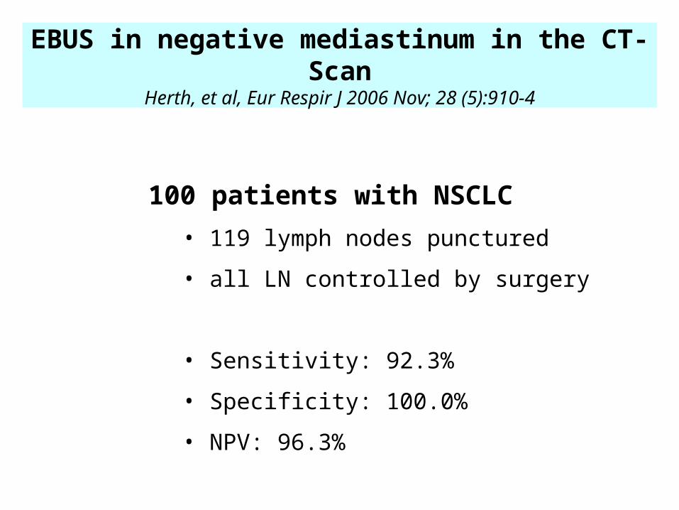
EBUS in negative mediastinum in the CT-ScanHerth, et al, Eur Respir J 2006 Nov; 28 (5):910-4
100 patients with NSCLC
• 119 lymph nodes punctured
• all LN controlled by surgery
• Sensitivity: 92.3%
• Specificity: 100.0%
• NPV: 96.3%
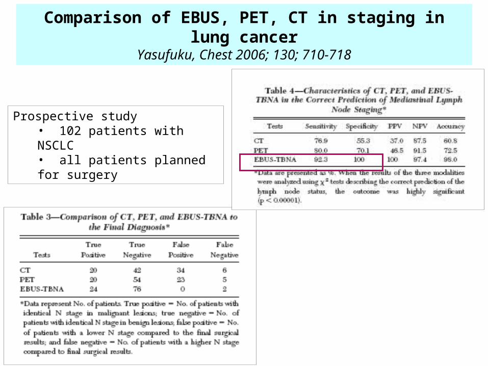
Comparison of EBUS, PET, CT in staging in lung cancerYasufuku, Chest 2006; 130; 710-718
Prospective study• 102 patients with NSCLC• all patients planned for surgery
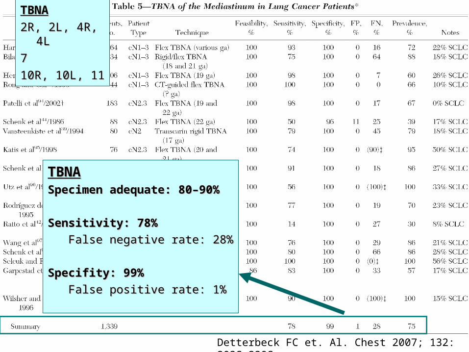
Detterbeck FC et. Al. Chest 2007; 132: 202S-220S
TBNATBNASpecimen adequate: 80–90%Specimen adequate: 80–90%
Sensitivity: 78%Sensitivity: 78%
False negative rate: 28%False negative rate: 28%
Specifity: 99%Specifity: 99%
False positive rate: 1%False positive rate: 1%
TBNATBNA
2R, 2L, 4R, 4L2R, 2L, 4R, 4L
77
10R, 10L, 1110R, 10L, 11
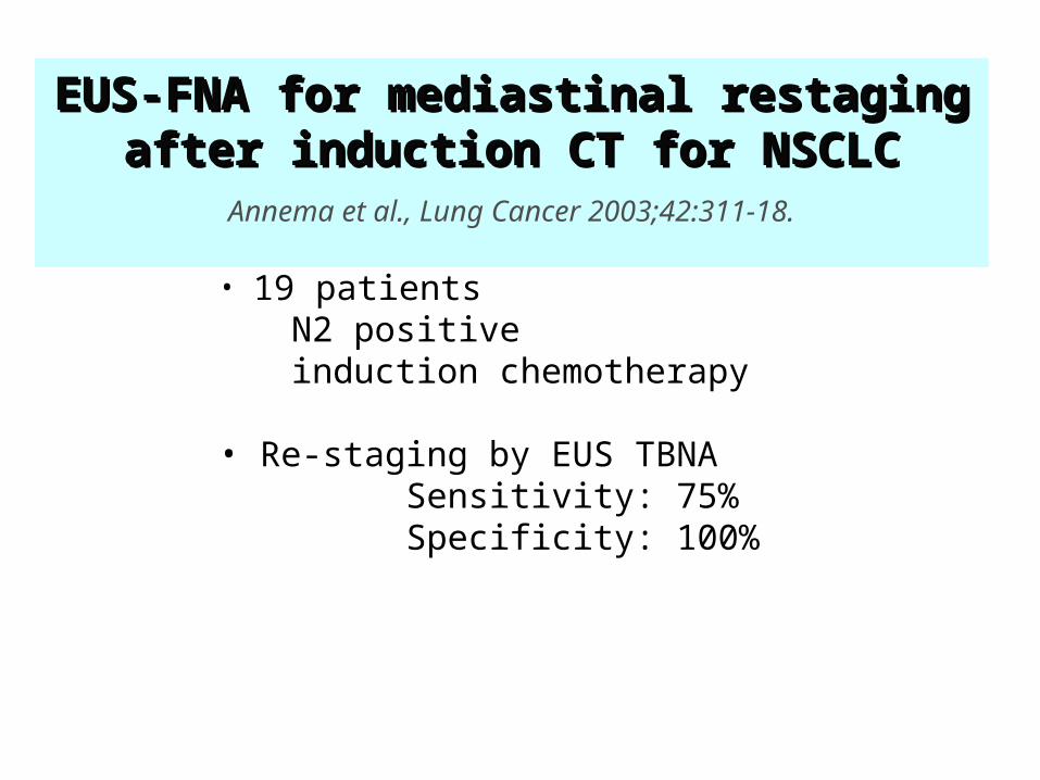
• 19 patients N2 positive induction chemotherapy
• Re-staging by EUS TBNA Sensitivity: 75% Specificity: 100%
EUS-FNA for mediastinal restaging after EUS-FNA for mediastinal restaging after induction induction CT CT for NSCLCfor NSCLC
Annema et al., Lung Cancer 2003;42:311-18.
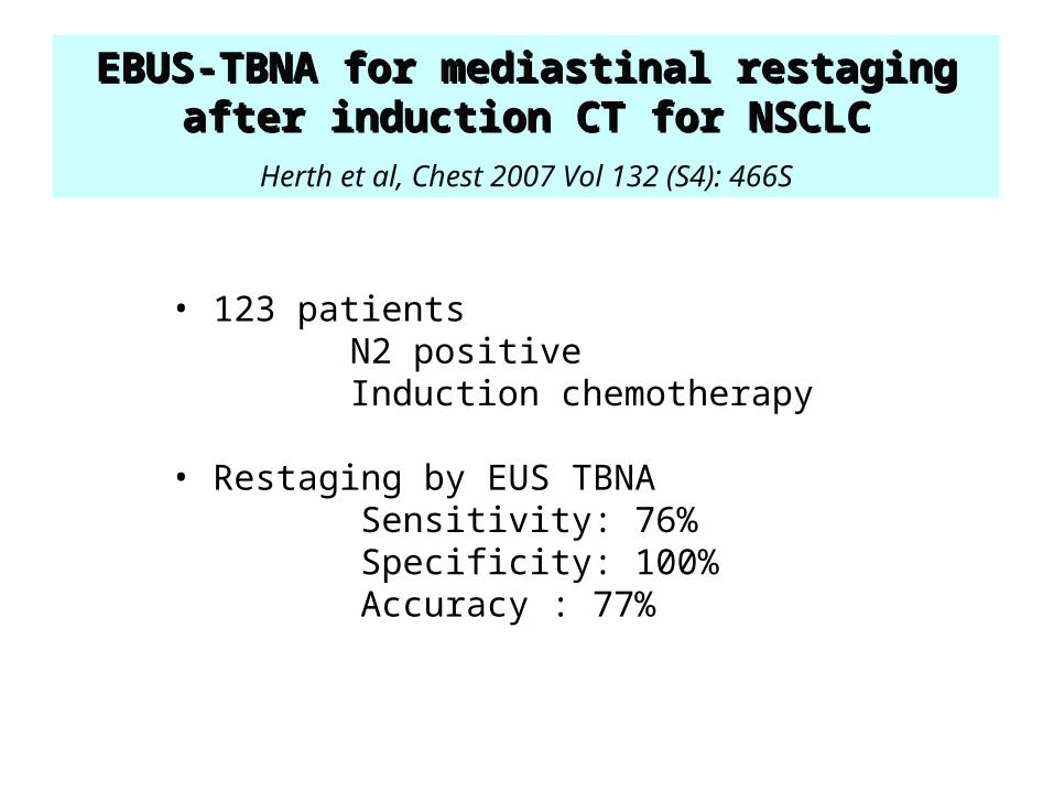
• 123 patients N2 positive Induction chemotherapy
• Restaging by EUS TBNA Sensitivity: 76% Specificity: 100% Accuracy : 77%
EBUS-TBNA for mediastinal restaging EBUS-TBNA for mediastinal restaging after induction after induction CT CT for NSCLCfor NSCLCHerth et al, Chest 2007 Vol 132 (S4): 466S
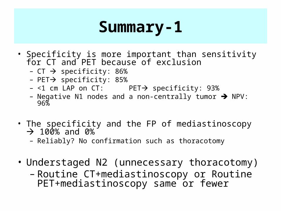
Summary-1
• Specificity is more important than sensitivity for CT and PET because of exclusion – CT specificity: 86%– PET specificity: 85%– <1 cm LAP on CT: PET specificity: 93%– Negative N1 nodes and a non-centrally tumor NPV: 96%
• The specificity and the FP of mediastinoscopy 100% and 0%– Reliably? No confirmation such as thoracotomy
• Understaged N2 (unnecessary thoracotomy)– Routine CT+mediastinoscopy or Routine
PET+mediastinoscopy same or fewer
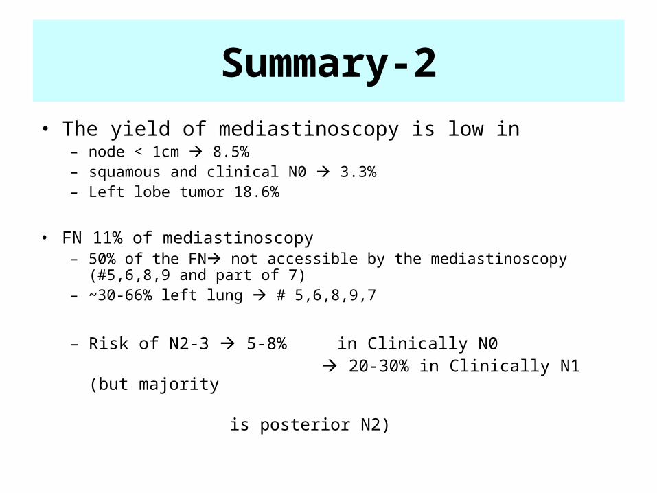
• The yield of mediastinoscopy is low in – node < 1cm 8.5%– squamous and clinical N0 3.3%– Left lobe tumor 18.6%
• FN 11% of mediastinoscopy– 50% of the FN not accessible by the mediastinoscopy (#5,6,8,9 and
part of 7)– ~30-66% left lung # 5,6,8,9,7
– Risk of N2-3 5-8% in Clinically N0 20-30% in Clinically N1 (but majority is posterior N2)
Summary-2
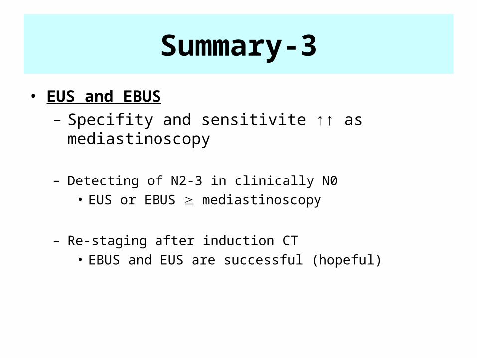
• EUS and EBUS– Specifity and sensitivite ↑↑ as mediastinoscopy
– Detecting of N2-3 in clinically N0• EUS or EBUS mediastinoscopy
– Re-staging after induction CT• EBUS and EUS are successful (hopeful)
Summary-3
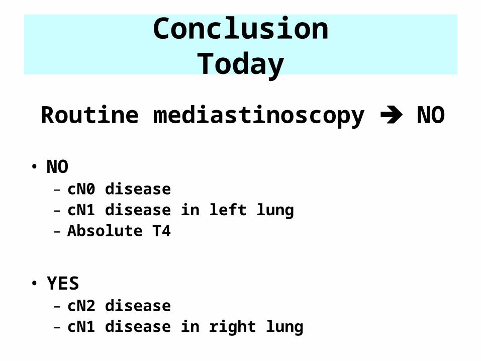
ConclusionToday
Routine mediastinoscopy NO
• NO– cN0 disease– cN1 disease in left lung– Absolute T4
• YES – cN2 disease– cN1 disease in right lung
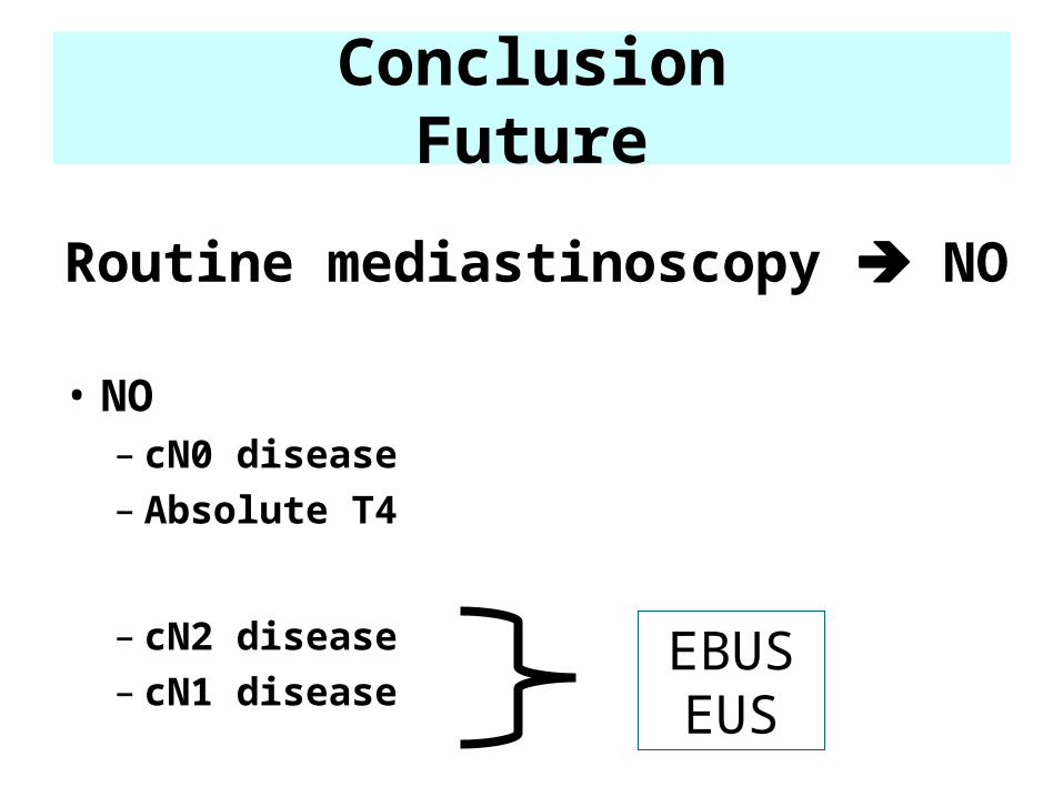
ConclusionFuture
Routine mediastinoscopy NO
• NO– cN0 disease– Absolute T4
– cN2 disease– cN1 disease
EBUS EUS