Embed Size (px)
Citation preview
Medical and SurgicalMedical and SurgicalManagement of SpinalManagement of Spinal
InfectionsInfections
Cesar A. Serrano-AlmeidaCesar A. Serrano-AlmeidaJune 14, 2007June 14, 2007
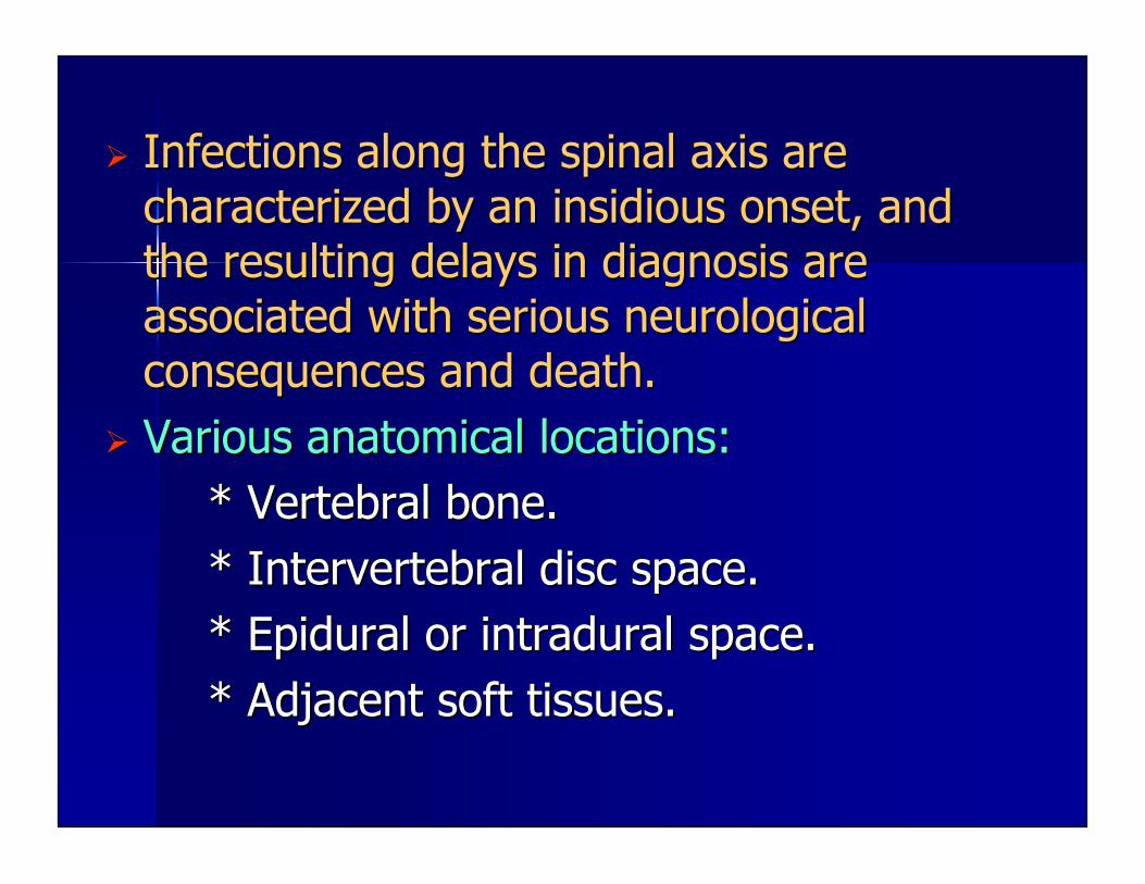
Infections along the spinal axis areInfections along the spinal axis arecharacterized by an insidious onset, andcharacterized by an insidious onset, andthe resulting delays in diagnosis arethe resulting delays in diagnosis areassociated with serious neurologicalassociated with serious neurologicalconsequences and death.consequences and death.
Various anatomical locations:Various anatomical locations:* Vertebral bone.* Vertebral bone.* * IntervertebralIntervertebral disc space. disc space.* Epidural or * Epidural or intraduralintradural space. space.* Adjacent soft tissues.* Adjacent soft tissues.
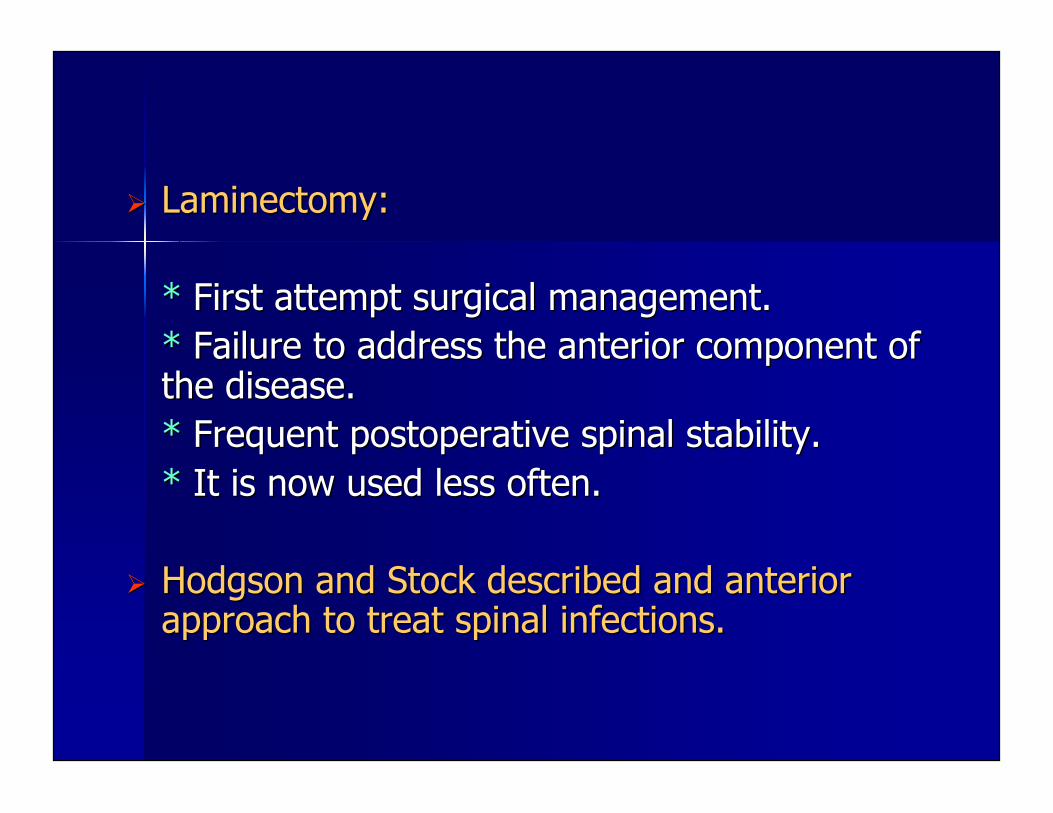
LaminectomyLaminectomy::
* * First attempt surgical management.First attempt surgical management.* * Failure to address the anterior component ofFailure to address the anterior component ofthe disease.the disease.* * Frequent postoperative spinal stability.Frequent postoperative spinal stability.* * It is now used less often.It is now used less often.
Hodgson and Stock described and anteriorHodgson and Stock described and anteriorapproach to treat spinal infections.approach to treat spinal infections.
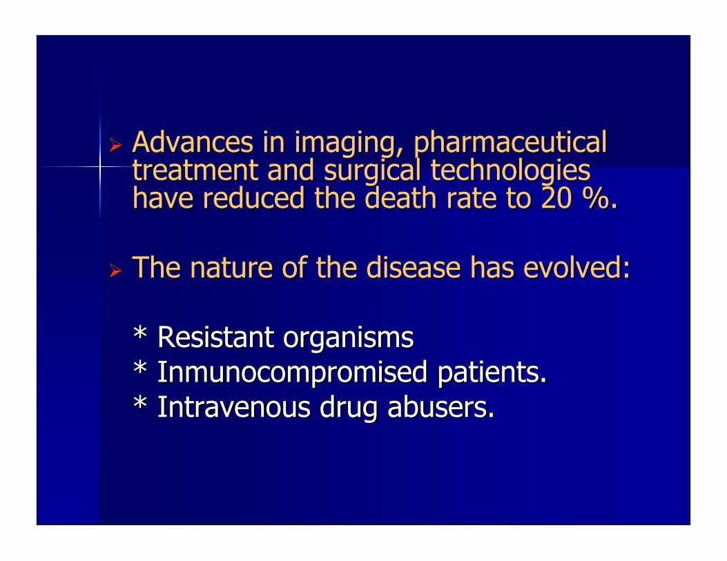
Advances in imaging, pharmaceuticalAdvances in imaging, pharmaceuticaltreatment and surgical technologiestreatment and surgical technologieshave reduced the death rate to 20 %.have reduced the death rate to 20 %.
The nature of the disease has evolved:The nature of the disease has evolved:
* Resistant organisms* Resistant organisms* * InmunocompromisedInmunocompromised patients. patients.* Intravenous drug abusers.* Intravenous drug abusers.
CLASSIFICATION, PATHOGENESIS,CLASSIFICATION, PATHOGENESIS,AND PRESENTATION.AND PRESENTATION.
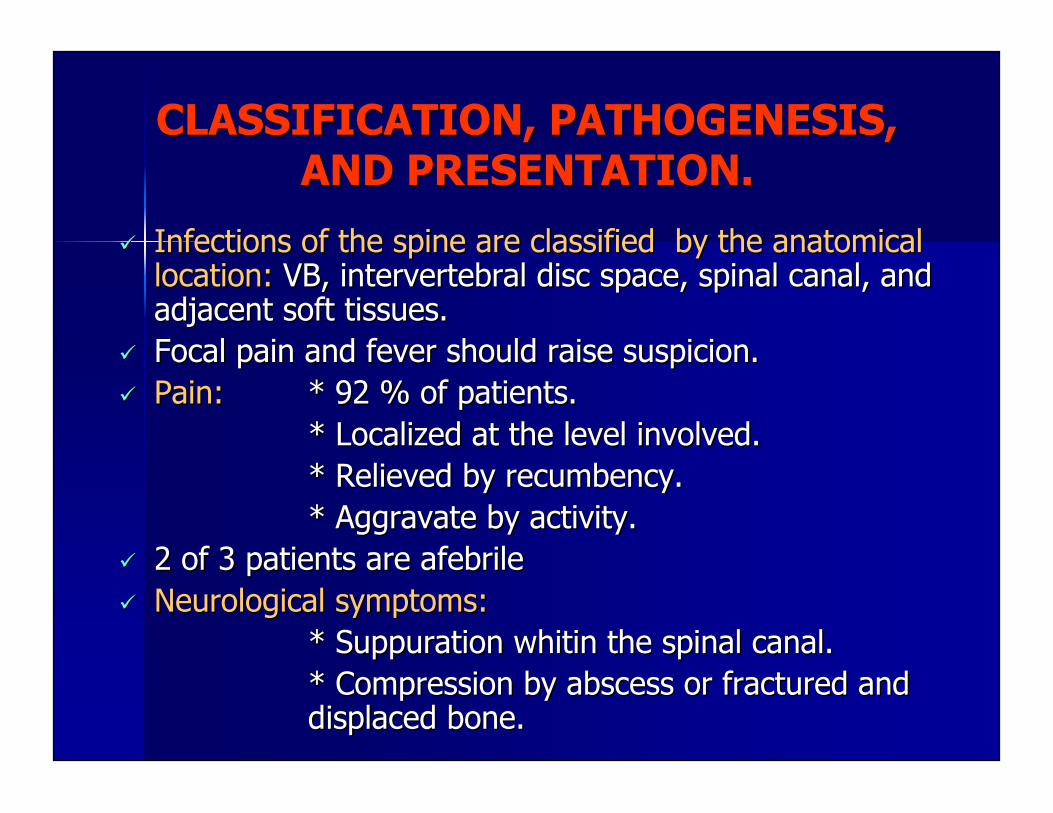
Infections of the spine are classified by the anatomicalInfections of the spine are classified by the anatomicallocation:location: VB, VB, intervertebralintervertebral disc space, spinal canal, and disc space, spinal canal, andadjacent soft tissues.adjacent soft tissues.
Focal pain and fever should raise suspicion.Focal pain and fever should raise suspicion. Pain:Pain: * 92 % of patients.* 92 % of patients.
* Localized at the level involved.* Localized at the level involved.* Relieved by * Relieved by recumbencyrecumbency..* Aggravate by activity.* Aggravate by activity.
2 of 3 patients are 2 of 3 patients are afebrileafebrile Neurological symptoms:Neurological symptoms:
* Suppuration * Suppuration whitinwhitin the spinal canal. the spinal canal.* Compression by abscess or fractured and* Compression by abscess or fractured anddisplaced bone.displaced bone.
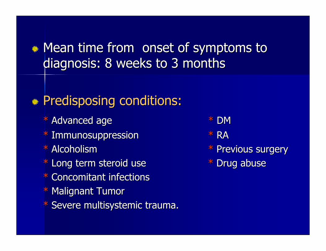
Mean time from onset of symptoms toMean time from onset of symptoms todiagnosis: 8 weeks to 3 monthsdiagnosis: 8 weeks to 3 months
Predisposing conditions:Predisposing conditions:** Advanced age Advanced age ** DM DM
** ImmunosuppressionImmunosuppression ** RA RA** Alcoholism Alcoholism ** Previous surgery Previous surgery** Long term steroid use Long term steroid use ** Drug abuse Drug abuse** Concomitant infections Concomitant infections** Malignant Tumor Malignant Tumor** Severe Severe multisystemicmultisystemic trauma. trauma.
SPINAL CANAL INFECTIONSSPINAL CANAL INFECTIONS
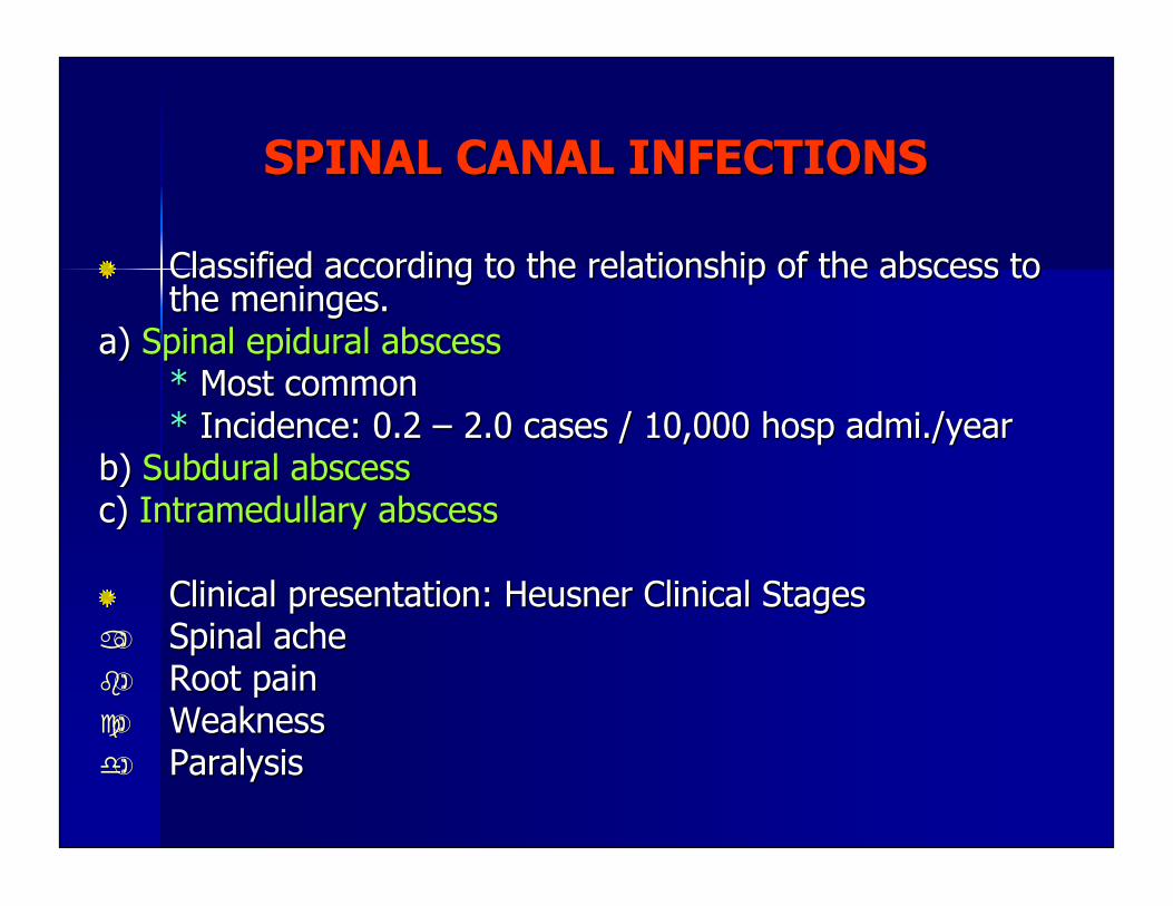
Classified according to the relationship of the abscess toClassified according to the relationship of the abscess tothe the meningesmeninges..
a) a) Spinal epidural abscessSpinal epidural abscess** Most common Most common** Incidence: 0.2 Incidence: 0.2 –– 2.0 cases / 10,000 hosp 2.0 cases / 10,000 hosp admiadmi./year./year
b) b) SubduralSubdural abscess abscessc) c) IntramedullaryIntramedullary abscess abscess
Clinical presentation: Clinical presentation: HeusnerHeusner Clinical Stages Clinical Stages Spinal acheSpinal ache Root painRoot pain WeaknessWeakness ParalysisParalysis
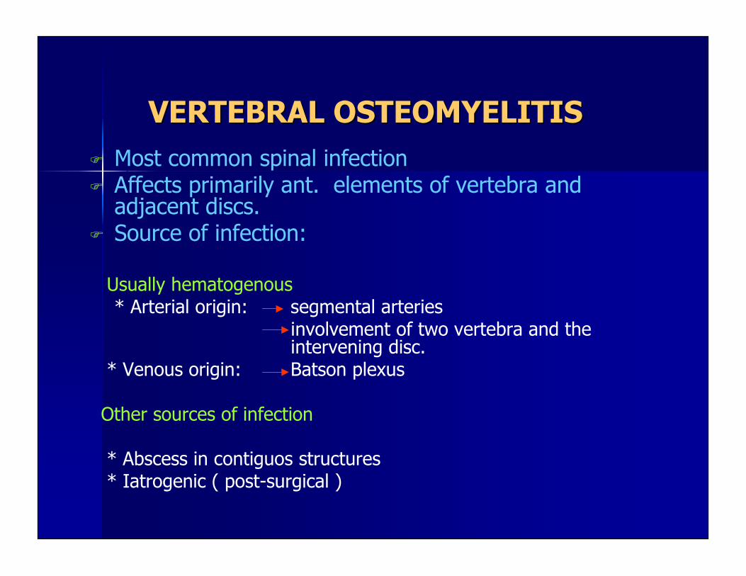
VERTEBRAL OSTEOMYELITISVERTEBRAL OSTEOMYELITIS Most common spinal infection Affects primarily ant. elements of vertebra and
adjacent discs. Source of infection:
Usually hematogenous* Arterial origin: segmental arteries
involvement of two vertebra and the intervening disc.
* Venous origin: Batson plexus
Other sources of infection
* Abscess in contiguos structures * Iatrogenic ( post-surgical )
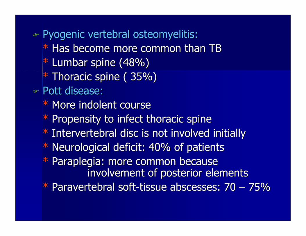
PyogenicPyogenic vertebral vertebral osteomyelitisosteomyelitis::** Has become more common than TB Has become more common than TB** Lumbar spine (48%) Lumbar spine (48%)** Thoracic spine ( 35%) Thoracic spine ( 35%)
PottPott disease: disease:** More indolent course More indolent course** Propensity to infect thoracic spine Propensity to infect thoracic spine** IntervertebralIntervertebral disc is not involved initially disc is not involved initially** Neurological deficit: 40% of patients Neurological deficit: 40% of patients** Paraplegia: more common because Paraplegia: more common because
involvement of posterior elementsinvolvement of posterior elements** ParavertebralParavertebral soft-tissue abscesses: 70 soft-tissue abscesses: 70 –– 75% 75%
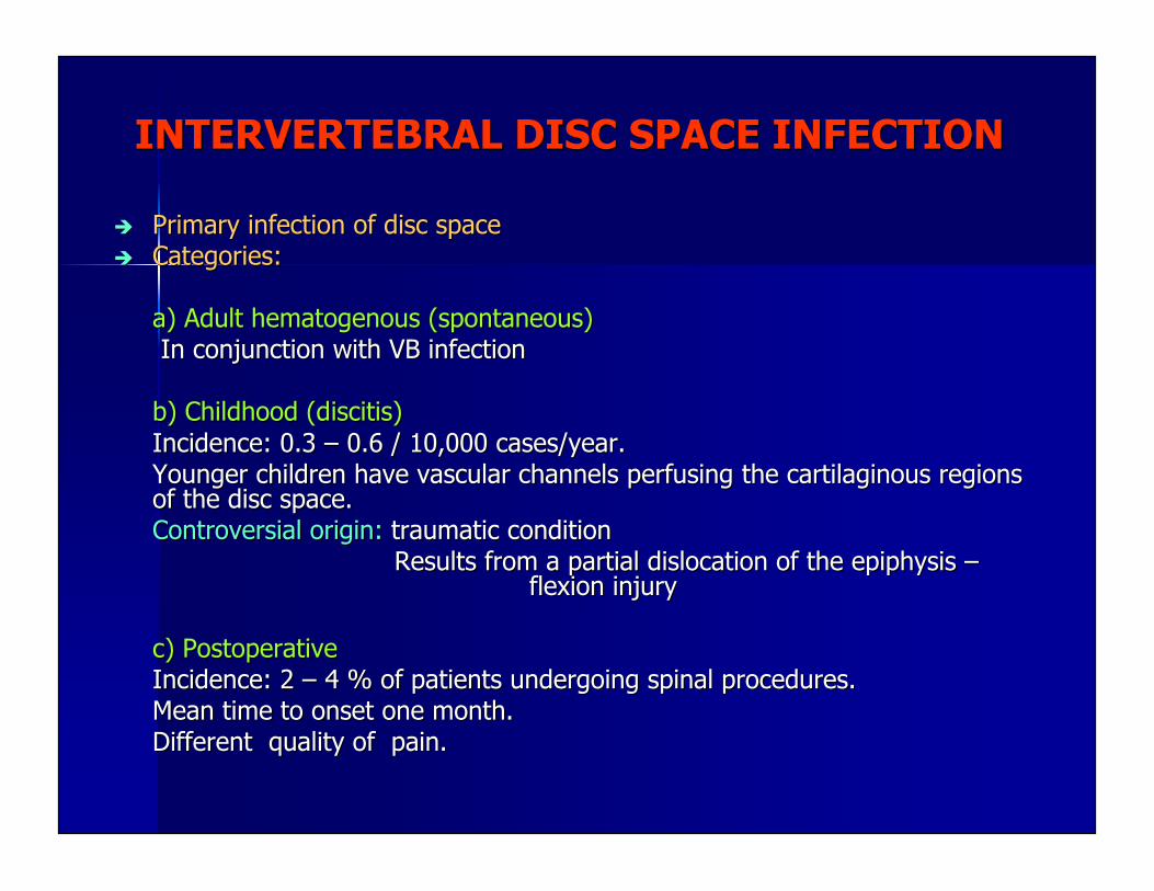
INTERVERTEBRAL DISC SPACE INFECTIONINTERVERTEBRAL DISC SPACE INFECTION
Primary infection of disc spacePrimary infection of disc space Categories:Categories:
a) Adult a) Adult hematogenoushematogenous (spontaneous) (spontaneous) In conjunction with VB infection In conjunction with VB infection
b) Childhood (b) Childhood (discitisdiscitis))Incidence: 0.3 Incidence: 0.3 –– 0.6 / 10,000 cases/year. 0.6 / 10,000 cases/year.Younger children have vascular channels Younger children have vascular channels perfusingperfusing the cartilaginous regions the cartilaginous regionsof the disc space.of the disc space.Controversial origin:Controversial origin: traumatic condition traumatic condition
Results from a partial dislocation of the epiphysis Results from a partial dislocation of the epiphysis ––flexion injuryflexion injury
c) Postoperativec) PostoperativeIncidence: 2 Incidence: 2 –– 4 % of patients undergoing spinal procedures. 4 % of patients undergoing spinal procedures.Mean time to onset one month.Mean time to onset one month.Different quality of pain.Different quality of pain.
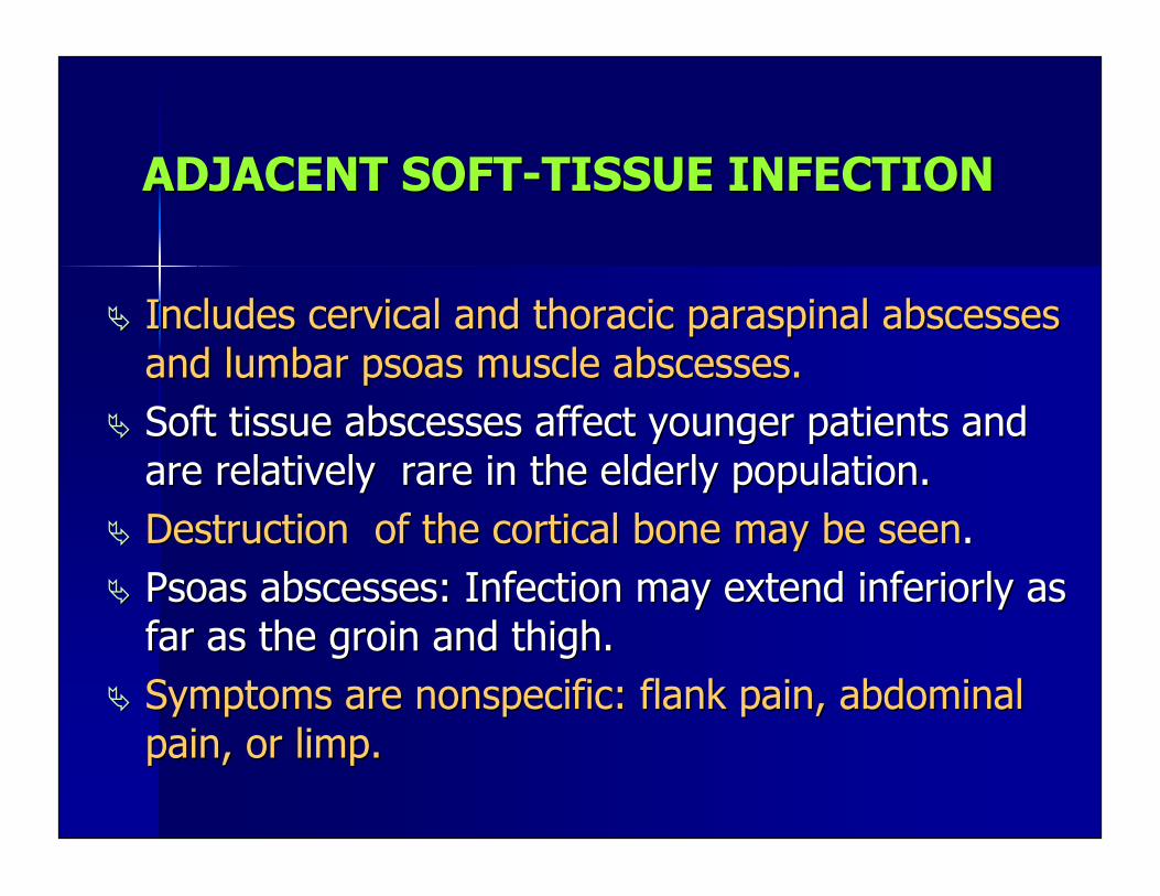
ADJACENT SOFT-TISSUE INFECTIONADJACENT SOFT-TISSUE INFECTION
Includes cervical and thoracic Includes cervical and thoracic paraspinalparaspinal abscesses abscessesand lumbar and lumbar psoaspsoas muscle abscesses. muscle abscesses.
Soft tissue abscesses affect younger patients andSoft tissue abscesses affect younger patients andare relatively rare in the elderly population.are relatively rare in the elderly population.
Destruction of the cortical bone may be seenDestruction of the cortical bone may be seen.. PsoasPsoas abscesses: Infection may extend inferiorly as abscesses: Infection may extend inferiorly as
far as the groin and thigh.far as the groin and thigh. Symptoms are nonspecific: flank pain, abdominalSymptoms are nonspecific: flank pain, abdominal
pain, or limp.pain, or limp.
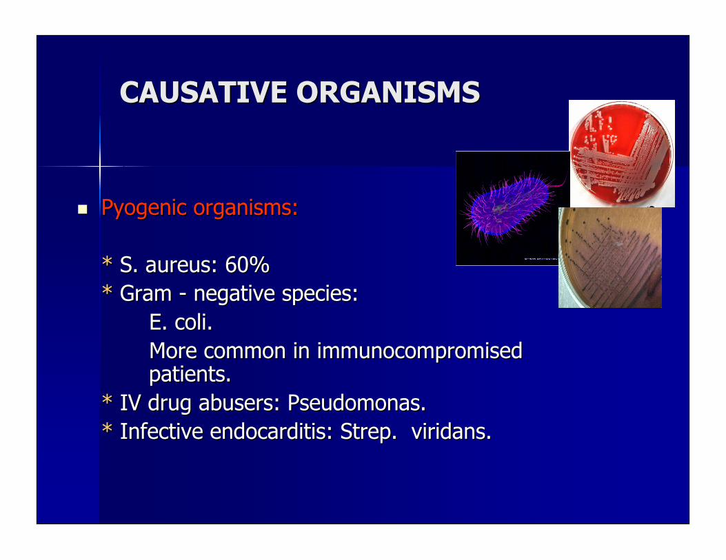
CAUSATIVE ORGANISMSCAUSATIVE ORGANISMS
PyogenicPyogenic organisms: organisms:
** S. S. aureusaureus: 60%: 60%** Gram - negative species: Gram - negative species:
E. coli. E. coli. More common in More common in immunocompromisedimmunocompromised patients. patients.
** IV drug abusers: Pseudomonas. IV drug abusers: Pseudomonas.** Infective Infective endocarditisendocarditis: Strep. : Strep. viridansviridans..
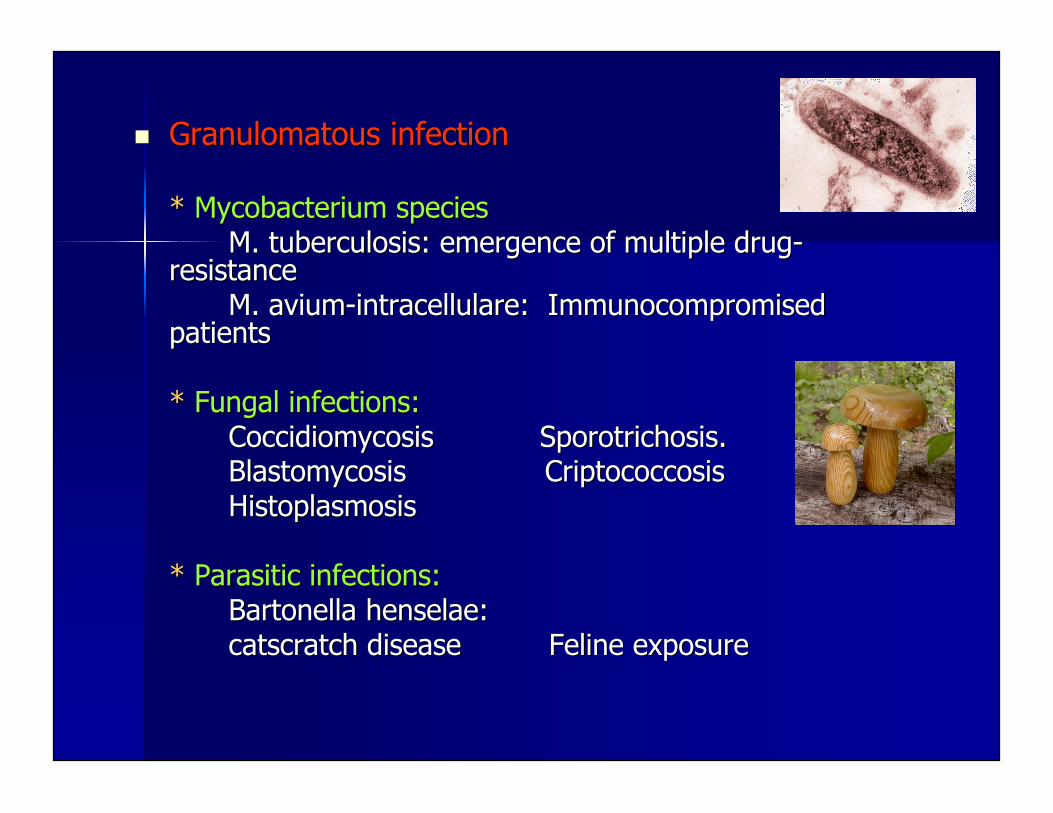
GranulomatousGranulomatous infection infection
** Mycobacterium speciesMycobacterium speciesM. tuberculosis: emergence of multiple drug-M. tuberculosis: emergence of multiple drug-
resistanceresistanceM. M. avium-intracellulareavium-intracellulare: : ImmunocompromisedImmunocompromised
patientspatients
** Fungal infections:Fungal infections:CoccidiomycosisCoccidiomycosis SporotrichosisSporotrichosis..BlastomycosisBlastomycosis CriptococcosisCriptococcosisHistoplasmosisHistoplasmosis
** Parasitic infections:Parasitic infections:BartonellaBartonella henselaehenselae::catscratchcatscratch disease disease Feline exposure Feline exposure
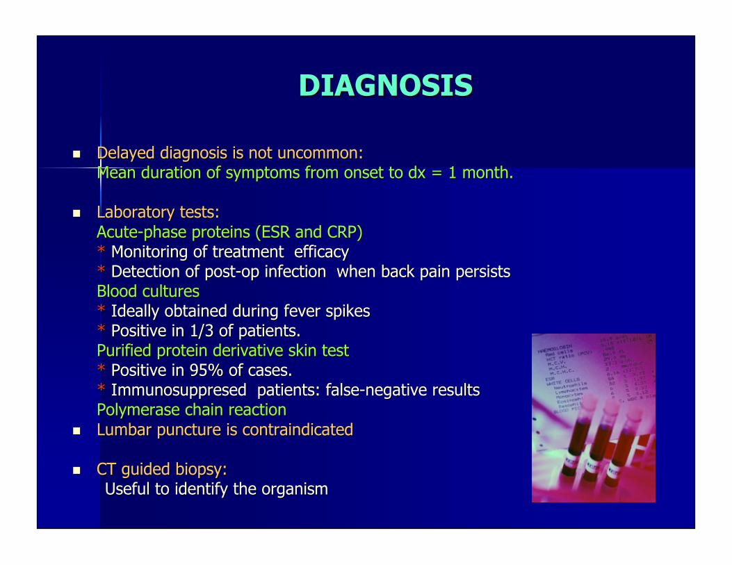
DIAGNOSISDIAGNOSIS
Delayed diagnosis is not uncommon:Delayed diagnosis is not uncommon:Mean duration of symptoms from onset to Mean duration of symptoms from onset to dxdx = 1 month. = 1 month.
Laboratory tests:Laboratory tests:Acute-phase proteins (ESR and CRP)Acute-phase proteins (ESR and CRP)** Monitoring of treatment efficacy Monitoring of treatment efficacy** Detection of post-op infection when back pain persists Detection of post-op infection when back pain persistsBlood culturesBlood cultures** Ideally obtained during fever spikes Ideally obtained during fever spikes** Positive in 1/3 of patients. Positive in 1/3 of patients.Purified protein derivative skin testPurified protein derivative skin test** Positive in 95% of cases. Positive in 95% of cases.** ImmunosuppresedImmunosuppresed patients: false-negative results patients: false-negative resultsPolymerase chain reactionPolymerase chain reaction
Lumbar puncture is contraindicatedLumbar puncture is contraindicated
CT guided biopsy:CT guided biopsy:Useful to identify the organismUseful to identify the organism
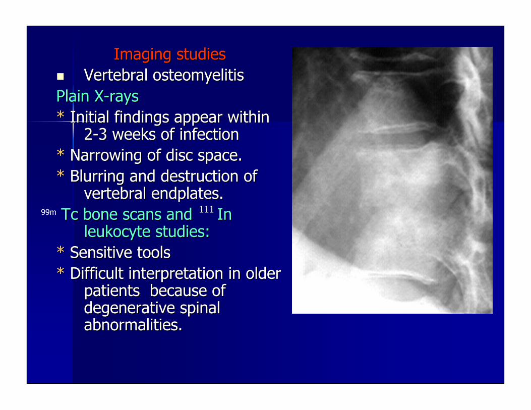
Imaging studiesImaging studies Vertebral Vertebral osteomyelitisosteomyelitisPlain X-raysPlain X-rays** Initial findings appear within Initial findings appear within
2-3 weeks of infection2-3 weeks of infection** Narrowing of disc space. Narrowing of disc space.** Blurring and destruction of Blurring and destruction of
vertebral endplates.vertebral endplates. TcTc bone scans and In bone scans and In
leukocyte studies:leukocyte studies:** Sensitive tools Sensitive tools** Difficult interpretation in older Difficult interpretation in older
patients because ofpatients because ofdegenerative spinaldegenerative spinalabnormalities.abnormalities.
99m 111
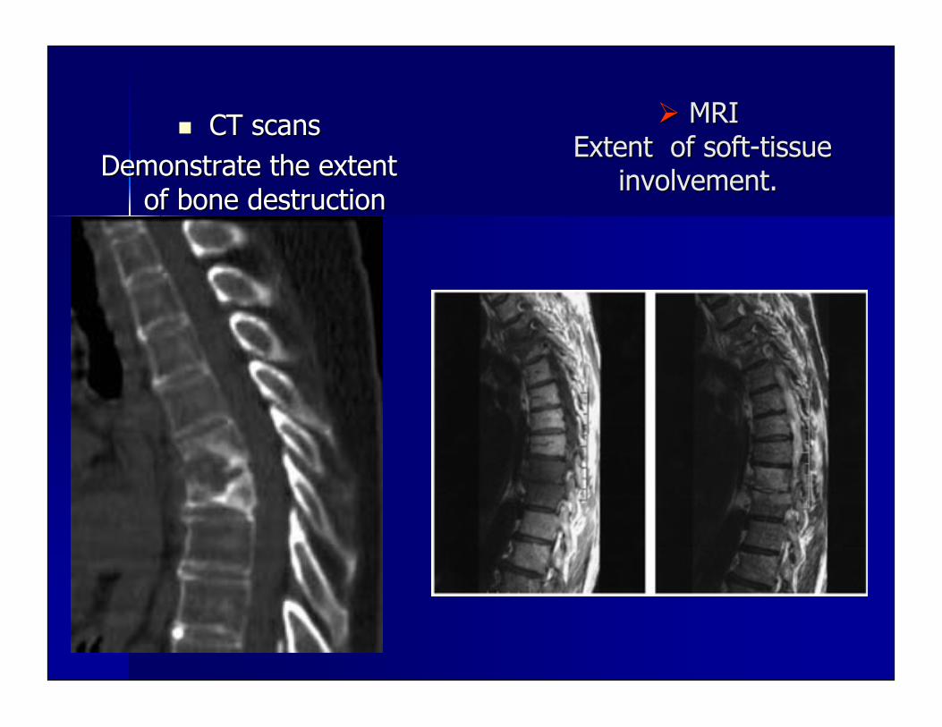
MRI MRI Extent of soft-tissue Extent of soft-tissue
involvement.involvement.
CT scansCT scansDemonstrate the extentDemonstrate the extent
of bone destructionof bone destruction
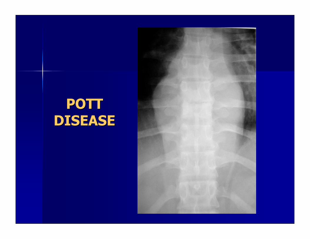
POTTPOTTDISEASEDISEASE
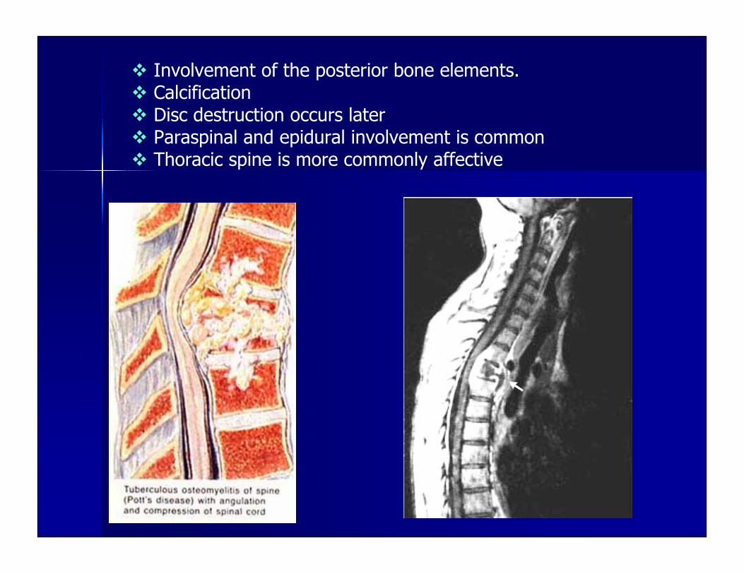
Involvement of the posterior bone elements. Calcification Disc destruction occurs later Paraspinal and epidural involvement is common Thoracic spine is more commonly affective
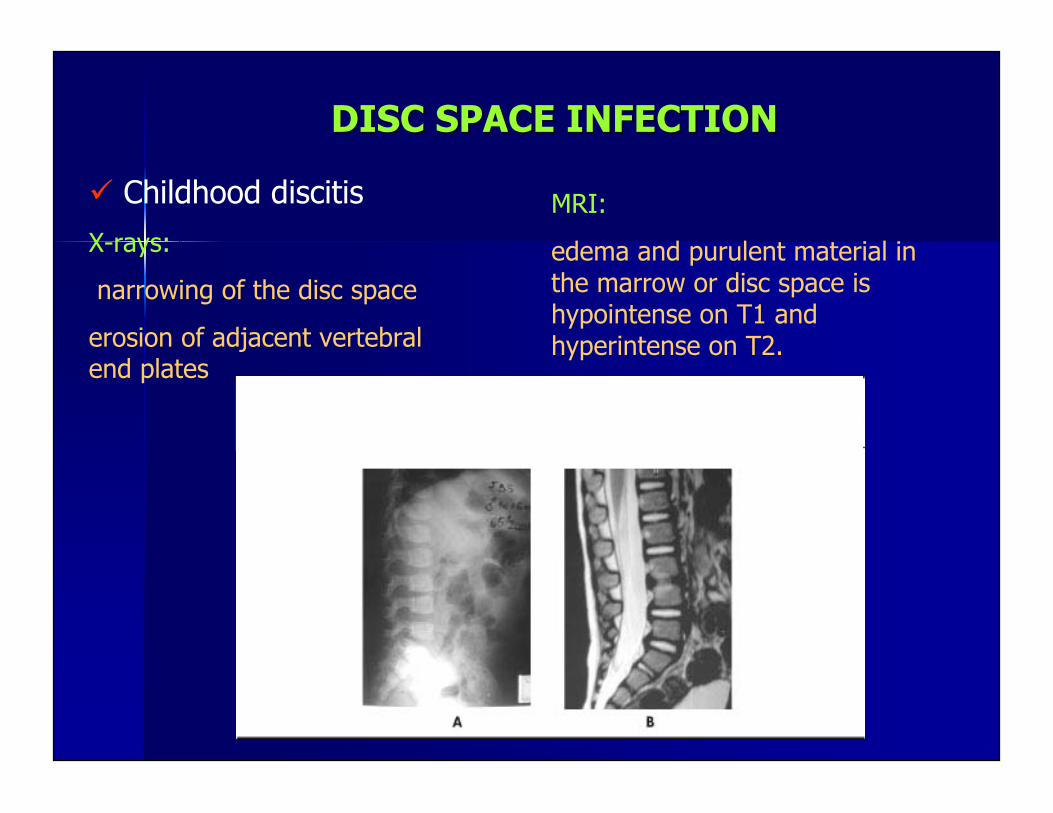
DISC SPACE INFECTIONDISC SPACE INFECTION
Childhood discitis
X-rays:
narrowing of the disc space
erosion of adjacent vertebralend plates
MRI:
edema and purulent material inthe marrow or disc space ishypointense on T1 andhyperintense on T2.
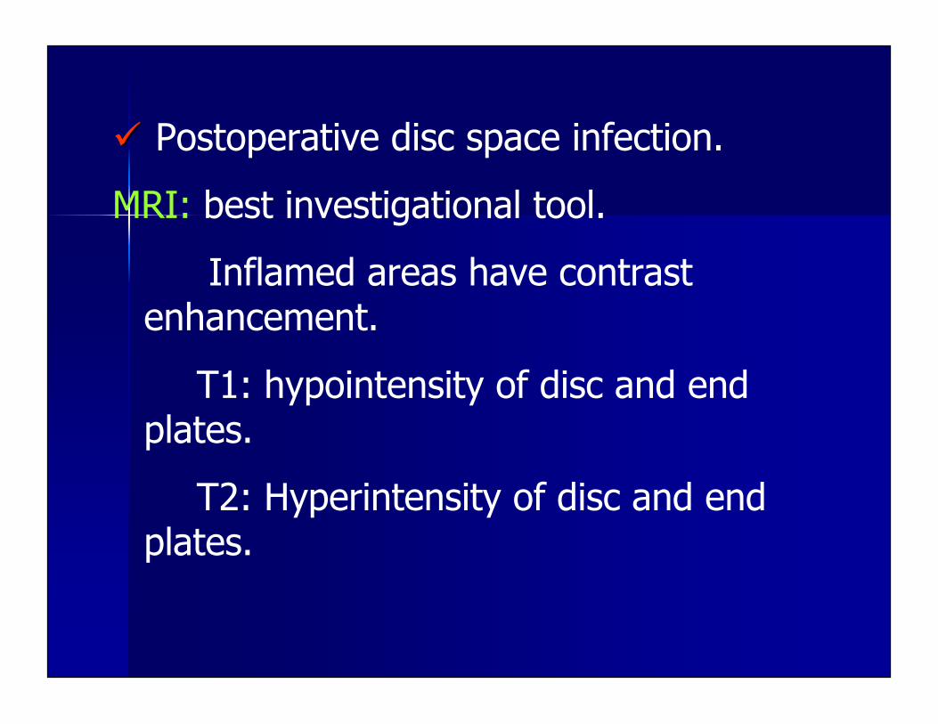
Postoperative disc space infection.
MRI: best investigational tool.
Inflamed areas have contrast enhancement.
T1: hypointensity of disc and end plates.
T2: Hyperintensity of disc and end plates.
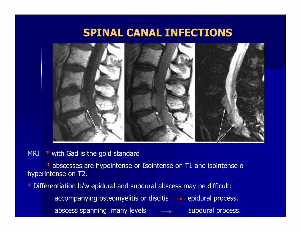
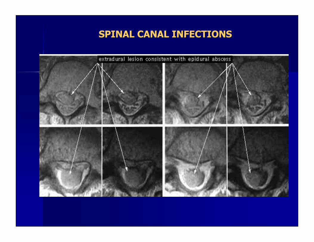
SPINAL CANAL INFECTIONSSPINAL CANAL INFECTIONS
MRI * with Gad is the gold standard
* abscesses are hypointense or Isointense on T1 and isointense o hyperintense on T2.
* Differentiation b/w epidural and subdural abscess may be difficult:
accompanying osteomyelitis or discitis epidural process.
abscess spanning many levels subdural process.
SPINAL CANAL INFECTIONSSPINAL CANAL INFECTIONS
MANAGEMENTMANAGEMENTBasic goals:Basic goals: Eradication of infectionEradication of infection Prevention or reversal of neurological deficitPrevention or reversal of neurological deficit Relief of pain.Relief of pain.NonsurgicalNonsurgical management: management: Indicated in patients with no neurological deficit and no significant Indicated in patients with no neurological deficit and no significant kyphotickyphotic deformity or pathological deformity or pathological
fracture.fracture. Medical treatment: Medical treatment: AbxAbx and rest. and rest. Immobilization Immobilization
** T&L spine: TLSO T&L spine: TLSO** C spine: hard collar. C spine: hard collar.
Identification of specific causative agentIdentification of specific causative agent** CT guided biopsy CT guided biopsy** Blood cultures Blood cultures** Repeat CT guided biopsy and blood cultures if prior results negative Repeat CT guided biopsy and blood cultures if prior results negative** Open biopsy if again negative results. Open biopsy if again negative results.
Duration of Duration of AbxAbx** PyogenicPyogenic osteomyelitisosteomyelitis: 6-8 weeks: 6-8 weeks** POTT disease: 12 months POTT disease: 12 months
Failure of medical management:Failure of medical management: if symptoms persist or increase after: if symptoms persist or increase after:* 1 month * 1 month pyogenicpyogenic infection infection* 3 months * 3 months PottPott disease. disease.
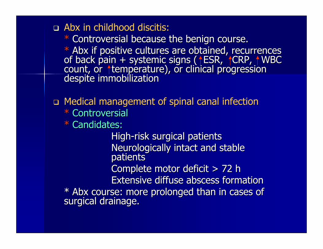
AbxAbx in childhood in childhood discitisdiscitis::** Controversial because the benign course. Controversial because the benign course.** AbxAbx if positive cultures are obtained, recurrences if positive cultures are obtained, recurrencesof back pain + systemic signs ( ESR, CRP, WBCof back pain + systemic signs ( ESR, CRP, WBCcount, or temperature), or clinical progressioncount, or temperature), or clinical progressiondespite immobilizationdespite immobilization
Medical management of spinal canal infectionMedical management of spinal canal infection** ControversialControversial** Candidates:Candidates:
High-risk surgical patientsHigh-risk surgical patientsNeurologically intact and stable Neurologically intact and stable patientspatientsComplete motor deficit > 72 hComplete motor deficit > 72 hExtensive diffuse abscess formationExtensive diffuse abscess formation
* * AbxAbx course: more prolonged than in cases of course: more prolonged than in cases ofsurgical drainage.surgical drainage.
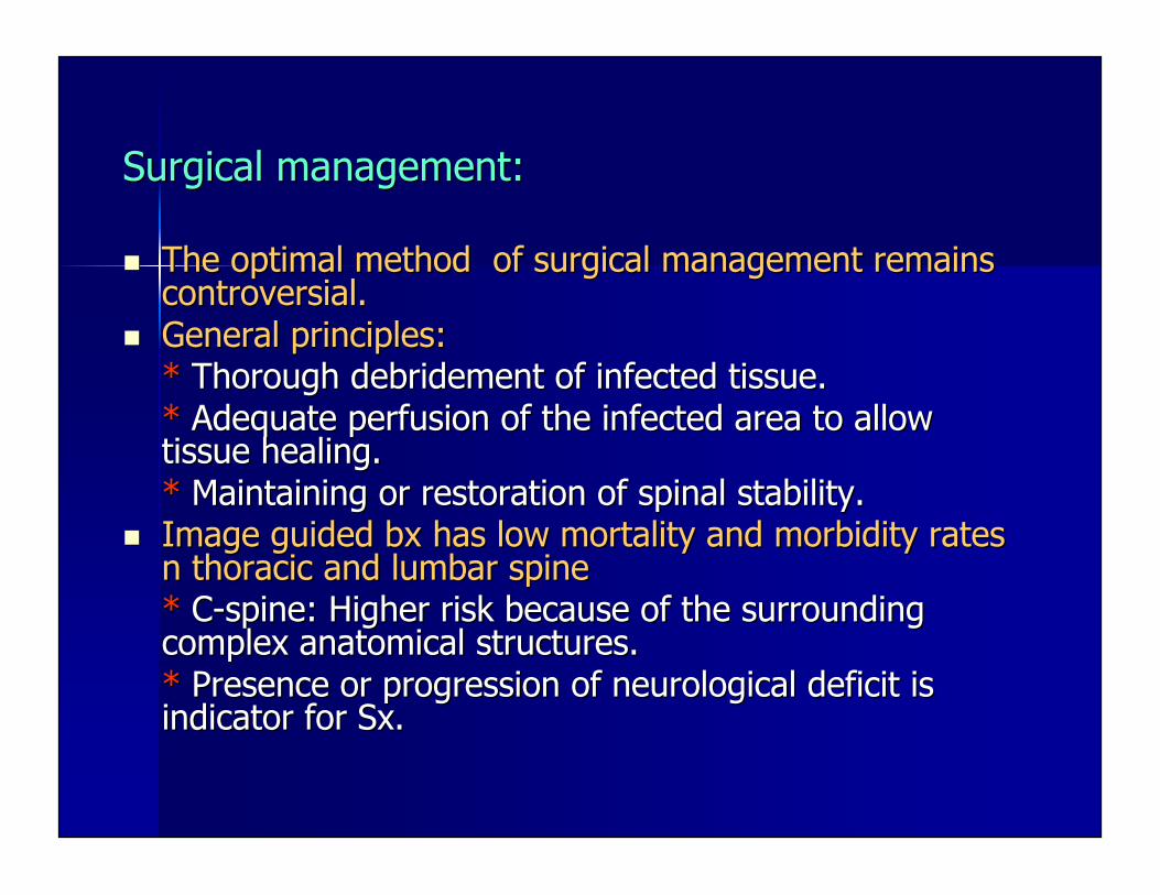
Surgical management:Surgical management:
The optimal method of surgical management remainsThe optimal method of surgical management remainscontroversial.controversial.
General principles:General principles:** Thorough Thorough debridementdebridement of infected tissue. of infected tissue.* * Adequate perfusion of the infected area to allowAdequate perfusion of the infected area to allowtissue healing.tissue healing.** Maintaining or restoration of spinal stability. Maintaining or restoration of spinal stability.
Image guided Image guided bxbx has low mortality and morbidity rates has low mortality and morbidity ratesn thoracic and lumbar spinen thoracic and lumbar spine** C-spine: Higher risk because of the surrounding C-spine: Higher risk because of the surroundingcomplex anatomical structures.complex anatomical structures.** Presence or progression of neurological deficit is Presence or progression of neurological deficit isindicator for indicator for SxSx..
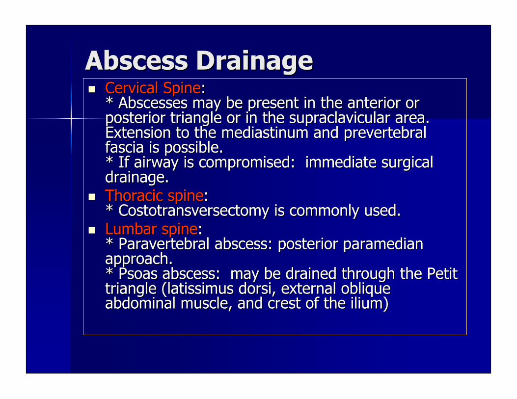
Abscess DrainageAbscess Drainage Cervical SpineCervical Spine::
* Abscesses may be present in the anterior or* Abscesses may be present in the anterior orposterior triangle or in the posterior triangle or in the supraclavicularsupraclavicular area. area.Extension to the Extension to the mediastinummediastinum and and prevertebralprevertebralfascia is possible.fascia is possible.* If airway is compromised: immediate surgical* If airway is compromised: immediate surgicaldrainage.drainage.
Thoracic spineThoracic spine::* * CostotransversectomyCostotransversectomy is commonly used. is commonly used.
Lumbar spineLumbar spine::* * ParavertebralParavertebral abscess: posterior abscess: posterior paramedianparamedianapproach.approach.* * PsoasPsoas abscess: may be drained through the Petit abscess: may be drained through the Petittriangle (triangle (latissimuslatissimus dorsidorsi, external oblique, external obliqueabdominal muscle, and crest of the abdominal muscle, and crest of the iliumilium))
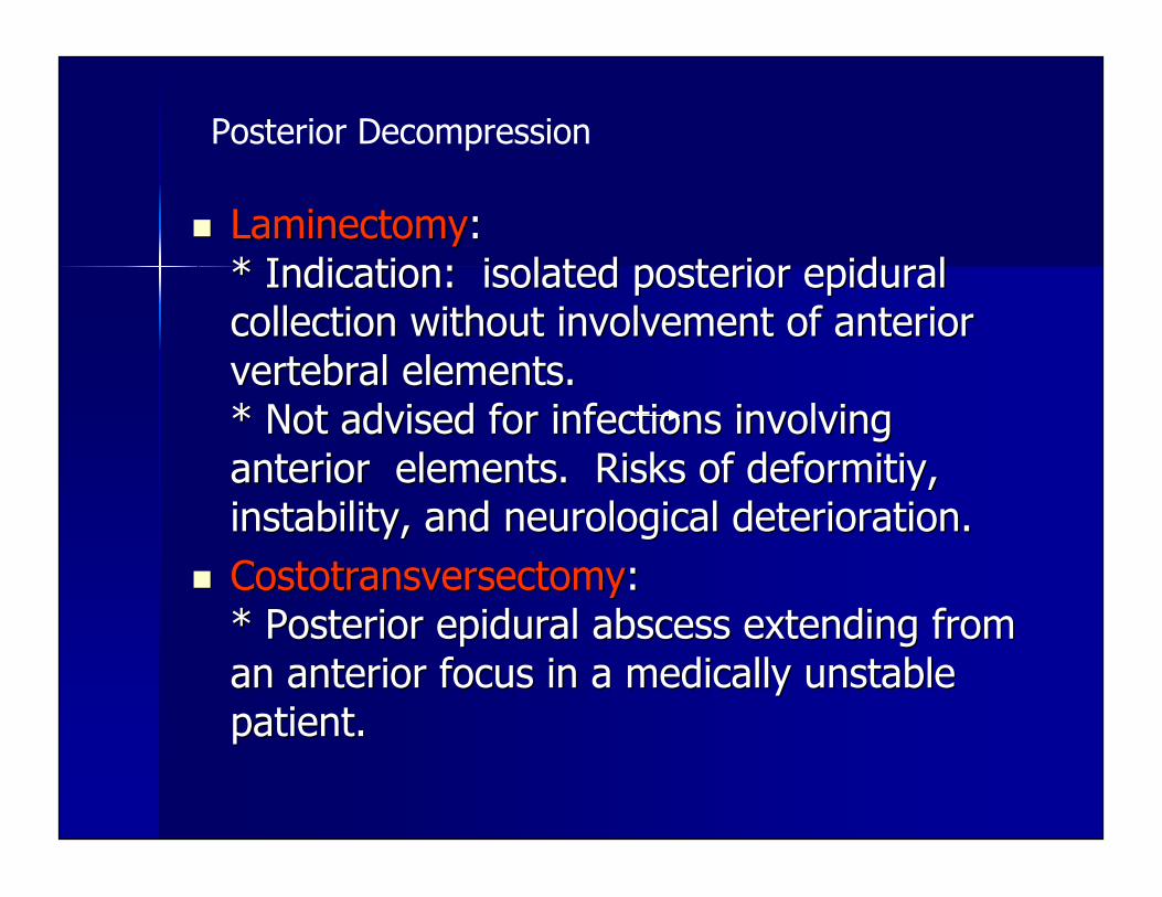
LaminectomyLaminectomy::* Indication: isolated posterior epidural* Indication: isolated posterior epiduralcollection without involvement of anteriorcollection without involvement of anteriorvertebral elements.vertebral elements.* Not advised for infections involving* Not advised for infections involvinganterior elements. Risks of anterior elements. Risks of deformitiydeformitiy,,instability, and neurological deterioration.instability, and neurological deterioration.
CostotransversectomyCostotransversectomy::* Posterior epidural abscess extending from* Posterior epidural abscess extending froman anterior focus in a medically unstablean anterior focus in a medically unstablepatient.patient.
Posterior Decompression
Anterior DecompressionAnterior Decompression ModalitiesModalities::
1) Anterior decompression with and1) Anterior decompression with andwithout without autologousautologous bone graft. bone graft.2) Anterior decompression and fusion2) Anterior decompression and fusionwith posterior stabilization.with posterior stabilization.3) Anterior decompression and fusion3) Anterior decompression and fusionwith anterior instrumentation.with anterior instrumentation.
Postoperative SpinalPostoperative SpinalInfections After PlacementInfections After Placementof Instrumentationof Instrumentation.. Incidence of spinal infection after Incidence of spinal infection after laminectomylaminectomy or or
discectomydiscectomy: 1%: 1% Incidence of spinal infection after instrumentation:Incidence of spinal infection after instrumentation:
2.1 - 8.5 %2.1 - 8.5 % Strategies:Strategies:
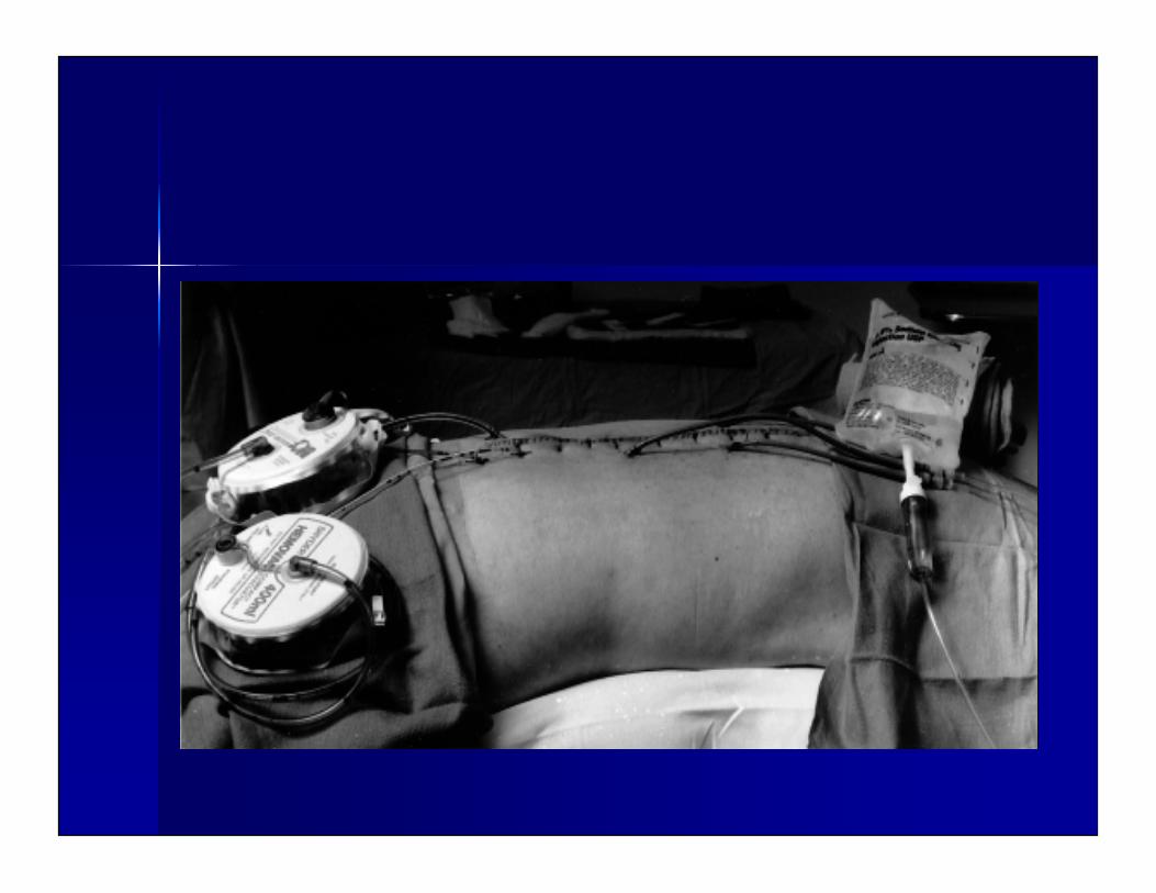
* Multiple surgical * Multiple surgical debridementsdebridements with removal of with removal ofhardware.hardware.*Antibiotic-impregnated beads with multiple*Antibiotic-impregnated beads with multiplereoperationsreoperations..* Surgical * Surgical debridementdebridement and insertion of an and insertion of anantibiotic delivery irrigation-suction system for 5antibiotic delivery irrigation-suction system for 5days. days. Instrumentation is not removedInstrumentation is not removed..
RHEUMATOID ARTHRITIS:RHEUMATOID ARTHRITIS:
RA is a debilitating RA is a debilitating polyarthropaticpolyarthropatic degenerative degenerativecondition.condition.
86% of patients have C-spine involvement.86% of patients have C-spine involvement. If left untreated a large percentage of patientsIf left untreated a large percentage of patients
progress toward complex instability patterns.progress toward complex instability patterns. Once Once myelopathymyelopathy occurs, prognosis for occurs, prognosis for neurologicneurologic
recovery and long term survival is poor.recovery and long term survival is poor.
RA is a progressive RA is a progressive inflamatoryinflamatory condition conditionexhibiting exhibiting polyarthropathicpolyarthropathic degeneration and degeneration andsystemic manifestations resulting from both thesystemic manifestations resulting from both thedisease process and management-associateddisease process and management-associatedmedication.medication.
Clinical manifestations of the disease are theClinical manifestations of the disease are theresult of an autoimmune process that attacks theresult of an autoimmune process that attacks thejoint joint synoviumsynovium, resulting in the release of, resulting in the release ofcytokines that stimulate inflammatory reactioncytokines that stimulate inflammatory reactioneventually leading to join destruction andeventually leading to join destruction andperiarticularperiarticular erosions. erosions.
PATHOPHYSIOLOGYPATHOPHYSIOLOGY The T-lymphocyte-mediated process results inThe T-lymphocyte-mediated process results in
an inflammatory an inflammatory synovitissynovitis and rheumatoid and rheumatoidpannuspannus that can damage: that can damage:** ligaments ligaments * * bonesbones** synovialsynovial joints in the cervical spine joints in the cervical spine** potentially resulting in potentially resulting in subluxationsubluxation** instability and brainstem or cord instability and brainstem or cordcompression.compression.
Because the large number of Because the large number of synovialsynovialarticulations present and the increase range ofarticulations present and the increase range ofmotion required in the upper C-spine themotion required in the upper C-spine theoccipito-atlantoaxialoccipito-atlantoaxial joints are the greatest risk joints are the greatest riskfor pathological involvement.for pathological involvement.
Most common patterns of instability:Most common patterns of instability:** AtlantoAtlanto axial axial subluxationsubluxation (AAS) (AAS)** Cranial settling Cranial settling** SubaxialSubaxial subluxationsubluxation (SAS) (SAS)
AtlantoAtlanto-axial joint is most commonly affected because:-axial joint is most commonly affected because:** The consistent involvement of the The consistent involvement of the synovialsynovial-lined-linedbursa found b/w the bursa found b/w the odontoidodontoid and the transverse and the transverseligament.ligament.** Subsequent weakening and rupture of the Subsequent weakening and rupture of thetransverse, transverse, alaralar, and apical ligaments combined with, and apical ligaments combined witherosions of the erosions of the atlantoaxialatlantoaxial joint can lead to anterior joint can lead to anteriorAAS.AAS.** The increased The increased atlantoaxialatlantoaxial motion + motion + periodontoidperiodontoidrheumatoid rheumatoid pannuspannus can result in can result in catatastrophiccatatastrophic spinal spinalcord and/or brain stem injury.cord and/or brain stem injury.
Posterior AASPosterior AAS** Represents 6.7% of all AASRepresents 6.7% of all AAS** Results from either C1 ant. Arch defects or from Results from either C1 ant. Arch defects or from odontoidodontoiderosions/fractures.erosions/fractures.
Lateral AAS:Lateral AAS:* * Secondary to a combination of rotational deformities.Secondary to a combination of rotational deformities.Frequency of AASFrequency of AAS** anterior > lateral (20%) > Posterior (10%) anterior > lateral (20%) > Posterior (10%)
CranealCraneal settling: settling:
** AKA: Basilar settling, basilar AKA: Basilar settling, basilar invaginationinvagination, , atlantoaxialatlantoaxial invaginationinvagination,,atlantoaxialatlantoaxial impaction and sup. migration of the dens. impaction and sup. migration of the dens.** Occurs as result of bone and cartilage destruction at the Occurs as result of bone and cartilage destruction at the occipito-occipito-atlantalatlantal and and atlantoaxialatlantoaxial joints. joints.** TipicallyTipically there is a destruction of the lateral masses of the atlas. there is a destruction of the lateral masses of the atlas.Less commonly the lateral masses of the axis and the occipital Less commonly the lateral masses of the axis and the occipital condylescondylesmay be involved.may be involved.** Bilateral destruction and collapse results in settling of the skull ontoBilateral destruction and collapse results in settling of the skull ontothe cervical spine with subsequent relative superior migration of thethe cervical spine with subsequent relative superior migration of theodontoidodontoid into the foramen magnum. into the foramen magnum.** Cranial setting is almost always preceded by AAS.Cranial setting is almost always preceded by AAS.
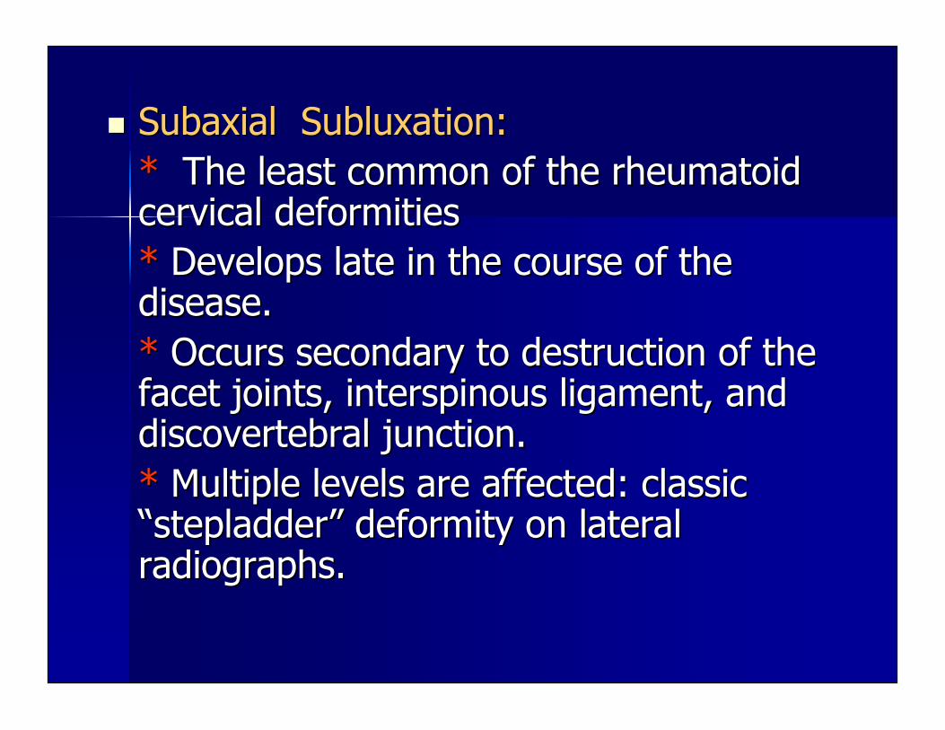
SubaxialSubaxial SubluxationSubluxation::* * The least common of the rheumatoid The least common of the rheumatoidcervical deformitiescervical deformities** Develops late in the course of the Develops late in the course of thedisease.disease.** Occurs secondary to destruction of the Occurs secondary to destruction of thefacet joints, facet joints, interspinousinterspinous ligament, and ligament, anddiscovertebraldiscovertebral junction. junction.** Multiple levels are affected: classic Multiple levels are affected: classic““stepladderstepladder”” deformity on lateral deformity on lateralradiographs.radiographs.
NATURAL HISTORYNATURAL HISTORY
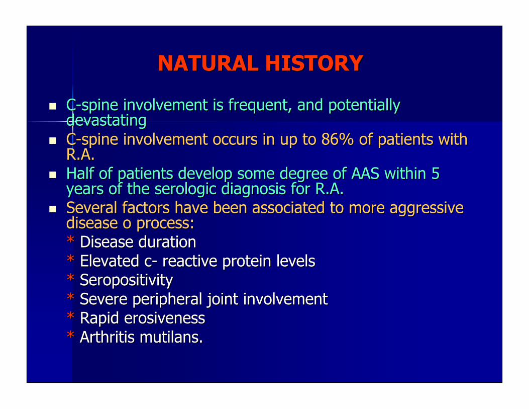
C-spine involvement is frequent, and potentiallyC-spine involvement is frequent, and potentiallydevastatingdevastating
C-spine involvement occurs in up to 86% of patients withC-spine involvement occurs in up to 86% of patients withR.A.R.A.
Half of patients develop some degree of AAS within 5Half of patients develop some degree of AAS within 5years of the serologic diagnosis for R.A.years of the serologic diagnosis for R.A.
Several factors have been associated to more aggressiveSeveral factors have been associated to more aggressivedisease o process:disease o process:** Disease duration Disease duration** Elevated c- reactive protein levels Elevated c- reactive protein levels** SeropositivitySeropositivity** Severe peripheral joint involvement Severe peripheral joint involvement** Rapid Rapid erosivenesserosiveness* * Arthritis Arthritis mutilansmutilans..
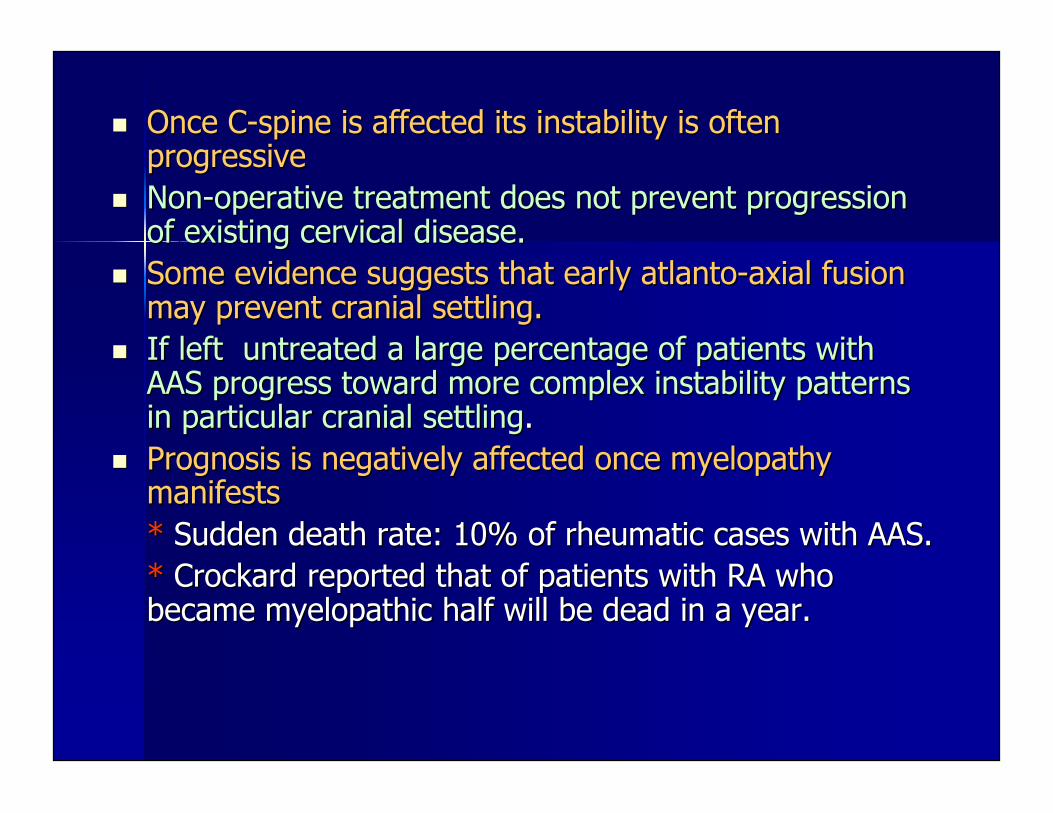
Once C-spine is affected its instability is oftenOnce C-spine is affected its instability is oftenprogressiveprogressive
Non-operative treatment does not prevent progressionNon-operative treatment does not prevent progressionof existing cervical disease.of existing cervical disease.
Some evidence suggests that early Some evidence suggests that early atlantoatlanto-axial fusion-axial fusionmay prevent cranial settling.may prevent cranial settling.
If left untreated a large percentage of patients withIf left untreated a large percentage of patients withAAS progress toward more complex instability patternsAAS progress toward more complex instability patternsin particular cranial settlingin particular cranial settling..
Prognosis is negatively affected once Prognosis is negatively affected once myelopathymyelopathymanifestsmanifests** Sudden death rate: 10% of rheumatic cases with AAS. Sudden death rate: 10% of rheumatic cases with AAS.** CrockardCrockard reported that of patients with RA who reported that of patients with RA whobecame became myelopathicmyelopathic half will be dead in a year. half will be dead in a year.
CLINICAL MANIFESTATIONSCLINICAL MANIFESTATIONS
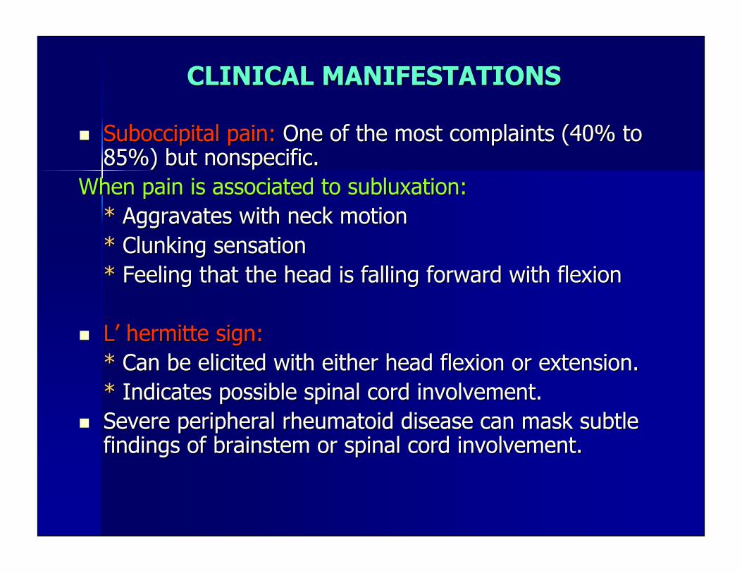
SuboccipitalSuboccipital pain: pain: One of the most complaints (40% to One of the most complaints (40% to85%) but nonspecific.85%) but nonspecific.
When pain is associated to When pain is associated to subluxationsubluxation::** Aggravates with neck motion Aggravates with neck motion** Clunking sensation Clunking sensation** Feeling that the head is falling forward with flexion Feeling that the head is falling forward with flexion
LL’’ hermittehermitte sign: sign:** Can be elicited with either head flexion or extension. Can be elicited with either head flexion or extension.** Indicates possible spinal cord involvement. Indicates possible spinal cord involvement.
Severe peripheral rheumatoid disease can mask subtleSevere peripheral rheumatoid disease can mask subtlefindings of brainstem or spinal cord involvement.findings of brainstem or spinal cord involvement.
First signs of early First signs of early myelopathymyelopathy::** Clumsiness of the hand Clumsiness of the hand** Gait disturbances Gait disturbances** Sensation of heaviness or fatigability in the legs. Sensation of heaviness or fatigability in the legs.
Cervical spine involvement should be suspected in patients whoCervical spine involvement should be suspected in patients whowere previously ambulatory even if with were previously ambulatory even if with crutchscrutchs or a walker who or a walker whobecame wheelchair dependent.became wheelchair dependent.
PE:PE:** Weakness Weakness** SpasticitySpasticity** Clumsiness Clumsiness** Pathologic reflexes: Pathologic reflexes: hyperrreflexiahyperrreflexia and positive and positive BabinskiBabinski and and
Hoffman signHoffman sign** Urinary and rectal disturbances: are unusual and occur late Urinary and rectal disturbances: are unusual and occur late
in the disease progress.in the disease progress. Patients with C-spine involvement may present with findingsPatients with C-spine involvement may present with findings
consistent with consistent with vertebrobasilarvertebrobasilar insufficiency: insufficiency:** vertigo vertigo ** vomiting vomiting ** dysarthriadysarthria** nausea nausea ** dysphagiadysphagia or or cerebellarcerebellar signs. signs.
IMAGINGIMAGING
Plain RadiographsPlain Radiographs Screening x-rays include:Screening x-rays include:* AP* AP * Lateral* Lateral* Open mouth * Open mouth odontoidodontoid * Dynamic flexion * Dynamic flexion
and extension lateral views.and extension lateral views. Particular attention should be paid to:Particular attention should be paid to:
* Anterior * Anterior atlantodentalatlantodental interval (AADI) interval (AADI)* Posterior * Posterior atlantodentalatlantodental interval (PADI) interval (PADI)* Superior migration of the tip of the * Superior migration of the tip of the odontoidodontoid* Degree of * Degree of subaxialsubaxial subluxationsubluxation..
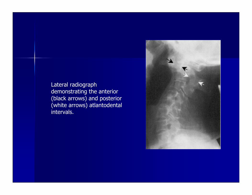
Lateral radiographdemonstrating the anterior(black arrows) and posterior(white arrows) atlantodentalintervals.
AtlantoAtlanto-axial complex integrity-axial complex integrity::* Evaluated by lateral flexion-* Evaluated by lateral flexion-extension radiographs.extension radiographs.* The amount of dynamic motion* The amount of dynamic motionpresent during flexion-extensionpresent during flexion-extensionradiographs is more relevant thanradiographs is more relevant thanvalues obtained from single neutralvalues obtained from single neutrallateral radiograph.lateral radiograph.
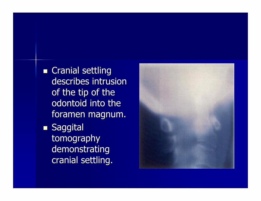
Cranial settlingCranial settlingdescribes intrusiondescribes intrusionof the tip of theof the tip of theodontoidodontoid into the into theforamen magnum.foramen magnum.
SaggitalSaggitaltomographytomographydemonstratingdemonstratingcranial settling.cranial settling.
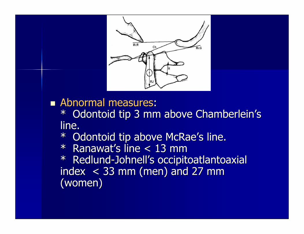
Abnormal measuresAbnormal measures::* * OdontoidOdontoid tip 3 mm above tip 3 mm above ChamberleinChamberlein’’ssline.line.* * OdontoidOdontoid tip above McRae tip above McRae’’s line.s line.* * RanawatRanawat’’ss line < 13 mm line < 13 mm* * Redlund-JohnellRedlund-Johnell’’ss occipitoatlantoaxialoccipitoatlantoaxialindex < 33 mm (men) and 27 mmindex < 33 mm (men) and 27 mm(women)(women)
Computed TomographyComputed Tomography
Provides valuable information aboutProvides valuable information aboutbony detail and osseous destruction.bony detail and osseous destruction.
Use of CT with and without contrastUse of CT with and without contrastprovides information aboutprovides information aboutinflammatory soft tissue changesinflammatory soft tissue changesdifferentiating b/w effusions anddifferentiating b/w effusions andhypervascularhypervascular pannuspannus..
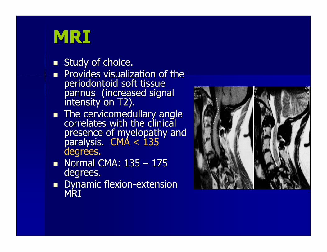
MRIMRI Study of choice.Study of choice. Provides visualization of theProvides visualization of the
periodontoidperiodontoid soft tissue soft tissuepannuspannus (increased signal (increased signalintensity on T2).intensity on T2).
The The cervicomedullarycervicomedullary angle anglecorrelates with the clinicalcorrelates with the clinicalpresence of presence of myelopathymyelopathy and andparalysis. paralysis. CMA < 135CMA < 135degrees.degrees.
Normal CMA: 135 Normal CMA: 135 –– 175 175degrees.degrees.
Dynamic flexion-extensionDynamic flexion-extensionMRIMRI
Indications for SurgeryIndications for Surgery Intractable pain.Intractable pain. Neurologic involvement:Neurologic involvement:
* Cervical collars do not prevent progression of* Cervical collars do not prevent progression ofsubluxationsubluxation..* Once * Once myelopathymyelopathy develops the prognosis is poor. develops the prognosis is poor.
Cervical instability with Cervical instability with ““impendingimpending”” neurologicneurologicinvolvement:involvement:* Most challenging group: patients with cervical* Most challenging group: patients with cervicalsubluxationsubluxation without without neurologicneurologic deficit and minimal deficit and minimalpain.pain.* Classical criteria for * Classical criteria for atlantoatlanto-axial and sub-axial-axial and sub-axialinstability should not be extrapolated to RAinstability should not be extrapolated to RApatients.patients.* Cut off for surgery: AADI > 10 mm, PADI < 14* Cut off for surgery: AADI > 10 mm, PADI < 14mm, Space available for the cord (SAC) < 13 mmmm, Space available for the cord (SAC) < 13 mm
Surgical ManagementSurgical Management
Main goalsMain goals::* Preserve or restore * Preserve or restore neurologicneurologic function. function.* Management of cervical instability and deformity* Management of cervical instability and deformityshould focus on restoring alignment and obtainingshould focus on restoring alignment and obtainingsolid solid arthrodesisarthrodesis..
AAS surgical techniquesAAS surgical techniques::* Modified * Modified GallieGallie graft. graft.* Brooks-Jenkins fusion.* Brooks-Jenkins fusion.* * MagerlMagerl C1-C2 C1-C2 transarticulartransarticular screw technique. screw technique.
SAS surgical techniquesSAS surgical techniques::* * InterspinousInterspinous wiring. wiring.* Lateral mass screw fixation.* Lateral mass screw fixation.* Pedicle screw fixation.* Pedicle screw fixation.