Embed Size (px)
Citation preview
siemens.com/healthineers
in the Age ofMedical Imaging
Artificial Intelligence
White Paper
In the next five to 10 years, artificial intelligence is likely to fundamentally transform diagnostic imaging. Although intelligent algorithms have been used for some time in segments of the imag-ing field, new methods of machine learning, based particularly on “deep learning”, are much more powerful. According to pilot studies, they offer concrete prospects for quantitative, standardized imaging and reporting that is, at the same time, person alized. AI will not by any means replace radiologists – rather it will provide them with tools to meet the rising demand for diagnostic imaging and actively shape the transformation of radiology into a data-driven research discipline. AI algorithms are expected to help speed up clinical workflows, prevent diagnostic errors and reduce missed billing opportunities, thus enabling sustained productivity increases. Above all, methods of artificial intelligence could lead to more precise results and more meaningful prognostic risk scores, and integrate diagnostic radiology even more into outcome-oriented clinical decision-making.
Adding Value with AI in Medical Imaging
Challenges for a Transforming Discipline . . . . . . . . . . . . . . . . . . . . . 4
Machine Learning is Entering a New Era . . . . . . . . . . . . . . . . . . . . . . . 5
Exploring the Clinical Value of AI . . . . . . . . . . . . . . . . . . . . . . . . . . . . 8
A Framework for the Future . . . . . . . . . . . . . . . . . . . . . . . . . . . . . . . . . . 9
Conclusion . . . . . . . . . . . . . . . . . . . . . . . . . . . . . . . . . . . . . . . . . . . . . . . . . 10
3
White Paper | Artificial Intelligence
In recent years artificial intelligence (AI) gained entrance into everyday life in various ways. Intelligent computer algorithms are utilized in Internet search engines and language recognition on smart phones, as well as in the analysis of genetic data, photographic images or financial transactions, humanoid robots and self-driving cars. Just recently, a machine-learning-based computer program (“AlphaGo”) defeated the world champion of Go the strategic board game, under tournament conditions, once again dem onstrating the potential of AI to a broad public (Mozur 2017).
Medical imaging, too, is likely to undergo a fundamental transformation in the near future due to the latest AI methods such as “deep learning,” which is based on artificial neural networks. “It is easy to predict that AI will be increasingly implemented in medical imaging systems,” Italian doctor Francesco Sardanelli commented in an editorial on dominant trends in radiology (Sardanelli 2017). Likewise, according to a recent poll more than 50% of global healthcare leaders expect an expanding role of AI in monitoring and diagnosis (The Economist 2017). Whereas the use of artificial intelligence is already common practice in segments of imaging, market analyses foresee a veritable boom in innovative AI applications over the next five to 10 years (Signify Research 2017). They have the potential to raise analysis and interpretation of digital medical images to a whole new level compared with older algorithms, and pave the way for quantitative, standardized yet also personalized diagnostics, while helping to prevent errors in diagnosis. Also, AI-based risk scores could revolutionize long-term management of chronic diseases. Numerous studies by academic research groups now confirm the basic clinical value of this second generation of artificial intelligence in medicine.
“Meaningful AI will improve quality, efficiency, and outcomes,” emphasized Keith Dreyer, vice chairman of radiology at Massachusetts General Hospital in Boston and associate professor at Harvard Medical School, at the 2017 Annual Meeting of the Society for Imaging Informatics in Medicine in Pittsburgh, Pennsylvania (Ridley 2017). Many experts, however, regard scenarios, which foresee diagnostic computer algorithms soon eliminating or replacing doctors with training in radiology, as highly unlikely (Chockley & Emanuel 2016). The general message is not “man versus AI,” but rather “man with AI.” Radiologists – always forerunners with regard to digital technologies in medicine – now have the opportunity to meet the challenges of their changing discipline with help from machine intelligence.
Introduction: Medical Imaging in the Age of AI
Source: Sardanelli 2017
“It is easy to predict that AI will be increasingly implemented in medical imaging systems.”
4
Artificial Intelligence | White Paper
There are several factors simultaneously driving integration of AI in radiology. Firstly, in many countries around the world there is a discrepancy between the number of doctors trained in radiology and the rising demand for diagnostic imaging. This leads to greater demands for work efficiency and productivity. For example, the number of radiology specialists (consultant work-force) in England went up 5% between 2012 and 2015, while in the same period the number of CT and MR scans increased by 29 and 26 percentage points respectively. In Scotland, the gap widened even further (The Royal College of Radiologists 2016). Today, the average radiologist is interpreting an image every three to four seconds, eight hours a day (Choi et al. 2016).
Secondly, the image resolution of today’s scanners is continuously improving – resulting in an ever greater volume of data. Indeed, the estimated overall medical data volume doubles every three years, making it harder and harder for radiologists to make good use of the available information without extra help from computerized digital processing. It is desirable, both in radiological research and in clinical diagnostics, to be able to quantitatively analyze this largely unexploited wealth of data and, for example, utilize new measurable imaging biomarkers to assess disease progression and prognosis (O’Connor et al. 2017). Experts see considerable future potential in the transformation of radiology from a discipline of qualitative interpretation to one of quantita-tive analysis, which derives clinically relevant information from extensive data sets (“radiomics”). “Images are more than pictures, they are data,” American radiologist Robert Gillies and his colleagues write (Gillies et al. 2016). Of course, this direction for radiology will require powerful, automated procedures, some of which at least will come under the field of artificial intelligence.
Last but not least, diagnostic errors are an unresolved problem. Studies show that erroneous interpretations occur in about 4% of all radiology diagnoses, with the error rate varying individu-ally and heavily dependent on the procedure. In abdominal and pelvic CT scans, the rate is much higher, for example (Radiology Quality Institute 2012; Berlin 2007). When radiologists are forced to work faster, their average interpretation error rate rises substantially (Sokolovskaya et al. 2015). It is further well known that radiologists differ not only from one another in image interpretations, even the very same examiner may come to different conclusions when a reading is repeated. If only images that actually show pathological changes are considered in the error analysis, the error rate rises as high as around 30%, meaning that in three out of 10 cases pathological structures are either incorrectly interpreted or simply overlooked (false negative findings). Given the distractions and high workloads in everyday clinical practice, this is not surprising, considering the physiologi-cal conditions needed for effective perception (Waite et al. 2017; Donald & Barnard 2012). “There are times when an experienced physician ‘sees’ a visible lesion clearly and times when he does not,” wrote American radiologist L. Henry Garland back in 1959 in a trailblazing article (Garland 1959). Particularly, incidental findings, for example on a chest CT, are hard to “see” and easy to overlook, which entails many liability issues.
Artificial intelligence will likely help to overcome all these challenges. AIgorithms could thus prove an indispensable aid both for efficient, data-based image analysis and diagnostics with few errors. In fact, the astonishing advances that arti ficial intelligence and machine learning methods have made in recent years justify this optimism.
Challenges for a Transforming Discipline
Source: Gillies et al. 2016
“Images are more than pictures, they are data.”
5
White Paper | Artificial Intelligence
Machine Learning is Entering a New Era
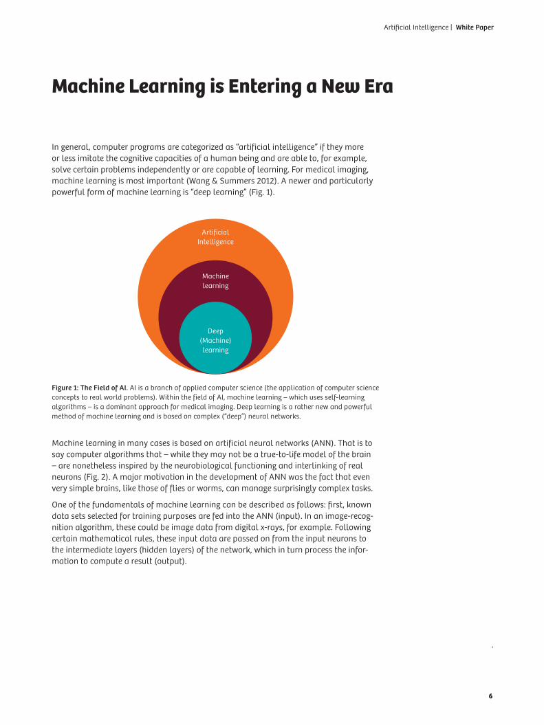
In general, computer programs are categorized as “artificial intelligence” if they more or less imitate the cognitive capacities of a human being and are able to, for example, solve certain problems independently or are capable of learning. For medical imaging, machine learning is most important (Wang & Summers 2012). A newer and particularly powerful form of machine learning is “deep learning” (Fig. 1).
Machine learning in many cases is based on artificial neural networks (ANN). That is to say computer algorithms that – while they may not be a true-to-life model of the brain – are nonetheless inspired by the neurobiological functioning and interlinking of real neurons (Fig. 2). A major motivation in the development of ANN was the fact that even very simple brains, like those of flies or worms, can manage surprisingly complex tasks.
One of the fundamentals of machine learning can be described as follows: first, known data sets selected for training purposes are fed into the ANN (input). In an image-recog-nition algorithm, these could be image data from digital x-rays, for example. Following certain mathematical rules, these input data are passed on from the input neurons to the intermediate layers (hidden layers) of the network, which in turn process the infor-mation to compute a result (output).
Figure 1: The Field of AI. AI is a branch of applied computer science (the application of computer science concepts to real world problems). Within the field of AI, machine learning – which uses self-learning algorithms – is a dominant approach for medical imaging. Deep learning is a rather new and powerful method of machine learning and is based on complex (“deep”) neural networks.
Deep (Machine) learning
Machine learning
Artificial Intelligence
6
Artificial Intelligence | White Paper
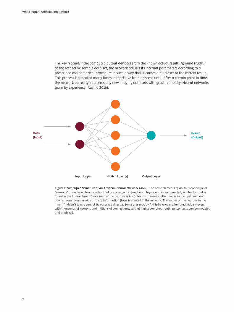
The key feature: if the computed output deviates from the known actual result (“ground truth”) of the respective sample data set, the network adjusts its internal parameters according to a prescribed mathematical procedure in such a way that it comes a bit closer to the correct result. This process is repeated many times in repetitive training steps until, after a certain point in time, the network correctly interprets any new imaging data sets with great reliability. Neural networks learn by experience (Rashid 2016).
Figure 2: Simplified Structure of an Artificial Neural Network (ANN). The basic elements of an ANN are artificial “neurons” or nodes (colored circles) that are arranged in functional layers and interconnected, similar to what is found in the human brain. Since each of the neurons is in contact with several other nodes in the upstream and downstream layers, a wide array of information flows is created in the network. The values of the neurons in the inner (“hidden”) layers cannot be observed directly. Some present-day ANNs have over a hundred hidden layers with thousands of neurons and millions of connections, so that highly complex, nonlinear contexts can be modeled and analyzed.
Data (Input)
Result (Output)
Input Layer Hidden Layer(s) Output Layer
7
White Paper | Artificial Intelligence
The use of machine learning in medical imaging is not new – algorithms today are, however, much more powerful than traditional applications (van Ginneken 2017). The ANNs on which deep learn-ing is based always have multiple functional layers, sometimes even exceeding a hundred, which can encompass thousands of neurons with millions of connections. (Simple ANNs with, say, only one interim layer are described in contrast as “shallow” networks.) All of these connections are adjusted during an ANN’s training by gradual changes of their respective parameters – in mathematical terms: their weights. In this way, deep networks feature a virtually unimaginable number of possible combinations for processing information, and can even model highly complex, non-linear contexts. During the training procedure, the different layers of an ANN increasingly structure the input data with each consecutive layer, developing a more abstract “understanding” of the information. Of course, such deep ANNs were only made possible by advanced mathe-matical methods and the availability of higher computational power and faster graphic processors (GPU) to compute the innumerable steps during the learning process. In 2013, the MIT Technology Review identified deep learning as one of the 10 Breakthrough Technologies of the Year (Hof 2013).
For image recognition, “deep convolutional neural networks” (a specific type of ANN) have proven to be especially efficient. Similar to the visual cortex in the brain, these networks first extract fundamental image characteristics from the input data, like corners, edges and shading. In multiple abstraction steps, they then settle independently on more complex image patterns and objects. When the best of these kinds of networks are tested on non-medical image databases, their error rate is now down to just a few percent (He et al. 2015). Moreover, different network architectures and methods may be combined (e.g. deep learning with “reinforcement learning”) to achieve an optimal result depending on the problem posed.
Given this development, experts anticipate significant changes in medical imaging (Lee et al. 2017). Unlike previous AI methods, introduced in the US beginning in the late 1990s especially for mammography screening with a lot of shortcomings (Morton et al. 2006; Fenton et al. 2007; Lehman et al. 2015), today’s algorithms will likely prove to be transformative technologies for clinical diagnostics.
8
Artificial Intelligence | White Paper
Exploring the Clinical Value of AI
The promise of AI in medical imaging lies not only in higher automation, productivity and standardi zation, but also in an unprecedented use of quantitative data beyond the limits of human cognition. This will support better, and more personalized, diagnostics and therapies. Today, artificial intelligence already plays an important role in the everyday practice of image acquisition, processing and interpretation. Siemens Healthineers, for example, has developed a pattern recognition algorithm (Automatic Landmarking and Parsing of Human Anatomy, ALPHA) for its 3D diagnostic software “syngo.via,” which automatically detects anatomical structures, independently numbers vertebrae and ribs, and also aids in precisely overlaying different exami-nation dates or even different modalities (AI-based landmark detection and image registration). This considerably helps simplify workflows in diagnostic imaging. The same is true for award- winning algorithms like “CT Bone Reading” for virtual unfolding (2D reformatting) of the rib cage or “eSie Valve” for simultaneous 3D visualization of heart valve anatomy and blood flow (R&D Magazine 2014; R&D 100 Conference 2015). AI applications like these are already an established part of available imaging software.
Numerous other applications are in development (Comaniciu et al. 2016) and can be expected from a range of companies in coming years (Signify Research 2017). The corporate research of Siemens Healthineers alone includes 400 patents and patent applications in the field of machine learning, 75 of them in deep learning.
No less significant is the availability of comprehensive open-source tools for developing AI applica-tions (Erickson et al. 2017). And many academic research groups are moving clinical implementa-tions of machine methods forward through pilot studies. Realistic scenarios for routine clinical use of AI might include, for example, improved assessments of chest ultrasound images and detection of pulmonary nodes in CT (Cheng et al. 2016), or quantitative analyses of neurological diseases through precise segmentation of brain structures (Akkus et al. 2017).
Intelligent algorithms would also benefit cardiac patients undergoing a coronary CT angiogram, since deep learning methods can be used to calculate the calcium score of their vessels at the same time. Until now, an additional CT scan is often performed for this purpose, with added radiation exposure (Wolterink et al. 2016).
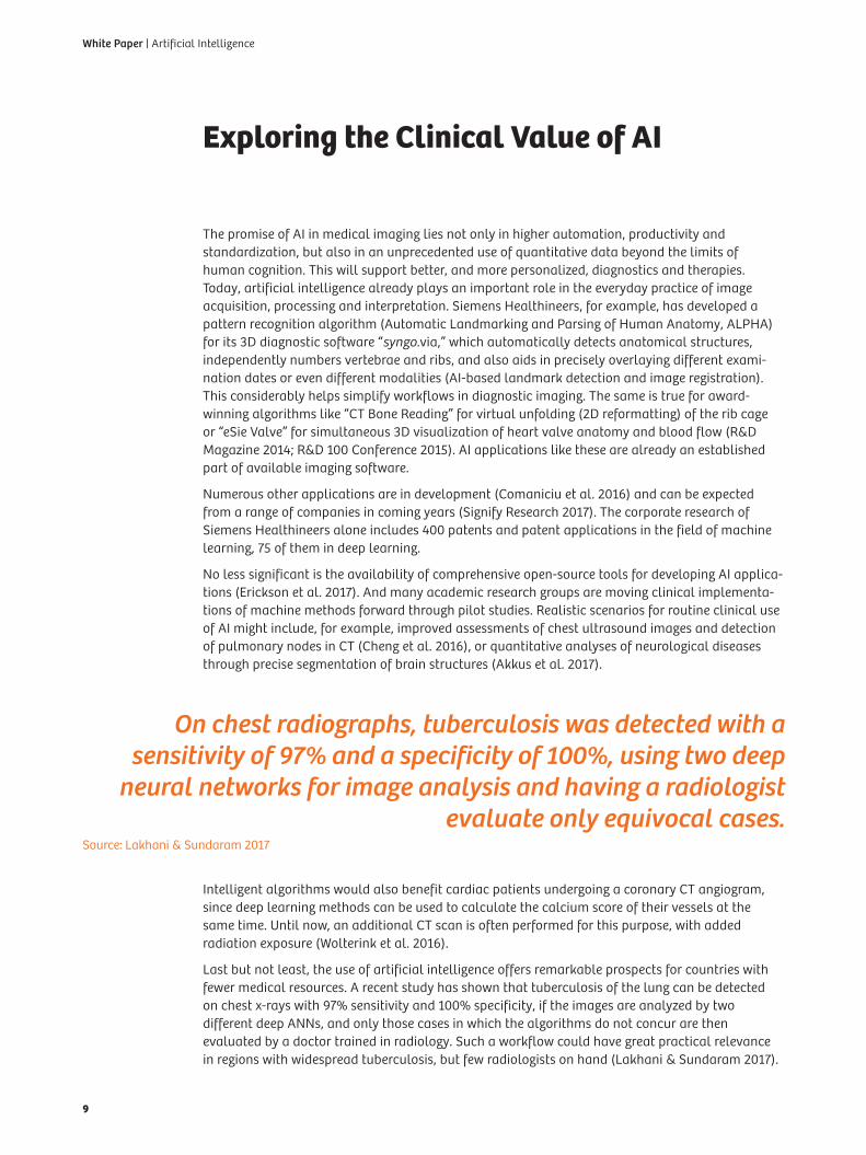
Last but not least, the use of artificial intelligence offers remarkable prospects for countries with fewer medical resources. A recent study has shown that tuberculosis of the lung can be detected on chest x-rays with 97% sensitivity and 100% specificity, if the images are analyzed by two different deep ANNs, and only those cases in which the algorithms do not concur are then evaluated by a doctor trained in radiology. Such a workflow could have great practical relevance in regions with widespread tuberculosis, but few radiologists on hand (Lakhani & Sundaram 2017).
Source: Lakhani & Sundaram 2017
On chest radiographs, tuberculosis was detected with a sensitivity of 97% and a specificity of 100%, using two deep
neural networks for image analysis and having a radiologist evaluate only equivocal cases.
9
White Paper | Artificial Intelligence
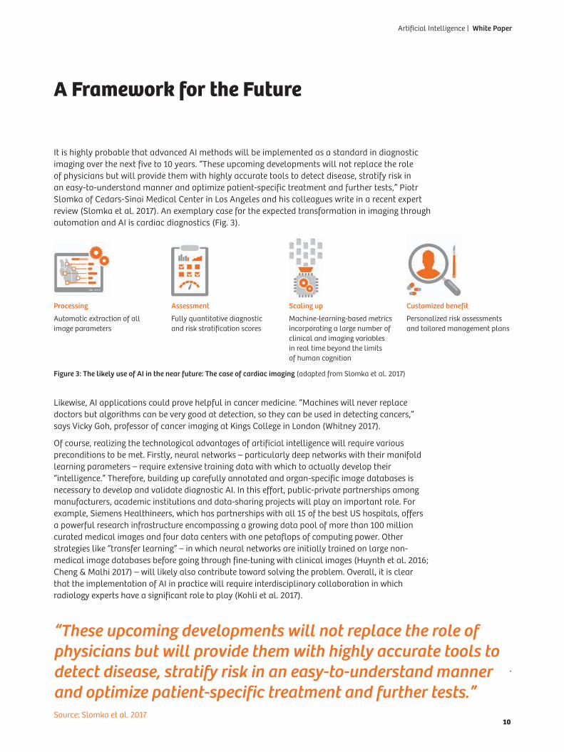
A Framework for the Future
It is highly probable that advanced AI methods will be implemented as a standard in diagnostic imaging over the next five to 10 years. “These upcoming developments will not replace the role of physicians but will provide them with highly accurate tools to detect disease, stratify risk in an easy-to-understand manner and optimize patient-specific treatment and further tests,” Piotr Slomka of Cedars-Sinai Medical Center in Los Angeles and his colleagues write in a recent expert review (Slomka et al. 2017). An exemplary case for the expected transformation in imaging through automation and AI is cardiac diagnostics (Fig. 3).
Likewise, AI applications could prove helpful in cancer medicine. “Machines will never replace doctors but algorithms can be very good at detection, so they can be used in detecting cancers,” says Vicky Goh, professor of cancer imaging at Kings College in London (Whitney 2017).
Of course, realizing the technological advantages of artificial intelligence will require various preconditions to be met. Firstly, neural networks – particularly deep networks with their manifold learning parameters – require extensive training data with which to actually develop their “intelligence.” Therefore, building up carefully annotated and organ-specific image databases is necessary to develop and validate diagnostic AI. In this effort, public-private partnerships among manufacturers, academic institutions and data-sharing projects will play an important role. For example, Siemens Healthineers, which has partnerships with all 15 of the best US hospitals, offers a powerful research infrastructure encompassing a growing data pool of more than 100 million curated medical images and four data centers with one petaflops of computing power. Other strategies like “transfer learning” – in which neural networks are initially trained on large non- medical image databases before going through fine-tuning with clinical images (Huynth et al. 2016; Cheng & Malhi 2017) – will likely also contribute toward solving the problem. Overall, it is clear that the implementation of AI in practice will require interdisciplinary collaboration in which radiology experts have a significant role to play (Kohli et al. 2017).
Figure 3: The likely use of AI in the near future: The case of cardiac imaging (adapted from Slomka et al. 2017)
Source: Slomka et al. 2017
“These upcoming developments will not replace the role of physicians but will provide them with highly accurate tools to detect disease, stratify risk in an easy-to-understand manner and optimize patient-specific treatment and further tests.”
Processing
Automatic extrac tion of all image parameters
Assessment
Fully quantitative diagnostic and risk stratification scores
Scaling up
Machine-learning-based metrics incorporating a large number of clinical and imag ing variables in real time beyond the limits of human cognition
Customized benefit
Personalized risk assess ments and tailored manage ment plans
10
Artificial Intelligence | White Paper
Secondly, it will be crucial to demonstrate the benefit of each new algorithm and account for the demands of licensing procedures and technological standards. In general, a transparent research and development process is needed to help win broad acceptance for a promising technology (Wachter et al. 2017).
ConclusionFrom Speeding Up Workflows to Better Diagnostics and Financial Gains
The acceleration of certain work steps in diagnostic imaging with artificial intelligence already is a reality today. For example, AI algorithms enable automated detection of anatomical structures, intelligent image registration and reformatting. These kind of efficiency gains will become increas-ingly important given the growing demand for diagnostic imaging and rising cost pressure.
In the longer term, AI-based image analyses with reproducible characteristic measurements (imaging biomarkers), indices and “lab-like” results will likely prevail, particularly in areas like cardiac imaging that are already quantitatively oriented. This will also favor the (semi-)automated drafting of radiology reports and the transformation of radiology to a data-driven research discipline (“radiomics”). Meanwhile, radiological data sets can not only be analyzed graphically by artificial intelligence, but they can also be annotated textually (Shin et al. 2016).
Moreover, AI algorithms could establish themselves as virtual “second readers” in a wide variety of disciplines (Zhao et al. 2012; Venkatesh et al. 2015). For example, they would call a doctor’s attention to pathological changes (computer-aided detection) or offer suggested or differential diagnoses (computer-aided diagnosis). In this way, clinically consequential and in some cases costly diagnostic errors could be avoided, at least in some cases, and missed billing opportunities could be reduced.
The implementation of AI offers particularly fascinating prospects for personalized diagnostics and treatment. Through interoperable information systems, for example, clinical patient data could be linked with imaging algorithms to pursue individualized scanning strategies. In addition, AI applications would likely enable more precise diagnoses and more meaningful risk scores by gathering together large quantities of information. As a large multicenter study recently showed, for example, the long-term risk of mortality of patients with suspected cardiovascular diseases can be estimated with much greater precision if manifold clinical and CT angiogram parameters are integrated into a personalized prognosis model using machine learning procedures (Motwani et al. 2017). Such AI-based approaches could in future better identify high-risk patients, but also help prevent unnecessary treatments, and thus involve diagnostic radiology more closely in outcome-oriented clinical decisions.
11
White Paper | Artificial Intelligence
1. Akkus Z, Galimzianova A, Hoogi A (2017) Deep Learning for Brain MRI Segmentation: State of the Art and Future Directions. J Digit Imaging. June 2, 2017. doi: 10.1007/s10278-017-9983-4 (Epub ahead of print)
2. Berlin L (2007) Accuracy of diagnostic procedures: has it improved over the past five decades? AJR Am J Roentgenol 188:1173-8
3. Cheng JZ, Ni D, Chou YH et al. (2016) Computer-Aided Diagnosis with Deep Learning Architecture: Applications to Breast Lesions in US Images and Pulmonary Nodules in CT Scans. Sci Rep 6:24454
4. Cheng PM, Malhi HS (2017) Transfer Learning with Convolutional Neural Networks for Classification of Abdominal Ultrasound Images. J Digit Imaging 30:234-243
5. Chockley K, Emanuel E (2016) The End of Radiology? Three Threats to the Future Practice of Radiology. J Am Coll Radiol 13:1415-1420
6. Choi E, Schuetz A, Stewart WF, Sun J (2016) Medical Concept Representation Learning from Electronic Health Records and its Application on Heart Failure Prediction. https://arxiv.org/abs/1602.03686 (accessed October 9, 2017)
7. Comaniciu D, Engel K, Georgescu B, Mansi T (2016) Shaping the future through innovations: From medical imaging to precision medicine. Med Image Anal 33:19-26
8. Donald JJ, Barnard SA (2012) Common patterns in 558 diagnostic radiology errors. J Med Imaging Radiat Oncol 56:173-8
9. Erickson BJ, Korfiatis P, Akkus Z et al. (2017) Toolkits and Libraries for Deep Learning. J Digit Imaging. March 17, 2017. doi: 10.1007/s10278-017-9965-6 (Epub ahead of print)
10. Fenton JJ, Taplin SH, Carney PA et al. (2007) Influence of computer-aided detection on performance of screening mammography. N Engl J Med 2007 356:1399-409
11. Garland LH (1959) Studies on the accuracy of diagnostic procedures. Am J Roentgenol Radium Ther Nucl Med 82:25-38
12. Gillies RJ, Kinahan PE, Hricak H (2016) Radiomics: Images Are More than Pictures, They Are Data. Radiology 278:563-77
13. He K, Zhang X, Ren S, Sun J (2015) Deep Residual Learning for Image Recogni-tion. https://arxiv.org/abs/1512.03385 (accessed July 16, 2017)
14. Hof, Robert D. (2013) “Deep Learning – With massive amounts of computational power, machines can now recognize objects and translate speech in real time. Artificial intelligence is finally getting smart.” https://www.technologyreview.com/s/513696/deep-learning/#comments (accessed July 16, 2017)
15. Huynh BQ, Li H, Giger ML (2016) Digital mammographic tumor classification using transfer learning from deep convolutional neural networks. J Med Imaging (Bellingham) 3:034501
16. Kohli M, Prevedello LM, Filice RW, Geis JR (2017) Implementing Machine Learning in Radiology Practice and Research. AJR Am J Roentgenol 208:754-760
17. Lakhani P, Sundaram B (2017) Deep Learning at Chest Radiography: Automated Classification of Pulmonary Tuberculosis by Using Convolutional Neural Networks. Radiology. April 24, 2017. doi: 10.1148/radiol.2017162326 (Epub ahead of print)
18. Lehman CD, Wellman RD, Buist DS et al. (2015) Diagnostic Accuracy of Digital Screening Mammography With and Without Computer-Aided Detection. JAMA Intern Med 175:1828-37
19. Lee JG, Jun S, Cho YW et al. (2017) Deep Learning in Medical Imaging: General Overview. Korean J Radiol 18:570-584
20. Morton MJ, Whaley DH, Brandt KR, Amrami KK (2006) Screening mammograms: interpretation with computer-aided detection – prospective evaluation. Radiology 239:375-83
21. Motwani M, Dey D, Berman DS et al. (2017) Machine learning for prediction of all-cause mortality in patients with suspected coronary artery disease: a 5-year multicentre prospective registry analysis. Eur Heart J 38:500-507
22. Mozur, Paul (May 23, 2017) “Google’s AlphaGo Defeats Chinese Go Master in Win for A.I.” https://www.nytimes.com/2017/05/23/business/google-deepmind-alphago-go-champion-defeat.html?module=ArrowsNav&contentCollection=Business%20Day&action=keypress®ion=FixedLeft&pgtype=article (accessed July 13, 2017)
23. O’Connor JPB, Aboagye EO, Adams JE et al. (2017) Imaging biomarker roadmap for cancer studies. Nature Reviews Clinical Oncology 14:169-86
24. R&D 100 Conference (2015) Getting to the Heart of Visualization. https://www.rd100conference.com/awards/winners-finalists/5542/getting-to-the-heart-of-visualization (accessed 16 July, 2017
25. R&D Magazine (August 20, 2014) Flattening Yields Faster CT. https://www.rdmag.com/award-winners/2014/08/flattening-yields-faster-ct (accessed 16 July, 2017)
26. Radiology Quality Institute (2012) Diagnostic Accuracy in Radiology: Defining a Literature-Based Benchmark. http://www.radisphereradiology.com/wp-content/uploads/Diagnostic-Accuracy-in-Radiology (accessed July 14, 2017)
27. Rashid, Tariq (2016) Make Your Own Neural Network. CreateSpace Independent Publishing Platform
28. Ridley, Erik L. (June 1, 2017) “SIIM: AI poised to enhance all aspects of radiology”. http://www.auntminnie.com/index.aspx?sec=ser&sub=def&pag=dis&ItemID=117495 (accessed July 13, 2017)
29. Sardanelli F (2017) Trends in radiology and experimental research. European Radiology Experimental 1:1 (DOI: 10.1186/s41747-017-0006-5)
30. Shin HC, Roberts K, Lu L et al. (2016) Learning to Read Chest X-Rays: Recurrent Neural Cascade Model for Automated Image Annotation. https://arxiv.org/abs/1603.08486 (accessed July 17, 2017)
31. Signify Research (2017) Machine Learning in Medical Imaging – World Market Report
32. Slomka PJ, Dey D, Sitek A et al. (2017) Cardiac imaging: working towards fully-automated machine analysis & interpretation. Expert Rev Med Devices 14: 197-212
33. Sokolovskaya E, Shinde T, Ruchman R et al. (2015) The Effect of Faster Reporting Speed for Imaging Studies on the Number of Misses and Interpretation Errors: A Pilot Study. J Am Coll Radiol 12:683-8
34. The Economist (2017) The future of healthcare – Realities or science fiction? http://thefutureishere.economist.com/thefutureofheathcare-infographic.html (accessed October 9, 2017)
35. The Royal College of Radiologists (2016) Clinical radiology UK workforce census 2015 report. https://www.rcr.ac.uk/system/files/publication/field_publication_files/bfcr166_cr_census.pdf (accessed July 13, 2017)
36. van Ginneken B (2017) Fifty years of computer analysis in chest imaging: rule-based, machine learning, deep learning. Radiol Phys Technol 10:23-32
37. Venkatesh SS, Levenback BJ, Sultan LR et al. (2015) Going beyond a First Reader: A Machine Learning Methodology for Optimizing Cost and Performance in Breast Ultrasound Diagnosis. Ultrasound Med Biol 41:3148-62
38. Wachter S, Mittelstadt B, Floridi L (2017) Transparent, explainable, and accountable AI for robotics. Science Robotics 2:eaan6080
39. Waite S, Scott J, Gale B et al. (2017) Interpretive Error in Radiology. AJR Am J Roentgenol 208:739-749
40. Wang S, Summers RM (2012) Machine learning and radiology. Med Image Anal 16:933-51
41. Whitney, Linda (February 2017) Artificial intelligence could transform radiology. Future of Healthcare: 6. https://www.bir.org.uk/media/346567/2017_future_of_imaging.pdf (accessed 9 October, 2017)
42. Wolterink JM, Leiner T, de Vos BD et al. (2016) Automatic coronary artery calcium scoring in cardiac CT angiography using paired convolutional neural networks. Med Image Anal 34:123-136
43. Zhao Y, de Bock GH, Vliegenthart R et al. (2012) Performance of computer-aided detection of pulmonary nodules in low-dose CT: comparison with double reading by nodule volume. Eur Radiol 22:2076-84
Additional References
Want more insights for adding value with AI in medical imaging? Visit: healthcare.siemens.com/artificial-intelligence
12
Artificial Intelligence | White Paper

Published by Siemens Healthcare GmbH · Order No. HOOD05162002714049 · Printed in Germany · 5390 1117.5 · © Siemens Healthcare GmbH, 2017
Siemens Healthineers HeadquartersSiemens Healthcare GmbH Henkestr. 127 91052 Erlangen, Germany Phone: +49 9131 84-0 siemens.com/healthineers


![Artificial Intelligence · Artificial Intelligence 2016-2017 Introduction [5] Artificial Brain: can machines think? Artificial Intelligence 2016-2017 Introduction [6] ... Deep Blue](https://img.pdfslide.net/doc/110x75/5f0538917e708231d411e192/artificial-intelligence-artificial-intelligence-2016-2017-introduction-5-artificial.jpg)