Embed Size (px)
Citation preview

1
MEDICAL MANAGEMENT OF INTERNALLY
RADIOCONTAMINATED PATIENTS
Carol S. Marcus, Ph.D., M.D. and Jeffry A. Siegel, Ph.D.
with Richard B. Sparks, Ph.D., M.B.A., Consultant
June, 2006
This work was supported by the Los Angeles County Department of Health Services Emergency Medical Services Agency
John Celentano, M.D., Disaster Medical Officer, MMRS Program Manager Kay Fruhwirth, R.N., M.S.N., Assistant Director
Jim Eads, Paramedic, Chief, Response Teams
Funding for this project was made possible by grant number EMW 2004-GR-0793 from the Department of Homeland Security Metropolitan Medical Response System

2
TABLE OF CONTENTS
MEDICAL MANAGEMENT OF INTERNALLY RADIOCONTAMINATED PATIENTS
1. Purpose of this manual…………………………………………………………4 2. Scenario descriptions…………………………………………………………..6 a) Radiological Dispersal Devices (RDDs)………………………………6 b) Surreptitious spread……………………………………………………7 c) Detonation of nuclear weapons………………………………………..8 3. Identification of radionuclides………………………………………………....9 4. Screening of patients for external radiocontamination……………………….11 a) Emergency Department screening of the injured…………………….12 b) Use of mass action decontamination solutions……………………….12 5. Screening of patients for internal radiocontamination………………………..14 a) Modes of internalization of radionuclides……………………………14 b) Determination of significant contamination: use of the annual limit on intake (ALI)…………………………………………………14 c) Detection of internal radiocontamination…………………………….15 d) Use of decorporation drugs…………………………………………...15 e) Alpha emitters………………………………………………………...16 f) Pure beta emitters……………………………………………………..17 g) Photon (gamma and x-ray) emitters………………………………….18 6. Estimation of internal radiocontamination with gamma (or other photon) emitters……………………………………………………………………….20 a) Summary table of externally-measurable radionuclides……………..22 b) Procedure for americium-241………………………………………..23 c) Procedure for cesium-137……………………………………………25 d) Procedure for cobalt-60……………………………………………...27 e) Procedure for iodine-125………………………………………….…29 f) Procedure for iodine-131…………………………………………….30 g) Procedure for iridium-192…………………………………………...31 h) Procedure for palladium-103………………………………………...33 i) Procedure for phosphorus-32…………………………………………35 j) Procedure for strontium-90…………………………………………...37 k) Procedure for yttrium-90……………………………………………..39 7. Medical management for internal contamination by specific radionuclide….41 a) Alphabetical list of radioelement and decorporation treatment

3
summary…………………………………………………………….41 Americium (Am)-241………………………………………………41 Cesium (Cs)-137……………………………………………………41 Cobalt (Co)-60……………………………………………………...41 Iodine (I)-125……………………………………………………….41 Iodine (I)-131……………………………………………………….41 Iridium (Ir)-192……………………………………………………..41 Palladium (Pd)-103…………………………………………………41 Phosphorus (P)-32…………………………………………………..41 Plutonium (Pu)-239…………………………………………………41 Radium (Ra)-226……………………………………………………41 Strontium (Sr)-90……………………………………………………41 Tritium (H)-3………………………………………………………..41 Uranium (U)-234, 235, and 238…………………………………….41 Yttrium (Y)-90………………………………………………………41 b) Alphabetical list of decorporation drugs……………………………41 Ammonium chloride………………………………………………...41 Calcium (oral)……………………………………………………….42 Calcium-DTPA……………………………………………………...42 Calcium gluconate…………………………………………………..42 Dimercaprol (British antilewisite, BAL)……………………………42. D-penicillamine……………………………………………………..42 Potassium iodide…………………………………………………….43 Propylthiouracil……………………………………………………..43 Prussian blue………………………………………………………...43 Sodium alginate……………………………………………………..43 Sodium bicarbonate…………………………………………………43 Sodium phosphate…………………………………………………...43 Zinc-DTPA………………………………………………………….43 c) Annual limits on intake (ALIs) for selected radionuclides………….43 8. Medical follow-up for internally radiocontaminated patients………………46 9. Medical management of externally irradiated patients (those who absorbed radiation directly, and were not radiocontaminated in the process)…………………..48 Appendix I.: Mathematical models for calculations of humanized constants...49

4
1. PURPOSE
The purpose of this manual is to aid medical personnel in managing patients who have been internally contaminated with one or more radionuclides (“internally radiocontaminated”). Most of the time, especially in a mass casualty situation, the physicians treating such patients will not have had prior education, training, or experience in managing these problems. For this reason, the Los Angeles County Department of Health Services-Emergency Medical Services Agency thought it important to produce a manual that covers the important aspects of such management, and the Department of Homeland Security agreed. In addition to medical management, this manual will also include important sources of help in the event of a radiological incident. For example, Los Angeles County has stockpiled drugs that help remove internalized radioactive material from the body. These drugs are called “decorporation” drugs, and they may be accessed by physicians managing radiocontaminated patients in whom decorporation appears to be clinically appropriate. A number of these drugs are not generally stocked in hospital pharmacies, which is why they have been stockpiled for an emergency. Some other counties and hospitals have stockpiled decorporation drugs also, and some of these drugs are in the Strategic National Stockpile, maintained by the Center for Disease Control and Prevention (CDC). This manual is not intended to cover in any significant detail the treatment of externally irradiated patients who were not radiocontaminated in the process. While such management may be essential, other references have covered this topic quite well. One handy reference is Medical Management of Radiological Casualties Handbook, 2nd edition, Military Medical Operations, Armed Forces Radiobiology Research Institute, Bethesda, MD, April, 2003. To request a copy of this handbook, e-mail [email protected] or telephone (301)295-0316, or write to Military Medical Operations, AFRRI, 8901 Wisconsin Avenue, Bethesda, MD 20889-5603. It fits in the pocket of a white coat. Another good reference is Waselenko JK, MacVittie TJ, Blakely WF, et al.: Medical management of the acute radiation syndrome: Recommendations of the Strategic National Stockpile Radiation Working Group: Ann Intern Med 140:1037-1051, 2004. There are numerous reports, papers, and manuals concerning the management of radiological accidents and terrorism available from federal, state, county, and military sources, as well as medical and health physics professionals and national and international radiation protection organizations. They are strikingly similar, and most any may be used. This manual has one advantage and one unique feature. The advantage is that it is short and simple, except that it includes detailed information about the use of decorporation drugs and was written specifically for Emergency Medicine physicians. The unique feature is that research was performed to create a simple procedure to estimate internal photon-emitting radionuclide contamination in exposed persons, and

5
that this procedure may be used anywhere. Humanized exposure rate constants were determined for a variety of radionuclides to facilitate the estimation of the degree of radiocontamination. This information may then be used to decide whether decorporation drug therapy is appropriate. No other publication at present contains these procedures, as they were developed for this manual. The Appendix concerning Models and Correction Factors for Calculations of Humanized Constants may be of more interest to a hospital’s medical physicist than a physician. It is included for completeness, and with the realization that Emergency Medicine personnel will likely be calling upon their hospital’s medical physicist or health physicist for help in a radiological incident. Certain excerpts from this section are duplicated in the sections primarily designed for physicians.

6
2. SCENARIO DESCRIPTIONS
Persons may become internally radiocontaminated in a number of situations. The most common one is intentional, comprising the millions of patients a year who are given radiopharmaceuticals for nuclear medicine procedures. Trace quantities of radiocontamination are present in everyone because of the ubiquitous existence of naturally-occurring radioactive potassium (K)-40 in all plant and animal foods and all sources of potassium. There are also tiny quantities of other naturally-occurring radionuclides in all of us, such as carbon (C)-14 and tritium (H)-3. And, there are tiny amounts of radionuclides such as strontium (Sr)-90 from nuclear weapons fallout and some nuclear accidents that contaminate us as well. The largest contributor to background radiation is radon (Rn)-222, which we all breathe in, all the time. Contamination events can occur accidentally in laboratory and industrial settings, usually affecting small numbers of workers. Rarely, an accident can involve many members of the public if it involves the breaching of a large sealed source without realizing it. This has happened a few times with the breaching of abandoned teletherapy sources in other countries. Criticality accidents, such as occurred at Chernobyl, caused the release of a large quantity of radioactive material and contaminated many people. A criticality accident in Japan a few years ago killed a couple of workers. The radionuclides of major concern in a nuclear weapons blast or a destroyed nuclear power plant are I-131 and Cs-137.
Radiological Dispersal Devices (RDDs) Concern about terrorist use of radioactive material has spawned other scenarios. While Chechen terrorists planted a “dirty bomb” in Moscow containing cesium (Cs)-137, it was found and dismantled by the Russians and no harm occurred. The technical name for a dirty bomb is a Radiological Dispersal Device (RDD), and this is basically conventional explosives laced with radioactive material. In this scenario, the patients injured by the blast would be expected to have the largest contamination with radioactive material, both internal, external, and possibly in wounds. From a medical standpoint, the treatment of blast injuries takes precedence over the radiation contamination considerations. It is unlikely that any patient will be radioactive enough to be any danger to medical personnel. The only exception would be a patient with radioactive shrapnel from a huge radioactive source. It is therefore wise to monitor these patients with an instrument that can detect high activities, such as an ion chamber. This equipment is usually in the Nuclear Medicine Department or the Radiation Safety Office. For example, assuming a 1 curie (Ci) Co-60 bit of shrapnel is present, the surgeon would receive 2.5 rem/hr (Smith JM, Ansari A, and Harper FT: Hospital

7
management of mass radiological casualties: Reassessing exposures from contaminated victims of an exploded radiological dispersion device. Health Physics 89(5)513-520, 2005). The maximum permissible dose to a radiation worker each year is 5 rem. In an emergency, doses of 50-75 rem may be accepted by individuals involved in lifesaving activities. However, if a patient had 100 Ci of Co-60 in his shrapnel, and the surgeon worked for two hours, the surgeon would himself have absorbed a lethal dose of radiation. It is highly likely that patients may reach an Emergency Department before anyone even knows that the blast was accompanied by dispersion of radioactive material, and probably well before the radioactive material is identified. Keeping radiation meters in the ED will at least help avoid a nasty surprise. However, radiation meters or not, it is very probable that the Emergency Department and other parts of the hospital will become contaminated, likely with low levels of radioactive material that are not a significant threat to anyone. Removal of the patient’s clothing usually takes care of most of the external contamination. Identify a shielded place in which bags of radioactive clothing may be temporarily stored. A large closet or small room, with additional concrete blocks as needed, would work well.
Surreptitious Spread
Radioactive material may be introduced into food or water supplies, where it will be consumed without knowledge of the contamination. At some future time, the chance pick-up of radioactivity by a detector or a telephone call from the perpetrators will alert the public to the event. Hundreds, thousands, or even more people could potentially be contaminated. An accident something like this occurred in Goiania, Brazil in 1987. A 1375 Curie (Ci) Cs-137 source from an abandoned teletherapy machine was taken apart out of curiosity. The rest of the teletherapy machine was sold as scrap metal. When water vapor from the air came in contact with the CsCl in the breached source, the radiation coming off, which is invisible, interacted with the water and a small amount of the energy was given off in the visible range (Cerenkov radiation). The CsCl emitted a beautiful blue glow, and this “magical” material was taken home as a curiosity and given to children to play with. It was also passed around to several households. Several patients presented to clinics with ulcers that were blamed on infections and insect bites at first. Finally, one physician considered radiation. A visiting medical physicist was asked to evaluate this possibility, and so, two weeks after the source was breached, the event was discovered. Goiania was a city of 1 million people at the time. One hundred twelve thousand people showed up at the local sports stadium to be monitored for contamination. External contamination was found in 249 people. Internal contamination was found in 129 people. Forty-six of these internally contaminated people were treated with Prussian blue (hexacyanoferrate, or RadiogardaseR ). Twenty-eight people suffered radiation burns, and 33 were hospitalized with acute radiation effects. There were six surgical interventions, composed of four skin debridements, one skin graft, and one amputation. There were four acute radiation

8
deaths. (Later deaths that might be due to radiation-induced cancer were not included in this total.)
Detonation of Nuclear Weapons
Detonation of an improvised nuclear device could cause the deaths of tens or hundreds of thousands of people; detonation of professionally constructed devices could kill even more. Of the injured survivors, external radiation, radiation burns, and blast injuries will completely overshadow internal radiocontamination, which will be of relatively minor importance. Eventually, once the chaos is under control, there might be some internal contamination issues to consider. Nuclear fission results in the formation of several hundred different radionuclides, many of which have short halflives. Long term internal radiocontamination with such radionuclides as Sr-90 and Cs-137 may be seen, but probably not at high levels. An entire issue of the journal Health Physics was devoted to radiological terrorism. It is volume 89, no. 5, November, 2005, and contains extensive technical information for those wishing to have more detailed information.

9
3. IDENTIFICATION OF RADIONUCLIDES
The radiation equipment used by HAZMAT teams and others to detect radiation does not identify the radionuclide(s) present. It just detects radioactivity. While this is all that is needed to know to begin external decontamination of patients, it is necessary to know which radionuclides are present in order to estimate internal body burden and institute appropriate treatment, if needed. It will also be necessary to have such information for various public health activities, such as monitoring food and water. The radionuclides with which we are most concerned are americium(Am)-241, cesium (Cs)-137, cobalt (Co)-60, iodine (I)-125, iodine (I)-131, iridium (Ir)-192, palladium (Pd)-103, phosphorus (P)-32, plutonium (Pu)-239, radium (Ra)-226, strontium (Sr)-90, tritium (H)-3, uranium (U)-234, 235, and 238, and yttrium (Y)-90. These radionuclides emit alpha particles (helium nuclei), and/or beta particles (electrons arising in the nucleus), and/or photons (gamma rays, which arise in the nucleus, or x-rays, which arise from orbital electron transitions). In addition, beta-emitters emit bremsstrahlung or “braking radiation” which consists of secondary photons emitted as the beta particles interact with matter. The easiest way to identify a radionuclide is by the characteristic energies of its photons and their relative frequencies. This is performed with a device called a spectrometer, coupled with a computer program that identifies radionuclides by their spectra. Spectrometers may be stationary or portable. There are various kinds of radiation detection materials used in spectrometers, but it is enough to know that these devices are commonly possessed by radiation regulators, some industries, and many universities and teaching hospitals. It is therefore reasonable to expect that once radioactive material is detected, it will likely take a number of hours to perhaps a day to identify the radionuclide(s) in question. Useful portable spectrometers include the ICS-4000 (see www.xrf.com), EasySpec by Canberra Industries, and GR-130 miniSPEC by Exploranium. In addition, the Dept. of Energy runs a service in which spectra and certain other information are sent to two of its laboratories that operate 24/7, and the radionuclide(s) are identified. This is known as the Triage Program. The collected spectra should be e-mailed to both laboratories at [email protected] and [email protected]. Telephone them at (202)586-8100 and ask for the Emergency Response Officer (ERO) to discuss Triage information. Medical physicists, nuclear physicists, nuclear engineers, and health physicists are usually the best people to ask to obtain spectra and get them identified. Medical physicists are usually affiliated with Radiation Oncology Departments, nuclear physicists

10
are often at universities, nuclear engineers are found at nuclear power plants, and health physicists often work as radiation safety officers in hospitals, colleges and universities, commercial entities that use radioactive material, and radiation regulatory agencies. For help in identifying radiation professionals and obtaining radiation services, call your State or local (if available) radiation regulators. In Los Angeles, call Radiation Management at (213)351-7897. The Director is Kathleen Kaufman; her number is (213)351-7387. In California, the State radiation regulators are in Sacramento. The main number is (916)327-5106. The Acting Chief is Gary Butner, at (916)440-7899 or (916)440-7909. If he is not available, ask for a senior health physicist such as Victor Anderson (916)440-7931, or the Chief of the Medical Section, Gonzalo Perez (916)440-7967.

11
4. SCREENING OF PATIENTS FOR EXTERNAL RADIOCONTAMINATION
In the event of uncontrolled release of radioactive material, members of the public will have to be screened for contamination. If the release is accompanied by an explosion, vehicular crash, or other traumatic event, it is probable that those persons close enough to the incident to be injured by blast or collapsing structures will be the most likely to have the most external contamination. These injured patients will be taken to hospitals, likely before it is even obvious that radioactivity was involved in the incident, and before the identification of the radionuclide(s) occurs. Persons who are not injured will be very concerned about contamination, and will likely flock to hospitals for screening unless other provisions are made for screening them elsewhere. In order to protect Emergency Departments from an onslaught of uninjured patients when attention must be paid to injured ones, there must be a rational and publicly acceptable plan for screening uninjured persons for radiocontamination. Local health officials should have a plan for directing uninjured persons to places where screening may be carried on. Sports stadiums and other locations with ample parking make good screening centers. Screening equipment, such as Geiger-Muller (G-M) detectors, ion chambers, and portal monitors, and persons trained to use this equipment, must be available. In the event that such plans have not been made or are incomplete at the time of a radioactive material release event, it would be wise for hospitals to set up a screening center outside the hospital, near a large parking area, to keep uninjured patients from coming into the Emergency Department. All hospitals which have nuclear medicine services and brachytherapy services have radiation detection instrumentation. The Emergency Department should have a plan to borrow this equipment, with qualified equipment operators, or get their own and have their people trained in advance. Some equipment will be needed in the Emergency Department for screening injured patients, and some will be needed for the outside area ambulatory screening. G-M detectors (“Geiger counters”) are the cheapest and simplest devices for screening, and the most commonly available. However, they are not necessarily very accurate. G-M detectors are calibrated yearly, usually against a Cs-137 source of known activity. So, they are accurate for a Cs-137 event, but will be quite inaccurate for pure beta emitters, low photon energy emitters, and alpha emitters. For this reason, use of G-M detectors for screening is not very useful quantitatively. Those who appear to be contaminated (their levels are at least three times the background levels) will need to be evaluated with other equipment or by other means, depending upon the radionuclide(s) involved. In the early 1980s, sanitary landfills in Los Angeles County installed radiation detectors at the entrance to the landfill to prevent any radioactive material from being dumped there. Other localities followed suit, and now landfills all over the country have radiation detectors, as do medical waste treatment facilities. In order to avoid having a hospital’s

12
garbage truck refused entry to a landfill, many hospitals in Los Angeles County installed portal monitors to check their trucks before leaving the hospital. These detectors are quite sensitive, and may be used to screen people as well as garbage trucks. For those hospitals which have them, calibrating them and planning to use them for screening people could be part of their RDD disaster plan. Due to the fact that there will inevitably be some delay before mass screening of uninjured persons is available, those uninjured persons in proximity to the radioactive material release event should go home, shower and wash their hair, wash their clothes in the washing machine, clean their shoes with a wet paper towel which should then be discarded, and then get screened. They may bring their newly-washed clothes and cleaned shoes along in a plastic bag to make sure that they are no longer significantly contaminated. Depending upon limited decontamination showering facilities from the Fire Department is unrealistic, because the waiting time will be huge. If one or more radioactive material release events occur in Los Angeles County, probably a million people will have to be screened.
Emergency Department Screening of the Injured
Removal of all clothing will generally remove about 90% of external contamination, and the treatment of injuries takes precedence over radiocontamination issues. It is highly unlikely that residual radiation levels from the patient will constitute a hazard to medical personnel. The only exception to this would be if a piece of metal from the covering of a high activity radioactive source penetrated the patient (radioactive shrapnel), it is possible that it could be extremely radioactive. Patients should therefore be monitored quickly to make sure that they are not a hazard to others. It is important to remember that G-M detectors are sensitive instruments that flood at significant radiation levels. By “flooding” we mean that they cannot function properly with the high countrate presented to them. Most will register “zero”. They may thus indicate that no radiation is present, when in fact the opposite is true. When monitoring patients with a G-M detector, start the monitoring at a significant distance from the patient, and at the highest setting, e.g. “x 100”, and then come closer. If the radiation readings fall as you get closer, have your health physicist bring an ion chamber that can give accurate readings at high radiation levels. There is one other rare exception to the assumption that the radioactivity on or in the patient will not be a significant hazard to the medical personnel. If there is a criticality accident and a worker gets a very high neutron dose, the neutrons may activate non-radioactive atoms in the patient’s body and create radioactive ones. With fatal doses from such an accident, the patient may be highly radioactive. Assuming that a patient’s non-radiation injuries are stabilized, decontamination of the patient should follow. Often soap and warm water are all that are necessary to remove most of the remaining external contamination. Never rub the skin so as to cause an abrasion, because external radioactive material can now become absorbed and internalized.

13
Use of Mass Action Decontamination Solutions
If soap and water do not remove all the contamination, there is the possibility that the contamination is internal. As most internal contamination comes in through inhalation and swallowing, the main areas of radioactivity will be the chest and abdomen. If residual radioactivity is on extremities or other areas that appear to represent external contamination, it is recommended that mass action decontamination solutions be used. These agents have been used to clean contaminated surfaces, and have been approved by the FDA for use on intact skin. There are three different solutions to choose from, and the choice depends upon the radioactive element involved. If the radioactive material has not yet been identified, try all three and see which works. A set of the three mass effect decontamination solutions (for halogens, actinides, and transition metals) is available, complete with instructions, from Dr. John Kuperus, a nuclear pharmacist in Tampa, FL. He may be reached at: John Kuperus, Ph.D., R.Ph. Radiationh Decontamination Solutions, LLC 101A Dunbar Ave. Oldsmar, FL 33634 Telephone: (800)995-4363 ext. 267 FAX: (800)697-5250 E-mail: At the time of this writing, the mass action decontamination solutions are not in the Strategic National Stockpile nor are they stockpiled by Los Angeles County Department of Health Services.

14
5. SCREENING OF PATIENTS FOR INTERNAL CONTAMINATION
Modes of Internalization of Radionuclides
Radioactive material may be inhaled, either as gases or particulates. Some will end up being swallowed, from mouth contamination, ciliary movement in the bronchial system that moves particulates to the mouth, or the eating or drinking of contaminated food. In addition, radioactive shrapnel from the destruction of a sealed source of radioactive material can become embedded in a wound. The treatment of radioactive shrapnel is its surgical removal, as quickly as possible. Precise localization with CT or gamma camera should be undertaken to minimize time spent in exploration. The shrapnel should only be touched with instruments, not fingers, and should be placed in a lead container (called a “pig”) for shielding purposes. The Nuclear Medicine service should have lead pigs available, or know how to obtain them. In the event that larger receptables are needed for a larger volume of radioactive articles, “caves” may be built out of lead bricks or concrete blocks with a “cover” of metal sheeting and bricks or blocks on top. A steel safe is often a good start, with extra shielding as needed. Inhaled radioactive gases have varying amounts of absorption into the blood. Inhaled particulates that are not coughed out or swept out by cilia can be gradually solubilized to some extent, and then absorbed, or deposited eventually in the tracheobronchial lymph nodes, where they stay virtually indefinitely. Radioactive material that is swallowed can be absorbed to some extent, depending upon what it is, and unabsorbed radioactive material is excreted in stool. Of material that is absorbed, some may be deposited in a variety of organs, and some may be excreted in urine.
Determination of Significant Contamination: Use of the Annual Limit on Intake (ALI)
Once the radionuclide(s) involved in the radiological incident have been identified, it will be possible to determine a method for ascertaining whether persons are internally contaminated to a significant extent. “Significant” is provisionally defined herein as being greater than the maximum quantity of internal radiocontamination permitted for radiation workers per year (the “annual limit on intake”, or ALI). This is determined by the Nuclear Regulatory Commission (NRC), and appears in 10 CFR Part 20 and later in this document for the radionuclides of concern. This is a hugely conservative approach with many approximations and inaccuracies, and in a mass casualty setting, “significant” may be taken to mean ten times that level, especially if resources are scarce. The limits set for workers by the NRC may recur yearly for, say, 40 years of employment, while a member of the public’s exposure will most likely be limited to a single accident or terrorist act. In a sense, this “builds in” a conservative factor of 40.

15
The NRC limits apply to workers who are over 18 years of age. There are no published limits for children or pregnant women who are members of the general public. The NRC limits do not imply hazard above the level of an ALI. They are calculated based upon a theoretical increased risk of cancer starting from an oversimplification suggesting that any amount of radiation can cause cancer, and the less radiation one receives, the lower the probability of such a cancer occurring. In fact, low doses of radiation have not been convincingly associated with increased cancer, and there is even an hormetic response in numerous situations (that is, low radiation doses result in a protective effect and result in less cancer than in persons absorbing no extra radiation dose at all). For these reasons, we find no scientific basis to alter the ALIs for pregnant women, older children, or teenagers. For infants and small children, it might make some empirical sense to decrease the ALI proportional to body mass, but there are no data supporting this concept. While some decorporation drugs have few, if any, side effects, others have definite risks. There is therefore the need to weigh the risks of the drugs, which are known, against the supposed risks of the radiation, which, at low doses, may have no actual risk at all. Therefore, it is not necessarily good medicine to be conservative and treat even very low levels of radiocontamination because one may do more harm with the treatment than by doing nothing.
Detection of Internal Radiocontamination Some internalized radionuclides will be easily detected in persons with simple external radiation detectors. Others will require nasal swabs as an indication of inhalation, and/or urine and stool samples.
Use of Decorporation Drugs In the event of an RDD, those closest to the explosion would in general be expected to have inhaled a greater quantity of the radionuclide(s) than those further away. Therefore those who are injured may well be among those with the greatest internal radioactive burden. If it is easy to estimate internal burden with external detectors once the radionuclide is identified, this should be done before deciding whether or not to use decorporation drugs. However, if estimation of internal burden is complicated, time-consuming, or specialized enough that it must be performed outside the hospital, once the radionuclide is identified the Emergency Medicine physician may elect to presumptively treat the patients who were close to the explosion with decorporation drugs, especially if the risk of the particular decorporation drug is very low. There is no all-purpose decorporation drug “cocktail” to take that will protect against all internal radiocontamination possibilities. Although decorporation drugs act most efficiently if given early, they are also effective if given late, even a few weeks late in some cases. Treatment may have to go on longer if there is a late start. One exception to this is potassium iodide (KI) for radioactive iodine internal contamination. If it isn’t used

16
within four to six hours, it will have significantly decreased effectiveness, and that effectiveness will approach zero after about 12-24 hours. Any physician in Los Angeles County needing one or more decorporation drugs may obtain them from the County using a rather simple process. First, call the Medical Alert Center (MAC) at (323)226-6619 or (323)722-8073. Ask to speak to the Medical Officer (there is one available 24/7). Describe your clinical need. The Medical Officer can then authorize drug delivery to you from the Los Angeles County stockpile, and the drugs will come to you by County ambulance or other rapid means. Detailed information on the use of the various decorporation drugs is given in the section on Medical Management.
Alpha Emitters
Information for the sections on alpha and pure beta emitters comes from Methods and Models of the Hanford Internal Dosimetry Program, issued Jan. 31, 2003 (http://www.pnl.gov/eshs/pub/pnnl860.html).
Of the sixteen radionuclides with which we are chiefly concerned, six are alpha emitters. They are Am-241, Pu-239, Ra-226, U-234, U-235, and U-238. The energies of the alphas are roughly similar, and travel only about 100 microns in tissue. Therefore, alpha particles arising internally cannot be externally detected. However, all six alpha emitters also emit photons (gamma rays and/or x-rays) that may be detected and identified by portable spectrometers (e.g the ICS-4000). All of these radionuclides are best detected outside the body as some of these photons are low energy and easily absorbed. Alpha particles are very biologically damaging, and the quantities permitted internally in radiation workers are very low. Even if there is an externally detectable photon, the quantity one needs to measure is so low that nasal swabs and excreta are probably the best way to detect all of them. Am-241 also emits a rather low energy photon, about 60 kev, and this can be externally detected. However, approximately every 5 cm (2 inches) of water (or tissue, approximately) reduces the intensity of the photons to half. That is, the half value layer of 60 kev photons in water is 5 cm. The photon intensity may thus be low enough that nasal swabs and excreta are still indicated for quantitation. Patients should have 24 hr urine collections shortly after exposure and then at 10 and 100 days post exposure. Pu-239 emits low percentage or low energy photons that are very difficult to detect inside the patient. Nasal swabs and excreta are the best ways to detect it. Nasal swabs may revert to background as early as 30-60 minutes post exposure. If patients are mouth breathers, the swabs will never be positive. Twenty-four hour urine samples should be collected after complete external decontamination to avoid collecting misleading evidence of internal contamination. Urine samples should be obtained shortly after exposure and again at 10 and 100 days post exposure.

17
Plutonium is actually found as a mixture of radioisotopes. The small quantity of Pu-241 present decays to Am-241, which has a 60 kev photon which may be detected externally. However, it is necessary to know the fractional isotopic composition of the Pu-239 in order to use the counts of the 60 kev photons to back calculate the quantity of Pu-239 present internally. Such analyses need to be performed in highly specialized laboratories such as the DOE labs in Hanford, WA or the Livermore Laboratory in CA. Ra-226 emits low percentage or low energy photons that are difficult to detect inside the patient. Nasal swabs and excreta are the best ways to detect it. U-234, U-235, and U-238 have such long halflives that they may be detected chemically more easily than radiologically. In any case, nasal swabs and excreta are the best ways to detect them. Due to environmental uranium in soil, plants, water, and animals, about 0.6 microgram/day is expected in the urine of ordinary adults. This is the median at Hanford, WA. Levels up to 0.2 microgram/day are considered environmental in origin. The halflives of the uranium isotopes are so long that the masses of the radionuclides per microcurie are high enough to be more easily detected chemically rather than radiologically. Spot urine samples taken several days after exposure and at about 10 and 100 days post exposure will be useful for chemical analyses. Chemical toxicity to the kidneys is more important than radiation effects. Direct in vivo chest counting with a planar Ge detector will often work but such equipment is hard to find.
Pure Beta Emitters
Pure beta emitters do not have associated gamma rays. They therefore cannot readily be identified by a spectrometer. As beta particles (electrons arising in the nucleus from radioactive decay) produce some low energy photons as they interact with matter (“bremsstrahlung”, or “braking radiation”), they may be detected with radiation detectors such as G-M counters. The beta particles themselves only travel on the order of a few mm in tissue (the higher the energy, the farther they travel), and these are thus absorbed in the body and seldom detected externally. They are easily absorbed by tissue, metal, glass, and plastic. If a G-M counter detects radioactivity, cover the detector with its cap (or turn the probe upside down if there is no cap or cover) and see if there is any more radioactivity detected. If not, the beta particles and bremsstrahlung have been absorbed by the cover or cap or the metal casing around the detector, and that basically tells you that you likely are dealing with a pure beta emitter. The pure beta emitters that are a likely concern with RDDs are Sr-90, Y-90, H-3 (tritium), and P-32. Sr-90 decays to Y-90, which is also radioactive. They are both pure beta emitters and are generally found in equilibrium together. (All Sr-90 sources are in equilibrium with Y-90, having approximately equal activities of both radionuclides.) The presence of Cs-137 in decayed fission products suggests the presence of Sr-90 as well. A urine sample should be taken shortly after exposure, and others at later times, such as 14 and 60 days. Ion

18
chamber readings or whole body counting in a gamma camera or whole body counter calibrated for bremsstrahlung may also be done to estimate internal radioactive burden. P-32 has a halflife of only two weeks, and is not a very serious RDD threat. A urine sample soon after exposure and others at approximately 7 and 14 days would be helpful. Bremsstrahlung counting by an ion chamber, gamma camera or a whole body counter may also be used. Tritium has an extremely weak beta, requires a liquid scintillation counter for detection (a G-M counter will not work), and is also not a very serious RDD threat. Therefore, if you suspect internal contamination with a pure beta emitter, a good bet is Sr-90/Y-90. However if tritium is suspected, a urine sample should be collected at least 2 hours after exposure and again at 10 days post exposure. As tritium is normally occurring in nature, it may be helpful to use a urine sample of a non-exposed family member to estimate “background”.
Photon (Gamma and X-ray) Emitters
Photon emitters emit at characteristic energies and may be identified by their energy spectrum, as long as the energies are high enough to pass through the body without significant absorption and the probability of emission per disintegration is high enough to be practical. They may also emit alpha or beta particles. Of the radionuclides of concern, Am-241, Cs-137, Co-60, I-131, and Ir-192 are photon emitters which may be identified by their spectrum. I-125 has a low energy photon emission which is poorly detected except if it is in the thyroid. Due to the thin tissue layer between the thyroid and an external detector, enough of the photons get through to permit detection and possible identification. However, with low activities present it may well be missed. Pd-103 has very low energy photon emission and is unlikely to be externally detected Portable spectrometers such as the ICX-4000 come with built-in spectra and the ability to immediately identify the photon emitter. In the event of contamination with a mixture of radionuclides, simple spectral identification is often much more difficult. The Department of Energy (DOE) has two laboratories which operate 24/7 and perform advanced spectral analysis on the spectrum you e-mail them. The ICX-400 spectrum can be converted to a computer file and e-mailed. In order to access the DOE Triage Program for Radionuclide Identification, telephone (202)586-8100 and ask for the Emergency Response Officer (ERO) in charge of Triage information. E-mail the spectrum to both [email protected] and [email protected]. Your Radiation Safety Officer (RSO) or Medical Physicist can probably take care of this. (All hospitals using radiation-producing machines and/or radioactive material have RSOs. Hospitals with Radiation Oncology services usually have a Medical Physicist.) Detecting the presence of radioactive material and identifying the radionuclide(s) present is the first step in treating patients. The identification process may also be done by the

19
people from the Los Angeles County-Department of Health Services Radiation Management group. Their telephone number is (213)351-7897. The Director is Kathleen (Cass) Kaufman. Estimating the activity of internal radionuclidic contamination is the next step in determining whether the use of decorporation drugs is advisable.

20
6. ESTIMATION OF INTERNAL RADIOCONTAMINATION WITH GAMMA (OR OTHER PHOTON) EMITTERS
Every photon-emitting radionuclide emits a characteristic quantity of radiation over a given time at a given radioactivity level as measured at a given distance from the source of the radioactivity. The source is generally assumed to be an unshielded point source. The characteristic quantity of radiation may be measured or calculated knowing the energy of the emissions and the yield of those emissions per radioactive decay. The quantity of radiation used is the roentgen, abbreviated R. Smaller quantities are a thousandth of a roentgen, a mR, or a millionth of a roentgen, a μR. The roentgen is defined as a quantity of radiation that causes a given amount of ionization in air at standard temperature and pressure. The radiation absorbed dose, abbreviated rad, is defined as the absorption of 100 ergs per gram of anything. The radiation absorbed dose corrected for the degree of harmfulness is called the roentgen-equivalent man, or rem. For all photons and beta particles, no correction for degree of harmfulness is needed. For alpha particles, the correction factor commonly used is 20. Because there is virtually no radiation repair of densely packed alpha particle damage, one rad of alpha radiation is equal to 20 rem. For photons and beta particles, one rad equals one rem. One roentgen (R) is approximately one rad due to how the units were defined. One R of a photon or beta emitter is approximately one rad or one rem, and the units are often assumed to be interchangeable for health physics purposes. In most of the rest of the world, the gray(Gy) and sievert (Sv) are used. One Gy = 100 rad, and 1 Sv = 100 rem. The commonly used unit of radioactivity in the USA is the curie, abbreviated Ci. One thousandth of a Ci is one mCi, and one millionth of a Ci is one μCi. A Ci is 3.7x10¹⁰ disintegrations/sec. Most of the rest of the world uses a more modern unit, the becquerel (Bq). One Bq is one disintegration/sec. This is such a tiny amount that the unit corresponding to a million (mega) Bq is often used, the MBq. One MBq = 27 μCi and 1 mCi = 37 MBq. The characteristic quantity of radiation described in the first paragraph, above, is called the specific gamma ray constant. These can be looked up in health physics books. If a drop of a known radionuclide fell on the floor, one could measure the radiation at any measured distance and back calculate the quantity of radioactivity on the floor if one knew the specific gamma ray constant. The units of the specific gamma ray constant are commonly expressed as either R-cm2/mCi-hr or mR-cm2/μCi-hr. In the event of a radiological incident, one would like to measure the radiation dose rate at a measured distance from a person, and do the same sort of calculation to find out how many μCi are in the person. For this we need humanized gamma ray constants (or humanized exposure constants) for people of different sizes. These values cannot be

21
looked up anywhere, as they do not exist. Until now. The research performed for this manual includes these calculations. They will permit the estimation of internal radioactivity in a person by measuring the radiation dose rate from the person with a calibrated ion chamber. An ion chamber is a common instrument in any hospital that performs nuclear medicine therapy. However, we need one that will read very low levels of radiation. Ion chambers can read in R/hr, mR/hr, and μR/hr. The nuclear medicine department equipment will probably read mainly in mR/hr. For most of the radionuclides and activities with which we are interested, one would need a μR meter. All dose rates at one ALI at two days are below one mR/hr except for 1-131, which is about 1.5 mR/hr. If your hospital has a gamma camera calibrated for the radionuclide involved in the incident, along with correction factors for human tissue absorption, then that will be more accurate than the following ion chamber procedure. However, at the time of the writing of this manual, this does not seem to have been done anywhere. If your hospital has a calibrated whole body counter that would probably be the most accurate method of all to use. However, it is highly unlikely to have one. If it has a calibrated portal monitor, this would also be reasonably accurate to use. The humanized exposure constants were calculated using the predicted biodistribution of each radionuclide of interest in the body two days after the radiological event. If the relative biodistribution in the individual remains the same or nearly the same for times after two days, the humanized exposure constants are still good, even if there has been excretion. A mass correction factor has been added to accommodate people of all sizes. The mass correction factor is multiplied by the humanized exposure constant to give a corrected humanized exposure constant for a person weighing other than 70 kg. Use of these correction factors tends to be conservative because they accurately reflect weight change but not height change. The humanized exposure constants are only good if the measurements are made 1 cm from the skin at the level of the xiphoid process, or in the case of the two radionuclides of iodine, 1 cm from the skin of the neck. A very useful value has been calculated, which is the dose rate (mR/hr) from a 70 kg person containing one ALI two days after the radiological event. For this value to be fairly accurate at times earlier or later than two days, the relative biodistribution in the body should not have changed significantly, there may not have been any significant excretion, and there may not be any significant loss of activity due to natural radioactive decay. If one is making measurements at times other than two days, and it is not known that the two-day value is accurate, divide the measured mR/hr reading on the ion chamber by the humanized exposure constant and that will yield μCi of internal contamination.

22
While it might be very useful to generate tables of these dose rate values for a person containing one ALI at other times in addition to two days, the generation of such tables was beyond the scope of this effort. However, it is possible to do this, and may be done in the future. The value of two days was selected for this manual as that was seen as the earliest that mass screening could take place, assuming a detailed plan was in existence and that radiation professionals had been recruited, trained in the emergency procedures, and sworn in as volunteers. This has not yet occurred in Los Angeles County, but it is under consideration. The following table summarizes basic data of externally measurable radionuclides:
SUMMARY TABLE OF EXTERNALLY-MEASURABLE RADIONUCLIDES
Radionuclide ALI (μCi)* Humanized Exposure Constant Dose Rate (mR/h) (mR/μCi h @ 1 cm) from 1 ALI ________________________________________________________________________ 60Co 30 0.00642 0.19 131I 50 0.200 1.51** 137Cs 200 0.00207 0.41 192Ir 200 0.00184 0.37 241Am 0.006 0.0000477 0.00000029 32P 400 0.000132 0.053 90Sr 20 0.00000709 0.00014 90Y 600 0.0000801 0.048 103Pd 4000 0.00000338 0.014 125I 60 0.0342 0.36** ________________________________________________________________________ * For those radionuclides with multiple ALIs, corresponding to D, W, and/or Y class, the smallest ALI value is given. ** The dose rates per ALI for the radioiodines were modified by the day 2 biodistribution data to reflect the activity present in the thyroid at that time.

23
Please note that the dose rate measurements need to be background-subtracted. Background in Los Angeles averages about 0.02 mR/h and ranges from approximately 0.009-0.04 mR/h. Background should be measured on each instrument prior to use.
Procedure for Americium-241
The dose rate from a person contaminated with one ALI at two days is so low that a μR meter will not be sensitive enough to detect it. This procedure cannot be used except for activity well above the ALI. Basic data for Am-241 may be found in the following table and graph:
For Am-241 Correction Factors for Humanized Exposure Rate Constant as function of weight Specific γ-ray constant = 0.1 mR/μCi h @ 1 cm Humanized constant for 70 kg person = 0.0000477 mR/μCi h @ 1 cm Correction Factors for
Absorbed Fraction Mass (kg) (1 - Absorbed Fraction) Humanized Constant 0.247 10 0.753 1.33 0.308 20 0.692 1.22 0.397 50 0.603 1.06 0.432 70 0.568 1.00 0.471 100 0.529 0.93 0.511 140 0.489 0.86 0.559 200 0.441 0.78
Correction Factor (CF) v Body Weight: Am-241
CF Humanized = 1.27 x e-0.0027 x weight (kg)
R2 = 0.94
0.0
0.5
1.0
1.5
0 50 100 150 200 250Body weight (kg)
Cor
rect
ion
Fact
or

24
Example 1: A 70 kg adult has an ion chamber reading of 0.003 mR/hr two days after the radiological event involving Am-241. The dose rate for one ALI is 0.00000029 mR/hr. 0.003/0.00000029 = 10,000 ALIs or 60 μCi. This patient should receive Ca/Zn-DTPA therapy. Example 2: The patient in the above example has been treated for a month. His ion chamber reading is now 0.001 mR/hr. His body burden is approximately 0.001/0.0000477 = 20 μCi. This patient should continue therapy. Example 3: A 20 kg child has an ion chamber reading of 0.001 mR/hr two days after the radiological event. Her body burden is 0.001/(0.0000477)(1.22) = 20 μCi. She is a candidate for Ca/Zn-DTPA therapy.

25
Procedure for Cesium-137
The mass correction factors for the humanized exposure constant are found in the table and graph below, along with other basic information:
For Cs-137 Correction Factors for Humanized Exposure Rate Constant as function of weight Specific γ-ray constant = 3.3 mR/μCi h @ 1 cm Humanized constant for 70 kg person = 0.00207 mR/μCi h @ 1 cm Correction Factors for
Absorbed Fraction Mass (kg) (1 - Absorbed Fraction) Humanized Constant 0.202 10 0.798 1.21
0.25 20 0.750 1.14 0.317 50 0.683 1.04 0.341 70 0.659 1.00 0.368 100 0.632 0.96 0.399 140 0.601 0.91 0.449 200 0.551 0.84
Correction Factor (CF) v Body Weight: Cs-137
CF Humanized = 1.17 x e-0.0018 x weight (kg)
R2 = 0.94
0.0
0.5
1.0
1.5
0 50 100 150 200 250Body weight (kg)
Corr
ectio
n Fa
ctor

26
Example 1: A 70 kg person has an ion chamber reading of 0.5 mR/hr two days after the radiological event. As the dose rate for one ALI is 0.41 mR/hr, this person is a candidate for Prussian blue. Example 2: The 70 kg person from the above example has been given Prussian blue for five days. A measurement made on Day 7 after the radiological event reads 0.2 mR/hr. This corresponds to an internal contamination of 0.2/0.00207 = 100 μCi. This patient now contains about half an ALI, and no longer needs treatment. Tell him that the treatment was successful, and that he should no longer be concerned. Example 3: A 10 kg child has an ion chamber reading of 3 mR/hr two days after the radiological event. His corrected ALI is (10/70)(200) = 30 μCi. His corrected humanized exposure constant is (0.00207)(1.21) = 0.0025. 3/0.0025 = 1000 μCi. This corresponds to 1000/30 = 30 ALIs. This child is a candidate for treatment with Prussian blue.

27
Procedure for Cobalt-60
The mass correction factors for the humanized exposure constant are found in the table and graph below, along with other basic information:
For Co-60 Correction Factors for Humanized Exposure Rate Constant as function of weight Specific γ-ray constant = 13.2 mR/μCi h @ 1 cm Humanized constant for 70 kg person = 0.00642 mR/μCi h @ 1 cm Correction Factors for
Absorbed Fraction Mass (kg) (1 - Absorbed Fraction) Humanized Constant 0.183 10 0.817 1.18 0.223 20 0.777 1.12 0.282 50 0.718 1.03 0.306 70 0.694 1.00 0.334 100 0.666 0.96 0.364 140 0.636 0.92 0.403 200 0.597 0.86
Correction Factor (CF) v Body Weight: Co-60
CF Humanized = 1.15 x e-0.0016 x weight (kg)
R2 = 0.94
0.0
0.5
1.0
1.5
0 50 100 150 200 250Body weight (kg)
Cor
rect
ion
Fact
or

28
Example 1: A 70 kg person has an ion chamber reading of 0.3 mR/hr two days after the radiological event. As the dose rate for one ALI is 0.19 mR/hr, this person is a possible candidate for decorporation therapy. Unfortunately, there is no good decorporation agent recognized for radionuclides of cobalt. Penicillamine could be tried, but it did not work in mice. Cobaltous DTPA reduced radioactive cobalt concentration by about 1/3 in mice, but it has never been tried in humans and it is not presently available. The patient should have accurate bioassay and then be entered into a national registry, presumably through the CDC or the DOE. They might then be available for clinical trials and/or for eventual therapy with a new drug.

29
Procedure for Iodine-125
There are no mass correction factors for radionuclides of iodine. After two days, almost all the radioiodine in the body is in the thyroid. Ion chamber measurements are made one cm from the surface of the neck. Specific γ-ray constant = 0.7 mR/μCi-hr @ 1 cm Humanized exposure constant for 70 kg person = 0.0342 mR/μCi-hr @ 1 cm Example 1: A 70 kg person has an ion chamber reading of 0.5 mR/hr two days after the radiological event. As the dose rate for one ALI is 0.36 mR/hr, this person is a possible candidate for decorporation therapy. However, potassium iodide (KI) needs to be administered almost immediately after intake. It is virtually useless after 12 hours, and has no effect after two days. KI may be purchased without prescription over the internet, and stockpiled at home. Due to the inherent delays in a screening program, waiting to use it after screening generally makes it a rather useless drug. However, workers who are cleaning up the environmental contamination and getting re-exposed should have KI administered before engaging in cleanup activities.

30
Procedure for Iodine-131
There are no mass correction factors for radionuclides of iodine. After two days, almost all the radioiodine in the body is in the thyroid. Ion chamber measurements are made one cm from the surface of the neck. Specific γ-ray constant = 2.2 mR/μCi-hr @ 1 cm Humanized exposure constant for 70 kg person = 0.200 mR/μCi-hr @ 1 cm Example 1: A large release of I-131 has occurred close to your hospital, and the air sampler in the nuclear medicine hot lab shows significant increase in radiation levels. A medical physicist at your hospital sees a peak in his spectrometer corresponding to the I-131 gamma ray, and tentatively identifies it as I-131. Your hospital has stockpiled only 20 doses of KI. What should you do with the KI? Without waiting for any ion chamber measurements, consider giving each newborn in the hospital nursery one dose of 16.25 mg KI. For the first two weeks of life, newborns have about a 75% thyroid uptake of internal iodine, as opposed to an uptake afterwards of about 15%, which is an average adult uptake as well. Please see notes under previous section for effective time of administration of KI.

31
Procedure for Iridium-192
The mass correction factors for the humanized exposure constant are found in the table and graph below, along with other basic information:
For Ir-192 Correction Factors for Humanized Exposure Rate Constant as function of weight Specific γ-ray constant = 4.8 mR/μCi h @ 1 cm Humanized constant for 70 kg person = 0.00184 mR/μCi h @ 1 cm Correction Factors for
Absorbed Fraction Mass (kg) (1 - Absorbed Fraction) Humanized Constant 0.2 10 0.800 1.21
0.245 20 0.755 1.14 0.312 50 0.688 1.04
0.34 70 0.660 1.00 0.371 100 0.629 0.95 0.405 140 0.595 0.90 0.446 200 0.554 0.84
Correction Factor (CF) v Body Weight: Ir-192
CF Humanized = 1.17 x e-0.0018 x weight (kg)
R2 = 0.94
0.0
0.5
1.0
1.5
0 50 100 150 200 250Body weight (kg)
Cor
rect
ion
Fact
or

32
Example 1: A 70 kg person has an ion chamber reading of 0.5 mR/hr two days after the radiological event. As the dose rate for one ALI is 0.37 mR/hr, this person is a candidate for decorporation therapy. Unfortunately, there is no known decorporation therapy for iridium. Oral penicillamine might work, but no one knows. The patient should have accurate bioassay and then be entered into a national registry, presumably through the CDC or the DOE. They might then be available for clinical trials and/or for eventual therapy with a new drug.

33
Procedure for Palladium-103
The mass correction factors for the humanized exposure constant are found in the table and graph below, along with other basic information:
For Pd-103 Correction Factors for Humanized Exposure Rate Constant as function of weight Specific γ-ray constant = 1.48 mR/μCi h @ 1 cm Humanized constant for 70 kg person = 0.00000338 mR/μCi h @ 1 cm Correction Factors for
Absorbed Fraction Mass (kg) (1 - Absorbed Fraction) Humanized Constant 0.83 10 0.170 1.87
0.868 20 0.132 1.45 0.9 50 0.100 1.10
0.909 70 0.091 1.00 0.918 100 0.082 0.90 0.929 140 0.071 0.78
0.94 200 0.060 0.66
Correction Factor (CF) v Body Weight: Pd-103
CF Humanized = 1.59 x e-0.0049 x weight (kg)
R2 = 0.88
0.0
0.5
1.0
1.5
2.0
0 50 100 150 200 250Body weight (kg)
Cor
rect
ion
Fact
or

34
Example 1: A 20 kg child has an ion chamber reading of 0.003 mR/hr seven days after the radiological event. 0.003/(0.00000338)(1.45) = 600 μCi. This is less than the ALI of 4000 μCi. If we elect to mass-correct the ALI, it becomes 4000(20)/(70) = 1000 μCi, still more than the actual body burden. No action is indicated. As there is no known decorporation drug for palladium, even if action was indicated, all one could do is try oral penicillamine.

35
Procedure for Phosphorus-32
The mass correction factors for the humanized exposure constant are found in the table and graph below, along with other basic information:
For P-32 Correction Factors for Humanized Exposure Rate Constant as function of weight Pure β emitter: specific bremsstrahlung constant = 0.007425 mR/μCi h @ 1 cm Humanized constant for 70 kg person = 0.000132 mR/μCi h @ 1 cm Correction Factors for
Absorbed Fraction Mass (kg) (1 - Absorbed Fraction) Humanized Constant 0.19 10 0.810 1.22
0.234 20 0.766 1.15 0.305 50 0.695 1.05 0.335 70 0.665 1.00 0.369 100 0.631 0.95 0.405 140 0.595 0.89 0.451 200 0.549 0.83
Example 1: A 50 kg person has an ion chamber reading of 0.1 mR/hr two days after the radiological event. As the dose rate for one ALI is 0.053 mR/hr, this person is a candidate for decorporation therapy using oral sodium or potassium phosphate. Example 2: The above individual was treated, and repeat monitoring was performed at 16 days post event. The ion chamber reading was 0.05 mR/hr. As the halflife of P-32 is
Correction Factor (CF) v Body Weight: P-32
CF Humanized = 1.18 x e-0.002 x weight (kg)
R2 = 0.95
0.0
0.5
1.0
1.5
0 50 100 150 200 250Body weight (kg)
Cor
rect
ion
Fact
or

36
14 days, all we are seeing is physical decay to ½ the level 14 days before. There has been no excretion since day two. Either the decorporation therapy is not having any effect, or the patient is not receiving it.

37
Procedure for Strontium-90
The mass correction factors for the humanized exposure constant are found in the table and graph below, along with other basic information:
For Sr-90 Correction Factors for Humanized Exposure Rate Constant as function of weight Pure β emitter: specific bremsstrahlung constant = 0.0030 mR/μCi h @ 1 cm Humanized constant for 70 kg person = 0.00000709 mR/μCi h @ 1 cm Correction Factors for
Absorbed Fraction Mass (kg) (1 - Absorbed Fraction) Humanized Constant 0.247 10 0.753 1.33 0.308 20 0.692 1.22 0.397 50 0.603 1.06 0.432 70 0.568 1.00 0.471 100 0.529 0.93 0.511 140 0.489 0.86 0.559 200 0.441 0.78
Example 1: A 70 kg person has an ion chamber reading of 0.02 mR/hr two days after a radiological event in which a large Sr-90 source is exploded. In working through the case of Sr-90, it is important to realize that Sr-90 decays into Y-90, that Y-90 is radioactive and much easier to detect than Sr-90, that Y-90 has a much
Correction Factor (CF) v Body Weight: Sr-90
CF Humanized = 1.27 x e-0.0027 x weight (kg)
R2 = 0.940.0
0.5
1.0
1.5
0 50 100 150 200 250Body weight (kg)
Cor
rect
ion
Fact
or

38
shorter halflife (64 hrs) than Sr-90 (28 yrs), and that Sr-90 and Y-90 activities reach equilibrium after about two weeks starting with pure Sr-90. This means that if you start with a 1000 Ci source of Sr-90, after about two weeks the source will also contain about 1000 Ci of Y-90, and that this equilibrium will remain the same as the Sr-90 decays. After 28 years, for example, the source will contain 500 Ci of Sr-90 and 500 Ci of Y-90. Notice that the humanized exposure constant for Y-90 is about ten times higher than that of Sr-90. This means that if one has an equal mixture of the two radionuclides, almost all of what one measures will be due to the Y-90. If we assume that the measured rate in this patient is essentially all due to Y-90, we see that the 0.02 reading is well below the dose rate from 1 ALI for Y-90, which is 0.048. So, we do not have to worry about treating for Y-90 contamination. However, the ALI for Y-90 (600 μCi) is much higher than the ALI for Sr-90 (20 μCi). So, let us use the measurement of the actual Y-90 body burden to tell us the actual Sr-90 body burden, which will be approximately the same. (They were the same concentration in the source, probably roughly similar in concentration in the explosive gases, therefore similar in concentration in the inhaled material, and might differ slightly because of differences in excretion over two days, but probably not significantly so.) The body burden of Y-90 is 0.02/0.0000801 = 250 μCi. We can then infer that the body burden of Sr-90 is also approximately 250 μCi, well above the ALI. This patient is therefore a candidate for Sr-90 decorporation. One more point about ALIs needs to be made. The ALI is calculated assuming that it is the only source of radiation to the individual. If there is more than one radionuclide present, then the ALI is lowered proportionally. In this case there are two radionuclides present in approximately equal activities, so the ALI of each is reduced by half.

39
Procedure for Yttrium-90
The mass correction factors for the humanized exposure constant are found in the table and graph below, along with other basic information:
For Y-90 Correction Factors for Humanized Exposure Rate Constant as function of weight Pure β emitter: specific bremsstrahlung constant = 0.01032 mR/μCi h @ 1 cm Humanized constant for 70 kg person = 0.0000801 mR/μCi h @ 1 cm Correction Factors for
Absorbed Fraction Mass (kg) (1 - Absorbed Fraction) Humanized Constant 0.195 10 0.805 1.22
0.24 20 0.760 1.15 0.309 50 0.691 1.04 0.338 70 0.662 1.00
0.37 100 0.630 0.95 0.405 140 0.595 0.90 0.449 200 0.551 0.83
Example 1: A 100 kg person has an ion chamber reading of 0.02 mR/hr two days after a radiological event in which a large Y-90 source is exploded. While almost any source of Sr-90 will contain Y-90 in equilibrium, it is possible to remove the Y-90 and therefore have essentially pure Y-90. It is not used as a sealed
Correction Factor (CF) v Body Weight: Y-90
CF Humanized = 1.18 x e-0.0019 x weight (kg)
R2 = 0.94
0.0
0.5
1.0
1.5
0 50 100 150 200 250Body weight (kg)
Cor
rect
ion
Fact
or

40
source, but, for example, in radiopharmaceutical therapy attached to monoclonal antibodies. In this case, 0.02/(0.95)(0.0000801) = 262 or about 300 μCi. The individual is below the annual ALI and is not a candidate for decorporation therapy.

41
7. MEDICAL MANAGEMENT FOR INTERNAL CONTAMINATION BY SPECIFIC RADIONUCLIDE
Alphabetical List of Radioelement and Decorporation Treatment Summary (see
specific details under alphabetical list of drugs)
Americium: parenteral Ca-DTPA, Zn-DTPA. Cesium: oral Prussian blue. Cobalt: nothing too good, but oral penicillamine worth trying. Iodine: KI within about first 4 hours. Consider PTU. Iridium: unknown; try oral penicillamine. Palladium: unknown; try oral penicillamine. Phosphorus: oral Na phosphate or K phosphate. Plutonium: parenteral Ca-DTPA, Zn-DTPA. Radium: oral calcium to reduce gastrointestinal absorption and increase urinary excretion. Alginates are also useful to reduce gastrointestinal absorption. Strontium: intravenous calcium gluconate, oral ammonium chloride for acidification. Alginates are useful to reduce gastrointestinal absorption. Tritium: force water to promote diuresis. Uranium: Ca-DTPA and Zn-DTPA within 4 hours only. Na bicarbonate to alkalinize urine. Yttrium: parenteral Ca-DTPA, Zn-DTPA.
Alphabetical List of Decorporation Drugs
Ammonium chloride: This orally administered salt causes acidification of the blood, and is useful for the removal of strontium from the body, especially when combined with intravenous calcium gluconate. Ammonium chloride is given p.o., 1-2 gm q.i.d., for up to 6 consecutive days. Check blood pH or serum CO2 which will be lowered due to acidification. While best results occur if given quickly after intake, some effect is seen if used up to two weeks afterwards. If used promptly
with calcium gluconate, radiostrontium levels can diminish 40-75 %. Nausea, vomiting, and gastric irritation are common. Avoid in patients with severe liver disease.

42
Calcium (oral): A variety of oral calcium supplements are available. One commonly
used one is TumsR. There are numerous others. Calcium is an alkaline earth, as are strontium, barium, and radium, and a mass effect from calcium can interfere with absorption of the other alkaline earths, and compete with their deposition in bone. In the event of internal contamination with Sr-90 or Ra-226, generous doses of oral calcium preparations should be beneficial.
Calcium-DTPA: This is a powerful and stable chelating agent, which has been used primarily to remove plutonium and americium. It chelates transuranic (Z>92) metals (plutonium, americium, curium, californium, and neptunium), rare earths such as cerium, yttrium, lanthanum, promethium, and scandium), and some transition metals (such as zirconium and niobium). In normal, healthy, non- pregnant adults with normal bone marrow and renal function, the dose to use is 1 gm in 250 ml normal saline or 5% dextrose in water, iv over 1 hour. No more than 1 dose per day should be used, and the dose should not be fractionated. May use for several days to a week in most cases without toxic effects. Toxicity is due to chelation of needed metals, such as Zn and Mn. Toxicity includes nausea, vomiting, chills, diarrhea, fever, pruritus, muscle cramps, and anosmia. After a couple of doses, the less toxic Zn-DTPA should be used instead. Zn-DTPA should be used exclusively in pregnant patients, if available. The same dose and dose schedule is used for Zn-DTPA as for Ca-DTPA. While the DTPA compounds are best used as quickly as possible after internal contamination, they are effective if given later, but therapy may go on for months or even years. The DTPA compounds are only effective if the metals one wishes to chelate are in ionic form. They are useless for highly insoluble compounds. Calcium gluconate: Intravenous calcium gluconate is indicated for Sr-90 contamination,
and probably Ra-226 contamination as well. Five ampoules, each containing approximately 500 mg calcium, may be administered in 0.5 liter D5W over a 4 hour period. This treatment may be administered daily for 6 consecutive days. It is contraindicated in patients who have a very slow heart rate, those on digoxin preparations, and those on quinidine.
Dimercaprol (British antilewisite, BAL): This agent effectively chelates radioactive and stable nuclides of mercury, lead, arsenic, gold, bismuth, chromium, and nickel. It is quite toxic, however, with about 50% of patients given 6 mg/kg IM developing reactions. These include systolic and diastolic hypertension, tachycardia, nausea, vomiting, chest pain, headache, and sterile abscess at the injection site. The dose to use is 2.5 mg/kg (or less) q4h x 2 days, then bid for 1 day, and then qd for days 5-10. It is available as 300 mg/vial for deep IM use (suspension in peanut oil). D-Penicillamine: This drug chelates nuclides of copper, iron, mercury, lead, gold, and possibly other heavy metals. The chelated metals are excreted in the urine. While this drug is relatively non-toxic, it probably has only limited usefulness for radionuclide decorporation, saving perhaps only 1/3 of the total radiation

43
absorbed dose that would have occurred without treatment. The adult dose is 250 mg p.o. qd between meals and at bedtime. May increase to 4 or 5 g qd in divided doses. Be very cautious if patient has a penicillin allergy. Potassium iodide: Useful for blocking radioiodine uptake by the thyroid, but needs to be administered almost immediately after intake. It is virtually useless after 12 hours following a contamination event. Adult dose is 130 mg p.o. ASAP and repeat dose daily as long as the contamination lingers in the environment. For children 4 to18y, the dose is 65 mg p.o.; 1 month to 3y, 32.5 mg, and <1 month, 16.25 mg mixed with a liquid such as low fat milk. Potassium phosphate: This drug would be used to block uptake of radioactive phosphate. K-PhosR Neutral contains 250 mg phosphorus per tablet. Usual adult dose is 1-2 tabs p.o. qid, with full glass of water each time, with meals and at bedtime. Pediatric patients over 4y, 1 tab qid. Contraindicated in hyperphosphatemia, renal insufficiency, and infected phosphate stones. Propylthiouracil: This drug is useful to decrease the thyroid’s retention of radioiodine, and may be considered if it is too late for KI to be effective. The adult dose is 50 mg tabs, 2 p.o. tid x 8 days. Prussian blue: This oral ion-exchange drug is indicated for decorporation of
cesium, thallium, and rubidium, and has been shown to be highly effective for Cs-137 contamination. It is benign, with the exception of occasional constipation. Stool turns blue. Usual dose starts at 0.5 g capsule, 2 caps p.o. tid for up to 3 weeks or longer as required. Doses up to 10-12 g/day for significantly contaminated adults may be used.
Sodium alginate: A derivative of kelp used in the manufacture of ice cream. Oral alginates efficiently bind strontium in the gastrointestinal tract, and prevent its absorption. The dose is 10 gm powder in a 30 cc vial, add water and drink. Sodium bicarbonate: Used to alkalinize the urine after uranium intake, which protects the kidneys from uranium deposition. Oral or intravenous, take as needed to maintain alkaline urine. The intravenous formulation is 8.9%, 100 or 200 cc vials. Sodium phosphate: See potassium phosphate. Also used for radioactive phosphate de- corporation. Zinc-DTPA: See Calcium-DTPA.
Annual Levels of Intake (ALIs) for Selected Radionuclides
These are the yearly legal limits for radiation workers, who may experience such intakes every year. Internally contaminated individuals with less than one ALI would not ordinarily be candidates for decorporation therapy, as their effective dose is not

44
significant enough to merit concern. The ALI limits differ for ingestion and inhalation routes because of biodistribution and kinetic differences leading to different effective radiation doses. One ALI gives an effective dose of about 5 rem, the annual limit of radiation dose permitted for a radiation worker, or the annual organ-specific limit, whichever is more dose limiting. Compounds may be represented as D, W, and Y signifying body retention times in Days, Weeks, or Years. If there is no such representation, the ALI is for all compounds. Ingestion (µCi) Inhalation (µCi) Americium-241: 8E-1 6E-3 Cesium-137: 1E+2 2E+2 Cobalt-60: W: 5E+2 W: 2E+2 Y: 2E+2 Y: 3E+1 Iodine-125: 4E+1 6E+1 Iodine-131: 3E+1 5E+1 Iridium-192: 9E+2 D: 3E+2 W: 4E+2 Y: 2E+2 Palladium-103: 6E+3 D: 6E+3 W: 4E+3 Y: 4E+3 Phosphorus-32: 6E+2 D: 9E+2 W: 4E+2 Plutonium-239: 8E-1 W: 6E-3 Y: 2E-2 Radium-226: 2E+0 6E-1 Strontium-90: 3E+1 D: 2E+1 Y: 4E+0 Tritium (hydrogen-3): 8E+4 8E+4 Uranium-233: 1E+1 D: 1E+0 W: 7E-1 Y: 4E-2

45
Uranium-234: 1E+1 D: 1E+0 W: 7E-1 Y: 4E-2 Uranium-235: 1E+1 D: 1E+0 W: 8E-1 Y: 4E-2 Yttrium-90: 4E+2 W: 7E+2 Y: 6E+2
References
1. Most of the material contained herein comes from NCRP Report No. 65, Management of Persons Accidentally Contaminated with Radionuclides, April 15, 1980. This publication may be ordered on line at www.ncrp.com for $50.00. 2. The ALI’s come from 10 CFR Part 20, the regulations of the Nuclear Regulatory Commission. 3. Information on Ca-DTPA, Zn-DTPA, and Prussian blue came from the REAC/TS web site, http://www.orau.gov/reacts/. 4. Medical Management of Radiological Casualties, 2nd Edition, Military Medical Operations, AFRRI, Bethesda, MD, April, 2003. 5. Physician’s Desk Reference for material on oral calcium supplements and potassium phosphate.

46
8. MEDICAL FOLLOW-UP FOR INTERNALLY CONTAMINATED PATIENTS
For patients who were shown to have internal contamination levels below the ALI, no medical follow-up is appropriate. These patients have very low levels for which there is no evidence of adverse effects. They need reassurance, possibly repeated reassurance, but no further studies or work-up. While many may hysterically demand studies to detect cancers, the radiation levels associated with such studies, such as CT scans, x-ray contrast studies, and some nuclear medicine procedures, may well exceed the radiation dose received in the initial radiation incident. Those who received decorporation drugs should have repeat measurements to determine whether or not treatment needs to be continued. These measurements may also help to establish biological halflife or halflives, which could later be used in making dosimetry estimates. Patients who received contamination levels above the ALI, and those to whom decorporation drugs were administered, need fairly accurate measurements of internal radioactivity levels and then calculated dosimetry estimates. The measurements may be performed at the hospital if there are calibrated gamma cameras. Otherwise, they need to go to specialized facilities which have such cameras or to whole body counting facilities. In Los Angeles County, the only appropriate whole body counter is at UCLA. There is another at the V.A. Wadsworth, but it is not certain how useful it would be. Once the internal contamination activity is known, and details of the kinetics are worked out from multiple counts at different times or multiple urine samples at different times, the data may be used to calculate radiation absorbed dose. These calculations are specialized, and would probably not be able to be done by professionals in community hospitals. However, they may be done by selected individuals in large teaching hospitals, by medical physics consultants, or by individuals employed by the DOE. Once the radiation absorbed dose estimates are in, a radiation biologist should be able to predict effects. Depending upon these predictions, further medical tests or measurements over time may be warranted. Emergency Departments may become the collectors of urine samples, blood samples, and the like, and careful labeling and dating of the samples must be performed, even if the analyses are done elsewhere. Labels should contain the patient’s name and identifying number (hospital ID number, Social Security number, driver’s license state and number, etc.), type of sample, date of collection, date of exposure, and the name and address of the hospital. Labels should be printed up ahead of time with the headings and the hospital’s name and address. Presumably the hospital will have contact information for the patient, and discussion of the dosimetry information and risk of adverse events should be done with the patient by the physician in the Emergency Department or other designated physician after that information is made available from outside laboratories, consultants, and/or other experts.

47
Emergency Departments may also have to direct patients to mental health and other specialists for post traumatic stress disorder or hysterical fear of radiation. Hopefully such services will be available. In the event that some patients absorbed high doses of radiation, high enough to manifest the acute radiation syndrome, the Emergency Medicine physician should refer the patient to a Hematologist-Oncologist, as this is the specialty most capable of treating the acute bone marrow syndrome. An excellent review of the treatment of the radiation-induced acute bone marrow syndrome (and other acute radiation syndromes and effects) is Waselenko JK, MacVittie TJ, Blakely WF, et al.: Medical management of the acute radiation syndrome: recommendations of the strategic national stockpile radiation working group. Ann Intern Med 2004; 140(12)1037-1051. See www.annals.org.

48
9. MEDICAL FOLLOW-UP OF EXTERNALLY IRRADIATED PATIENTS
Depending upon the type of radiological incident, there may be patients who absorbed radiation without being internally or externally contaminated with radioactive material. For example, a large sealed source may be abandoned or maliciously placed in a public area in an unshielded situation. While no radioactivity may escape, radiation, mainly gamma rays or other photon radiations, may get through the source covering and irradiate persons. Such irradiated persons are, of course, not radioactive. Their radiation absorbed doses must be estimated using clinical symptoms and hematological panels. For example, if the onset of vomiting is less than 12 hours after exposure, the corresponding radiation absorbed dose is 200-2000 rads. The lethal dose for 50% of the young, healthy adult population at 60 days is approximately 325 rads, while for children, the elderly, and the chronically ill it is lower, down to about 200 rads. If a blood smear shows depletion of peripheral blood lymphocytes less than 1.5 weeks after irradiation, the corresponding radiation absorbed dose is about 200-800 rads. Cytogenetic bioassay is the best method for determining radiation absorbed dose, but it is only done in a few laboratories in the country. One such place is the Armed Forces Radiobiology Research Institute, Bethesda, MD 20889-5603, http://www.afrri.usuhs.mil. The federal government is making provisions for the establishment of five or six laboratories nationwide which can accept these tasks. Assuming that provisions are made for using this or another facility for cytogenetic bioassay, the samples should be collected as follows: 10 ml of peripheral blood is drawn from the irradiated patient into a lithium-heparin or EDTA tube at about 24 hours or later after the exposure incident. The blood sample should immediately be kept at 4○C and be transported at this temperature to the cytogenetic laboratory. In a mass casualty situation, this is probably not feasible unless a system to accomplish this has been set up ahead of time. If patients have received a combination of external irradiation and internal contamination, then both cytogenetic bioassay and determination of internal activity, kinetics, and dosimetry must be performed to establish total radiation absorbed dose. Patients who may have absorbed large doses (over 200 rads) on the basis of time to vomiting, peripheral lymphocyte depletion, or radiation survey measurements made at the scene should be referred to a Hematologist-Oncologist for management of the effects of possible bone marrow depletion.

49
APPENDIX I. MATHEMATICAL AND KINETIC MODELS FOR CALCULATION OF HUMANIZED EXPOSURE CONSTANTS AND MASS
CORRECTION FACTORS
Background Material Radiological dispersal devices (RDDs) are candidates for terrorist acts. RDDs include radioactive material mixed with conventional explosives (“dirty bombs”), radioactive material released insidiously, as with an aerosol, or radioactive material surreptitiously placed, such as in a public water supply or food source. After persons are externally decontaminated following an RDD event and their significant non-radiation injuries are addressed, it becomes necessary to evaluate them for internal contamination with radioactive material. Such evaluation will also be necessary for uninjured persons at some distance from the event who nevertheless are concerned about internal radiocontamination, in part from prevailing wind currents. The primary reason for this evaluation is to identify patients with significant internal radiocontamination who could benefit from the use of decorporation drugs, that is, drugs that aid in the removal of radiocontaminants from the body. Generally speaking, the sooner decorporation drug therapy is begun, the more effective it is. Radiation absorbed dose resulting from exposure to the radionuclides that can potentially be employed for a RDD is due to both external and internal components. The external dose component is due to proximity to the sources and the internal dose component is due to intake by inhalation, ingestion, or through the skin. Only the internal dose component due to inhalation will be considered here. We are aware that the body retention of many of the radionuclides may vary as a function of age, gender, body weight, etc. In order to accurately assess the internal dose component, after radionuclide identification, it is necessary to estimate the activity of the radionuclide in the body. For this purpose, “humanized” gamma ray constants are required that reflect not only the biodistribution of the radionuclide in the body but also account for attenuation. Such constants have never been tabulated and are essential, along with suitable exposure measurements (using for example, calibrated ionization chamber survey meters, whole body counters, portal monitors, or gamma cameras), to estimate the internal radiocontamination activity for a particular radionuclide. We are calling these “humanized exposure constants” because they apply to gamma rays, x-rays, and bremsstrahlung. The determination of humanized exposure constants involves the following two steps: (1) determination of the biokinetic behavior of the radiocontaminant in the body; and (2) creation of anthropomorphic mathematical models and Monte Carlo radiation transport simulations to determine the humanized constants for various radionuclides. To perform step 1, the most appropriate biokinetic model for each radionuclide is needed. In addition to various ICRP reports, we have used as a reference the Hanford Internal Dosimetry Program (HIDP) within the Pacific Northwest National Laboratory (Methods and Models

50
of the Hanford Internal Dosimetry Program. January 31, 2003 available online at www.pnl.gov/eshs/pub/pnnl860.html). The accurate estimation of internal radiocontamination involves the appropriate dose rate measurement conversion to activity intake using the humanized constants. Decorporation drug treatment decisions, if any, can be made by comparison of the estimated activity intake to the Annual Limit on Intake for the particular radionuclide. The Annual Limits on Intake (ALIs) are used to represent the intake by inhalation, ingestion or through the skin of a given radionuclide in a year by reference man which would result in a committed dose equal to the relevant dose limit; either a stochastic or non-stochastic dose limit. The inhalation ALI is based on the assumption that the airborne radioactive material is in the form of particles with an activity median aerodynamic diameter (AMAD) of 1 micron. All ALIs were taken from the Nuclear Regulatory Commission (NRC) requirements given in 10 CFR Part 20. The ICRP 30 respiratory tract model is used. This model assumes that approximately 37% and 91% of the inhaled material will be exhaled for 1 μm and 5 μm AMAD aerosols, respectively (thus, only 63% and 9%, respectively, of the inhaled activity is initially retained). (The new respiratory tract model presented in ICRP 66 replaced the ICRP 30 concept of inhalation class D, W, and Y, with absorption types F, M, and S. Whereas the ICRP 30 inhalation classes described overall clearance, i.e., absorption and mechanical clearance, the ICRP 66 type refers only to the absorption characteristics, i.e., dissolution and absorption into blood. Although the ICRP 66 model has received considerable international acceptance for both technical and regulatory applications, we and others (HIDP) believe that it has not been sufficiently evaluated as a tool for implementation).

51
A. BIODISTRIBUTION OF POTENTIAL RDD RADIONUCLDES I. Lung Retention of Activity; ICRP Respiratory Tract Model
Region Compartment Class Day (D) Week (W) Year (Y) T (days) F T (days) F T (days) F__ N-P a 0.01 0.5 0.01 0.1 0.01 0.01 b 0.01 0.5 0.40 0.9 0.40 0.99 T-B c 0.01 0.95 0.01 0.5 0.01 0.01 d 0.2 0.05 0.2 0.5 0.2 0.99 P e 0.5 0.8 50 0.15 500 0.05 f NA NA 1.0 0.4 1.0 0.4 g NA NA 50 0.4 500 0.4 h 0.5 0.2 50 0.05 500 0.15 L i 0.5 1.0 50 1.0 1000 0.9 j -- 0.0 -- 0.0 -- 0.1

52
The above mathematical model is used to describe clearance from the respiratory tract; it is the International Commission on Radiological Protection (ICRP) publication 30 Respiratory Tract Model. The schematic drawing identifies the various clearance pathways from compartments a-j in the four respiratory regions: nasal passages (N-P), tracheal-bronchial tree (T-B), pulmonary (P), and lymph nodes (L). The values for the removal half-times, Ta-j, and compartmental fractions, Fa-j, are given in tabular form for each of the three classes (D, W, or Y) of retained materials. The deposition fractions, DN-P, DT-B, and DP, for aerosols with activity median aerodynamic diameters (AMAD) of 1 and 5 μm are given below (note that the remainder of the inhaled activity is assumed to be exhaled): Deposition Fraction AMAD 1 μm 5 μm DN-P 0.30 0.74 DT-B 0.08 0.08 DP 0.25 0.088 The respiratory tract model consists of 10 compartments linked by first order kinetics, with some material recycling. It is assumed that the outflow from one compartment to another is the inflow for that compartment (e.g., the outflow from compartment i is the inflow for compartment j). The mathematics describing this case is analogous to that for the production and decay of radioactive daughter products. The Bateman equations are applicable to chains of such relationships. The parameters of the involved 2 compartments can be distinguished by the subscripts i and j. for ki ≠ kj : qi(t) = qi(0) e –k
it
qj(t) = ij
i
kkk−
qi(0) [e – kit – e – k
jt]
where qi(t) is the amount of activity present in compartment i at time t, qj(t) is the amount of activity present in compartment j at time t, ki is the transfer rate constant (equal to ln 2/Ti, the removal half-time) for compartment i, and kj is the transfer rate constant for compartment j. If ki = kj, the typical Bateman equation results cannot be used because they result in the indeterminate form 0/0. In this case, application of L’Hospital’s rule gives the limiting form as kj → ki: for ki = kj: qj(t) = ki qi(0) t e – k
it

53
Using these relationships, we can derive the necessary lung retention fractions as a function of time, RFlung (t), for each class of retained material (i.e., D, W, or Y class) and for both 1 μm and 5 μm AMAD aerosols. The lung retention fractions are obtained by summing the N-P, T-B, P, and L retentions fractions at each time point. N-P retention fractions as a function of time: This region involves compartments a and b; DN-P is 0.30 and 0.74 for 1 μm and 5μm AMAD aerosols, respectively. 1. Day class RFN-P (t) = DN-P x (0.5 e – 69.3 x t + 0.5 e – 69.3 x t) 2. Week class RFN-P (t) = DN-P x (0.1 e – 69.3 x t + 0.9 e – 1.7325 x t) 3. Year class RFN-P (t) = DN-P x (0.01 e – 69.3 x t + 0.99 e – 1.7325 x t) T-B retention fractions as a function of time: This region involves compartments c and d (note that compartment d may contain activity from compartments f and g; DT-B is 0.08 for both 1 μm and 5μm AMAD aerosols. 1. Day class RFT-B(t) = DT-B x (0.95 e – 69.3 x t + 0.05 e – 3.465 x t) 2. Week class RFT-B (t) = DT-B x (0.5 e – 69.3 x t + 0.5 e – 3.465 x t)
+ DP x 0.4 x ⎟⎟⎠
⎞⎜⎜⎝
⎛− 693.0465.3693.0 x (e -0.693 x t – e -3.465 x t)

54
+ DP x 0.4 x ⎟⎟⎠
⎞⎜⎜⎝
⎛− 01386.0465.301386.0 x (e -0.01386 x t – e -3.465 x t)
3. Year class RFT-B (t) = DT-B x (0.01 e – 69.3 x t + 0.99 e – 3.465 x t)
+ DP x 0.4 x ⎟⎟⎠
⎞⎜⎜⎝
⎛− 693.0465.3693.0 x (e -0.693 x t – e -3.465 x t)
+ DP x 0.4 x ⎟⎟⎠
⎞⎜⎜⎝
⎛− 001386.0465.3001386.0 x (e -0.001386 x t – e -3.465 x t)
P retention fractions as a function of time: This region involves compartments e, f, g and h; DP is 0.25 and 0.088 for 1 μm and 5μm AMAD aerosols, respectively. 1. Day class RFP(t) = DP x 0.8 x e – 1.386 x t + DP x 0.2 x e – 1.386 x t 2. Week class RFP (t) = DP x 0.15 x e – 0.01386 x t + DP x 0.4 x e – 0.693 x t + DP x 0.4 x e – 0.01386 x t + DP x 0.05 x e – 0.01386 x t 3. Year class RFP (t) = DP x 0.05 x e – 0.001386 x t + DP x 0.4 x e – 0.693 x t + DP x 0.4 x e – 0.001386 x t + DP x 0.15 x e – 0.001386 x t L retention fractions as a function of time: This region involves compartments i and j.

55
1. Day class RFL(t) = DP x 1.0 x 0.2 x 1.386 x t x e – 1.386 x t 2. Week class RFL (t) = DP x 1.0 x 0.05 x 0.01386 x t x e – 0.01386 x t 3. Year class
RFL (t) = DP x 0.15 x ⎟⎟⎠
⎞⎜⎜⎝
⎛− 001386.0000693.0
001386.0 x (e -0.001386 x t – e -0.000693 x t)
+ (DP x 0.15 – DP x 0.15 x e -0.001386 x t) Lung retention fractions: These analyses utilize the lung retention fractions at 2 days post-uptake. This two-day time period was chosen as an appropriate time when exposed individuals are likely to be monitored after a radiological event. These fractions, RTlung (2 days), based on the above mathematical relationships for the involved compartments, are summarized below: Lung Retention Fractions at 2 days Day class Week class Year class 1 μm AMAD 0.0243 0.1863 0.190 5 μm AMAD 0.0086 0.0835 0.087

56
II. Retention of Activity in Rest of Body; ICRP 30 Biokinetic Model We are assuming that all uptakes occur following inhalation of airborne contamination and that the particle size is 1 μm AMAD. We are using 1 μm AMAD because the appropriate ALIs for each radionuclide are given for this particle size by the Nuclear Regulatory Commission in 10 CFR Part 20. Deposition in the lungs precedes systemic uptake and the lung retention functions were provided in the last section. The radionuclide can reach the transfer compartment (i.e., the bloodstream) either directly or from the GI tract (see preceding Respiratory Tract Model). The fraction of material passing through the GI tract that is absorbed into the blood is defined as the f1 factor. This factor is variable and specific values for the radionuclides considered are given below. Values for f1 Radionuclide Day Class Week Class Year Class 60Co --- 0.05 0.05 131I 1 --- --- 137Cs 1 --- --- 192Ir 0.01 0.01 0.01 241Am --- 0.001 --- 32P 0.8 0.8 --- 90Sr 0.3 --- 0.01 90Y --- 0.0001 0.0001 103Pd 0.005 0.005 0.005 125I 1 --- --- The higher the value of the f1 factor, the more material that passes into the GI tract will be absorbed into the blood. Thus, not all inhaled activity will reach the transfer compartment. Besides the activity that is exhaled from the lungs, activity reaching the blood from the GI tract is highly dependent on the f1 factor. The fraction of the total inhaled activity reaching the bloodstream has been estimated using the lung model and the f1 factors and is given in the following Table for 1 μm AMAD particles.

57
Activity Reaching Bloodstream (1 μm AMAD) Radionuclide Day Class Week Class Year Class 60Co --- 0.46 0.026 131I 0.61 --- --- 137Cs 0.61 --- --- 192Ir 0.46 0.075 0.0083 241Am --- 0.45 --- 32P 0.58 0.37 --- 90Sr 0.50 --- 0.0083 90Y --- 0.45 0.0039 103Pd 0.45 0.073 0.006 125I 0.61 --- ---
The rest of the body retention factors for each radionuclide, given below, are applied to the activity
reaching the transfer compartment, i.e., the bloodstream, to derive the necessary biokinetic model
result. The resulting biodistributions for each radionuclide at 2 days are used to derive the
humanized exposure constants at the level of the xiphoid process at a distance of 1 cm from the
contaminated individual, except for the radioiodines. For both 125I and 131I, the humanized
exposure constants are derived at the level of the thyroid, since at 2 days, this is where the majority
of the remaining activity is located. Lastly, to derive the humanized exposure constants of the
various total body biodistributions determined for each radionuclide, we used the derived total
body biodistribution for the shortest inhalation class for each radionuclide. For example, 60Co

58
compounds may either be inhalation Class Week or Year; we used the total biodistribution
obtained for the Week Class materials.

59
a. Photon-emitting radionuclides 1. 60Co Co-60 has a physical half-life of 5.27 years and decays by β- emission. The biokinetic model described in ICRP 30 is used to estimate the whole body retention, R(t), of cobalt:
R (t) = 0.5 x ⎥⎦⎤
⎢⎣⎡−
5.0693.0exp tx + 0.30 x ⎥⎦
⎤⎢⎣⎡−
6693.0exp tx + 0.10 x ⎥⎦
⎤⎢⎣⎡−
60693.0exp tx
+ 0.10 x ⎥⎦⎤
⎢⎣⎡−
800693.0exp tx
Of the cobalt entering the transfer compartment a fraction 0.5 is assumed to go directly to excretion, 0.05 to the liver and 0.45 is assumed to be uniformly distributed in all other organs and tissues of the body. Of the cobalt translocated from the transfer compartment to any tissue of the body, fractions 0.6, 0.2 and 0.2 are assumed to be retained with biological half-lives of 6, 60 and 800 days, respectively. Cobalt is assumed to be retained in the transfer compartment with a half-life of 0.5 days.
Whole body retention fractions:
Days Post Intake 1 μm AMAD
0 0.63
1 0.58
2 0.42 2. 131I I-131 has a physical half-life of 8.04 days and decays by β- emission. The biokinetic model described in ICRP 30 is used to estimate the whole body retention, R(t), of iodine:
R (t) = 0.3 x ⎥⎦⎤
⎢⎣⎡−
80693.0exp tx + 0.7 x ⎥⎦
⎤⎢⎣⎡−
5.0693.0exp tx
Distribution and retention models described in ICRP 30 and ICRP 54 are used to predict the uptake and retention following an intake of radioiodine. The gastrointestinal uptake (f1) factor for all forms of iodine is 1.0. Of the iodine entering the systemic compartment, a fraction, 0.3, is assumed to be translocated to the thyroid, while the remainder (0.7) is

60
assumed to go directly to excretion. Iodine in the thyroid is assumed to be retained with a biological half-life of 80 days.
Whole body retention fractions:
Days Post Intake 1 μm AMAD
1 0.27
2 0.19
Thyroid retention fractions:
Days Post Intake 1 μm AMAD
1 0.13
2 0.15
3. 137Cs
Cs-137 has a physical half-life of 30 years and decays by β- emission. Dosimetry methods used for radiocesium are based on the concepts of ICRP 30. From the blood, the activity is distributed uniformly in the body with no organ or tissues exhibiting a higher concentration. Cesium-137 is assumed to be completely and rapidly absorbed into the systemic circulation from both the respiratory and GI tracts. Some of the cesium is excreted into the intestine, reabsorbed from the gut into the blood, then goes to the liver, where some of it is excreted via bile into the intestine, reabsorbed from the gut into the blood, then to the liver, where some is excreted again into the gut (enterohepatic circulation).
The body retention of 137Cs is described as consisting of two components. Using the ICRP 30 model (two component biokinetic model with 10% of the initial intake exhibiting a clearance half-time of 2 days and 90% exhibiting a longer half-time of 110 days), the systemic compartment biokinetic model for stable cesium is represented by:
R (t) = 0.1 x ⎥⎦⎤
⎢⎣⎡−
2693.0exp tx + 0.9 x ⎥⎦
⎤⎢⎣⎡−
110693.0exp tx
where R(t) is the fraction of the initial uptake that is present in the body at t days post intake. (The ICRP 30 systemic model is also used in the more recent ICRP publications 68 and 78. Publication 78 notes that the biological clearance half-time from the transfer compartment to the systemic compartment is 0.25 days and that females may exhibit significantly shorter retention half-times in the long-term compartment than males.) For systemic excretion, according to ICRP 54, it is assumed that 80% of the 137Cs intake is excreted in the urine and

61
20% in feces since the main pathway of 137Cs excretion is known to be through glomerular filtration in the kidneys. There are sufficient data to identify an alternate function to the above systemic retention function. Based on the average measured data as given by the IAEA (Dosimetric and Medical Aspects of the Radiological Accident in Goiânia in 1987, IAEA-TECDOC-1009) and the FDA label in 137Cs-contaminated adults as a result of the Goiânia incident (short half-time component of 2 days and longer mean half-time component of 80 days), the following biokinetic model is used in this report to estimate the whole body retention, R(t), of cesium:
R (t) = 0.1 x ⎥⎦⎤
⎢⎣⎡−
2693.0exp tx + 0.9 x ⎥⎦
⎤⎢⎣⎡−
80693.0exp tx
Whole body retention fractions:
Days Post Intake 1 μm AMAD
1 0.62
2 0.60 4. 192Ir Ir-192 has a physical half-life of 73.831 days and decays by electron capture and β- emission. The biokinetic model described in ICRP 30 is used to estimate the whole body retention, R(t), of iridium:
R (t) = 0.2 x ⎥⎦⎤
⎢⎣⎡−
3.0693.0exp tx + 0.15 x ⎥⎦
⎤⎢⎣⎡−
8693.0exp tx + 0.65 x ⎥⎦
⎤⎢⎣⎡−
200693.0exp tx
It is assumed that of the iridium leaving the transfer compartment fractions 0.2, 0.04 and 0.02 are translocated to liver, kidney and spleen, respectively. A further fraction, 0.54, is assumed to be uniformly distributed throughout all other organs and tissues of the body. The remaining fraction of iridium leaving the transfer compartment is assumed to go directly to excreta. Of iridium deposited in any organ or tissue of the body fractions 0.2 and 0.8 are assumed to be retained with biological half-lives of 8 and 200 days, respectively. 5. 241Am Am-241 has a physical half-life of 432.2 years and decays by α emission (it emits several photons which can be detected, most notably a gamma ray with an energy of 59.54 keV).

62
The biokinetic model described in ICRP 30 is used to estimate the whole body retention, R(t), of americium:
R (t) = 0.5 x ⎥⎦⎤
⎢⎣⎡−
50693.0exp tx + 0.30 x ⎥⎦
⎤⎢⎣⎡−
9693.0exp tx + 0.20 x ⎥⎦
⎤⎢⎣⎡−
00136.0693.0exp tx
Dosimetry methods are based on the concepts of ICRP 48 and ICRP 67. Fractional uptake to blood from GI tract is assumed to be 5 x 10⁻⁴, as recommended in ICRP publications 67 and 78. The basic ICRP 48 model for distribution and retention of americium in the body is described as follows. For dissolved (ionic form) americium reaching the transfer compartment (i.e., the bloodstream), 50% of the activity distributes to the bone with a clearance half-time of 50 years and 30% to the liver with a clearance half-time of 9 years. Activity deposited in bone is assumed to be deposited uniformly over bone surfaces of both cortical and trabecular bone. A small fraction is permanently retained in the gonads (0.035% for testes and 0.011% for ovaries). The remaining 20% is assumed to go directly to excretion or short-term holdup in other body tissues. Americium deposited in the pulmonary parenchyma after inhalation of the oxide is mostly cleared with a half-time of 10-20 days (80%), but the clearance half-time of the remaining material has been estimated to vary between a few tens of days to almost 1000 days. These differences may reflect the degree of solubility of the Am-241 in lung fluids which, in turn, is a reflection of the composition of the oxide. According to ICRP 30, a clearance half-time of 28 days was estimated in a worker who had inhaled Am-241 in the oxide form.
Lung retention fractions:
Days Post Intake 1 μm AMAD
0 0.33
1 0.21
2 0.18

63
b. Weak photon-emitting radionuclides and non-photon-emitting radionuclides that may be detectable within the body by an external detector 1. 32P P-32 has a physical half-life of 14.26 days and decays by β- emission (its bremsstrahlung photons may be detectable); specific bremsstrahlung constant, ΓP-32 = 4.05 x 10⁻³ R cm2 /mCi h in soft tissue and 1.08 x 10⁻² R cm2/mCi h in bone (Zanzonico et al. JNM 1999; 40:1024-1028). The biokinetic model described in ICRP 30 is used to estimate the whole body retention, R(t), of phosphorus:
R (t) = 0.15 x ⎥⎦⎤
⎢⎣⎡−
5.0693.0exp tx + 0.15 x ⎥⎦
⎤⎢⎣⎡−
2693.0exp tx + 0.40 x ⎥⎦
⎤⎢⎣⎡−
19693.0exp tx + 0.30
These 4 terms have been associated with blood plasma, intracellular fluids, soft tissues and mineral bone, respectively. Phosphorus entering the transfer compartment is assumed to be retained there with a half-life of 0.5 days. Of this, 0.15 is assumed to go directly to excretion, 0.15 to intracellular fluids where it is retained with a half-life of 2 days, 0.40 to soft tissue where it is assumed to be retained with a half-life of 19 days and 0.30 to mineral bone where it is assumed to be permanently retained. P-32 going either to intracellular fluids or to soft tissues is assumed to be uniformly distributed throughout all organs and tissues of the body excluding mineral bone, where it is assumed to be retained on the bone surfaces. 2. 90Sr Sr-90 has a physical half-life of 29.12 years and decays by β- emission (its bremsstrahlung photons may be detectable); specific bremsstrahlung constant, ΓSr-90 = 1.05 x 10⁻³ R cm2 /mCi h in soft tissue and 3.0 x 10⁻³ R cm2/mCi h in bone (determined using method of Zanzonico et al. JNM 1999; 40:1024-1028). The biokinetic model used for the distribution, retention, and excretion of stable strontium is the ICRP alkaline earth model. It is assumed that stable strontium is uniformly distributed throughout the bone volume, where it is retained and internally recycled according to a series of exponential terms. The alkaline earth excretion model assumes that the fraction of excreted uptake occurring by the urinary pathway and by the fecal pathway is 0.8 and 0.2, respectively. Urine sample analysis is the easiest and most common bioassay method. Direct in vivo detection is possible by bremsstrahlung counting (indications are that a retained quantity in the skeleton of about 100 nCi might be detectable by head counting; however, there is no calibration for this measurement). The absorption coefficient (f1) used for the GI tract absorption of readily transportable (inhalation class D) forms of strontium is 0.3 (for class Y, the ICRP 30 value of 0.01 should be used).

64
Urine excretion fractions:
Days Post Intake 1 μm AMAD
1 0.048
2 0.038 3. 90Y Y-90 has a physical half-life of 64 hours and decays by β- emission (its bremsstrahlung photons may be detectable); specific bremsstrahlung constant, ΓY-90 = 5.64 x 10⁻³ R cm2 /mCi h in soft tissue and 1.50 x 10⁻² R cm2/mCi h in bone (Zanzonico et al. JNM 1999; 40:1024-1028). The biokinetic model described in ICRP 30 is used to estimate the whole body retention, R(t), of yttrium:
R (t) = 0.25 x ⎥⎦⎤
⎢⎣⎡−
64693.0exp tx + 0.5 x ⎥⎦
⎤⎢⎣⎡−
64693.0exp tx
+ 0.15 x ⎥⎦⎤
⎢⎣⎡−
64693.0exp tx + 0.10 x ⎥⎦
⎤⎢⎣⎡−
64693.0exp tx
Of the yttrium leaving the transfer compartment 0.25 goes directly to excreta, 0.5 is translocated to the skeleton, 0.15 is translocated to the liver and 0.1 is uniformly distributed throughout all other organs and tissues of the body. It is also assumed that yttrium not going from the transfer compartment directly to excretion is retained indefinitely in the body. 4. 103Pd Pd-103 has a physical half-life of 16.991 days and decays by electron capture (it emits x-rays that may be detectable with energies of approximately 20 keV). The biokinetic model described in ICRP 30 is used to estimate the whole body retention, R(t), of palladium:
R (t) = ⎥⎦⎤
⎢⎣⎡−
15693.0exp tx
The retention of palladium in the body is assumed to be approximated by a single exponential with a biological half-life of 15 days. Of the palladium leaving the transfer compartment, it is assumed that 0.3 goes directly to excretion, 0.45 is translocated to the liver, 0.15 is translocated to the kidneys, 0.07 is translocated to mineral bone (Pd-103 is assumed to be uniformly distributed throughout the volume of mineral bone) and 0.03 is uniformly distributed throughout all other organs and tissues of the body. Palladium translocated to any organ or tissue is assumed to be retained there with a biological half-life of 15 days.

65
5. 125I I-125 has a physical half-life of 60.14 days and decays by electron capture (it emits several x-rays which can be detected, most notably at energies of 27.20 keV, 27.47 keV, and 31.0 keV). For biokinetic model see 131I above.
Whole body retention fractions:
Days Post Intake 1 μm AMAD
1 0.29
2 0.21
Thyroid retention fractions:
Days Post Intake 1 μm AMAD
1 0.14 2 0.17 Summary Tables are given below for the model-determined biodistributions of each of the above radionuclides at 2 days for 1 μm AMAD particles. % Inhaled Activity at Two Days (1 μm AMAD) Day Class Radionuclide Source Region 60Co 131I 137Cs 192Ir 241Am 32P 90Sr 90Y 103Pd 125I Bone 15.70 11.58 0.36 Kidneys 1.72 0.78 Liver 8.61 2.33 Lungs 2.19 2.19 2.19 2.19 2.19 2.19 2.19 Spleen 0.86 Thyroid 15.09 17.52 Remainder 0.56 56.83 23.25 23.88 20.13 1.71 0.65 Total 17.8 59.0 36.6 41.8 33.9 7.4 20.36
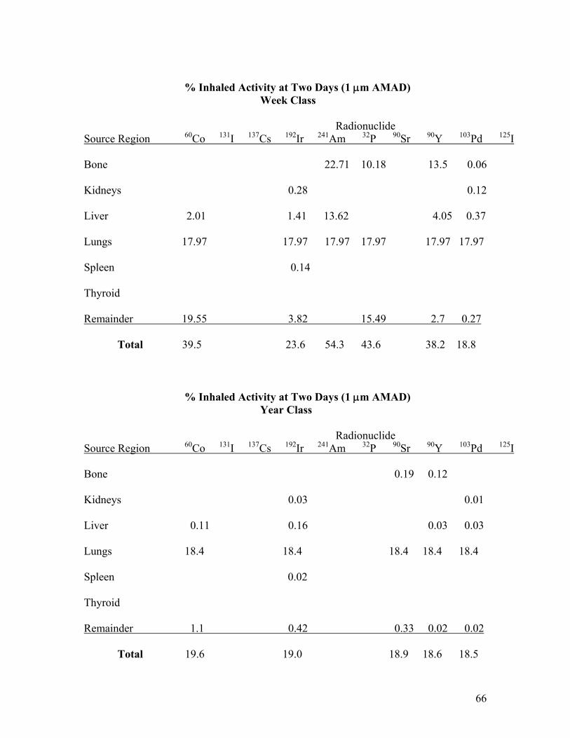
66
% Inhaled Activity at Two Days (1 μm AMAD) Week Class Radionuclide Source Region 60Co 131I 137Cs 192Ir 241Am 32P 90Sr 90Y 103Pd 125I Bone 22.71 10.18 13.5 0.06 Kidneys 0.28 0.12 Liver 2.01 1.41 13.62 4.05 0.37 Lungs 17.97 17.97 17.97 17.97 17.97 17.97 Spleen 0.14 Thyroid Remainder 19.55 3.82 15.49 2.7 0.27 Total 39.5 23.6 54.3 43.6 38.2 18.8 % Inhaled Activity at Two Days (1 μm AMAD) Year Class Radionuclide Source Region 60Co 131I 137Cs 192Ir 241Am 32P 90Sr 90Y 103Pd 125I Bone 0.19 0.12 Kidneys 0.03 0.01 Liver 0.11 0.16 0.03 0.03 Lungs 18.4 18.4 18.4 18.4 18.4 Spleen 0.02 Thyroid Remainder 1.1 0.42 0.33 0.02 0.02 Total 19.6 19.0 18.9 18.6 18.5

67
c. Non-photon-emitting radionuclides that cannot be detected within the body by an external detector 1. 3H H-3 has a physical half-life of 12.33 years and decays by β- emission. The metabolic model for tritium is described in ICRP 30. Tritiated water is assumed to be uniformly distributed among all soft tissues at any time following intake. Its retention, R(t), is described as a single exponential with an effective clearance half-time of 10 days: R (t) = e - 0.693 x t/10 2. 226Ra
Ra-226 has a physical half-life of 1600 years and decays by α emission. Since radium is an alkaline earth element, it can be assumed that the biokinetic model is the same as for strontium above. 3. 234,235,238U U-234 has a physical half-life of 2.455x10⁵ years and decays by α emission; U-235 has a physical half-life of 7.038x10⁸ years and decays by α emission; and U-238 has a physical half-life of 4.468x10⁹ years and decays by α emission. For material entering the systemic circulation, fractions 0.2 and 0.023 are assumed to go to mineral bone and be retained there with half-lives of 20 and 5000 days, respectively; fractions 0.12 and 0.00052 are assumed to go to the kidneys and to be retained with half-lives of 6 and 1500 days, respectively; and fractions 0.12 and 0.00052 are assumed to go to all other tissues of the body. The remaining fraction of the uranium entering the systemic circulation, 0.54, is assumed to go directly to excretion. Lung retention fractions: Days Post Intake Class D Class W Class Y 1 μm 5 μm 1 μm 5 μm 1 μm 5 μm 0 0.33 0.17 0.33 0.17 0.33 0.17 1 0.080 0.028 0.21 0.075 0.21 0.077 2 0.024 0.0086 0.18 0.063 0.18 0.064

68
Urine excretion fractions: Days Post Intake Class D Class W Class Y 1 μm 5 μm 1 μm 5 μm 1 μm 5 μm 1 0.10 0.084 0.0125 0.020 0.00080 0.0016 2 0.041 0.028 0.0039 0.0060 0.00027 0.00050 4. 239Pu Pu-239 has a physical half-life of 24,110 years and decays by α emission. For dissolved (ionic form) plutonium reaching the transfer compartment (i.e., the bloodstream), the ICRP 30 model distributes 45% to the bone surfaces from which it clears with a biological half-time of 50 years and 45% to the liver with a biological clearance half-time of 20 years. The activity deposition in bone is assumed to be uniformly distributed over the bone surfaces of both cortical and trabecular bone. A small radioactivity fraction is permanently retained in the gonads (0.035% for testes and 0.011% for ovaries). The remaining 10% is assumed to go directly to excretion; for purposes of dosimetry this component is considered to be an insignificant contributor to effective dose equivalent and is generally ignored.

69
B. ANTHROPOMORPHIC GEOMETRIC MODELS AND MONTE CARLO RADIATION TRANSPORT SIMULATIONS
Monte Carlo simulations allow for realistic modeling of source and target composition and
geometry. In addition, realistic radiation transport and nonuniform distribution of the activity in the
source can be modeled. The adult male anthropomorphic phantom model as developed by Cristy
and Eckerman (Cristy et al. 1987) was created for use with the Monte Carlo transport software
code MCNP4B (Briesmeister 1993). Some regions which were not relevant to this calculation
were excluded, but all relevant regions, including all bone, kidneys, liver, spleen, lungs, thyroid,
and remainder tissues were modeled. Soft tissue, bone, and lung elemental composition and
density were also modeled according to the values given in the Cristy-Eckerman phantom series.
The detector was modeled as a cylindrical disk located 1 cm from the front edge of the phantom, at
the midline and center of the trunk region, i.e., at the level of the xiphoid process. Thus, these
simulations resulted in various “humanized” exposure rate constants at a distance of 1 cm from the
anterior surface of the body. The distance of 1 cm was chosen in order to ensure the detectability
of the various radionuclides. For example, at a more standard distance of 1 m, the resulting
humanized exposure rate constants would have been extremely low indicating that measurements
at 1 m would probably not be able to detect the presence of one ALI of the particular radionuclide
in the body of a contaminated individual. The distribution of activity in the source phantom was
modeled in the various organs and tissues using the biodistribution results obtained for each
radionuclide in the previous section. Monte Carlo radiation transport simulations were performed
for each of the following radionuclides: 60Co, 131I , 137Cs, 192Ir, 241Am, 32P, 90Sr, 90Y, 103Pd, and 125I.
Photon and electron histories as appropriate were run using MCNP4B in order to obtain absorbed
fractions necessary for the determination of the humanized exposure rate constants for each
source-“detector” pair. The MCNP4B criteria for statistical precision is a standard error of less
than 10% for non-point detectors. In this problem, standard error levels were generally kept below

70
5%, and in all cases kept below 10%. Between 1.5 million and 400 million photon histories were
required for this level of accuracy.
Whole Body Source Regions
The model consisted of an external shape of head, neck, legs, and trunk, where the trunk contained
kidneys, liver, spleen, thyroid, bone, and lungs as described below.
Head and Neck:
The head and neck of each phantom was modeled as an ellipsoidal cylinder joined to an ellipsoid
to represent the neck and head, and limited on the lower end by a bounding plane as follows (All
dimensions in cm):
x8
⎡ ⎣ ⎢
⎤ ⎦ ⎥
2
+y
10⎡ ⎣ ⎢
⎤ ⎦ ⎥
2
≤ 1 and 70 ≤ z ≤ 86.85
and
x8
⎡ ⎣ ⎢
⎤ ⎦ ⎥
2
+y
10⎡ ⎣ ⎢
⎤ ⎦ ⎥
2
+z − 86.85
7.15⎡ ⎣ ⎢
⎤ ⎦ ⎥
2
≤ 1 and z f 86.85
Trunk:
The trunk of each phantom was modeled as an ellipsoidal cylinder with upper and lower bounding
planes as follows (All dimensions in cm):
11020
22
≤⎥⎦⎤
⎢⎣⎡+⎥⎦
⎤⎢⎣⎡ yx
and 0 ≤ z ≤ 70

71
Legs:
The legs on the phantom were represented by the frustums of two circular cones as follows (All
dimensions in cm):
⎥⎦⎤
⎢⎣⎡ +±≤+ zxyx
100202022
and −80 ≤ z ≤ 0
Thyroid Region:
The thyroid region was represented as an ellipsoid in the neck of the phantom as follows (All
dimensions in cm):
15.2
5.72378.1
6.7378.1
222
≤⎥⎦⎤
⎢⎣⎡ −
+⎥⎦⎤
⎢⎣⎡ +
+⎥⎦⎤
⎢⎣⎡ zyx
Liver:
The liver region was represented as an elliptical cylinder as follows (All dimensions in cm):
185.16
22
≤⎥⎦⎤
⎢⎣⎡+⎥⎦
⎤⎢⎣⎡ yx
1434535
−≤⎥⎦⎤
⎢⎣⎡−⎥⎦
⎤⎢⎣⎡+⎥⎦
⎤⎢⎣⎡ zyx
27 ≤ z ≤ 43
Lungs:

72
The lung regions were represented as two half-ellipsoids with a section removed. The equation for
a single lung was defined as follows (All dimensions in cm):
124
5.435.75
5.8 222
≤⎥⎦⎤
⎢⎣⎡ −
+⎥⎦⎤
⎢⎣⎡+⎥⎦
⎤⎢⎣⎡ + zyx
z ≥ 43.5
Spleen:
The spleen region was represented as an ellipsoid defined as follows (All dimensions in cm):
1637
23
5.311 222
≤⎥⎦⎤
⎢⎣⎡ −
+⎥⎦⎤
⎢⎣⎡ −
+⎥⎦⎤
⎢⎣⎡ − zyx
Kidneys:
The kidneys were represented as two ellipsoids cut by a plane. The equation for a kidney was
defined as follows (All dimensions in cm):
15.532
5.16
5.46 222
≤⎥⎦⎤
⎢⎣⎡ −
+⎥⎦⎤
⎢⎣⎡ −
+⎥⎦⎤
⎢⎣⎡ ± zyx
| x |≥ 3
Skeletal Regions:

73
The composite skeletal source region was represented by combining the individual bone regions
represented by the following equations.
Arm Bones:
Each arm bone was represented as the frustrum of an elliptical cone that was defined by the
following inequalities:
( ) ( ) ( ) 22
2
13869138
7.24.1
4.1869138
4.1
⎥⎦⎤
⎢⎣⎡ −+
≤⎥⎦⎤
⎢⎣⎡+
⎥⎥⎥⎥
⎦
⎤
⎢⎢⎢⎢
⎣
⎡±+−⎥⎦
⎤⎢⎣⎡
zyxz
0 ≤ z ≤ 69
Pelvis:
The pelvis was represented by a portion of the volume between two non-concentric elliptical
cylinders that were defined by the following inequalities:
112
312
22
≤⎥⎦⎤
⎢⎣⎡ +
+⎥⎦⎤
⎢⎣⎡ yx
13.11
8.33.11
22
≥⎥⎦⎤
⎢⎣⎡ +
+⎥⎦⎤
⎢⎣⎡ yx
y ≥ −3 0 ≤ z ≤ 22 y ≤ 5 if z ≤14
Spine:

74
The spine was defined as an elliptical cylinder using the following inequalities:
15.2
5.52
22
≤⎥⎦⎤
⎢⎣⎡ −
+⎥⎦⎤
⎢⎣⎡ yx
22 ≤ z ≤ 80.54
Skull:
The skull region consisted of the cranium and the facial skeleton. The cranium was
defined as the volume between two concentric ellipsoids using the following inequalities:
[ ] 1
75.585.1670
6.86.6
222
≥⎥⎦⎤
⎢⎣⎡ +−
+⎥⎦⎤
⎢⎣⎡+⎥⎦
⎤⎢⎣⎡ zyx
[ ] 19.075.585.1670
9.06.89.06.6
222
≤⎥⎦⎤
⎢⎣⎡
++−
+⎥⎦⎤
⎢⎣⎡
++⎥⎦
⎤⎢⎣⎡
+zyx
The facial skeleton was represented using a portion of the volume between two
concentric elliptical cylinders. The segment that intersects the cranium and brain was not
included in the above volume. The following inequalities were used:
10.90.7
22
≤⎥⎦⎤
⎢⎣⎡+⎥⎦
⎤⎢⎣⎡ yx
19.00.99.00.7
22
≥⎥⎦⎤
⎢⎣⎡
−+⎥⎦
⎤⎢⎣⎡
−yx
y ≤ 0 70 + 4.0 ≤ z ≤ 70 + 14.73
[ ] 165.6
85.16705.92.15
222
>⎥⎦⎤
⎢⎣⎡ +−
+⎥⎦⎤
⎢⎣⎡+⎥⎦
⎤⎢⎣⎡ zyx
Rib Cage:

75
The ribs were defined using a series of bands between two concentric, right-vertical,
elliptical cylinders. This region was then sliced by a series of equispaced horizontal
planes into slabs. Every other one of these was considered a rib. The following conditions
had to be satisfied as part of this definition:
18.917
22
≤⎥⎦⎤
⎢⎣⎡+⎥⎦
⎤⎢⎣⎡ yx
150.08.950.017
22
≥⎥⎦⎤
⎢⎣⎡
−+⎥⎦
⎤⎢⎣⎡
−yx
35.10 ≤ z ≤ 67.30
Integer ⎥⎦⎤
⎢⎣⎡ −
40.110.35z is even
Clavicles:
The clavicles were represented by two portions of a torus lying along a circular arc. The
following inequalities were used:
( ) ( ) 6214.010.112025.682
222 ≤⎥⎦⎤
⎢⎣⎡ −+−+− yxz
.89415 ≤11.1− y
x≤ 7.0342
y < 0
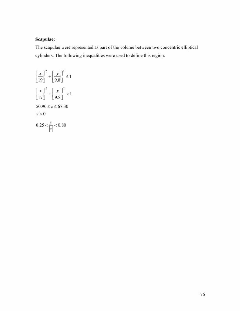
76
Scapulae:
The scapulae were represented as part of the volume between two concentric elliptical
cylinders. The following inequalities were used to define this region:
18.919
22
≤⎥⎦⎤
⎢⎣⎡+⎥⎦
⎤⎢⎣⎡ yx
18.917
22
>⎥⎦⎤
⎢⎣⎡+⎥⎦
⎤⎢⎣⎡ yx
50.90 ≤ z ≤ 67.30 y > 0
0.25 <yx
< 0.80

77
C. HUMANIZED EXPOSURE CONSTANTS OF POTENTIAL RDD RADIONUCLDES The conventional dose rate, DR, equation is given by:
DR (mR/h) = 2dAxΓ
where Γ is the specific gamma-ray constant, or in the case of pure beta-emitters, the specific bremsstrahlung constant (R cm2/mCi h), A is the activity (mCi) contained in the contaminated individual, and d is the distance (cm) at which the dose rate measurement is made. Since the potential RDD radionuclides include both photon and beta-emitters, we will refer to Γ as the specific exposure rate constant from this point on. This equation can be rearranged to estimate the activity contained in an individual:
A (mCi) = Γ
2dxDR
The dose rate measurement should be made at the level of the xiphoid process at a distance of 1 cm from the contaminated individual for all considered radionuclides with the exception of the radioiodines. For both 125I and 131I, the dose rate measurement should be made at the level of the thyroid gland at a distance of 1 cm since essentially all activity remaining at day 2 will be located in the thyroid. Dividing the measured dose rate by the specific exposure rate constant would result in the activity contained in the individual. However, use of Γ for this purpose is highly conservative as it represents a point-source, in air measurement. It does not account for attenuation or the distribution of the radioactivity in the body of the contaminated individual; therefore its use will significantly underestimate the activity contained in the individual (i.e., the uncorrected Γ is a value that is too high). We have used ICRP 30 biodistribution models for lung and total body retention coupled with the adult male anthropomorphic mathematical phantom and state-of-the art Monte Carlo radiation transport techniques to determine the appropriate correction factors to be applied to the dose rate measurements for each considered radionuclide in order to account for attenuation and scatter in the contaminated individual. These represent the humanized exposure rate constants we have determined at the level of the xiphoid process at a distance of 1 cm from the contaminated individual (for 125I and 131I the humanized constants are at the level of the thyroid gland at 1 cm). These humanized constants may also be considered S values. These values will put external radiation dose calculations on an equal par to those conventionally used for internal absorbed dose estimation using the well-known Medical Internal Radiation Dose (MIRD) schema (Siegel et al. J Nucl Med 2002; 43:1241-1244). As an example, it has been demonstrated that use of the uncorrected Γ resulted in a 60% overestimate for 131I dose rate measurements (Sparks et al. Health Phys 1998; 75:385-

78
388). Thus, use of the humanized constants will result in more realistic determinations since their calculation is based on more realistic assumptions; in essentially all modeling scenarios, the more the model reflects the actual physical characteristics and configuration of the problem, the greater the accuracy. The humanized exposure rate constants for the considered radionuclides are given the Table below. These values were obtained using the ICRP model-determined biodistributions at 2 days along with the adult male anthropomorphic mathematical phantom and Monte Carlo radiation transport simulations for each radionuclide.
SUMMARY TABLE OF HUMANIZED EXPOSURE RATE CONSTANTS Radionuclide Humanized Exposure Rate Constant (mR/μCi h @ 1 cm) ________________________________________________________________________ 60Co 0.00642 131I 0.0103 137Cs 0.00207 192Ir 0.00184 241Am 0.0000477 32P 0.000132 90Sr 0.00000709 90Y 0.0000801 103Pd 0.00000338 125I 0.00352 In order to help validate and serve as a check on our Monte Carlo results, a somewhat simpler and less mathematically rigorous technique was utilized for all radionuclides except the radioiodines (see below for assumptions made for both 125I and 131I). An approximate method for determining the necessary humanized exposure rate constants is to first multiply the individual radionuclide Γ factors by a term given by 1- φ(TB←TB), where φ(TB←TB) is the total body-to-total body absorbed fraction representing the fraction of energy that is not absorbed within the total body and thus approximates the

79
effect of shielding by the individual (Zanzonico et al. J Nucl Med 1999; 40:1024-1028). The needed absorbed fractions can be obtained from MIRD Pamphlet No. 3, Table 9 (an assumed uniform distribution of activity in unit density ellipsoids of various sizes). This homogeneous ellipsoidal model has principal axes in the ratio of 1/1.8/9.27. Bob Loevinger stated in 1956 that the human body may be approximated by an ellipsoid of ratio of axes 5:1 which is a ratio of length-to-maximum diameter; this agrees well with this model, which has a 5.15:1 ratio of length-to-maximum diameter. Secondly, since all calculations were performed at a distance of 1 cm from the body of a contaminated individual, these “absorbed fraction-corrected values” have to be multiplied by the factor 0.018 because the activity distribution in the body is NOT a point source, but rather a distributed source. This total body distributed source can be approximated as a line source and we have shown that the line-to-point source ratio for a whole-body distribution at a measurement distance of 1 cm is 0.018 (Siegel et al. J Nucl Med 2002; 43:1241-1244). Further, because the total body activity distribution is on average at a depth of 10 cm, division by an additional factor of 10 is necessary (the dose rate falloff with small distances is linear, not inverse square, for a line source). Lastly, a correction reflecting the activity remaining in the body at day 2 is necessary since the day 2 biodistribution data were utilized in the Monte Carlo simulations. In performing these calculations, we considered only the energy of the most abundant penetrating emission for each radionuclide in determining the appropriate value for the absorbed fraction. In the case of the pure β-emitters, 32P, 90Sr, and 90Y, the mean energy of the bremsstrahlung radiation was found by multiplying the maximum initial kinetic energy of the β-ray by a factor of 0.11 (Zanzonico et al. J Nucl Med 1999; 40:1024-1028. The specific bremsstrahlung constants for 32P and 90Y were taken from this article and we derived the needed constant for 90Sr). The purpose of performing these calculations, as previously stated, was as a rough check on our Monte Carlo results. We do not expect the approximate values to agree closely since they are based on a uniform distribution of activity but nevertheless are a useful exercise. These approximate humanized exposure rate constants are given in the following Table and compared to those obtained by Monte Carlo simulation. A different procedure was used to generate the approximate humanized exposure rate constants for both 125I and 131I since the activity distribution of these radionuclides is essentially confined to the thyroid gland at day 2. The individual radionuclide Γ factors were first multiplied by a term given by e-μd, where μ is the linear attenuation coefficient for the respective radionuclide (equal to 0.41 cm-1 and 0.10 cm-1 for 125I and 131I, respectively) and d is the depth of center of the thyroid gland from the anterior surface of the neck (assumed to be 2 cm). This factor represents the fraction of energy that is absorbed within the body and thus approximates the effect of shielding by the individual. Due to this depth of the thyroid gland in the neck, an inverse square correction was then applied.

80
SUMMARY TABLE OF APPROXIMATE AND MONTE CARLO- DETERMINED HUMANIZED EXPOSURE RATE CONSTANTS
Radionuclide Humanized Exposure Rate Constant (mR/μCi h @ 1 cm) Monte Carlo Approximate ________________________________________________________________________ 60Co 0.00642 0.00652 131I 0.0103 0.200 137Cs 0.00207 0.00230 192Ir 0.00184 0.00209 241Am 0.0000477 0.0000554 32P 0.000132 0.00000372 90Sr 0.00000709 0.00000105 90Y 0.0000801 0.00000470 103Pd 0.00000338 0.0000179 125I 0.00352 0.0342 Considering the assumptions and approximations necessary in determining the approximate values, the agreement between the approximate and Monte-Carlo based humanized exposure rate constants is excellent in most cases. The exception is for the radioiodines. We believe that the Monte Carlo results for the radioiodines are not realistic since the phantom thyroid could not be centered at a distance closer than 2.5 cm from the anterior surface of the neck. Thus, we will use the Monte Carlo results for all radionuclides in the following sections of this report except for the radioiodines, where we will use the approximate results as generated by the methods discussed above. The Monte Carlo-derived humanized constants represent values obtained in a standard 70 kg person. In this project, it was not practical to derive humanized constants for the range of weights of contaminated individuals that might be encountered for all the considered radionuclides. In order to approximate the necessary adjustments of these values for individuals of differing weights, we considered ellipsoids corresponding to total body

81
masses of 10, 20, 50, 70, 100, 140, and 200 kg. Based on the observed mass dependence of 1-φ(TB←TB) as given in MIRD Pamphlet No. 3, Table 9, correction factors for the Monte-Carlo derived humanized constants were obtained. It must be emphasized that these correction factors are based on the assumption that the radioactivity is uniformly distributed throughout the body. All values are provided for each radionuclide. These correction factors are important as it is necessary to adjust the humanized exposure rate constants on a weight basis, particularly when the contaminated individual weighs a significantly different amount than that of the standard 70 kg individual. Note that this weight adjustment was assumed to not be necessary for the radioiodines as the activity is confined to the thyroid gland, i.e., no weight adjustment of the humanized exposure rate constants are necessary for 125I and 131I.

82
D. ANNUAL LIMITS ON INTAKE (ALIs) AND DETECTABILITY OF POTENTIAL RDD RADIONUCLDES All ALIs were taken from the NRC’s 10 CFR Part 20 for 1 μm AMAD particles. 1. 60Co ICRP 30 and the Nuclear Regulatory Commission (NRC) pursuant to 10 CFR Part 20, classify 60Co depending on its chemical form as inhalation class W (weeks) or Y (years) for oxides, hydroxides, halides and nitrates. The annual limits on intake (ALI) for 60Co due to inhalation are 7.4 MBq (200 μCi) and 1.11 MBq (30 μCi) for Class W and Y, respectively. These ALIs are defined by the stochastic dose limit. 2. 131I The annual limit on intake (ALI) for 131I due to inhalation is 1.85 MBq (50 μCi) pursuant to Nuclear Regulatory Commission (NRC) requirements given in 10 CFR Part 20. This ALI is defined by the non-stochastic dose limit, i.e., the ALI is based on a committed dose equivalent of 0.5 Gy to an organ or tissue (in this case, thyroid). ICRP 30 and the Nuclear Regulatory Commission (NRC) pursuant to 10 CFR Part 20, classify 131I in any chemical form as inhalation class D (days), indicating that inhaled material will be absorbed rapidly from the respiratory tract into the circulatory system. 3. 137Cs The annual limit on intake (ALI) for 137Cs due to inhalation is 7.4 MBq (200 μCi) pursuant to Nuclear Regulatory Commission (NRC) requirements given in 10 CFR Part 20. Because cesium is assumed to be distributed evenly throughout all tissues in the body, the stochastic dose limit or ALI is limiting, i.e., the ALI is based on a committed effective dose equivalent of 0.05 Gy. ICRP 30 and the Nuclear Regulatory Commission (NRC) pursuant to 10 CFR Part 20, classify 137Cs in any chemical form as inhalation class D (days), indicating that inhaled material will be absorbed rapidly from the respiratory tract into the circulatory system. 4. 192Ir ICRP 30 and the Nuclear Regulatory Commission (NRC) pursuant to 10 CFR Part 20, classify 192Ir depending on its chemical form as inhalation class D (days), W (weeks) for halides, nitrates and metallic iridium, or Y (years) for oxides and hydroxides. The annual limits on intake (ALI) for 192Ir due to inhalation are 11.1 MBq (300 μCi), 14.8 MBq (400 μCi), and 7.4 MBq (200 μCi) for Class D, W, and Y, respectively. These ALIs are defined by the stochastic dose limit.

83
5. 241Am The annual limit on intake (ALI) for 241Am due to inhalation is 0.22 kBq (0.006 μCi) pursuant to Nuclear Regulatory Commission (NRC) requirements given in 10 CFR Part 20. This ALI is defined by the non-stochastic dose limit, i.e., the ALI is based on a committed dose equivalent of 0.5 Gy to an organ or tissue (in this case, bone surface). ICRP 30 and the Nuclear Regulatory Commission (NRC) pursuant to 10 CFR Part 20, classify 241Am in any chemical form as inhalation class W (weeks), indicating that inhaled material will be retained in the pulmonary region of the lung for 10 to 100 days. 6. 32P ICRP 30 and the Nuclear Regulatory Commission (NRC) pursuant to 10 CFR Part 20, classify 32P depending on its chemical form as inhalation class D (days) or W (weeks) for phosphates of Zn2+, S3+, Mg2+, Fe3+, Bi3+, and lanthanides. The annual limits on intake (ALI) for 32P due to inhalation are 33.3 MBq (900 μCi) and 14.8 MBq (400 μCi) Class D and W, respectively. These ALIs are defined by the stochastic dose limit. 7. 90Sr The annual limit on intake (ALI) for 90Sr due to inhalation is 0.74 MBq (20 μCi) pursuant to Nuclear Regulatory Commission (NRC) requirements given in 10 CFR Part 20. This ALI is defined by the non-stochastic dose limit, i.e., the ALI is based on a committed dose equivalent of 0.5 Gy to an organ or tissue (in this case, bone surface). All intakes of strontium are considered to be inhalation class D, with the exception that strontium titanate is the only compound identified by the ICRP as belonging to inhalation class Y. 8. 90Y ICRP 30 and the Nuclear Regulatory Commission (NRC) pursuant to 10 CFR Part 20, classify 90Y depending on its chemical form as inhalation class W (weeks) or Y (years) for oxides and hydroxides. The annual limits on intake (ALI) for 90Y due to inhalation are 25.9 MBq (700 μCi) and 22.2 MBq (600 μCi) Class W and Y, respectively. These ALIs are defined by the stochastic dose limit. 9. 103Pd ICRP 30 and the Nuclear Regulatory Commission (NRC) pursuant to 10 CFR Part 20, classify 103Pd depending on its chemical form as inhalation class D (days), W (weeks) for nitrates or Y (years) for oxides and hydroxides. The annual limits on intake (ALI) for 103Pd due to inhalation are 222 MBq (6000 μCi), 148 MBq (4000 μCi), and 148 MBq (4000 μCi) Class D, W and Y, respectively. These ALIs are defined by the stochastic dose limit.

84
10. 125I The annual limit on intake (ALI) for 125I due to inhalation is 2.22 MBq (60 μCi) pursuant to Nuclear Regulatory Commission (NRC) requirements given in 10 CFR Part 20. Each of the above radionuclides can potentially be detected by an external dose rate measurement at 1 cm using a calibrated ion chamber. The expected dose rates (mR/h) for each of the above radionuclides, based on their respective ALIs and humanized exposure constants at 1 cm, are summarized in the Table below.
SUMMARY TABLE OF EXTERNALLY-MEASURABLE RADIONUCLIDES Radionuclide ALI (μCi)* Humanized Exposure Constant Dose Rate (mR/h) (mR/μCi h @ 1 cm) from 1 ALI ________________________________________________________________________ 60Co 30 0.00642 0.19 131I 50 0.200 1.51** 137Cs 200 0.00207 0.41 192Ir 200 0.00184 0.37 241Am 0.006 0.0000477 0.00000029 32P 400 0.000132 0.053 90Sr 20 0.00000709 0.00014 90Y 600 0.0000801 0.048 103Pd 4000 0.00000338 0.014 125I 60 0.0342 0.36** ________________________________________________________________________ * For those radionuclides with multiple ALIs, corresponding to D, W, and/or Y class, the smallest ALI value is given. ** The dose rates per ALI for the radioiodines were modified by the day 2 biodistribution data to reflect the activity present in the thyroid at that time. Note: Dose rate measurements need to be background-subtracted. Background in Los Angeles averages about 0.02 mR/h and ranges from approximately 0.009 - 0.04 mR/h. Background should be measured on each instrument prior to use.

85
Dose rate measurements for most of the radionuclides listed in the preceding Table will be able to assess the level of one ALI in a given contaminated individual. That is, a measured dose rate obtained in a potentially contaminated individual at or above the expected dose rate in the Table indicates the presence of at least one ALI of the particular radionuclide. To obtain the estimated activity (μCi) contained in the body of the internally contaminated individual all that is necessary is to divide the measured dose rate at 1 cm (at the level of the xiphoid process except for the radioiodines where the measurements should be performed at the level of the thyroid) by the humanized exposure rate constant for the radionuclide in question. For the case of the radioiodines, the measured dose rate must be divided by the humanized constant adjusted for the activity remaining in the thyroid at day 2 as indicated in the Table on page 17. The activity estimates may have to be adjusted up or down depending upon the individual’s weight. We have provided the necessary mass-adjustments for each radionuclide, except for the radioiodines as previously discussed. It should be noted that accurate dose rate measurements may not be possible for 241Am, 103Pd, and the pure β-emitter 90Sr due to the low expected dose rates per ALI for these radionuclides. It is important to state that although we have used the best available models, mathematical phantoms and Monte Carlo radiation transport simulation techniques, the resulting humanized exposure rate constants are estimates. They are highly dependent on the extent to which the biodistribution models estimate the radioactivity biokinetics in an exposed individual, how well the anthropomorphic mathematical phantom represents a particular internally contaminated individual, and the time and distance at which the dose rate measurement is actually made. The following radionuclides do not emit photons and therefore cannot be detected by external means: 11. 3H The annual limit on intake (ALI) for 3H due to inhalation is 2.96 GBq (80 mCi) and is defined by the stochastic dose limit. 12. 226Ra The annual limit on intake (ALI) for 226Ra due to inhalation is 22.2 kBq (0.6 μCi) pursuant to Nuclear Regulatory Commission (NRC) requirements given in 10 CFR Part 20. This ALI is defined by the stochastic dose limit. ICRP 30 and the Nuclear Regulatory Commission (NRC) pursuant to 10 CFR Part 20, classify 226Ra in any chemical form as inhalation class W (weeks). 13. 234,235,238U

86
ICRP 30 and the Nuclear Regulatory Commission (NRC) pursuant to 10 CFR Part 20, classify uranium depending on its chemical form as inhalation class D (days) for UF6, UO2F2, UO2(NO3)2, W (weeks) for UO3, UF4, UCl4 or Y (years) for UO2 and U3O8. The annual limits on intake (ALI) for 234U due to inhalation are 37 kBq (1 μCi for bone surfaces), 25.9 kBq (0.7 μCi), and 1.48 kBq (0.04 μCi) Class D, W and Y, respectively. The annual limits on intake (ALI) for 235U due to inhalation are 37 kBq (1 μCi for bone surfaces), 29.6 kBq (0.8 μCi), and 1.48 kBq (0.04 μCi) Class D, W and Y, respectively. The annual limits on intake (ALI) for 238U due to inhalation are 37 kBq (1 μCi for bone surfaces), 29.6 kBq (0.8 μCi), and 1.48 kBq (0.04 μCi) Class D, W and Y, respectively. 14. 239Pu The annual limit on intake (ALI) for 239Pu due to inhalation is 0.22 kBq (0.006 μCi) and 0.74 kBq (0.02 μCi) for inhalation class W and Y, respectively, pursuant to Nuclear Regulatory Commission (NRC) requirements given in 10 CFR Part 20. These ALIs are defined by the non-stochastic dose limit, i.e., the ALI is based on a committed dose equivalent of 0.5 Gy to an organ or tissue (in this case, bone surface). ICRP 30 and the Nuclear Regulatory Commission (NRC) pursuant to 10 CFR Part 20, classify 239Pu in any chemical form as inhalation class W (weeks) and Y (years) for PuO2. Also, as stated above, 241Am, 103Pd, and 90Sr may not be detectable by external exposure rate measurements.

87
For Co-60 Correction Factors for Humanized Exposure Rate Constant as function of weight Specific γ-ray constant = 13.2 mR/μCi h @ 1 cm Humanized constant for 70 kg person = 0.00642 mR/μCi h @ 1 cm Correction Factors for
Absorbed Fraction Mass (kg) (1 - Absorbed Fraction) Humanized Constant 0.183 10 0.817 1.18 0.223 20 0.777 1.12 0.282 50 0.718 1.03 0.306 70 0.694 1.00 0.334 100 0.666 0.96 0.364 140 0.636 0.92 0.403 200 0.597 0.86
Correction Factor (CF) v Body Weight: Co-60
CF Humanized = 1.15 x e-0.0016 x weight (kg)
R2 = 0.94
0.0
0.5
1.0
1.5
0 50 100 150 200 250Body weight (kg)
Cor
rect
ion
Fact
or

88
For Cs-137 Correction Factors for Humanized Exposure Rate Constant as function of weight Specific γ-ray constant = 3.3 mR/μCi h @ 1 cm Humanized constant for 70 kg person = 0.00207 mR/μCi h @ 1 cm Correction Factors for
Absorbed Fraction Mass (kg) (1 - Absorbed Fraction) Humanized Constant 0.202 10 0.798 1.21
0.25 20 0.750 1.14 0.317 50 0.683 1.04 0.341 70 0.659 1.00 0.368 100 0.632 0.96 0.399 140 0.601 0.91 0.449 200 0.551 0.84
Correction Factor (CF) v Body Weight: Cs-137
CF Humanized = 1.17 x e-0.0018 x weight (kg)
R2 = 0.94
0.0
0.5
1.0
1.5
0 50 100 150 200 250Body weight (kg)
Corr
ectio
n Fa
ctor

89
For Ir-192 Correction Factors for Humanized Exposure Rate Constant as function of weight Specific γ-ray constant = 4.8 mR/μCi h @ 1 cm Humanized constant for 70 kg person = 0.00184 mR/μCi h @ 1 cm Correction Factors for
Absorbed Fraction Mass (kg) (1 - Absorbed Fraction) Humanized Constant 0.2 10 0.800 1.21
0.245 20 0.755 1.14 0.312 50 0.688 1.04
0.34 70 0.660 1.00 0.371 100 0.629 0.95 0.405 140 0.595 0.90 0.446 200 0.554 0.84
Correction Factor (CF) v Body Weight: Ir-192
CF Humanized = 1.17 x e-0.0018 x weight (kg)
R2 = 0.94
0.0
0.5
1.0
1.5
0 50 100 150 200 250Body weight (kg)
Cor
rect
ion
Fact
or

90
For Am-241 Correction Factors for Humanized Exposure Rate Constant as function of weight Specific γ-ray constant = 0.1 mR/μCi h @ 1 cm Humanized constant for 70 kg person = 0.0000477 mR/μCi h @ 1 cm Correction Factors for
Absorbed Fraction Mass (kg) (1 - Absorbed Fraction) Humanized Constant 0.247 10 0.753 1.33 0.308 20 0.692 1.22 0.397 50 0.603 1.06 0.432 70 0.568 1.00 0.471 100 0.529 0.93 0.511 140 0.489 0.86 0.559 200 0.441 0.78
Correction Factor (CF) v Body Weight: Am-241
CF Humanized = 1.27 x e-0.0027 x weight (kg)
R2 = 0.94
0.0
0.5
1.0
1.5
0 50 100 150 200 250Body weight (kg)
Cor
rect
ion
Fact
or

91
For P-32 Correction Factors for Humanized Exposure Rate Constant as function of weight Pure β emitter: specific bremsstrahlung constant = 0.007425 mR/μCi h @ 1 cm Humanized constant for 70 kg person = 0.000132 mR/μCi h @ 1 cm Correction Factors for
Absorbed Fraction Mass (kg) (1 - Absorbed Fraction) Humanized Constant 0.19 10 0.810 1.22
0.234 20 0.766 1.15 0.305 50 0.695 1.05 0.335 70 0.665 1.00 0.369 100 0.631 0.95 0.405 140 0.595 0.89 0.451 200 0.549 0.83
Correction Factor (CF) v Body Weight: P-32
CF Humanized = 1.18 x e-0.002 x weight (kg)
R2 = 0.95
0.0
0.5
1.0
1.5
0 50 100 150 200 250Body weight (kg)
Cor
rect
ion
Fact
or
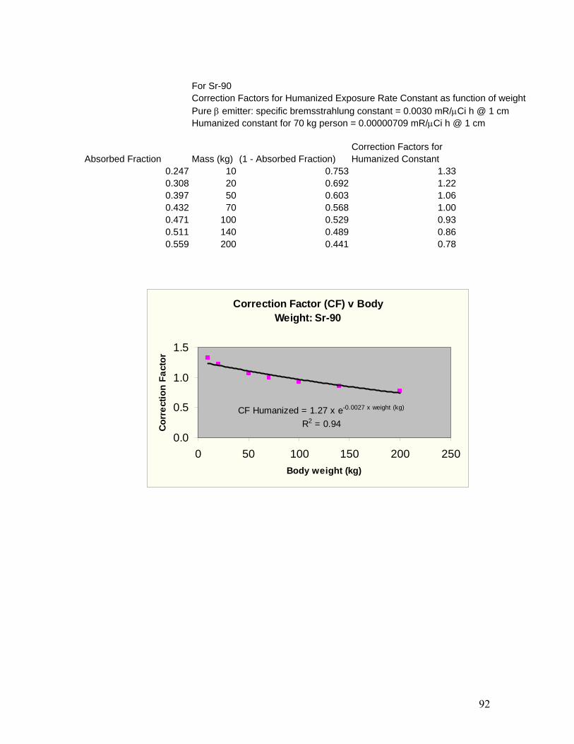
92
For Sr-90 Correction Factors for Humanized Exposure Rate Constant as function of weight Pure β emitter: specific bremsstrahlung constant = 0.0030 mR/μCi h @ 1 cm Humanized constant for 70 kg person = 0.00000709 mR/μCi h @ 1 cm Correction Factors for
Absorbed Fraction Mass (kg) (1 - Absorbed Fraction) Humanized Constant 0.247 10 0.753 1.33 0.308 20 0.692 1.22 0.397 50 0.603 1.06 0.432 70 0.568 1.00 0.471 100 0.529 0.93 0.511 140 0.489 0.86 0.559 200 0.441 0.78
Correction Factor (CF) v Body Weight: Sr-90
CF Humanized = 1.27 x e-0.0027 x weight (kg)
R2 = 0.940.0
0.5
1.0
1.5
0 50 100 150 200 250Body weight (kg)
Cor
rect
ion
Fact
or

93
For Y-90 Correction Factors for Humanized Exposure Rate Constant as function of weight Pure β emitter: specific bremsstrahlung constant = 0.01032 mR/μCi h @ 1 cm Humanized constant for 70 kg person = 0.0000801 mR/μCi h @ 1 cm Correction Factors for
Absorbed Fraction Mass (kg) (1 - Absorbed Fraction) Humanized Constant 0.195 10 0.805 1.22
0.24 20 0.760 1.15 0.309 50 0.691 1.04 0.338 70 0.662 1.00
0.37 100 0.630 0.95 0.405 140 0.595 0.90 0.449 200 0.551 0.83
Correction Factor (CF) v Body Weight: Y-90
CF Humanized = 1.18 x e-0.0019 x weight (kg)
R2 = 0.94
0.0
0.5
1.0
1.5
0 50 100 150 200 250Body weight (kg)
Cor
rect
ion
Fact
or

94
For Pd-103 Correction Factors for Humanized Exposure Rate Constant as function of weight Specific γ-ray constant = 1.48 mR/μCi h @ 1 cm Humanized constant for 70 kg person = 0.00000338 mR/μCi h @ 1 cm Correction Factors for
Absorbed Fraction Mass (kg) (1 - Absorbed Fraction) Humanized Constant 0.83 10 0.170 1.87
0.868 20 0.132 1.45 0.9 50 0.100 1.10
0.909 70 0.091 1.00 0.918 100 0.082 0.90 0.929 140 0.071 0.78
0.94 200 0.060 0.66
Correction Factor (CF) v Body Weight: Pd-103
CF Humanized = 1.59 x e-0.0049 x weight (kg)
R2 = 0.88
0.0
0.5
1.0
1.5
2.0
0 50 100 150 200 250Body weight (kg)
Cor
rect
ion
Fact
or