Embed Size (px)
Citation preview

Megan Larson and Michelle Stanek
Wuchereria bancrofti
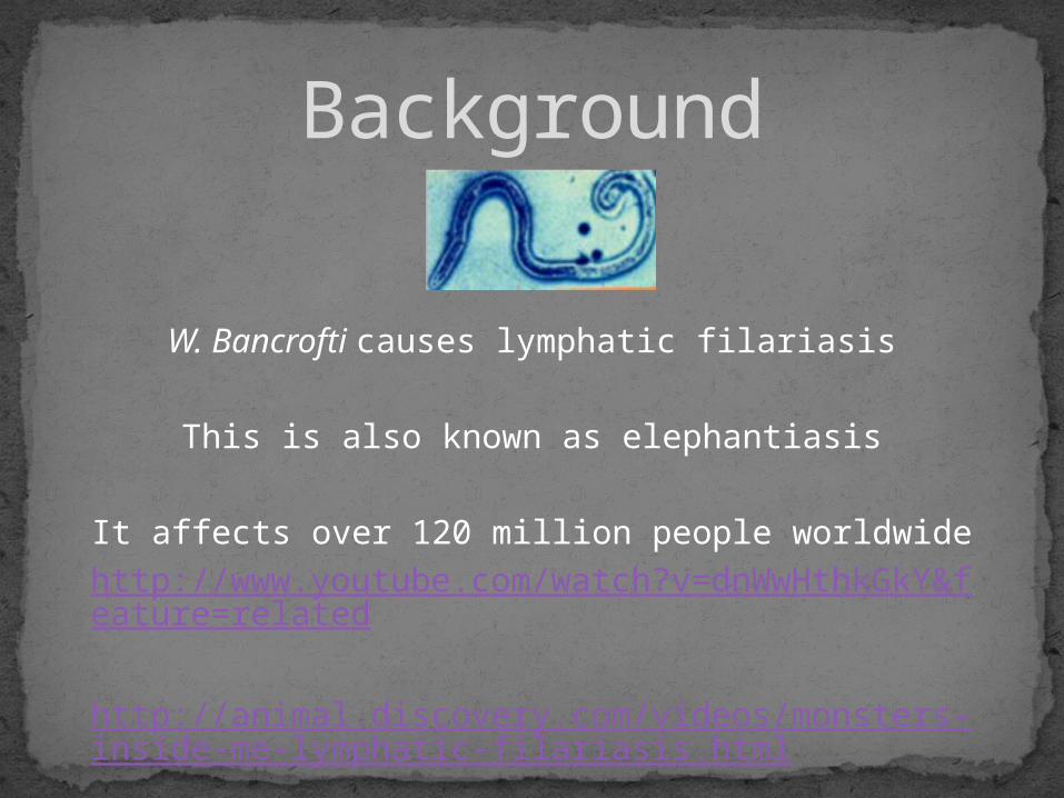
W. Bancrofti causes lymphatic filariasis
This is also known as elephantiasis
It affects over 120 million people worldwidehttp://www.youtube.com/watch?v=dnWwHthkGkY&feature=related
http://animal.discovery.com/videos/monsters-inside-me-lymphatic-filariasis.html
Background

Definitive Host: HumansIntermediate Host: Mosquitoes (especially night-
feeding mosquitoes) Species: Anopheles, Aedes, Culex, Mansonia
Morphology:Males:
40 mm long & 100 μm wideFingerlike tail
Females:6-10 cm long & 300 μm wideThey are viviparous Their vulva is near the level of the middle of their esophagus
Wuchereria bancrofti

Sub-Saharan AfricaEgyptSouthern AsiaWestern Pacific
islandsNortheastern coast of
BrazilGuyanaCaribbean island of
Hispaniola
Geographic Distribution
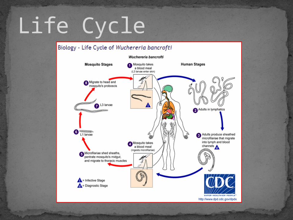
Life Cycle

1. 1.The mosquito takes a blood meal, transferring the L3 larva into the skin.
2. The L3 larva grow into adults in the lymph ducts. 3. The adults reproduce, producing sheathed microfilariae;
the microfilariae migrate into the blood and lymph channels
4. A mosquito takes a blood meal, ingesting the microfilariae.5. Once in the mosquito, the sheath of the microfilariae is
sloughed off. The microfilariae then penetrates the midgut of the mosquito, making its way into the thoracic muscles.
6. L1 larvae form7. L3 larvae form8. The L3 larvae are the infective stage, traveling to the
mosquito’s head and proboscis.
Life Cycle Continued
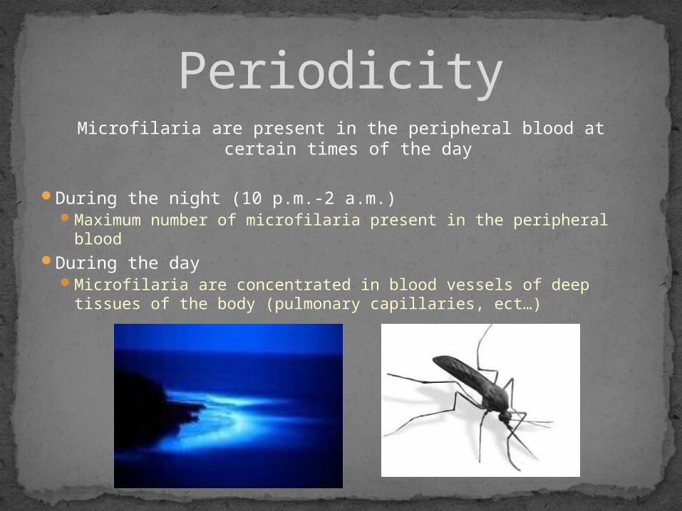
Microfilaria are present in the peripheral blood at certain times of the day
During the night (10 p.m.-2 a.m.) Maximum number of microfilaria present in the peripheral blood
During the day Microfilaria are concentrated in blood vessels of deep tissues of
the body (pulmonary capillaries, ect…)
Periodicity
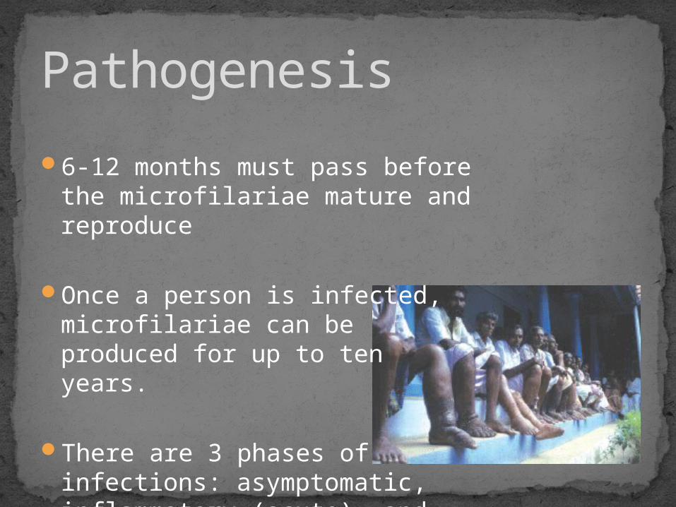
6-12 months must pass before the microfilariae mature and reproduce
Once a person is infected, microfilariae can be produced for up to ten years.
There are 3 phases of infections: asymptomatic, inflammatory (acute), and obstructive.
Pathogenesis

Asymptomatic Phase: High levels of microfilaria in the blood No symptoms present
Inflammatory (Acute) Phase: Inflammatory responses happen in response to antigens from
adult worms Lymphedema—swelling due to blockage of lymph vessels Orchitis—inflammation of the testes Epididymitis—inflammation of the spermatic cord
Obstructive Phase: Lymph varices—enlarged lymph vessels (synonymous with
varicose veins) Lymph scrotum Chyluria—lymph in urine (milky and sometimes bloody urine) Elephantiasis—enlargement of limbs and thickening of the skin
due to repeated inflammatory episodes
Pathogenesis Continued…

Wuchereria bancrofti
Elephantiasis
Lymphedema
Orchitis
Chyluria
Thick blood smear Juveniles must be present in
peripheral blood
Polymerase chain reaction (PCR) Distinguishes between other
similar species
Ultrasonography Detects vigorous movement of
adults known as “filaria dance sign”
X-rays Detects dead, calcified worms
Diagnosis
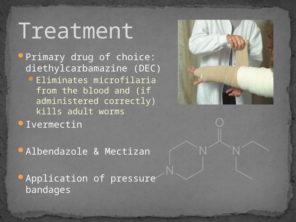
Primary drug of choice: diethylcarbamazine (DEC)Eliminates microfilaria from
the blood and (if administered correctly) kills adult worms
Ivermectin
Albendazole & Mectizan
Application of pressure bandages
Treatment

Administration of Albendazole & Mectizan to control the spread of the disease.
There are currently no vaccines Elimination of common mosquito
breeding grounds Fallen coconuts Any small container filled with water
They hope to eradicate it by year 2020; so far, the diseased population has declined significantly
http://www.youtube.com/watch?v=dnWwHthkGkY&feature=related
Control

CDC. http://www.cdc.gov/parasites/lymphaticfilariasis/biology_w_bancrofti.html
Guyen Et. al. “Evidence for Wolbachia symbiosis in microfilariae of Wuchereria bancrofti from West Bengal, India.” <http://www.ias.ac.in/jbiosci/gayen150.pdf>.
Schmidt, GD; Roberts, LS. Foundations of Parasitology. Seventh ed. P. 461-66.
http://www.youtube.com/watch?v=dnWwHthkGkY&feature=related
http://animal.discovery.com/videos/monsters-inside-me-lymphatic-filariasis.html
Sources

Questions?
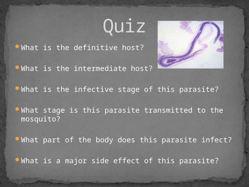
What is the definitive host?
What is the intermediate host?
What is the infective stage of this parasite?
What stage is this parasite transmitted to the mosquito?
What part of the body does this parasite infect?
What is a major side effect of this parasite?
Quiz