Embed Size (px)
Citation preview

Pancreatic Cancer Basics
Meghan McGurk, PA-C, MMSc
Physician Assistant
Department of Medical Oncology
Yale Cancer Center

Case Study
� Mr.T is a 36 yo male who presented in the fall
of 2003, at the age of 33, with complaints of
abdominal discomfort and “generally feeling
ill”
� Seen by PCP, symptoms thought to be
secondary to irritable bowel syndrome or
GERD, and was started on Prilosec

Case Study
� Symptoms persisted despite Prilosec, and the patient ultimately developed jaundice with markedly elevated LFTs
� An abdominal ultrasound was performed at Lawrence & Memorial hospital confirming biliary dilatation
� ERCP was performed and a biliary stent was placed – pt was d/c’d home with presumed gallbladder diease
Case Study
� Patient subsequently presented to YNHH ED
with severe abdominal pain in Jan 2004,
repeat abdominal ultrasound revealed
enlargement in HOP
� Follow up CT scan confirmed mass in HOP,
4.5x2.5cm, with dilation of the intrahepatic
and pancreatic ducts
� EUS with FNA of pancreas was performed

Case Study
� Diagnosis was made as adenocarcinoma
arising from the head of the pancreas
� Staging CT scans revealed no evidence for
metastatic disease, therefore, patient was
referred to surgical oncology for consideration
of Whipple procedure

Epidemiology
� 33,730 cases of pancreatic cancer are
anticipated in 2006, with 32,200 expected
deaths
� 4th leading cause of cancer-related death in the
U.S.; second only to CRC as a cause of
digestive cancer-related death
� Mortality rates closely follow incidence rates
due to poor prognosis

Epidemiology
� Incidence rare before the age of 45, however,
sharply rises thereafter
� Incidence higher in men than women
� Increased incidence in blacks compared to the
general population

Pancreatic/Biliary Anatomy
� The pancreas = retroperitoneal organ located
deep within the abdominal cavity that serves
both exocrine and endocrine function
� Exocrine – secretes digestive enzymes including
amylase and lipase
� Endocrine – secretes metabolic mediators
including insulin, glucagon, and somatostatin

Pancreatic/Biliary Anatomy

Pathophysiology
� Pancreatic tumors can arise from both the
exocrine and endocrine portions of the
pancreas, however, of pancreatic tumors, 95%
develop in the exocrine portion
� These include: ductal epithelium, acinar cells,
connective tissue, and lymphatic tissue

Histology
� Of all pancreatic cancers, 80% are adenocarcinomasof the ductal epithelium
� Only 2% of tumors from the exocrine pancreas are benign
� Less common types of exocrine pancreatic tumors include: giant cell carcinoma, adenosquamouscarcinoma, cystadenocarcinoma, papillary cystic carcinoma
� The most common types of endocrine pancreatic tumors include: insulinoma, gastrinoma, VIPoma

Genetic Factors
� Up to 5-10% of patients with pancreatic carcinoma have some genetic predisposition to developing the disease
� Inherited disorders that increase the risk of developing the disease include: hereditary pancreatitis, hereditary breast/ovarian cancer, multiple endocrine neoplasia, familial atypical multiple mole melanoma syndrome (FAMM), HNPCC, FAP/Gardner syndrome, Peutz-Jegherssyndrome

Risk Factors
� Age
� Chronic pancreatitis
� Tobacco
� DM
� Hereditary predisposition
� Obesity/Diet

Screening
� USPSTF (United States Task Force) –
recommends against routine screening in
asymptomatic individuals
� Consideration may be given to screening with
EUS and/or CT scan in the high-risk
population

Clinical Features
� Signs and symptoms vary depending on the
anatomic location of the tumor (head vs.
body/tail)
� If head of pancreas is involved, symptoms
may include:
� Obstruction of the bile duct to the small intestine
or stomach may occur, associated with jaundice,
pruritis, and/or vomiting

Clinical Features
� Conversely, if the body/tail of the pancreas is
involved, symptoms may include: upper
abdominal/back pain, weight loss (usually
substantial, 30-50lb), and fatigue
� In general, 90% of the time, the tumor has already
metastasized outside the region of the pancreas
� The pain may be intermittent, worse with eating,
and usually associated with a poor prognosis

Clinical Features

Clinical Features
� Other symptoms include the following:
� Anorexia
� Early satiety
� Diarrhea
� Steatorrhea

Diagnosis/Staging
� Diagnosis is made based upon one or more of the
following:
� Routine labs tests – may suggest a rise in the serum
bilirubin or alk phos, or the presence of mild anemia,
however the dx is typically made radiographically and
histologically
� Ultrasound/EUS
� CT scan
� MRI/MRCP
� Serum tumor markers – elevated CA 19-9, CEA

Diagnosis/Staging
� Laparoscopy is emerging as a new staging
modality for periampullary tumors to detect
small occult mets, not seen on CT scan
� Can potentially reduce the number of apparent
operable cases that are found unresectable at
laparotomy

StagingTNM staging:
T (Tumor)
TX – primary tumor cannot be assessed
TO – No evidence of primary tumor
Tis – In situ carcinoma
T1 – Tumor limited to pancreas, 2cm or less
T2 – Tumor limited to pancreas, more than 2cm
T3 – Tumor extends beyond pancreas, but without involvement of celiac axis or SMA
T4 – Tumor involves the celiac axis or the SMA (unresectable)
N (Nodes)
NX – Regional lymph nodes cannot be assessed
NO – No regional lymph node metastasis
N1 – Regional lymph node metastasis
M (Metastasis)
MX – Distant metastasis cannot be assessed
MO – No distant metastasis
M1 – Distant metastasis
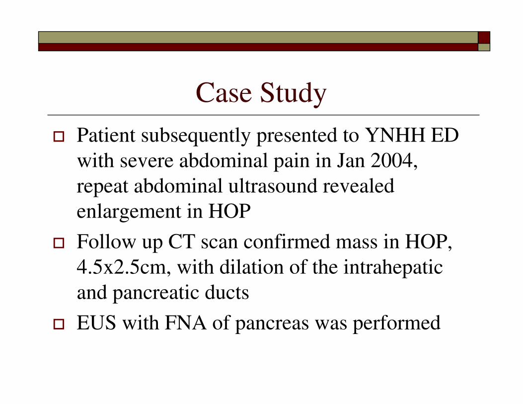
Staging
� Stage 0 – Tis N0M0
� Stage IA – T1N0M0
� Stage IB – T2N0M0
� Stage IIA – T3N0M0
� Stage IIB – T1-3N1M0
� Stage III – T4Any NM0
� Stage IV – Any T Any NM1

Treatment
� Resectable vs. Locally-Advanced vs.
Metastatic disease
� Adjuvant vs. Neoadjuvant treatment
� Surgery vs. Chemotherapy vs. Radiation
therapy

Surgery
� Surgical resection is the only potentially
curable treatment option, however, even then,
the 5 yr survival rate is only about 25-30% for
node-negative disease and <10% for node-
positive disease
� Due to the late presentation, only approx. 15-
20% of cases are resectable at the time of
diagnosis

Determining Resectibility
� Absolute contraindications include: mets to liver, peritoneum, omentum, or any extrabdominal site
� Most surgeons require that the tumor does not involve sites that would not be encompassed within the resection, and does not involve the adjacent critical structures such as SMA/SMV, portal vein, celiac axis, or hepatic artery

Potentially Resectable on CT scan

Unresectable on CT scan

Liver Mets

Surgical Resection
� The standard operation for
pancreas cancer is
pancreaticoduonectomy, or
Whipple procedure
� The outcome of resection
depends mainly on surgical
margins, nodal status, and
extent of local invasion

Case Study
� Mr. T met with the surgeon, and planned for
Whipple procedure on 1/23/04
� Intraoperatively, he was found to have hepatic
arterial involvement, and deemed
unresectable
� The surgery was aborted, and the patient was
referred to Medical Oncology for
consideration of alternate treatment options
Chemotherapy
� Chemotherapy regimens typically include:
� Gemcitabine
� 5-FU
� Tarceva
� Xeloda
� Oxaliplatin
� CPT-11
� Taxotere
� Bevacizumab
� Cetuximab

Radiation therapy
� Radiation is a useful modality, however, there
is no clear consensus on the role of radiation,
and optimal timing for its use
� Side effects include fatigue, diarrhea, abdominal
pain, myelosuppression

Advanced Disease
� Clinical benefit response – early studies
suggested that patients who lacked an
objective response often had improvement in
their symptoms (ie, pain, weight loss) and
performance status
� If patient is hesitant about considering
chemotherapy, the above should be discussed
at length

Complications/Palliation of Symptoms
� Jaundice – biliary stent vs. bypass procedure
� Duodenal obstruction – duodenal stent vs. bypass procedure
� Delayed gastric emptying – prokinetic agents may be helpful
� Pain – celiac plexus block, narcotics, palliative radiation
� Depression – adequate tx with antidepressants and emotional support
� Malabsorption/Cachexia – consider pancreatic enzyme replacement
� Ascites – palliative paracentesis, gentle diruesis

Celiac Plexus Nerve Block

Case study
� Mr. T initially started therapy on ECOG 1200
with weekly Gemcitabine and concomitant
radiation therapy with the potential to achieve
surgical resection
� This was followed by Gemcitabine in
combination with Oxaliplatin
� His CT scan did not suggest a marked
response, so Cetuximab was added

Case study
� He ultimately required cessation of
Oxaliplatin therapy related to peripheral
neuropathy, and continued Gemcitabine and
Cetuximab
� Finally, in November 2004, surgical resection
was again attempted with Whipple procedure

Case studyPANCREATIC HEAD, DUODENUM, PANCREATICODUODENECTOMY:
- PANCREAS WITH CHRONIC PANCREATITIS, FIBROSIS AND REACTIVE CHANGES,
SEE NOTE
- NO RESIDUAL TUMOR IDENTIFIED
- STROMAL RADIATION CHANGES ARE NOTED
- SURGICAL RESECTION MARGINS ARE NEGATIVE FOR TUMOR
- DUODENUM AND ANTRUM WITH REACTIVE EPITHELIAL CHANGES
- EIGHTEEN BENIGN LYMPH NODES (0/18)
- POST CHEMOTHERAPY AND RADIATION HISTOPATHOLOGIC STAGE AND GRADE(AJCC
2002): T0 NO MX, STAGE 0
NOTE: No residual tumor is identified. In the head of the pancreas close to
the distal bile duct , an areas of dense fibrous tissue is identified ( 1.5
cm in maximum dimension) and possibly represents site of prior tumor. There
is extensive intersitial fibrosis in and around the pancreas with acinar
atrophy.

Clinical Trials at Yale for Pancreatic
Cancer
1. HIC 0602001069 Wasif Saif M.D. A Phase II Clinical Trial
of Genexol-PM in Patients with Advanced Pancreatic
Cancer (closed to enrollment)
Phase I studies including:
1. HIC 0509000642 Wasif Saif M.D. A Phase I, Open-Label
Study Evaluating the Pharmacokinetics of Components of S-
1 in Patients with Impaired Hepatic Function
2. HIC 0509000643 Wasif Saif M.D. A Phase I, Open-Label
Study Evaluating the Pharamacokinetics of Components of
S-1 in Patients with Varying Degrees of Renal Function
3. HIC 0604001305 Wm. Kevin Kelly M.D. Open Label,
Dose Escalation Trial of Oral PXD101 in patients with
Advanced Solid Tumors

Final Points
� Pancreatic cancer is the 4th leading cause of cancer-
related death in the U.S.; second only to CRC as a
cause of digestive cancer-related death
� Surgical resection is the only potentially curable
treatment option
� Palliation of symptoms in advanced disease is key
� Clinical trials should always be considered given the
poor prognosis, and an additional potential benefit to
the patient

THANK YOU
QUESTIONS??? 370-7617, 785-2341