Embed Size (px)
Citation preview
The Rockefeller University Press $30.00J. Cell Biol. Vol. 197 No. 7 877–885www.jcb.org/cgi/doi/10.1083/jcb.201201100 JCB 877
JCB: Report
E. Llano and Y. Herrán contributed equally to this paper.Correspondence to Alberto M. Pendás: [email protected]; or Elena Llano: [email protected] used in this paper: AE, axial element; dKO, double knockout; DSB, double-strand break; LE, lateral element; RPA, replication protein A; SC, synaptonemal complex.
IntroductionIn mammalian spermatogenesis, a subset of spermatogonia undergoes a terminal round of DNA replication and then enters meiosis. Meiosis is a specialized process in which two successive rounds of chromosome segregation and cell divi-sions occur without intervening DNA replication. This re-duces the number of each chromosome from four copies in the meiocyte to one copy in haploid gametes. At the initiation of the prophase I, a proteinaceous structure called the axial element (AE) begins to form along replicated sister chroma-tids. Subsequently, the AEs of homologues become juxta-posed by transverse element proteins (e.g., SYCP1, beginning in zygonema), and the paired axes joined by transverse ele-ments form the tripartite synaptonemal complexes (SCs) that connect all homologues at pachynema (Yang and Wang, 2009). This dynamic protein complex provides the structural framework in which homologous chromosomes undergo close juxtaposition and repair of double-strand breaks (DSBs) by recombination. Subsequently, but before the first meiotic division, homologues desynapse but retain stable connections
formed by resolution of certain recombination events as crossovers (visible as chiasmata). In most organisms, chias-mata are required to enable proper orientation of homologues on the meiosis I spindle before the first meiotic division. Sister chromatid cohesion distal to crossovers maintains chiasmata at their initial positions until anaphase I.
During meiosis, sister chromatid cohesion is lost in two consecutive steps. Loss of chromosome arm cohesion in ana-phase I releases the linkage between homologues, allowing them to segregate to opposite poles (Page and Hawley, 2003). However, maintenance of centromeric cohesion by the action of Shugoshin-like–2 in mammals ensures the generation of tension by the proper attachment of sister chromatids to the meiosis II spindle, enabling their proper segregation to opposite poles (Llano et al., 2008; Gutiérrez-Caballero et al., 2012).
During the mitotic cell cycle, sister chromatid cohesion is mediated by the multisubunit cohesin complex between S phase and anaphase (Gruber et al., 2003; Unal et al., 2007; Haering et al., 2008; Zhang et al., 2008). Structurally, the mi-totic cohesin complex comprises four core proteins: SMC1, SMC3, RAD21, and a HEAT repeat domain protein (STAG1
Cohesin is a conserved multisubunit protein com-plex that participates in chromosome segregation, DNA damage repair, chromatin regulation, and
synaptonemal complex (SC) formation. Yeast, but not mice, depleted of the cohesin subunit Rec8 are defective in the formation of the axial elements (AEs) of the SC, sug-gesting that, in mammals, this function is not conserved. In this paper, we show that spermatocytes from mice lacking
the two meiosis-specific cohesin subunits RAD21L and REC8 were unable to initiate RAD51- but not DMC1-mediated double-strand break repair, were not able to assemble their AEs, and arrested as early as the leptotene stage of prophase I, demonstrating that cohesin plays an essential role in AE assembly that is conserved from yeast to mammals.
Meiotic cohesin complexes are essential for the formation of the axial element in mice
Elena Llano,1,2 Yurema Herrán,1 Ignacio García-Tuñón,1 Cristina Gutiérrez-Caballero,1 Enrique de Álava,1 José Luis Barbero,4 John Schimenti,5 Dirk G. de Rooij,6 Manuel Sánchez-Martín,3 and Alberto M. Pendás1
1Instituto de Biología Molecular y Celular del Cáncer, 2Departamento de Fisiología, and 3Departamento de Medicina, Consejo Superior de Investigaciones Científicas-Universidad de Salamanca, 37007 Salamanca, Spain
4Departamento de Proliferación Celular y Desarrollo. Centro de Investigaciones Biológicas, Consejo Superior de Investigaciones Científicas, 28040 Madrid, Spain5Center for Vertebrate Genomics, Cornell University, Ithaca, NY 148506Center for Reproductive Medicine, Academic Medical Center, University of Amsterdam, 1105 AZ Amsterdam, Netherlands
© 2012 Llano et al. This article is distributed under the terms of an Attribution–Noncommercial–Share Alike–No Mirror Sites license for the first six months after the pub-lication date (see http://www.rupress.org/terms). After six months it is available under a Creative Commons License (Attribution–Noncommercial–Share Alike 3.0 Unported license, as described at http://creativecommons.org/licenses/by-nc-sa/3.0/).
TH
EJ
OU
RN
AL
OF
CE
LL
BIO
LO
GY
on August 9, 2013
jcb.rupress.orgD
ownloaded from
Published June 18, 2012
http://jcb.rupress.org/content/suppl/2012/06/14/jcb.201201100.DC1.html Supplemental Material can be found at:

JCB • VOLUME 197 • NUMBER 7 • 2012 878
Results and discussionMice lacking RAD21L and REC8 develop normally but are infertileMice lacking either of two meiosis-specific cohesin subunits, REC8 or RAD21L, show similar defects in meiosis. To test for possible overlap in the functions of these two kleisin proteins, we generated kleisin double-knockout (dKO) mice. Rad21l/ Rec8/, henceforth dKO-kls animals, were obtained from crosses of double heterozygotes (Rec8+/ and Rad21l+/) in the expected Mendelian ratios and analyzed. dKO-kls mice developed nor-mally and displayed no overt defects besides infertility. As ex-pected from the phenotypes of the single Rec8 and Rad21l mutants, all the dKO-kls mice (n = 11) were infertile (unpub-lished data; Bannister et al., 2004; Herrán et al., 2011). The testes from dKO-kls mice were reduced in size, weighing 30 ± 3 mg at 6–8 wk of age compared with 103 ± 8 mg in wild-type males (Fig. 1 A and Table S1). Furthermore, histological exam-ination of testes from 6–8-wk-old dKO-kls males revealed sem-iniferous tubules that were always devoid of postmeiotic cell types, despite the presence of spermatogonia, and Sertoli and Leydig cells (Fig. 1 B and not depicted). Similar defects were also observed in the Rec8-deficient and Rad21L-deficient mice (Fig. 1, A and B; Bannister et al., 2004; Herrán et al., 2011).
Meiotic kleisins are essential for the formation of AEs in spermatocytesWe staged and examined spermatocyte spreads by immuno-localization of SYCP3. In the absence of RAD21L and REC8, AE assembly and synapsis between homologues were disrupted very early (Fig. 1, C and D). dKO-kls spermatocytes arrested at a leptotene-like stage (100% of cells) were characterized by the punctate aggregates of SYCP3. In contrast, thin threads were observed in late leptotene spermatocytes from wild-type and single mutant mice (Fig. 1, C and D). SYCP2, another axial pro-tein, colocalized with SYCP3 in these aggregates (Fig. 1 D). Antibodies against SYCP1 were used to determine whether trans-verse components of the SC assembled in dKO-kls mutant sper-matocytes. The aggregates of AE proteins did not show reactivity with SYCP1 antibodies, indicating an absence of transverse fila-ment assembly (n = 120; Fig. 1 D). In agreement with previous studies, spermatocytes of Rad21l and Rec8 single mutant mice arrested at a zygotene-like stage, displaying several fragmented AEs and some partially synapsed lateral elements (LEs) that never progressed to the expected 19 fully synapsed autosomal bivalent chromosomes observed in wild-type pachynema (un-published data; Bannister et al., 2004; Herrán et al., 2011).
Tubule section in wild-type mice can be categorized in stages running from I to XII according to the spectrum of germ cell types that are present (Russell et al., 1990). Following these criteria, dKO-kls, Rad21l, and Rec8 mutant mice appeared to be arrested at stage IV of the seminiferous epithelium cycle (Fig. 1 B; Herrán et al., 2011). In wild-type tubules, stage IV typically corre-sponds to midpachynema. At this stage, mutant spermatocytes that fail to complete recombination and/or chromosome synap-sis will usually undergo apoptosis (de Rooij and de Boer, 2003). As examples, MSH5-, DMC1-, or SPO11-deficient spermatocytes
and STAG2; Watanabe, 2005; Hirano, 2006). SMC1 and SMC3 are members of the structural maintenance of a chro-mosome family of ATPases, which heterodimerize in an anti-parallel orientation. The -kleisin subunit RAD21 closes the ring cohesin complex and is the substrate of the protease sepa-rase (Uhlmann et al., 2000). There are meiosis-specific mam-malian paralogues of RAD21, SMC1, and STAG1-2, namely, REC8 and RAD21L, SMC1, and STAG3, respectively (Parisi et al., 1999; Prieto et al., 2001; Gruber et al., 2003; Gutiérrez-Caballero et al., 2011), which lead to a variety of meiosis-specific cohesin complexes (Ishiguro et al., 2011; Gutiérrez-Caballero et al., 2011). Interestingly, yeast Rec8 is necessary for stepwise release of sister chromatid cohesion in meiosis, and this role is widely conserved across eukaryotes (Klein et al., 1999; Golubovskaya et al., 2006; Severson et al., 2009). Furthermore, the analysis of yeast mutants of Rec8 revealed additional functions in AE assembly, pairing of homologues, synapsis, and recombination (Klein et al., 1999; Watanabe and Nurse, 1999).
In mammals, cohesins colocalize and interact with the structural AE components SYCP3 and SYCP2 (Suja and Barbero, 2009). However, there is disagreement as to whether the mammalian cohesin complex is an integral part of the AE itself (Eijpe et al., 2003) or constitutes a different structure of the chromosomal core (Pelttari et al., 2001). Mouse sper-matocytes lacking SMC1, REC8, or RAD21L are able to assemble AEs but undergo meiotic arrest at the zygotene or early pachytene stages, with partially synapsed chromosomes (Bannister et al., 2004; Revenkova et al., 2004; Herrán et al., 2011). REC8 is also dispensable for AE and SC assembly in many higher eukaryotes, suggesting that cohesin may not be universally required for AE assembly (Bhatt et al., 1999; Bannister et al., 2004). However, Caenorhabditis elegans depleted of the three meiosis-specific kleisins (Rec8, COH-3, and COH-4) are unable to form AEs, similar to yeast bear-ing rec8 or smc3 alleles (Klein et al., 1999; Severson et al., 2009). These results suggest that this AE assembly function might be obscured in other higher eukaryotes, such as mammals, owing to the involvement of multiple kleisins (Gutiérrez-Caballero et al., 2011; Ishiguro et al., 2011; Lee and Hirano, 2011).
To better understand the role that cohesins play in AE and SC assembly in mammals, we performed a genetic depletion of the two meiosis-specific kleisins REC8 and RAD21L in mice (Bannister et al., 2004; Herrán et al., 2011). Our results reveal that either of these kleisins is each suffi-cient for association of the AE proteins SYCP3 and SYCP2 with chromosomes and that AE formation fails only in mice lacking both kleisins. This failure to assemble AEs leads to accumulation of cells with leptotene-like morphology, which is, to the best of our knowledge, the earliest arrest of mouse spermatogenesis ever reported. This evidence indicates that meiotic cohesin complexes are essential structural components of the AE from yeast to mammals. In addition, we show that mei-otic cohesins function downstream of the SPO11-mediated DSB formation and upstream of RAD51- but not DMC1-mediated DSB repair.
on August 9, 2013
jcb.rupress.orgD
ownloaded from
Published June 18, 2012
879Cohesins are essential for axial element assembly • Llano et al.
Figure 1. Absence of RAD21L and REC8 arrest mouse spermatogenesis in early prophase I. (A) dKO-kls mice show a 70% reduction in testes size compared with wild type. Similar reductions are observed in Rad21l/ and Rec8/ males. (B) Mutation of both Rad21l and Rec8 elicits an arrest of spermatogenesis at stage IV characterized by intermediate spermatogonia (arrows) in a representative section of a seminiferous tubule. Massive apoptosis of spermatocytes (condensed nuclei indicated by asterisks) and absence of mature spermatozoa/spermatids are observed in the dKO-kls tubules. A similar arrest is observed in seminiferous tubules from singly mutant Rad21l and Rec8 mice. (C) Immunolabeling for SYCP3 in spermatocytes from a wild-type mouse at early and late leptonema and spermatocytes arrested at a leptotene-like stage from a dKO-kls mouse. (D) dKO-kls spermatocytes arrested at the leptotene-like stage show absence of chromosomal synapsis. Double immunolabeling for SYCP3 and SYCP2 or SYCP1 shows SYCP3/SYCP2 aggregates without synapsis as indicated by the lack of SYCP1 labeling in double mutant spermatocytes. Spermatocytes from Rad21l/ (zygotene-like arrest), Rec8/ (zygotene-like arrest), and wild-type (zygotene stage) mice show AEs and synapsed LEs with stretches of SYCP1. Bars: (A) 5 mm; (B) 25 µm; (C and D) 100 µm.
on August 9, 2013
jcb.rupress.orgD
ownloaded from
Published June 18, 2012
JCB • VOLUME 197 • NUMBER 7 • 2012 880
leads to absence of AEs, which elicits spermatocyte death after an extended leptotene-like stage.
DSBs are formed but not repaired in dKO-kls male miceWe next sought to determine how recombination mechanisms are affected by the absence of RAD21L and REC8. Programmed DSBs that initiate meiotic recombination are normally generated by the nuclease SPO11 at the early leptotene stage (Keeney, 2001).
arrest at stage IV, but the terminal stage was described as zygo-nema, late zygonema, and midpachynema, respectively, based on the progression of synapsis (Yoshida et al., 1998; de Vries et al., 1999; Baudat et al., 2000). Interestingly, dKO-kls sper-matocytes arrest at a stage best described as leptonema be-cause they lack AEs yet remain viable up to stage IV. The absence of later stages indicates that these spermatocytes then undergo apoptosis, likely caused by activation of meiotic checkpoints. Thus, disruption of the meiotic cohesin complexes
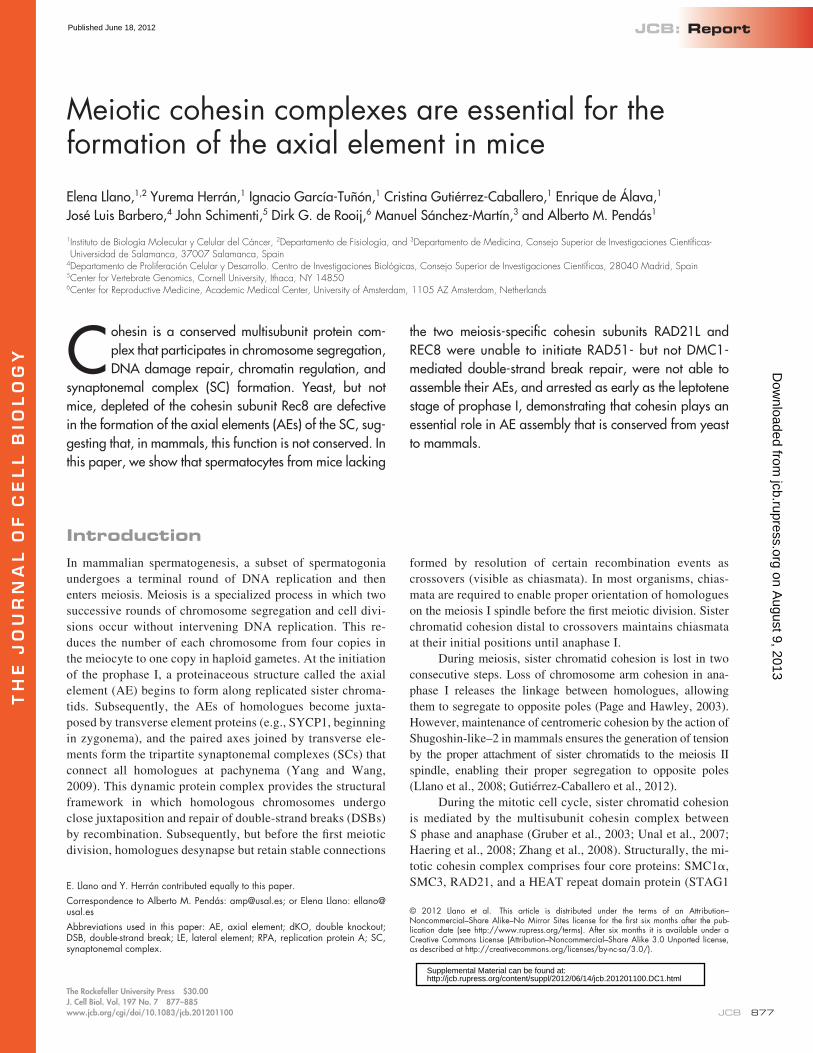
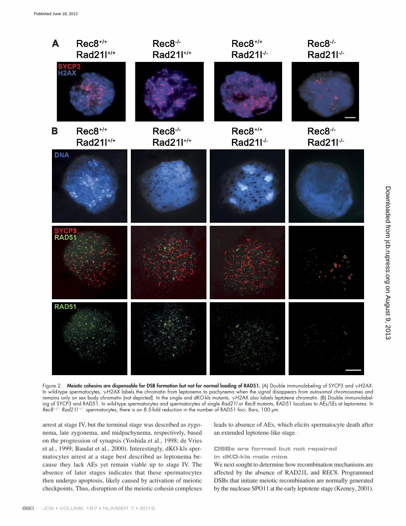
Figure 2. Meiotic cohesins are dispensable for DSB formation but not for normal loading of RAD51. (A) Double immunolabeling of SYCP3 and -H2AX. In wild-type spermatocytes, -H2AX labels the chromatin from leptonema to pachynema when the signal disappears from autosomal chromosomes and remains only on sex body chromatin (not depicted). In the single and dKO-kls mutants, -H2AX also labels leptotene chromatin. (B) Double immunolabel-ing of SYCP3 and RAD51. In wild-type spermatocytes and spermatocytes of single Rad21l or Rec8 mutants, RAD51 localizes to AEs/LEs at leptonema. In Rec8/ Rad21l/ spermatocytes, there is an 8.5-fold reduction in the number of RAD51 foci. Bars, 100 µm.
on August 9, 2013
jcb.rupress.orgD
ownloaded from
Published June 18, 2012

881Cohesins are essential for axial element assembly • Llano et al.
We analyzed break formation in mutant spermatocytes using antibodies against the -H2AX, a histone variant that is phos-phorylated during early prophase I in response to SPO11-induced DSBs in an ATM-dependent manner (Mahadevaiah et al., 2001). As seen in Fig. 2 A, 100% of leptotene-like arrested spermato-cytes from dKO-kls mice showed a positive staining that was similar to that observed in the wild-type mice (87 ± 30 vs. 92 ± 56; Table S1). This suggests that the formation of programmed DSBs is not markedly altered in dKO-kls mice. However, the presence of -H2AX staining in all the arrested spermato-cytes from dKO-kls animals indicates that breaks are not repaired efficiently.
We further analyzed the recombination process to ad-dress why DSBs are not repaired in the double mutant sper-matocytes. After DSBs are induced, the broken ends are resected, and the strand invasion enzymes RAD51 and DMC1 are recruited to the resulting single-strand DNA overhangs to promote homologue pairing and DNA repair (Symington and Gautier, 2011). In wild-type leptotene spermatocytes, RAD51 and DMC1 assemble on the AEs/LEs of bivalents and dis-appear toward pachynema, with the exception of the unsyn-apsed portions of the sex chromosome AEs (Tarsounas et al., 1999). As shown in Fig. 2 B, wild-type spermatocytes showed 70–150 RAD51 foci (111 ± 32). In contrast, the dKO-kls con-tained only a few RAD51 foci (14 ± 5, P < 0.05; Table S1). Next, we monitored the loading of DMC1, a meiosis-specific para-logue of RAD51. In wild-type spermatocytes, both recombinases colocalize extensively such that most recombination-associated foci contain both proteins (Tarsounas et al., 1999). Intriguingly, immunofluorescence with a DMC1-specific antibody revealed no detectable difference in numbers of DMC1 foci between wild type and dKO-kls in leptonema (52 ± 20 vs. 54 ± 19; Table S1 and Fig. 3 A). The presence of an equivalent number of DMC1 foci in double mutant leptotene spermatocytes im-plies that the -H2AX labeling in the dKO-kls spermatocytes is very likely generated by the meiotic SPO11 nuclease.
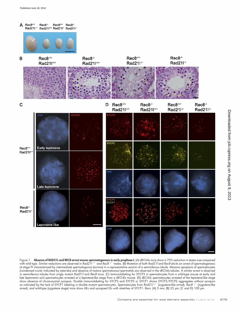
Next, we determined the distribution of the replication protein A (RPA) in dKO-kls spermatocytes. RPA is a single-stranded DNA-binding protein that enhances the formation of RAD51 and DMC1 filaments in vitro. RPA is first detected in few foci at leptonema. The initial binding of RPA to single-strand DNA at the resected ends of DSBs is supposed to be too transient to be cytologically detected by immunofluorescence because RPA is rapidly displaced by RAD51/DMC1 (Yang et al., 2008). Subsequently, after RAD51/DMC1 loading, abundant RPA foci are detected in the synapsed regions of the LEs at zygonema (Krogh and Symington, 2004; Moens et al., 2007). Intriguingly, despite failure to proceed beyond leptonema, RPA foci were increased threefold in the dKO-kls spermatocytes rel-ative to wild type (55 ± 19 vs. 18 ± 10, P < 0.05; Table S1 and Fig. 3 B), which is very likely caused by the sharp reduction in the loading of RAD51 (Fig. 2 B; Roig et al., 2010). TRIP13 is the mammalian orthologue of the yeast Pch2 (pachytene check-point 2) gene, and its deletion leads to a block of spermatogen-esis and oogenesis because of defects in DSB repair (Li and Schimenti, 2007; Roig et al., 2010). Interestingly, Trip13 mutant spermatocytes accumulate RPA foci in leptonema and also show
Figure 3. Normal loading of DMC1 and RPA accumulation at leptonema-arrested dKO-kls spermatocytes. Double immunolabeling of SYCP3 and DMC1 or RPA. (A) In wild-type and dKO-kls spermatocytes, DMC1 protein localizes to AEs/LEs at leptonema. (B) In dKO-kls spermatocytes, there is a threefold increase in the numbers of RPA foci at the leptotene-like arrest in comparison with wild-type spermatocytes. Bars, 100 µm.
on August 9, 2013
jcb.rupress.orgD
ownloaded from
Published June 18, 2012

JCB • VOLUME 197 • NUMBER 7 • 2012 882
can be genetically dissected. Collectively, our results indicate that meiotic cohesins are dispensable for the loading of DMC1 but are essential for normal loading of RAD51 (Xu et al., 2003; Sharan et al., 2004; Yang et al., 2008; Roig et al., 2010).
a sharply reduced number of RAD51 foci and an accumulation of DMC1 foci at leptonema (Roig et al., 2010). Thus, the RAD51 and DMC1 homologous recombination pathways, although very similar biochemically (Kagawa and Kurumizaka, 2010),
Figure 4. Cohesin complexes in the absence of RAD21L and REC8. Double immunofluorescence of SYCP3 and either RAD21, SMC3, SMC1, or STAG3. In wild-type leptotene spermatocytes, the cohesins RAD21, SMC3, SMC1, and STAG3 colocalize with SYCP3 along the AEs of the chromosomes. In Rad21l and Rec8 single mutant leptotene spermatocytes, RAD21, SMC3, SMC1, and STAG3 colocalize also with SYCP3 along the AEs/LEs of the chro-mosomes. In spermatocytes from dKO-kls, arrested at leptonema, SMC1 and STAG3 are not detected by immunofluorescence, whereas immunolabeling for RAD21 and SMC3 renders robust and very faint fluorescence signals, respectively. Bar, 100 µm.
on August 9, 2013
jcb.rupress.orgD
ownloaded from
Published June 18, 2012
883Cohesins are essential for axial element assembly • Llano et al.
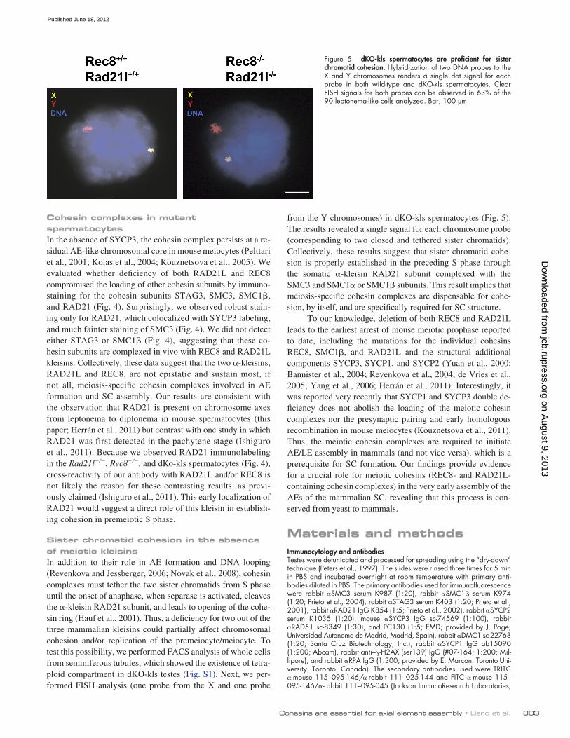
from the Y chromosomes) in dKO-kls spermatocytes (Fig. 5). The results revealed a single signal for each chromosome probe (corresponding to two closed and tethered sister chromatids). Collectively, these results suggest that sister chromatid cohe-sion is properly established in the preceding S phase through the somatic -kleisin RAD21 subunit complexed with the SMC3 and SMC1 or SMC1 subunits. This result implies that meiosis-specific cohesin complexes are dispensable for cohe-sion, by itself, and are specifically required for SC structure.
To our knowledge, deletion of both REC8 and RAD21L leads to the earliest arrest of mouse meiotic prophase reported to date, including the mutations for the individual cohesins REC8, SMC1, and RAD21L and the structural additional components SYCP3, SYCP1, and SYCP2 (Yuan et al., 2000; Bannister et al., 2004; Revenkova et al., 2004; de Vries et al., 2005; Yang et al., 2006; Herrán et al., 2011). Interestingly, it was reported very recently that SYCP1 and SYCP3 double de-ficiency does not abolish the loading of the meiotic cohesin complexes nor the presynaptic pairing and early homologous recombination in mouse meiocytes (Kouznetsova et al., 2011). Thus, the meiotic cohesin complexes are required to initiate AE/LE assembly in mammals (and not vice versa), which is a prerequisite for SC formation. Our findings provide evidence for a crucial role for meiotic cohesins (REC8- and RAD21L-containing cohesin complexes) in the very early assembly of the AEs of the mammalian SC, revealing that this process is con-served from yeast to mammals.
Materials and methodsImmunocytology and antibodiesTestes were detunicated and processed for spreading using the “dry-down” technique (Peters et al., 1997). The slides were rinsed three times for 5 min in PBS and incubated overnight at room temperature with primary anti-bodies diluted in PBS. The primary antibodies used for immunofluorescence were rabbit SMC3 serum K987 (1:20), rabbit SMC1 serum K974 (1:20; Prieto et al., 2004), rabbit STAG3 serum K403 (1:20; Prieto et al., 2001), rabbit RAD21 IgG K854 (1:5; Prieto et al., 2002), rabbit SYCP2 serum K1035 (1:20), mouse SYCP3 IgG sc-74569 (1:100), rabbit RAD51 sc-8349 (1:30), and PC130 (1:5; EMD; provided by J. Page, Universidad Autonoma de Madrid, Madrid, Spain), rabbit DMC1 sc-22768 (1:20; Santa Cruz Biotechnology, Inc.), rabbit SYCP1 IgG ab15090 (1:200; Abcam), rabbit anti–-H2AX (ser139) IgG (#07-164; 1:200; Mil-lipore), and rabbit RPA IgG (1:300; provided by E. Marcon, Toronto Uni-versity, Toronto, Canada). The secondary antibodies used were TRITC -mouse 115–095-146/-rabbit 111–025-144 and FITC -mouse 115–095-146/-rabbit 111–095-045 (Jackson ImmunoResearch Laboratories,
Cohesin complexes in mutant spermatocytesIn the absence of SYCP3, the cohesin complex persists at a re-sidual AE-like chromosomal core in mouse meiocytes (Pelttari et al., 2001; Kolas et al., 2004; Kouznetsova et al., 2005). We evaluated whether deficiency of both RAD21L and REC8 compromised the loading of other cohesin subunits by immuno-staining for the cohesin subunits STAG3, SMC3, SMC1, and RAD21 (Fig. 4). Surprisingly, we observed robust stain-ing only for RAD21, which colocalized with SYCP3 labeling, and much fainter staining of SMC3 (Fig. 4). We did not detect either STAG3 or SMC1 (Fig. 4), suggesting that these co-hesin subunits are complexed in vivo with REC8 and RAD21L kleisins. Collectively, these data suggest that the two -kleisins, RAD21L and REC8, are not epistatic and sustain most, if not all, meiosis-specific cohesin complexes involved in AE formation and SC assembly. Our results are consistent with the observation that RAD21 is present on chromosome axes from leptonema to diplonema in mouse spermatocytes (this paper; Herrán et al., 2011) but contrast with one study in which RAD21 was first detected in the pachytene stage (Ishiguro et al., 2011). Because we observed RAD21 immunolabeling in the Rad21l/, Rec8/, and dKo-kls spermatocytes (Fig. 4), cross-reactivity of our antibody with RAD21L and/or REC8 is not likely the reason for these contrasting results, as previ-ously claimed (Ishiguro et al., 2011). This early localization of RAD21 would suggest a direct role of this kleisin in establish-ing cohesion in premeiotic S phase.
Sister chromatid cohesion in the absence of meiotic kleisinsIn addition to their role in AE formation and DNA looping (Revenkova and Jessberger, 2006; Novak et al., 2008), cohesin complexes must tether the two sister chromatids from S phase until the onset of anaphase, when separase is activated, cleaves the -kleisin RAD21 subunit, and leads to opening of the cohe-sin ring (Hauf et al., 2001). Thus, a deficiency for two out of the three mammalian kleisins could partially affect chromosomal cohesion and/or replication of the premeiocyte/meiocyte. To test this possibility, we performed FACS analysis of whole cells from seminiferous tubules, which showed the existence of tetra-ploid compartment in dKO-kls testes (Fig. S1). Next, we per-formed FISH analysis (one probe from the X and one probe
Figure 5. dKO-kls spermatocytes are proficient for sister chromatid cohesion. Hybridization of two DNA probes to the X and Y chromosomes renders a single dot signal for each probe in both wild-type and dKO-kls spermatocytes. Clear FISH signals for both probes can be observed in 63% of the 90 leptonema-like cells analyzed. Bar, 100 µm.
on August 9, 2013
jcb.rupress.orgD
ownloaded from
Published June 18, 2012

JCB • VOLUME 197 • NUMBER 7 • 2012 884
Investigación Sanitaria and Formación de Personal Investigador fellowships, respectively. E. Llano is the recipient of a Ramón y Cajal Research contract.
Submitted: 18 January 2012Accepted: 21 May 2012
ReferencesBannister, L.A., L.G. Reinholdt, R.J. Munroe, and J.C. Schimenti. 2004.
Positional cloning and characterization of mouse mei8, a disrupted allelle of the meiotic cohesin Rec8. Genesis. 40:184–194. http://dx.doi.org/ 10.1002/gene.20085
Baudat, F., K. Manova, J.P. Yuen, M. Jasin, and S. Keeney. 2000. Chromosome synapsis defects and sexually dimorphic meiotic progression in mice lacking Spo11. Mol. Cell. 6:989–998. http://dx.doi.org/10.1016/S1097-2765 (00)00098-8
Bhatt, A.M., C. Lister, T. Page, P. Fransz, K. Findlay, G.H. Jones, H.G. Dickinson, and C. Dean. 1999. The DIF1 gene of Arabidopsis is required for meiotic chromosome segregation and belongs to the REC8/RAD21 cohesin gene family. Plant J. 19:463–472. http://dx.doi.org/10.1046/j.1365-313X.1999.00548.x
de Rooij, D.G., and P. de Boer. 2003. Specific arrests of spermatogenesis in ge-netically modified and mutant mice. Cytogenet. Genome Res. 103:267–276. http://dx.doi.org/10.1159/000076812
de Vries, F.A., E. de Boer, M. van den Bosch, W.M. Baarends, M. Ooms, L. Yuan, J.G. Liu, A.A. van Zeeland, C. Heyting, and A. Pastink. 2005. Mouse Sycp1 functions in synaptonemal complex assembly, meiotic re-combination, and XY body formation. Genes Dev. 19:1376–1389. http://dx.doi.org/10.1101/gad.329705
de Vries, S.S., E.B. Baart, M. Dekker, A. Siezen, D.G. de Rooij, P. de Boer, and H. te Riele. 1999. Mouse MutS-like protein Msh5 is required for proper chromosome synapsis in male and female meiosis. Genes Dev. 13:523–531. http://dx.doi.org/10.1101/gad.13.5.523
Disteche, C.M., U. Tantravahi, S. Gandy, M. Eisenhard, D. Adler, and L.M. Kunkel. 1985. Isolation and characterization of two repetitive DNA fragments located near the centromere of the mouse X chromosome. Cytogenet. Cell Genet. 39:262–268. http://dx.doi.org/10.1159/000132155
Eijpe, M., H. Offenberg, R. Jessberger, E. Revenkova, and C. Heyting. 2003. Meiotic cohesin REC8 marks the axial elements of rat synaptonemal complexes before cohesins SMC1 and SMC3. J. Cell Biol. 160:657–670. http://dx.doi.org/10.1083/jcb.200212080
Golubovskaya, I.N., O. Hamant, L. Timofejeva, C.J. Wang, D. Braun, R. Meeley, and W.Z. Cande. 2006. Alleles of afd1 dissect REC8 functions during meiotic prophase I. J. Cell Sci. 119:3306–3315. http://dx.doi.org/10 .1242/jcs.03054
Gruber, S., C.H. Haering, and K. Nasmyth. 2003. Chromosomal cohesin forms a ring. Cell. 112:765–777. http://dx.doi.org/10.1016/S0092-8674(03)00162-4
Gutiérrez-Caballero, C., Y. Herrán, M. Sánchez-Martín, J.A. Suja, J.L. Barbero, E. Llano, and A.M. Pendás. 2011. Identification and molecular character-ization of the mammalian -kleisin RAD21L. Cell Cycle. 10:1477–1487. http://dx.doi.org/10.4161/cc.10.9.15515
Gutiérrez-Caballero, C., L.R. Cebollero, and A.M. Pendás. 2012. Shugoshins: from protectors of cohesion to versatile adaptors at the centromere. Trends Genet. http://dx.doi.org/10.1016/j.tig.2012.03.003.
Haering, C.H., A.M. Farcas, P. Arumugam, J. Metson, and K. Nasmyth. 2008. The cohesin ring concatenates sister DNA molecules. Nature. 454:297–301. http://dx.doi.org/10.1038/nature07098
Hauf, S., I.C. Waizenegger, and J.M. Peters. 2001. Cohesin cleavage by separase required for anaphase and cytokinesis in human cells. Science. 293:1320–1323. http://dx.doi.org/10.1126/science.1061376
Herrán, Y., C. Gutiérrez-Caballero, M. Sánchez-Martín, T. Hernández, A. Viera, J.L. Barbero, E. de Álava, D.G. de Rooij, J.Á. Suja, E. Llano, and A.M. Pendás. 2011. The cohesin subunit RAD21L functions in meiotic synap-sis and exhibits sexual dimorphism in fertility. EMBO J. 30:3091–3105. http://dx.doi.org/10.1038/emboj.2011.222
Hirano, T. 2006. At the heart of the chromosome: SMC proteins in action. Nat. Rev. Mol. Cell Biol. 7:311–322. http://dx.doi.org/10.1038/ nrm1909
Ishiguro, K., J. Kim, S. Fujiyama-Nakamura, S. Kato, and Y. Watanabe. 2011. A new meiosis-specific cohesin complex implicated in the cohesin code for homologous pairing. EMBO Rep. 12:267–275. http://dx.doi.org/10.1038/ embor.2011.2
Kagawa, W., and H. Kurumizaka. 2010. From meiosis to postmeiotic events: uncovering the molecular roles of the meiosis-specific recombinase Dmc1. FEBS J. 277:590–598. http://dx.doi.org/10.1111/j.1742-4658.2009 .07503.x
Inc.; all 1:100). Slides were visualized at room temperature using a micro-scope (Axioplan 2; Carl Zeiss) with 63× objectives with an aperture of 1.4 (Carl Zeiss). Images were taken with a digital camera (ORCA-ER; Hamamatsu Photonics) and processed with OPENLAB 4.0.3 (PerkinElmer) and Photo-shop (Adobe).
MiceThe mutation at the Rad21l/ locus is a null allele generated by gene tar-geting using an insertional targeting vector (Herrán et al., 2011). Mice were genotyped by genomic Southern blot analysis of tail genomic DNA digested with SpeI and hybridized with a 5 external probe. The probe (1 kb) was generated by PCR using the primers 5-ACTAGTCTAAATAAAGGTCTT-3 and 5-GATTTAAGCATGAATGAAGTAAC-3. The mutation at the Rec8 locus is a null allele (premature stop codon at exon 6) isolated in a forward genetic screen for mouse infertility (Bannister et al., 2004). Mice were gen-otyped by direct sequencing of the PCR-amplified exon 6 from genomic DNA using the primers 5-CCTTTACATCCCTGTTCTC-3 and 5-ACAG-GAACACCAACTAACTC-3. dKO-kls mice were obtained by genetic crossing of double heterozygote mice (Rec8+/ Rad21l+/) and compared with single mutant and wild-type littermates. All animal experiments were reviewed and approved by the Consejo Superior de Investigaciones Cientí-ficas (Spanish Research Council) and the Universidad de Salamanca National Committee on Bioethics.
FISH analysisThe mouse Y-specific probes were obtained by PCR using the following three set of primers: 1S, 5-TAGGATGGTAAGCCCAATGC-3; 1AS, 5-TTG-GTTGGTTAATTGTTTGGG-3; 2S, 5-CATGCCCCTTGGACTGAC-3; 2AS, 5-CTTTTTTTTTCCCCCTCTGG-3; 3S, 5-TCCTCTTGCAGAGAAGGGAC-3; and 3AS, 5-CCTCCGCTCCAATCCTATCA-3 (Navin et al., 1996). The X-specific DNA probe is a pericentromeric DNA fragment cloned in a plas-mid (Disteche et al., 1985). Both probes were labeled by Nick translation with Dig-11–deoxy-UTP and Bio-16–deoxy-UTP and hybridized to sper-matocyte spreads following standard procedures. In brief, slides were pre-treated with pepsin (0.005% in 0.01 N HCl for 5 min at 37°C), dehydrated, and RNase treated (0.1 mg/ml in PBS for 1 h at 37°C). After denaturation in 70% formamide during 3 min at 75°C, slides were dehydrated in ice-cold ethanol and hybridized overnight at 37°C to a denatured DNA probe. After washing (50% formamide and 2× SSC at 42°C), biotin- and digoxi-genin-labeled probes were immunodetected using streptavidin-Cy3/goat biotinylated antistreptavidin/streptavidin-Cy3 (Jackson ImmunoResearch Laboratories, Inc.) and mouse antidigoxigenin-FITC/goat anti–mouse/rabbit anti–mouse-FITC (Roche), respectively (Pendás et al., 1994).
FACS analysisWild-type, Rad21l/, and dKO-kls testicular cell preparation and their DNA content measurement were performed as previously described (Malkov et al., 1998). In brief, testes were dissected in separation me-dium (4 mM l-glutamine, 1.5 mM sodium pyruvate, 10% fetal calf serum, and 75 mg/ml ampicillin in DME containing nonessential amino acids). After decapsulation, the tubules were then treated with collage-nase for 5 min at 37°C. The sedimented seminiferous cords were washed in separation medium and treated with 2.5 µg/ml trypsin and 1 U/ml DNase I for 2 min at 37°C. Using a pair of scalpels, the tubules were disintegrated, and the resulting tissue suspension was passed through a 50-µm nylon mesh, washed twice by centrifugation, and counted. Cells were brought to a concentration of 2 × 10⁶ cells/ml in separation medium, diluted 1:1 with propidium iodide solution (10 mM Tris, pH 8, 1 mM NaCl, 0.1% Nonidet P-40, 0.7 mg/ml RNase A, and 0.05 mg/ml propidium iodide), and freshly analyzed by a cell-sorting instrument (FACSort; BD).
Online supplemental materialFig. S1 shows that the absence of RAD21L and REC8 does not impair DNA replication and does not provoke loss of chromatid cohesion in dKO-kls sper-matocytes. Table S1 shows the number of early recombination-associated foci in spermatocytes and the weight of testis from wild-type and dKO-kls mice. Online supplemental material is available at http://www.jcb.org/ cgi/content/full/jcb.201201100/DC1.
We wish to express our sincere thanks to E. Marcon and J. Page for providing antibodies (RPA and RAD51, respectively), I. Ramos-Fernández and Teresa Hernandez for excellent technical assistance, and C. López-Otín for his sup-port. We specially appreciate the comments from an anonymous reviewer.
This work was supported by grant SAF2011-25252 and Junta de Castilla y León. C. Gutiérrez-Caballero and Y. Herrán are supported by Fondo de
on August 9, 2013
jcb.rupress.orgD
ownloaded from
Published June 18, 2012

885Cohesins are essential for axial element assembly • Llano et al.
mammalian mitotic cohesins are implicated in meiosis. EMBO Rep. 3:543–550. http://dx.doi.org/10.1093/embo-reports/kvf108
Prieto, I., C. Tease, N. Pezzi, J.M. Buesa, S. Ortega, L. Kremer, A. Martínez, C. Martínez-A, M.A. Hultén, and J.L. Barbero. 2004. Cohesin component dy-namics during meiotic prophase I in mammalian oocytes. Chromosome Res. 12:197–213. http://dx.doi.org/10.1023/B:CHRO.0000021945.83198.0e
Revenkova, E., and R. Jessberger. 2006. Shaping meiotic prophase chromo-somes: cohesins and synaptonemal complex proteins. Chromosoma. 115:235–240. http://dx.doi.org/10.1007/s00412-006-0060-x
Revenkova, E., M. Eijpe, C. Heyting, C.A. Hodges, P.A. Hunt, B. Liebe, H. Scherthan, and R. Jessberger. 2004. Cohesin SMC1 beta is required for mei-otic chromosome dynamics, sister chromatid cohesion and DNA recombina-tion. Nat. Cell Biol. 6:555–562. http://dx.doi.org/10.1038/ncb1135
Roig, I., J.A. Dowdle, A. Toth, D.G. de Rooij, M. Jasin, and S. Keeney. 2010. Mouse TRIP13/PCH2 is required for recombination and normal higher-order chromosome structure during meiosis. PLoS Genet. 6:e1001062. http://dx.doi.org/10.1371/journal.pgen.1001062
Russell, L.D., H.P. Ren, I. Sinha Hikim, W. Schulze, and A.P. Sinha Hikim. 1990. A comparative study in twelve mammalian species of volume den-sities, volumes, and numerical densities of selected testis components, emphasizing those related to the Sertoli cell. Am. J. Anat. 188:21–30. http://dx.doi.org/10.1002/aja.1001880104
Severson, A.F., L. Ling, V. van Zuylen, and B.J. Meyer. 2009. The axial element protein HTP-3 promotes cohesin loading and meiotic axis assembly in C. elegans to implement the meiotic program of chromosome segregation. Genes Dev. 23:1763–1778. http://dx.doi.org/10.1101/gad.1808809
Sharan, S.K., A. Pyle, V. Coppola, J. Babus, S. Swaminathan, J. Benedict, D. Swing, B.K. Martin, L. Tessarollo, J.P. Evans, et al. 2004. BRCA2 defi-ciency in mice leads to meiotic impairment and infertility. Development. 131:131–142. http://dx.doi.org/10.1242/dev.00888
Suja, J.A., and J.L. Barbero. 2009. Cohesin complexes and sister chromatid co-hesion in mammalian meiosis. Genome Dyn. 5:94–116. http://dx.doi .org/10.1159/000166622
Symington, L.S., and J. Gautier. 2011. Double-strand break end resection and repair pathway choice. Annu. Rev. Genet. 45:247–271. http://dx.doi .org/10.1146/annurev-genet-110410-132435
Tarsounas, M., T. Morita, R.E. Pearlman, and P.B. Moens. 1999. RAD51 and DMC1 form mixed complexes associated with mouse meiotic chromo-some cores and synaptonemal complexes. J. Cell Biol. 147:207–220. http://dx.doi.org/10.1083/jcb.147.2.207
Uhlmann, F., D. Wernic, M.A. Poupart, E.V. Koonin, and K. Nasmyth. 2000. Clea-vage of cohesin by the CD clan protease separin triggers anaphase in yeast. Cell. 103:375–386. http://dx.doi.org/10.1016/S0092-8674(00)00130-6
Unal, E., J.M. Heidinger-Pauli, and D. Koshland. 2007. DNA double-strand breaks trigger genome-wide sister-chromatid cohesion through Eco1 (Ctf7). Science. 317:245–248. http://dx.doi.org/10.1126/science.1140637
Watanabe, Y. 2005. Sister chromatid cohesion along arms and at centromeres. Trends Genet. 21:405–412. http://dx.doi.org/10.1016/j.tig.2005.05.009
Watanabe, Y., and P. Nurse. 1999. Cohesin Rec8 is required for reductional chromosome segregation at meiosis. Nature. 400:461–464. http://dx.doi .org/10.1038/22774
Xu, X., O. Aprelikova, P. Moens, C.X. Deng, and P.A. Furth. 2003. Impaired meiotic DNA-damage repair and lack of crossing-over during spermato-genesis in BRCA1 full-length isoform deficient mice. Development. 130:2001–2012. http://dx.doi.org/10.1242/dev.00410
Yang, F., and P.J. Wang. 2009. The mammalian synaptonemal complex: a scaffold and beyond. Genome Dyn. 5:69–80. http://dx.doi.org/10.1159/000166620
Yang, F., R. De La Fuente, N.A. Leu, C. Baumann, K.J. McLaughlin, and P.J. Wang. 2006. Mouse SYCP2 is required for synaptonemal complex as-sembly and chromosomal synapsis during male meiosis. J. Cell Biol. 173:497–507. http://dx.doi.org/10.1083/jcb.200603063
Yang, F., S. Eckardt, N.A. Leu, K.J. McLaughlin, and P.J. Wang. 2008. Mouse TEX15 is essential for DNA double-strand break repair and chromosomal synapsis during male meiosis. J. Cell Biol. 180:673–679. http://dx.doi .org/10.1083/jcb.200709057
Yoshida, K., G. Kondoh, Y. Matsuda, T. Habu, Y. Nishimune, and T. Morita. 1998. The mouse RecA-like gene Dmc1 is required for homologous chro-mosome synapsis during meiosis. Mol. Cell. 1:707–718. http://dx.doi .org/10.1016/S1097-2765(00)80070-2
Yuan, L., J.G. Liu, J. Zhao, E. Brundell, B. Daneholt, and C. Höög. 2000. The murine SCP3 gene is required for synaptonemal complex assembly, chro-mosome synapsis, and male fertility. Mol. Cell. 5:73–83. http://dx.doi .org/10.1016/S1097-2765(00)80404-9
Zhang, N., S.G. Kuznetsov, S.K. Sharan, K. Li, P.H. Rao, and D. Pati. 2008. A handcuff model for the cohesin complex. J. Cell Biol. 183:1019–1031. http://dx.doi.org/10.1083/jcb.200801157
Keeney, S. 2001. Mechanism and control of meiotic recombination initiation. Curr. Top. Dev. Biol. 52:1–53. http://dx.doi.org/10.1016/S0070-2153(01)52008-6
Klein, F., P. Mahr, M. Galova, S.B. Buonomo, C. Michaelis, K. Nairz, and K. Nasmyth. 1999. A central role for cohesins in sister chromatid cohesion, formation of axial elements, and recombination during yeast meiosis. Cell. 98:91–103. http://dx.doi.org/10.1016/S0092-8674(00)80609-1
Kolas, N.K., L. Yuan, C. Hoog, H.H. Heng, E. Marcon, and P.B. Moens. 2004. Male mouse meiotic chromosome cores deficient in structural proteins SYCP3 and SYCP2 align by homology but fail to synapse and have possible impaired specificity of chromatin loop attachment. Cytogenet. Genome Res. 105:182–188. http://dx.doi.org/10.1159/000078188
Kouznetsova, A., I. Novak, R. Jessberger, and C. Höög. 2005. SYCP2 and SYCP3 are required for cohesin core integrity at diplotene but not for centromere cohesion at the first meiotic division. J. Cell Sci. 118:2271–2278. http://dx.doi.org/10.1242/jcs.02362
Kouznetsova, A., R. Benavente, A. Pastink, and C. Höög. 2011. Meiosis in mice without a synaptonemal complex. PLoS ONE. 6:e28255. http://dx.doi .org/10.1371/journal.pone.0028255
Krogh, B.O., and L.S. Symington. 2004. Recombination proteins in yeast. Annu. Rev. Genet. 38:233–271. http://dx.doi.org/10.1146/annurev.genet.38.072902 .091500
Lee, J., and T. Hirano. 2011. RAD21L, a novel cohesin subunit implicated in linking homologous chromosomes in mammalian meiosis. J. Cell Biol. 192:263–276. http://dx.doi.org/10.1083/jcb.201008005
Li, X.C., and J.C. Schimenti. 2007. Mouse pachytene checkpoint 2 (trip13) is re-quired for completing meiotic recombination but not synapsis. PLoS Genet. 3:e130. http://dx.doi.org/10.1371/journal.pgen.0030130
Llano, E., R. Gómez, C. Gutiérrez-Caballero, Y. Herrán, M. Sánchez-Martín, L. Vázquez-Quiñones, T. Hernández, E. de Alava, A. Cuadrado, J.L. Barbero, et al. 2008. Shugoshin-2 is essential for the completion of meio-sis but not for mitotic cell division in mice. Genes Dev. 22:2400–2413. http://dx.doi.org/10.1101/gad.475308
Mahadevaiah, S.K., J.M. Turner, F. Baudat, E.P. Rogakou, P. de Boer, J. Blanco-Rodríguez, M. Jasin, S. Keeney, W.M. Bonner, and P.S. Burgoyne. 2001. Recombinational DNA double-strand breaks in mice precede synapsis. Nat. Genet. 27:271–276. http://dx.doi.org/10.1038/85830
Malkov, M., Y. Fisher, and J. Don. 1998. Developmental schedule of the postna-tal rat testis determined by flow cytometry. Biol. Reprod. 59:84–92. http://dx.doi.org/10.1095/biolreprod59.1.84
Moens, P.B., E. Marcon, J.S. Shore, N. Kochakpour, and B. Spyropoulos. 2007. Initiation and resolution of interhomolog connections: crossover and non-crossover sites along mouse synaptonemal complexes. J. Cell Sci. 120:1017–1027. http://dx.doi.org/10.1242/jcs.03394
Navin, A., R. Prekeris, N.A. Lisitsyn, M.M. Sonti, D.A. Grieco, S. Narayanswami, E.S. Lander, and E.M. Simpson. 1996. Mouse Y-specific repeats isolated by whole chromosome representational difference analysis. Genomics. 36:349–353. http://dx.doi.org/10.1006/geno.1996.0473
Novak, I., H. Wang, E. Revenkova, R. Jessberger, H. Scherthan, and C. Höög. 2008. Cohesin Smc1 determines meiotic chromatin axis loop organiza-tion. J. Cell Biol. 180:83–90. http://dx.doi.org/10.1083/jcb.200706136
Page, S.L., and R.S. Hawley. 2003. Chromosome choreography: the meiotic bal-let. Science. 301:785–789. http://dx.doi.org/10.1126/science.1086605
Parisi, S., M.J. McKay, M. Molnar, M.A. Thompson, P.J. van der Spek, E. van Drunen-Schoenmaker, R. Kanaar, E. Lehmann, J.H.J. Hoeijmakers, and J. Kohli. 1999. Rec8p, a meiotic recombination and sister chromatid cohesion phosphoprotein of the Rad21p family conserved from fission yeast to humans. Mol. Cell. Biol. 19:3515–3528.
Pelttari, J., M.R. Hoja, L. Yuan, J.G. Liu, E. Brundell, P. Moens, S. Santucci-Darmanin, R. Jessberger, J.L. Barbero, C. Heyting, and C. Höög. 2001. A meiotic chromosomal core consisting of cohesin complex pro-teins recruits DNA recombination proteins and promotes synapsis in the absence of an axial element in mammalian meiotic cells. Mol. Cell. Biol. 21:5667–5677. http://dx.doi.org/10.1128/MCB.21.16.5667-5677 .2001
Pendás, A.M., P. Moran, J.P. Freije, and E. Garcia-Vazquez. 1994. Chromosomal mapping and nucleotide sequence of two tandem repeats of Atlantic salmon 5S rDNA. Cytogenet. Cell Genet. 67:31–36. http://dx.doi.org/10 .1159/000133792
Peters, A.H., A.W. Plug, M.J. van Vugt, and P. de Boer. 1997. A drying-down technique for the spreading of mammalian meiocytes from the male and female germline. Chromosome Res. 5:66–68. http://dx.doi.org/10.1023/ A:1018445520117
Prieto, I., J.A. Suja, N. Pezzi, L. Kremer, C. Martínez-A, J.S. Rufas, and J.L. Barbero. 2001. Mammalian STAG3 is a cohesin specific to sister chroma-tid arms in meiosis I. Nat. Cell Biol. 3:761–766. http://dx.doi.org/10.1038/ 35087082
Prieto, I., N. Pezzi, J.M. Buesa, L. Kremer, I. Barthelemy, C. Carreiro, F. Roncal, A. Martinez, L. Gomez, R. Fernandez, et al. 2002. STAG2 and Rad21
on August 9, 2013
jcb.rupress.orgD
ownloaded from
Published June 18, 2012