Embed Size (px)
DESCRIPTION
Mekanisme dan patofisiologi brain injury
Citation preview

BRAIN METABOLISM, THE PATHOPHYSIOLOGY OF BRAIN INJURY, AND POTENTIAL BENEFICIAL AGENTS AND TECHNIQUES
Chapter 1
Ira S. Kass • James E. Cottrell • Baiping Lei
1
Brain metabolism involves both the production and the utili-zation of energy; catabolism is the breakdown and anabolism is the synthesis of components and molecules in the cells. For energy formation the main catabolic process is the breakdown of glucose with the ultimate formation of high-energy phos-phate in the form of adenosine triphosphate (ATP). Other catabolic processes break down structural and enzymatic proteins, lipids, and carbohydrates; these processes are neces-sary to replace damaged and nonfunctional molecules. These molecules are resynthesized by anabolic processes that renew the cells and maintain optimal function. Cellular function also requires the maintenance of ionic homeostasis, which for neu-rons requires a large amount of energy. The pathophysiologic mechanisms of brain injury are incompletely understood but ultimately represent a failure of anabolic processes to maintain normal cell function. In this chapter we explore the putative mechanisms of brain injury. The causes of neuronal damage are multifaceted, and one pathway alone cannot explain how the injury occurs. Some pathophysiologic mechanisms are common to damage caused by ischemic, epileptogenic, and traumatic injury, whereas others are discrete for each of these processes. This review focuses on some common triggers of neuronal damage, such as altered ionic gradients, and explores how they in turn lead to long-term damage. We also discuss pharmacologic agents and clinical procedures that may lead to a reduction in long-term brain damage.
BRAIN METABOLISM
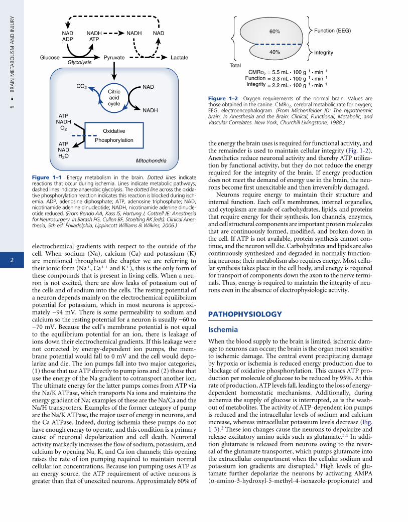
The main substance used for energy production in the brain is glucose. Because glucose is not freely permeable across the blood-brain barrier, it requires a transporter to enter the brain. This transporter does not require energy and can move glucose only down its concentration gradient, from a higher to a lower concentration. Normally the blood levels of glu-cose are well regulated so glucose concentrations in the brain are adequate; however, if blood levels of glucose fall, there can be net movement of glucose out of the brain. Thus adequate blood glucose levels are critical for normal brain activity. Dur-ing insulin shock or other conditions that cause a reduction in blood glucose, unconsciousness can result from insufficient energy due to low brain glucose levels. When glucose and oxygen levels are sufficient, glucose is metabolized to pyru-vate in the glycolytic pathway (Fig. 1-1). This biochemical
process generates ATP from adenosine diphosphate (ADP) and inorganic phosphate and produces nicotinamide adenine dinucleotide reduced (NADH) from nicotinamide adenine dinucleotide (NAD). Pyruvate from this reaction then enters the citric acid cycle which, with regard to energy production, primarily generates NADH from NAD. The mitochondria use oxygen to couple the conversion of NADH back to NAD with the production of ATP from ADP and inorganic phos-phate. This process, called oxidative phosphorylation, forms three ATP molecules for each NADH converted and yields a maximum of 38 ATP molecules for each glucose molecule metabolized.1 Because numerous parts of this pathway supply other metabolic requirements, such as amino acid synthesis and the formation of reducing equivalents for other synthetic pathways, the normal yield of this energy pathway is approxi-mately 30 to 35 ATP molecules for each glucose molecule.
This pathway requires oxygen; if oxygen is not present the mitochondria can neither make ATP nor regenerate NAD from NADH. The metabolism of glucose requires NAD as a cofactor and is blocked in its absence. Thus, in the absence of oxygen, glycolysis proceeds by a modified pathway termed “anaerobic glycolysis”; this modification involves the conver-sion of pyruvate to lactate, regenerating NAD. This process produces hydrogen ion, which may accentuate neuronal dam-age if the intracellular pH falls. A major problem with anaer-obic glycolysis, in addition to lowering pH, is that only two molecules of ATP are formed for each molecule of glucose metabolized. This level of ATP production is insufficient to meet the brain’s energy needs. In addition, ischemia cuts off the supply of glucose so even anaerobic glycolysis is blocked.
When the oxygen supply to a neuron is reduced, mecha-nisms that reduce and/or slow the fall in ATP levels include the following: (1) the utilization of phosphocreatine stores (a high-energy phosphate that can donate its energy to main-tain ATP levels), (2) the production of ATP at low levels by anaerobic glycolysis, and (3) a rapid cessation of spontaneous electrophysiologic activity.
CELLULAR PROCESSES THAT REQUIRE ENERGY
Pumping ions across the cell membrane is the largest energy requirement in the brain. The sodium, potassium, and cal-cium concentrations of a neuron are maintained against large
2
1 •
BRA
IN M
ETA
BOLI
SM A
ND
INJU
RY
ecattrtapmc−tinbl(uTteNathcacrcag
Frdtenoft
lectrochemical gradients with respect to the outside of the ell. When sodium (Na), calcium (Ca) and potassium (K) re mentioned throughout the chapter we are referring to heir ionic form (Na+, Ca++ and K+), this is the only form of hese compounds that is present in living cells. When a neu-on is not excited, there are slow leaks of potassium out of he cells and of sodium into the cells. The resting potential of neuron depends mainly on the electrochemical equilibrium otential for potassium, which in most neurons is approxi-ately −94 mV. There is some permeability to sodium and
alcium so the resting potential for a neuron is usually −60 to 70 mV. Because the cell’s membrane potential is not equal
o the equilibrium potential for an ion, there is leakage of ons down their electrochemical gradients. If this leakage were ot corrected by energy-dependent ion pumps, the mem-rane potential would fall to 0 mV and the cell would depo-
arize and die. The ion pumps fall into two major categories, 1) those that use ATP directly to pump ions and (2) those that se the energy of the Na gradient to cotransport another ion. he ultimate energy for the latter pumps comes from ATP via
he Na/K ATPase, which transports Na ions and maintains the nergy gradient of Na; examples of these are the Na/Ca and the a/H transporters. Examples of the former category of pump
re the Na/K ATPase, the major user of energy in neurons, and he Ca ATPase. Indeed, during ischemia these pumps do not ave enough energy to operate, and this condition is a primary ause of neuronal depolarization and cell death. Neuronal ctivity markedly increases the flow of sodium, potassium, and alcium by opening Na, K, and Ca ion channels; this opening aises the rate of ion pumping required to maintain normal ellular ion concentrations. Because ion pumping uses ATP as n energy source, the ATP requirement of active neurons is reater than that of unexcited neurons. Approximately 60% of
NADADP
NADHATP
NADH
GlucoseGlycolysis
Pyruvate
Citricacidcycle
Oxidative
Phosphorylation
Mitochondria
CO2
ATPNADH
O2
ATPNADH2O
Lactate
NAD
NAD
NADH
igure 1–1 Energy metabolism in the brain. Dotted lines indicate eactions that occur during ischemia. Lines indicate metabolic pathways, ashed lines indicate anaerobic glycolysis. The dotted line across the oxida-ive phosphorylation reaction indicates this reaction is blocked during isch-mia. ADP, adenosine diphosphate; ATP, adenosine triphosphate; NAD, icotinamide adenine dinucleotide; NADH, nicotinamide adenine dinucle-tide reduced. (From Bendo AA, Kass IS, Hartung J, Cottrell JE: Anesthesia or Neurosurgery. In Barash PG, Cullen BF, Stoelting RK [eds]: Clinical Anes-hesia, 5th ed. Philadelphia, Lippincott Williams & Wilkins, 2006.)
the energy the brain uses is required for functional activity, and the remainder is used to maintain cellular integrity (Fig. 1-2). Anesthetics reduce neuronal activity and thereby ATP utiliza-tion by functional activity, but they do not reduce the energy required for the integrity of the brain. If energy production does not meet the demand of energy use in the brain, the neu-rons become first unexcitable and then irreversibly damaged.
Neurons require energy to maintain their structure and internal function. Each cell’s membranes, internal organelles, and cytoplasm are made of carbohydrates, lipids, and proteins that require energy for their synthesis. Ion channels, enzymes, and cell structural components are important protein molecules that are continuously formed, modified, and broken down in the cell. If ATP is not available, protein synthesis cannot con-tinue, and the neuron will die. Carbohydrates and lipids are also continuously synthesized and degraded in normally function-ing neurons; their metabolism also requires energy. Most cellu-lar synthesis takes place in the cell body, and energy is required for transport of components down the axon to the nerve termi-nals. Thus, energy is required to maintain the integrity of neu-rons even in the absence of electrophysiologic activity.
PATHOPHYSIOLOGY
Ischemia
When the blood supply to the brain is limited, ischemic dam-age to neurons can occur; the brain is the organ most sensitive to ischemic damage. The central event precipitating damage by hypoxia or ischemia is reduced energy production due to blockage of oxidative phosphorylation. This causes ATP pro-duction per molecule of glucose to be reduced by 95%. At this rate of production, ATP levels fall, leading to the loss of energy-dependent homeostatic mechanisms. Additionally, during ischemia the supply of glucose is interrupted, as is the wash-out of metabolites. The activity of ATP-dependent ion pumps is reduced and the intracellular levels of sodium and calcium increase, whereas intracellular potassium levels decrease (Fig. 1-3).2 These ion changes cause the neurons to depolarize and release excitatory amino acids such as glutamate.3,4 In addi-tion glutamate is released from neurons owing to the rever-sal of the glutamate transporter, which pumps glutamate into the extracellular compartment when the cellular sodium and potassium ion gradients are disrupted.5 High levels of glu-tamate further depolarize the neurons by activating AMPA (α-amino-3-hydroxyl-5-methyl-4-isoxazole-propionate) and
60%
40%
Total
Function (EEG)
Integrity
CMRO2
FunctionIntegrity
= 5.5 mL • 100 g� 1 • min� 1
= 3.3 mL • 100 g� 1 • min� 1
= 2.2 mL • 100 g� 1 • min� 1
Figure 1–2 Oxygen requirements of the normal brain. Values are those obtained in the canine. CMRo2, cerebral metabolic rate for oxygen; EEG, electroencephalogram. (From Michenfelder JD: The hypothermic brain. In Anesthesia and the Brain: Clinical, Functional, Metabolic, and Vascular Correlates. New York, Churchill Livingstone, 1988.)

3
1 •
BRAIN
META
BOLISM
AN
D IN
JURY
NMDA (N-methyl-d-aspartate) receptors, increasing sodium and potassium ion conductance.6,7 The NMDA receptor also allows calcium to enter, triggering additional damaging path-ways. Glutamate activates metabotropic receptors, which via second-messenger systems can increase the release of calcium from intracellular stores and activate other biochemical processes.8,9 The damage due to excess glutamate has been termed
excitotoxicity and is caused by activation of glutamate receptors and the accompanying ionic and biochemical changes.10
In addition to increased influx through membrane chan-nels, cytosolic calcium is increased through reduced calcium pumping from the cell and the enhanced release of calcium from intracellular organelles such as the mitochondria and the endoplasmic reticulum (Fig. 1-4).11,12 The high cytoplasmic calcium level is thought to trigger a number of events that lead to the ischemic damage. These include increasing the activ-ity of proteases and phospholipases. Phospholipases raise the levels of free fatty acids, such as arachidonic acid, and free radicals. Free radicals are also generated by incomplete mito-chondrial oxidation.11 One of the most damaging free radi-cals is peroxynitrite, which is formed by the combination of nitric oxide and another free radical.11 Free radicals are known to damage proteins and lipids, whereas free fatty acids inter-fere with membrane function. There is a buildup of lactate and hydrogen ions during ischemia, and this decrease in pH can lead to further formation of free radicals.13 All of these processes, coupled with the reduced ability to synthesize proteins and lipids, contribute to the irreversible damage that occurs with ischemia (Box 1-1).
Additionally, phospholipase activation leads to the produc-tion of excess arachidonic acid, which upon reoxygenation can form eicosanoids, including thromboxane, prostaglandins, and leukotrienes. These substances can cause strong vasocon-striction, reduce blood flow in the postischemic period, alter the blood-brain barrier, and enhance free radical formation after reperfusion.14,15
Procedures that protect against ischemic damage should interfere with the cellular changes brought on by ischemia (Box 1-2). In addition to these direct triggering events, there is long-term damage that becomes apparent hours and days after the ischemic insult. Some of this delayed damage is necrotic; lysis of the cells causes microglial activation.16 Lymphocytes,
Hypoxia/Ischemia
ATP
Ion pumps
[K]extracell [Ca]intracell [Na]intracell
Depolarization
Glutamate release
Primaryeffect
Positivefeedback
Figure 1–3 Line diagram of cellular ionic events occurring during anoxia or ischemia. The events indicated are the primary triggers of events leading to neuronal cell death. Positive feedback loops are unsta-ble and rapidly worsen events. ATP, adenosine triphosphate; extracell, extracellular; intracell, intracellular; ↑, increase; ↓, decrease.
Glutamatenon-NMDA receptor
Neurotransmitter-gated channels
Voltage-sensitivechannels
GlutamateNMDA receptor
Ca-activatedphospholipase
GlutamateMetabotrophic
receptor
Glutamate
Free fatty acids(arachidonic acid)
Membranelipids
ProstaglandinsThromboxaneLeukotrienes
Na,Ca
K K K
Na Na Ca
Ca
Ca
Ca
Ca
Ca CaNa
Na
Na
Ca
Free radicals
Free radicals
Endoplasmic reticulum
Mitochondria
ATP
H
K
ATP
Na
ATP
v v v
KHCO2
[Na] ,[K] ,[Ca] ,[ATP] ,[H]
Events During Ischemia
Figure 1–4 The effect of ischemia on ion and metabolite levels in neurons. For clarity, ion channels are shown on the top membrane and ion pumps on the bottom membrane; their actual location can be on any membrane surface. Circles indicate energy-driven pumps; an x through a circle indicates that the pump is blocked or has reduced activity during ischemia. V indicates a voltage-dependent channel. ATP, adenosine triphosphate; NMDA, N-methyl-d-aspartate. (Modified from Bendo AA, Kass IS, Hartung J, Cottrell JE: Anesthesia for neurosurgery. In Barash PG, Cullen BF, Stoelting RK [eds]: Clinical Anesthesia, 5th ed. Philadelphia, Lippincott Williams & Wilkins, 2006.)

4
1 •
BRA
IN M
ETA
BOLI
SM A
ND
INJU
RY
polymorphonuclear cells, and macrophages can invade the ner-vous system, leading to additional damage.17,18 Although hista-mine receptor activation is generally associated with immune system activation, the histamine receptor involved with this is the H1 receptor. In the central nervous system, the H2 recep-tor is the one primarily activated, and it reduces immunologic processes and improves recovery from is chemia.19-21 Indeed, blocking immune system activation can reduce damage.19 It is clear there is also genetically programmed cell death as a result of the insult.22 This programmed cell death, which is similar to apoptotic cell death during neuronal development, can occur days after the initial insult.
Necrosis versus Apoptosis
There are two major processes leading to neuronal death. The first, necrosis, is due to a more severe insult in which mito-chondrial function is lost; it is characterized by a disintegra-tion of the cell and an activation of microglia and the immune response.16 The immune response and inflammation activate and recruit neutrophils and macrophages, which produce free radicals and damage adjacent neurons. This process expands the lesion in volume and time, allowing for continued and expanded neuronal damage.16 In the second, apoptosis, the cell dies without breaking apart and there is no microg-lial or immune system involvement with the potential for excess damage to adjacent neurons. This process is frequently delayed and can lead to the activation of immediate early genes (IEGs) such as c-Jun and c-Fos; these genes are thought to affect gene expression and lead to the production of apop-
BOX 1–1 �Brain�Metabolism�and�Cell�Death:�Triggers,�Effectors,�and�Functional�Changes
Triggers
Adenosine triphosphate ↓Extracellular potassium ↑Intracellular sodium ↑Intracellular calcium ↑Free radical levels ↑Depolarization ↑Glutamate level ↑
Effectors
Protease activity ↑Free radical action ↑DNA damage ↑Phospholipase activity ↑Mitochondrial factors ↑ (cytochrome c → caspase activation)
Critical Functional Changes
Mitochondrial damage ↑Apoptotic cascade activation ↑Antiapoptotic factors ↓Protein damage ↑Protein synthesis ↓Cytoskeletal damage ↑
End Stage
Apoptosis ↑ (programmed cell death)Necrosis ↑ (cell disintegration)
↑, increases; ↓, decreases; →, leads to.Adapted from Lipton P. Ischemic cell death in brain neutrons. Physiol Rev 1999;79:1431-1568.
totic or antiapoptotic proteins, which determine whether the neurons will survive or die.22,23 One set of proteins that lead to neuronal death are the cysteine proteinases, referred to as caspases. These enzymes are expressed as proenzymes, which undergo proteolytic processing to yield active enzymes that degrade important proteins in the cell (Fig. 1-5).24,25 Blockade of caspases has been shown to block apoptosis.26 Because these enzymes are now known to be present as proenzymes before ischemia, new protein synthesis is not needed to induce apop-tosis.27 However, proapoptotic proteins are synthesized under certain conditions, and their synthesis may lead to delayed neuronal cell death. Another set of proteins can be induced that block apoptosis and promote neuronal survival after ischemia; examples of these proteins are neuronal apoptosis inhibitory protein, heat shock proteins, and Bcl-2 family proteins.28,29 Thus the fate of ischemic neurons rests in balance between apoptotic inhibitory and activating processes (Fig. 1-6).29,30 The synthesis of certain trophic factors can improve neuronal survival by inhibiting apoptosis (see Fig. 1-5). The activation and release of certain cytokines, such as tumor necrosis factor and interleukin-1β, are thought to be damaging.31,32
Thus necrosis and apoptosis can be contrasted, with the for-mer being a result of more severe ischemia and leading to damage of adjacent tissue (Fig. 1-7). Apoptosis is subject to modulation, so once started down the apoptotic pathway, cells have a chance of being rescued by trophic substances (see Fig. 1-6).
Global versus Focal Ischemia
Ischemia can be either global or focal in nature; an example of the former would be cardiac arrest, and of the latter, localized stroke. Although the mechanisms leading to neuronal dam-age are probably similar for the two types of ischemia, there are important distinctions between them. In focal ischemia there are three regions. The first region receives no blood flow and responds the same as globally ischemic tissue; the sec-ond region, called the penumbra, receives collateral flow and is partially ischemic; the third region is normally perfused. If the insult is maintained for a prolonged period, the neurons in the penumbra die. More neurons in the penumbra region survive if collateral blood flow is increased or if reperfusion is established in a timely manner by opening of the blocked vessel. With total global ischemia, the time until the circulation
BOX 1–2 Consequences�of�Ischemia
Vascular Changes
VasospasmRed cell sludgingHypoperfusion
Neuronal Changes
Adenosine triphosphate reductionSodium influxPotassium effluxIntracellular acidosisHigh cellular calcium concentrationsCalcium-activated proteasesCaspase activationPhospholipase activationArachidonic acid formation and breakdownFree radical productionExcitatory amino acid releaseDisruption of ion and amino acid transporters

1 •
BRAIN
META
BOLISM
AN
D IN
JURY
Absence of trophic factor:Caspase activation
(a) Presence of trophic factor:Inhibition of caspase activation
(b)
Trophic factor receptor Trophic factor1
Procaspase 3
Death
Cleavage ofsubstrates
Procaspase 9
Caspase 3
Caspase 9
Apaf 1Procaspase 9
Apaf 1Cyt c
Cyt c
Cyt c
PI-3kinase
Procaspase 3PI-3
kinase
Aktkinase
AktkinaseATP
ADP
Bad
Bad
Ions
Bcl-2 Bcl-xl
Bcl-xl
Bax
Bax
BaxBcl-2
14-3-3
14-3-3
4
3
12
Bax
3
2
P
Plasma membrane
Outermitochondrial
membrane
Figure 1–5 Trophic factors and apoptosis. ADP, adenosine diphosphate; ATP, adenosine triphosphate; Cyt c, cytochrome c; PI, phosphoinositide; other abbreviations (Akt, Apaf, Bad, Bax, Bcl, 14-3-3) are names of proteins. The numbers on the diagram refer to the apoptotic pathway and its inhibi-tion a) activated apoptotic pathway: 1) Bad protein inhibits Bcl-2, Bcl-xl proteins; 2) these proteins can no longer inhibit Bax and therefore Bax allows ion flow into the mitochondria; 3) this leads to cytochrome c release and the activation of Apaf 1 which finally 4) activates caspase 9 and apoptosis. b) apoptosis inhibited 1) trophic factor binds to a receptor and activates protein kinases; 2) this leads to the phophorylation of Bad and its inactivation; 3) Bad can no longer inhibit Bcl-2 and Bcl-xl and these 2 proteins can now inhibit Bax, blocking ion flow and apoptosis. (From Lodish H, Berk A, Mat-sudaira P, et al [eds]: Molecular Cell Biology, 5th ed. New York, WH Freeman and Co, 2004: page 929, as adapted from Pettmann B, Henderson CE: Neuronal cell death. Neuron 1998;20:633-647.)
5
is reestablished is critical, and only very short ischemic times (on the order of minutes) are survivable. The selective neu-rologic damage after survival subsequent to global is chemia is mainly due to the differential sensitivity of certain neurons and brain regions. The hippocampus, especially the cornus ammonis 1 (CA1) pyramidal cell region, is extremely vulner-able to ischemic damage; loss of learning and memory is com-mon after global ischemia and hypoxia.33,34Genetic Influences on Neuronal Damage
Genetic factors play an important role in an individual’s susceptibility to ischemic brain injury. Both environmental (such as diet and stress) and genetic factors combine to deter-mine the risk of stroke. A study of the Icelandic population found that polymorphisms (genetic changes) in genetic locus ALOX5AP, which encodes 5-lipoxygenase–activating protein, and PDE4D, which encodes phosphodiesterase 4D, increase the susceptibility to stroke.35,36 In addition polymorphisms of both apolipoprotein B and apolipoprotein E have been found to enhance the susceptibility to stroke.37,38 The genetic factors could target neuronal risk but more likely raise the vascular risk, which is associated with an increase in both stroke and cardiac disease. If a patient’s genetic susceptibility to injury were known, it would be possible to choose therapeutic strate-gies individually for the patient, especially if those strategies carry their own morbidity or are costly.
In addition to neuronal dysfunction following stroke and global cerebral ischemia, postoperative cognitive deficit is frequently found after anesthesia and surgery even in the
absence of ischemia. This deficit also shows genetic variation. The genetic alleles that reduced serum C-reactive protein and platelet activation were associated with a reduction in cogni-tive deficit after cardiac surgery. These studies indicate that the immune response may enhance postoperative cognitive deficits and that targeting the immune activation in certain patients may be beneficial.39
POTENTIAL TREATMENTS FOR CEREBRAL ISCHEMIA
Reperfusion Strategies
The most successful technique for improving recovery from embolic stroke is prompt restoration of spontaneous perfu-sion through the use of thrombolytic agents (such as tissue plasminogen activator [TPA]) or other anticlotting drugs in the period directly after the onset of a stroke (<3 hours); as one would predict, hemorrhagic strokes are made markedly worse by this treatment.40,41 Thus detecting, classifying, and treating stroke rapidly after its onset is critical to a successful outcome.42 Thrombolytics can not be used during or recently after surgery because of the risk of bleeding. The use of anti-clotting agents such as warfarin is also thought to reduce the incidence and severity of subsequent strokes.43
The major side effect of this strategy is intracerebral hemor-rhage, which can be devastating. It is essential that a noncon-trast computed tomography scan be performed and analyzed shortly after patient presentation to the hospital in order to

6
1 •
BRA
IN M
ETA
BOLI
SM A
ND
INJU
RY
Feit↑
Hypoxia/Ischemia
ATP
DepolarizationMitochondria [Na+]intracell
[Ca++]intracell Kinase
Proteases
Phospholipase
Endonuclease
DNA breakdown
Glutamate
Free radicals Kinase Kinase
Mitochondrial cytochrome c release
Caspase Proapoptotictranscription
factors
Antiapoptotictranscription
factors
Antiapoptoticproteins
MitochondrialKATP channel
Proapoptoticproteins
Cytoskeletal changesapoptotic bodies
Apoptosis
Cell death • Membrane intact • No immune reponse
Block apoptosis
Block cytochrome c releaseendonuclease caspase
Cell survival
Trophic factors
igure 1–6 Apoptosis subsequent to hypoxia or ischemia: The apoptotic cascade of biochemical changes evoked by hypoxia or ischemia. Similar vents may also be occurring during epileptic and trauma-induced damage; they lead to depolarization, reduced adenosine triphosphate (ATP), sodium nflux, and high cytosolic calcium levels. There is no cellular membrane disruption during apoptosis, and inflammation is not triggered. The apop-otic biochemical cascade can be modulated and opposed by trophic factors. intracell, intracellular; KATP channel, ATP-sensitive potassium channel; , increase; ↓, decrease; the large open arrows indicate damaging pathways, the large closed arrows indicate protective pathways.
rule out a hemorrhagic stroke, because TPA must be admin-istered within 3 hours of an occlusive stroke onset to be effec-tive. Other drugs are being evaluated for reperfusion strategy, such as desmoteplase, which is more fibrin specific and has a longer half-life, but only TPA has been approved by the U.S. Food and Drug Administration for this indication.
Although these strategies have shown benefit only when administered to patients within 3 hours of stroke onset, there are currently studies to identify patients who might benefit from these agents at later time points. The key is to identify patients in whom an area of reduced perfusion has not progressed to irreversible neuronal damage. These stud-ies use advanced imaging to identify at-risk tissue that can still be salvaged if reperfusion can be established. Diffusion-weighted magnetic resonance imaging (DWI) identifies core ischemic areas where water has shifted into the intracellu-lar compartment and has reduced diffusibility. Areas that have not yet converted to core ischemia can potentially be rescued from irreversible damage. Perfusion-weighted mag-netic resonance imaging indicates regions with reduced perfusion that will ultimately proceed to irreversible dam-age if not reperfused. A region with reduced perfusion that
has not yet progressed to irreversible damage (as shown on diffusion-weighted imaging) would benefit from delayed reperfusion therapy.44 It is likely that patients in this cat-egory would benefit from delayed TPA; the issue is under current investigation and some centers have extended the time for tPA to 4.5 hours. (ref 45) However it is clear that the sooner TPA is given the better the outcome is likely to be. Some studies are being done to examine intra-arterial application of TPA after unsuccessful intravenous TPA some studies have found no significance but a trend toward infarct reduction.45 These results were encouraging but not enough to recommend a change in clinical practice. A study with a relatively small number of patients indicates that administration of desmoteplase 3 to 9 hours after the onset of an embolic stroke yields a high rate of reperfusion and better clinical outcome than administration of pla-cebo in carefully selected patients with perfusion /diffusion mismatch.46 However a larger well designed study was not able to confirm a benefit of desmoteplase under these same conditions, this indicates the difficulties and complications in finding agents that improve outcome.47 It also indicates the importance of cautiously interpreting positive studies of

7
1 •
BRAIN
META
BOLISM
AN
D IN
JURY
Hypoxia/Ischemia
ATP Mitochondria
[Na+]intracell
Lysosomerelease
[Ca++]intracell
Phospholipase
Protein synthesis inhibitionCytoskeletal
damage
Proteindamage DNA damage
Protein synthesisinitiation factor
Membrane damage
Glutamate Protease Protease
Long-term ATP Nuclease
Free radicals
Freeradicals
Freeradicals Mitochondrial damage
Depolorization
Necrosis
Membrane disruption • Immune response • Damage adjacent neurons
Figure 1–7 Necrosis subsequent to hypoxia and ischemia: The necrotic cascade of biochemical changes evoked by hypoxia or ischemia. Similar events may also be occurring during epileptic and trauma-induced damage; they lead to depolarization, reduced adenosine triphosphate (ATP), sodium influx, and high cytosolic calcium levels. These changes are more severe than those with apoptosis and lead to the disruption of the cells and activation of inflammation. The damage cannot be reversed, and surrounding intact neurons can be damaged by secondary processes. intracell, intracellular; ↑, increase; ↓, decrease; large arrows indicate damaging pathways.
clinical brain protection in which small groups of patients are examined.
In addition to chemical dissolution of the clot, mechanically assisted recanalization is possible. The Merci Retriever (Con-centric Medical, Inc., Mountain View, CA) and the Penumbra System (Penumbra, Inc., Alameda, CA) are approved by the U.S. Food and Drug Administration for mechanical removal of clots. The Merci Retriever has been shown to provide a higher rate of good clinical outcome compared to historical controls, although the intracranial hemorrhage rate was 7.8%.48 The Penumbra System successfully reopened 82% of the treated ves-sels, however the symptomatic intracerebral hemorrhage rate was 11%. The authors concluded that the Penumbra System allowed safe and effective revascularization up to 8 hours after symptom onset.49
Both intra-arterial thrombolysis and mechanical removal are confined to specialist centers and clinical trials. The only widely used treatment is administration TPA within 3 hours of stroke onset, however most recently it has been extended for up to 4.5 hours for certain patients.50 This treatment is underutilized, in part because of the time restraints for imag-ing and the frequent delay in presentation of patients to the hospital after the onset of symptoms. The latter problem can and is being addressed by community education about the signs of stroke and the emergency nature of transport to an
appropriate hospital with a stroke center. The guidelines for early management and treatment of stroke are frequently updated and should be consulted and examined for the latest recommendations.45
Hypothermia
Profound hypothermia has long been used in neonatal heart surgery to provide protection against irreversible brain injury when the heart is stopped. It has also been used during the repair of giant aneurysms. However there are numerous complications of profound hypothermia (27° C or lower) that limit its usefulness (Box 1-3). Profound hypo-thermia reduces cerebral metabolism to such an extent that the brain can survive relatively long periods without perfu-sion (Box 1-4). Experimental studies indicate that moder-ate hypothermia has a protective effect without many of the complications of profound hypothermia, although myocar-dial depression has been documented.51-53 There are many in vitro and in vivo animal studies to support the use of moderate hypothermia to protect against ischemic damage, and a prospective clinical study is beginning. Indeed mod-erate hypothermia has come into common use even though it has not been unequivocally shown to improve recovery in a major clinical trial.54,55 A European study published in

8
1 •
BRA
IN M
ETA
BOLI
SM A
ND
INJU
RY
2002 indicates that mild hypothermia, target temperature 32° to 34° C, after hospital cardiac arrest improves neuro-logic outcome and survival 6 months after the arrest.56 Even patients with out-of-hospital cardiac arrest benefit from mild hypothermia.57
It is clear that even minor amounts of hyperthermia worsen clinical outcome of ischemia and increase neuronal damage, and so must be carefully guarded against.58,59
Glucose
Glucose is the main source of energy for neurons in the brain, and some in vitro studies reported improved recov-ery with hyperglycemia. However, in vivo and clinical stud-ies found a clear worsening of damage with hyperglycemia, which is thought to be due to enhanced cellular acidosis.13,60 The precise mechanism by which hyperglycemia exacerbates damage is not known. Clinical recommendations are to maintain normal serum glucose levels and to treat hypergly-cemia in order to reduce the glucose value to near normal range.61 It is important for the patient not to be hypogly-cemic, as hypoglycemia would also worsen outcome. The difficulty in doing this and the overall recommendations for glucose management are likely to be effected by a recent study on intensive glucose control which demonstrated a higher mortality in patients managed from 81-108 mg per
BOX 1–3 Complications�of�Deep�Hypothermia�
Cardiovascular Complications
Myocardial depressionDysrhythmia including ventricular fibrillationHypotensionInadequate tissue perfusionIschemia
Coagulation
ThrombocytopeniaFibrinolysisPlatelet dysfunctionIncreased bleeding
Metabolism
Slowed metabolism of anesthetic agentsProlonged neuromuscular blockadeIncreased protein catabolism
Shivering
Increased oxygen consumptionIncreased carbon dioxide productionIncreased cardiac outputArterial oxygen desaturationHemodynamic instability
BOX 1–4 �Proposed�Mechanisms�of�Protection�by�Hypothermia
Decrease in cerebral metabolismDelayed anoxic/ischemic depolarizationPreservation of ion homeostasisDecrease excitatory neurotransmissionPrevention or reduction of damaging secondary biochemical changes
deciliter compared to patients managed to a target of 180 mg or less per deciliter.62
Pharmacologic Agents
Many drugs have been proposed as potential agents to reduce permanent neuronal damage subsequent to isch-emia, but very few have proved useful in clinical trials.58,63 The theoretical basis for choosing drugs that block specific damaging pathways is sound. Blocking one pathway to damage may not be efficacious, however, owing to the many parallel paths that lead to permanent damage (see Fig. 1-6). For example, one can block voltage-sensitive calcium chan-nels but cytoplasmic calcium can increase through influx via the NMDA receptor ion channel or release from intra-cellular organelles. Thus effective therapy might require multiple agents to block all of the parallel pathways simul-taneously.
It must be recognized that no pharmacologic agent has been shown to improve neurologic recovery clinically after a stroke. Pharmacologic agents that have been studied in clinical trials have not been shown to be efficacious for the improve-ment of stroke outcome. This is an extremely controversial field, as evidenced by two 2008 editorials in the journal Stroke, entitled “Neuroprotection: Still achievable in humans”64 and “Neuroprotection does not work!”65 That said, some agents appear promising in animal studies and may prove efficacious clinically.59,65,68
One major problem with stroke treatments and a reason for the discrepancy between animal and human results is that most animal studies apply the protective agents either before or during the insult, whereas clinical stroke treatment is always delayed. In the perioperative environment, drugs and treatments can be applied before an insult, at the begin-ning of a high-risk surgery; thus, agents that fail to protect against stroke when used after the insult may be efficacious if given before surgery. Because only very few patients undergo-ing high-risk surgery will suffer an ischemic insult, the agents used must have a high safety factor and/or must be required for surgery (e.g., anesthetics). The deleterious effects will be shared among all patients, but the protective effects will ben-efit only ischemic patients.
Sodium Blockade
Blocking sodium influx during anoxia and ischemia has been shown to improve recovery both in vivo and in vitro.67-
69 The neuronal depolarization during anoxia and isch-emia leads to massive flux of sodium and calcium into the neurons and of potassium out of the neurons.70-73 Block-ing sodium influx delays and attenuates the depolarization and delays the drop in ATP during anoxia and ischemia.74-76 Lidocaine has improved recovery by delaying and attenuat-ing the anoxic/ischemic depolarization and reducing anoxic sodium influx when given at concentrations that do not block sodium channels under normal conditions.74,77 In one study, lidocaine reduced the infarct size and improved neu-rologic outcome following focal cerebral ischemia.78 This agent appears to work, at least in part, by blocking apop-totic pathways in the penumbra.79 When lidocaine appli-cation was delayed until 45 minutes after the onset of focal cerebral ischemia, there was improved neuronal survival in the core and penumbra, but the size of the infarct was not significantly reduced.82 Studies in our laboratory have found that an antiarrhythmic dose of lidocaine administered

9
1 •
BRAIN
META
BOLISM
AN
D IN
JURY
30 minutes before during and 60 minutes after ischemia improves the survival of CA1 pyramidal neurons after tem-porary global cerebral ischemia; there was enhanced neuronal survival at both 1 week and 4 weeks following the ischemia.83 We also found that performance on a cognitive task that requires hippocampal involvement was improved with lido-caine treatment.82 Two small clinical studies of lidocaine have indicated better cognitive outcome following cardiac surgery; a larger clinical study examining the effect of this agent on cerebral ischemia is required to demonstrate its efficacy for stroke.83,84
Calcium Blockade
Blockers of the voltage sensitive calcium channel such as nimodipine have been demonstrated to improve recovery from subarachnoid hemorrhage; they are recommended. Along with hypertension, hypervolemia and hemodilution to treat vasospasm.85 A large clinical study of the effectiveness of nimodipine after stroke was discontinued due to higher mortality in the nimodipine group.86 Clearly nimodipine can not be recommended subsequent to cerebral ischemia. Indeed during ischemia and anoxia calcium channels are already inhibited and direct protection of neurons with nimodipine was not observed in in vitro preparations.87,88 Magnesium, an agent that blocks many voltage sensitive and transmitter activated channels which allow the influx of calcium and other ions, has recently been shown to be of benefit during focal cerebral ischemia, however a clini-cal trial did not show benefit from intravenous magnesium after stroke.89-91 There was a subgroup of lacunar stroke that did show benefit with magnesium, but this would require a large new trial to confirm.92 A study of magnesium in pre-term birth to protect infant brains from damage did not yield significant improvement with magnesium treatment.93 Clearly further studies are needed before it can be recom-mended. A major problem is its limited access to the central nervous system due to poor permeability across the blood-brain barrier. Blockade of secondary calcium activated path-ways during and after ischemia appears promising in animal studies.94
Free Radical Scavenging
Free radicals have been implicated as causing cellular dam-age and leading to neuronal damage after ischemia. Both apoptotic damage and necrotic damage are thought to have a component of free radical damage. Use of free radi-cal scavengers such as NXY-059, alpha-tocopherol and N-tert-alpha-phenyl-butyl nitrone (PBN) have shown to improve ischemia in animals.95-98 However, an ini-tial encouraging clinical result with NXY-05999 was not confirmed in a later study.100 Tirilazad has shown some promise in in vivo studies and with clinical trials for trauma and subarachnoid hemorrhage.101 However, clini-cal studies have found that patients given methylpredniso-lone for 48 hours had a better outcome after spinal cord injury than patients given either methylprednisolone for 24 hours or tirilazad for 48 hours.102 Neither methylpred-nisolone nor tirilazad has been demonstrated to improve recovery after cortical trauma or any ischemic lesion, and current practice is to use methylprednisolone only after spinal cord trauma.103,104 Corticosteroids suppress the immune system, increase infection, and may actu-ally enhance some free radical damage, so their use is not without consequences. Nitric oxide has been implicated
as enhancing neuronal damage, and lubeluzole, an inhibi-tor of nitric oxide formation, has shown some promise in animals105,106 but has not be found to benefit patients with ischemia.107
Excitotoxicity
Excitatory amino acids are implicated in the damaging cascade following ischemia, trauma, and epilepsy. Although blockers of NMDA and AMPA glutamate receptors have improved recovery in vitro and in vivo in a number of preparations, the results of clinical trials have been disappointing.63,107 It appears that these agents are toxic and may themselves cause neuronal damage. Indeed, clinical trials with some of these agents have been terminated early owing to adverse out-come.108,109 Magnesium, previously described as a potential protective agent for calcium blockade, is known to reduce NMDA receptor activation, perhaps one of its potential pro-tective effects. As stated previously, magnesium has not been shown to improve clinical outcome if administered after a stroke.
Antiapoptotic Agents
Work with apoptosis indicates that specific blockers of cas-pases and modulators of apoptosis might improve recovery after ischemia, trauma, or epilepsy.24,110,111,112 Although in vivo animal experiments are encouraging, these agents have not been shown to improve outcome clinically. A more useful technique might be to encourage neurons to synthesize anti-apoptotic proteins, such as Bcl-2 and Bcl-xl, by inducing pre-conditioning with certain volatile anesthetics.
Cytokines and Trophic Factors
Cytokines such as tumor necrosis factor-alpha and interleukin-1β can activate the immune system and enhance damage; indeed, antibodies to these compounds have been shown to reduce cerebral ischemic damage in some animals.63 However, tumor necrosis factor-alpha can also be beneficial, assisting neuronal survival in some circumstances, so targeting it can have mixed results.
Neurons have receptors for trophic factors such as nerve growth factor, neurotrophins, and brain-derived growth factor, which are required for neuronal survival even in the absence of any injury. These factors activate receptors that phosphorylate amino acids on certain proteins, thereby inhib-iting apoptosis.30,113 If these growth factors are not present, the receptors are not activated and the proteins are not phos-phorylated; then the neurons do undergo apoptosis.31 The loss of growth factors subsequent to neuronal degeneration after ischemia can exacerbate the delayed neuronal loss.
Erythropoietin is a trophic factor for blood cells that is also present in the nervous system. Animal studies indicate that it may protect neurons from apoptosis after ischemia by activat-ing the trophic factor antiapoptotic pathway.114 This is cur-rently an area of active investigation for the protection against ischemic neuronal damage, and at least one small clinical trial has indicated that it is effective.115
Anesthetic Agents
Anesthetic agents have been examined for their ability to improve recovery from ischemia. The intuitive theory is that they reduce neuronal activity and metabolic rate and therefore should lower energy demand, enhance energy supply, and attenuate ischemic damage (Table 1-1).

10
1 •
BRA
IN M
ETA
BOLI
SM A
ND
INJU
RY
However the different anesthetics also have specific actions, including effects on intracellular signalling pathways, ion conductances and neurotransmitters; these other actions may help explain their differential effects on neuronal dam-age (Table 1-2).Barbiturates
Barbiturates are the only anesthetics that have been shown to have protective efficacy clinically, and in a highly specific context.112 The mechanism of their protection has not been established and indeed may be multifaceted. Among its many
Table 1–1 Effects of Anesthetics on Cerebral Blood Flow (CBF) and the Cerebral Metabolic Rate for Oxygen (CMRo2)
Anesthetic CBF CMRo2
Direct Cerebral Vasodilation
Halothane ↑ ↑ ↑ ↓ Yes
Enflurane ↑↑ ↓ Yes
Isoflurane ↑ ↓↓ Yes
Desflurane ↑ ↓↓ Yes
Sevoflurane ↑ ↓↓ Yes
N2O ↑ ↑ —
N2O with NO volatile anesthetics
↑↑ ↑ —
N2O with NO intra-venous anesthetics
0 0 —
Thiopental ↓↓↓ ↓↓↓ No
Etomidate ↓↓ ↓↓ No
Propofol ↓↓ ↓↓ No
Midazolam ↓ ↓ No
Ketamine ↑↑ ↑ No
Fentanyl ↓/0 ↓/0 No
↑, increases; ↓, decreases (number of arrows indicates relative strength of effect); 0, no effect; —, not determined.
From Bendo AA, Kass IS, Hartung J, Cottrell JE: Anesthesia for Neuro-surgery. In Barash PG, Cullen BF, Stoelting RK (eds): Clinical Anesthe-sia, 5th ed. Philadelphia, Lippincott Williams & Wilkins, 2006.
actions, thiopental blocks Na, K, and calcium fluxes, scavenges free radicals, blocks seizures, improves regional blood flow, and decreases intracranial pressure (ICP).117,118 Perhaps it is the multiple actions blocking parallel damaging pathways that allow this agent to protect against ischemic damage. It is important to note that very high barbiturate coma doses were needed to dem-onstrate clinical improvement after cardiac surgery.116 In vitro studies also showed improved efficacy with very high doses.117
Etomidate and Propofol
Both etomidate and propofol, like thiopental, reduce the cere-bral metabolic rate if given to burst suppression doses; how-ever, they do not share many of thiopental’s other actions. Etomidate has not been demonstrated to improve recovery from ischemic and anoxic damage under normal condi-tions.54,118-121 Animal studies indicate that propofol may reduce ischemic damage, but it may not be as potent as thio-pental.122,123 It cannot be assumed that an anesthetic that reduces the cerebral metabolic rate to the same extent as thio-pental will provide the same cerebral protection.
Nitrous Oxide
Nitrous oxide has been demonstrated to reduce recovery from ischemia and anoxia in comparison with other anesthetics, and its use should probably be avoided when perfusion of the brain might be compromised.124,125
Benzodiazepines
The most commonly used benzodiazepine in anesthesia practice is midazolam, and its effect on cerebral metabolism and ischemic damage has been examined. Benzodiazepines enhance neuronal inhibition in the nervous system and reduce brain metabolism by potentiating the effect of the neuronal transmitter gamma-aminobutyric acid (GABA) on the GABAA receptor. High doses of midazolam have been shown to reduce cerebral metabolism and cerebral blood flow, an effect that is reversed by the benzodi-azepine antagonist flumazenil.126,127 Midazolam improved neu-ronal recovery after anoxia and ischemia in animals, but there are no studies showing a better clinical outcome.127-131 Flumaze-nil should be used cautiously, if at all, to reverse benzodiazepine effects in situations in which an increase in cerebral metabolic rate is undesirable, because this agent has been shown to increase the cerebral metabolic rate, cerebral blood flow, and ICP.126
Table 1–2 Effect of Anesthetics on Recovery after and Biochemical Changes during Hypoxia*
AgentProtects Physiologic Response
Delays Hypoxic Depolarization
Reduce Na+ in
Improves Adenosine Triphosphate
Reduce Ca2+ in
Thiopental (600 μM) Yes Yes Yes Yes Yes
Midazolam (100 μM) Yes — — Yes Yes
Propofol (20 μg/mL) No No Yes Yes Yes
Etomidate (3μg/mL) No — No No —
Lidocaine (10 μM) Yes Yes Yes Yes No
Lidocaine (100 μM) Yes Yes Yes Yes Yes
Nitrous oxide (50%) No — No No No
Isoflurane (2%) No No Yes Yes No
Sevoflurane (4%) Yes Yes Yes Yes Yes
Desflurane (6%) Yes Yes Yes Yes Yes
—, not determined, experiment not done; Na+ in, cytosolic sodium; Ca2+ in, cytosolic calcium. *Data from rat hippocampal slices at 37° C, in references 11,74,113,114,120,124,126.

11
1 •
BRAIN
META
BOLISM
AN
D IN
JURY
Volatile Anesthetic Agents
Isoflurane is a volatile anesthetic whose protective efficacy is controversial. It does not cause greater damage and appears to have a better outcome than fentanyl–nitrous oxide anes-thesia.54,118,131,132 The advantage of volatile anesthetics are that they allow rapid awakening, for example, as might be required for an intraoperative wake-up test of neurologic function.
Sevoflurane and desflurane are two new volatile agents with metabolic and blood flow effects similar to those of isoflurane, and they have been reported to have neuroprotective effects (see Box 1-1).133 There is an indication that isoflurane, but not sevoflurane, increases cytosolic Ca levels and can be cytotoxic to neurons in cell culture.134 Sevoflurane was found to improve recovery in brain slices; it delayed and attenuated the hypoxic/ischemic depolarization and reduced the Ca and Na rises inside neurons. At equal minimal alveolar concentrations, sevoflurane was more effective than isoflurane.135 It demonstrated sus-tained improvement after cerebral ischemia in comparison with nitrous oxide–fentanyl anesthesia.136 Desflurane was also pro-tective in brain slices and after cerebral ischemia in vivo.137-141
Preconditioning
In both heart and brain tissue, ischemic preconditioning—a short period of ischemia that allows recovery—can make tis-sue more resistant to a longer, normally damaging, period of is chemia. However ischemic preconditioning can lead to subtle damage.136 Anesthetics, when given before ischemia, have been shown to induce preconditioning; these agents are most likely less damaging than ischemic preconditioning. There is much evidence indicating that isoflurane improves recovery from cerebral ischemia by preconditioning the neurons, but most of the studies have been done on male animals. A later study indicates that male, but not female mice, have better recovery from isoflurane-induced preconditioning administered 1 day before ischemia.143 Thus it is important to remember there may be gender-specific aspects to protection from ischemic brain damage. This is a topic requiring further investigation.
Both anesthetic preconditioning and ischemic precondition-ing have two time courses: Delayed preconditioning is demon-strated beginning a day after the preconditioning stimulus and lasts for several days; immediate preconditioning requires treat-ment only minutes to an hour before the ischemia.144 Sevoflu-rane has been shown to induce preconditioning in vitro and in vivo if present only shortly before the ischemia.34 The mechanism and extent of protection with sevoflurane in vitro was similar to that when sevoflurane was present before and during hypoxia; this finding suggests that a major portion of sevoflurane-induced protection is due to an alteration of biochemical pathways before the insult.34 Sevoflurane preconditioning for 60 minutes, start-ing 90 minutes before the ischemia, with either 4% or 2% sevo-flurane, increased the number of surviving CA1 pyramidal cells 6 weeks after temporary global cerebral ischemia in rats (Fig. 1-8).34 Anesthetic preconditioning must be demonstrated clini-cally before it can be applied widely; however, if one is choosing an anesthetic in a patient at risk for ischemia, it might be prudent to use an agent that has been shown to be beneficial in animals.
Treatment
In summary, anesthetics have differential effects on neu-ronal metabolism, ionic fluxes, and membrane potentials. Anesthetics have multiple mechanisms of action; this feature
complicates scientific studies but may enhance clinical protec-tion. Hypothermia, lidocaine, thiopental, and sevoflurane have been shown to be protective against ischemia in animal studies.
Clinically, hypothermia and lidocaine show the most prom-ise, but there is no conclusive evidence that they improve recov-ery from ischemia in patients. Thiopental requires very high barbiturate coma concentrations to exert its protection, whereas sevoflurane needs further study to see whether it will protect patients when given in clinically usable concentrations. Because anesthesia is required during surgery, it may prove prudent to choose an anesthetic that appears protective in animals even if it has not been demonstrated to be protective clinically; sevoflu-rane might be a good choice because it appears to be protective in the clinical dose range. Thiopental requires too high a dose to be used as an anesthetic in these cases, because awakening would be delayed; however, its use in the critical care setting, in which awakening is not an issue, might be beneficial. Limiting the isch-emia by improving perfusion is the most effective mechanism of preventing neuronal damage from stroke. Thrombolysis and the prevention of clot formation are effective strategies.42,145
EPILEPTOGENIC DAMAGE
Epileptic activity is sudden, excessive, and synchronous dis-charges of large numbers of neurons.146 Aside from those patients with established epilepsy, this massive increase in activity is seen in patients with ionic and electrolyte imbalances, disorders of brain metabolism, infection, brain tumor, brain trauma, and elevated body temperature. The electroencephalo-graphic record shows spikes, which are rapid changes in voltage corresponding to excess activity in many neurons. During the epileptiform activity, sodium and calcium ions enter the cells, and potassium leaves. Thus the cells use more energy (ATP) for ion pumping. High extracellular potassium may be responsible for the large and progressive depolarization of the neurons that is commonly found. The mechanisms that lead to permanent neuronal damage with epilepsy may be similar to those that damage cells during ischemia. The enhanced excitability leads to glutamate release and excitotoxicity, which can exacerbate damage and contribute to enhanced activity due to NMDA receptor activation.146 Activation of metabotropic glutamate receptors can contribute to the excess excitability and prolong seizures.147 Intracellular calcium levels rise, possibly precipi-tating the damage. There is evidence that at least some of the permanent neuronal damage is apoptotic-like. It is clear that during epileptiform activity the energy demand and, therefore, the cerebral metabolic rate and blood flow increase greatly. Thus, in conditions in which blood flow to the brain may be compromised, it is imperative to avoid excess brain activity. Anticonvulsant medications increase neuronal inhibition or reduce excitatory processes in the brain.148 Epileptic activity may be accompanied by systemic lactic acidosis, reduced arte-rial oxygenation, and increased carbon dioxide; therefore it is important to maintain ventilation, oxygenation, and blood pressure in a patient with such activity.149 Prolonged or recur-ring epileptic activity can lead to profound brain damage.
Epileptic Treatment
For patients in status epilepticus, immediate treatment is nec-essary. Benzodiazepines such as midazolam and lorazepam are used to rapidly stop the seizure. If these agents do not stop the epileptiform activity, barbiturates (e.g., phenobarbital) are

12
1 •
BRA
IN M
ETA
BOLI
SM A
ND
INJU
RY
FfcCbemt(Pais
A
C
B
D
* *
Naive
200
150
100
50
0
Inta
ct C
A1
pyra
mid
al c
ells
untr 2%Sevo
4%Sevo
**
Naive
200
150
100
50
0
Inta
ct C
A1
pyra
mid
al c
ells
untr 2%Sevo
4%SevoE F
igure 1–8 CA1 pyramidal neurons after global cerebral ischemia. A to D, Representative hematoxylin and eosin–stained cryostat sections (16 μm) rom the CA1 pyramidal cell layer of the experimental groups 6 weeks after the global cerebral ischemia are shown at approximately ×250 magnifi-ation. A, Tissue from naïve rats not subjected to ischemia. B, Tissue from rats subjected to 10 minutes of global ischemia without preconditioning. , Tissue from rats preconditioned with 2% sevoflurane for 1 hour before ischemia. D, Tissue from rats preconditioned with 4% sevoflurane for 1 hour efore ischemia. E and F, The data were quantitated through counting the number of intact CA1 neurons per 475-μm length of stratum pyramidale in ach hemisphere at the same level of coronal section under light microscopy (250× magnification); the observer was blind to the experimental treat-ent. The numbers were averaged across both hemispheres to yield a single value for each rat and expressed as number/mm (mean ± standard devia-
ion). There were significantly more intact CA1 pyramidal cells in the rats with sevoflurane treatment (Sevo) than in the untreated ischemic group (untr) < .01) at both 1 week (E) and 6 weeks (F) after ischemia. From Wang J, Lei B, Popp S, Cottrell JE, Kass IS. Sevoflurane immediate preconditioning lters hypoxic membrane potential changes in rat hippocampal slices and improves recovery of CA1 pyramidal cells after hypoxia and global cerebral chemia. Neuroscience 145: 1097-1107, 2007.
indicated. Loading with maintenance anticonvulsants, such as phenytoin and fosphenytoin, may proceed after acute control has been achieved.149 In combination with ischemia, seizures can cause rapid and devastating neuronal damage and should be treated aggressively.
TRAUMA
Head trauma can lead directly to permanent physical neuronal damage. This primary damage can be caused by direct neuro-nal injury, brain herniation, or severing of blood vessels in the brain that results in hematoma or direct ischemia. Reversal of
the primary damage is not possible; however, much of the brain injury in trauma patients is secondary, occurring after the ini-tial insult.150,151 Calcium influx resulting from the trauma has been implicated as a trigger for the damage. It is important to prevent the secondary ischemia that frequently follows brain trauma and is possibly due to the release of vasoconstrictive substances during reperfusion. Cerebral edema commonly follows head trauma and can lead to a marked increase in ICP. This leads to hypoperfusion of the brain even if blood pressure is maintained, as shown in the following equation:
Cerebral perfusion pressure = Mean arterial blood pressureIntra− ccranial pressure

13
1 •
BRAIN
META
BOLISM
AN
D IN
JURY
A cornerstone of treatment for cerebral edema and its subsequent high ICP is diuretic therapy with agents such as mannitol, hypertonic saline, and furosemide to remove excess fluid from the brain. In addition hemorrhage may increase intracranial blood volume and ICP, also reducing cerebral perfusion pressure. The intracranial blood can cause damage by directly promoting free radical formation using the iron in hemoglobin. Secondary damage may be reduced with proper monitoring and treatment. Hypotension has been associated with markedly worse outcome, so one of the most impor-tant interventions for improving outcome in trauma patients is maintaining normal blood pressure to prevent secondary cerebral ischemia, which is a common finding in histologic studies of terminal trauma cases.152
Trauma Treatment
Treatment involves lowering ICP, maintaining blood flow, reducing vasospasm, removing blood from the subarachnoid space, and, perhaps, using pharmacologic agents that interfere with the cascade of events leading to neuron damage.151 High-dose corticosteroids, specifically methylprednisolone, have been shown to improve recovery after spinal cord trauma, although they have not been shown to be effective in treating cortical trauma.102 These agents do increase the risk of infection and pneumonia, and some studies indicate that they may actu-ally enhance free radical injury.153,154 Magnesium sulfate may also improve recovery following trauma, but no large clinical studies have substantiated this observation.155 Nimodipine has not been shown to improve recovery after head trauma but has been shown to be beneficial in patients with subarachnoid hemorrhage.85 Although the prophylactic use of barbiturates to prevent high ICP is not justified, barbiturates did improve outcome in patients in whom high ICP was refractory to stan-dard agents such as mannitol and furosemide.156,157
SUMMARY
For several pathophysiologic events in the brain, ionic imbal-ance (particularly, high intracellular calcium levels) and energy depletion have been implicated as possible triggers of the brain damage. In neurons subsequent to a pathophysio-logic insult, molecular biological and biochemical changes are triggered, which can lead to either apoptotic-like or necrotic cell death. Thus common mechanisms of neuronal cell death for various pathophysiologic events may exist.
Thrombolysis is recommended for embolic stroke if it can be instituted within 4.5 hours of onset; this treatment worsens hemorrhagic stroke, so careful diagnosis is required. Very high-dose barbiturates and mild hypothermia have also been shown to improve ischemic outcome; other encouraging agents such as sodium channel blockers, free radical scaven-gers, and antiapoptosis agents have not yet been shown to have clear clinical benefits. Clinical trials with NMDA and calcium channel blockers have been disappointing; however, some encouraging results have been noted with magnesium, which blocks both NMDA and calcium channels. Methyl-prednisolone has been shown to improve recovery after spi-nal cord trauma, but not brain trauma; therefore it is only recommended after the former. Prevention of hypoperfusion and ischemia after trauma is important to reduce secondary injury. Anticonvulsant medications should be used to imme-diately arrest status epilepticus in surgical patients. Thus a
number of treatments can be employed with some hope of reducing permanent brain damage.
References
1. Alberts B, Bray D, Johnson A, et al: How cells obtain energy from food. In Essential Cell Biology: An Introduction to the Molecular Biology of the Cell, New York, 1998, Garland, pp 107–124.
2. Hansen AJ: Effect of anoxia on ion distribution in the brain, Physiol Rev 65:101–148, 1985.
3. Rothman SM, Olney JW: Glutamate and the pathophysiology of hypoxic-ischemic brain damage, Ann Neurol 19:105–111, 1986.
4. Choi DW: Excitotoxic cell death, J Neurobiol 23:1261–1276, 1992. 5. Roettger V, Lipton P: Mechanism of glutamate release from rat hippo-
campal slices during in vitro ischemia, Neuroscience 75:677–685, 1996. 6. Watkins JC, Evans RH: Excitatory amino acid transmitters, Annu Rev
Pharmacol Toxicol 21:165–204, 1981. 7. MacDermott AB, Dale N: Receptors, ion channels and synaptic poten-
tials underlying the integrative actions of excitatory amino acids, Trends Neurosci 10:280–284, 1987.
8. Berridge MJ: Regulation of ion channels by inositol triphosphate and diacylglycerol, J Exp Biol 124:323, 1986.
9. Maiese K, Swiriduk M, TenBroeke M: Cellular mechanisms of protec-tion by metabotropic glutamate receptors during anoxia and nitric acid toxicity, J Neurochem 66:2419–2428, 1996.
10. Olney JW: Inciting excitotoxic cytocide among central neurons, Adv Exp Med Biol 203:631–645, 1986.
11. Kristian T, Siesjo BK: Calcium in ischemic cell death, Stroke 29:705–718, 1998.
12. Tymianski M, Tator CH: Normal and abnormal calcium homeostasis in neurons: A basis for the pathophysiology of traumatic and ischemic central nervous system injury, Neurosurgery 38:1176–1195, 1996.
13. Siesjo BK, Katsura K, Kristian T: Acidosis-related damage, Adv Neurol 71:209–236, 1996.
14. Betz AL: Alterations in cerebral endothelial cell function in ischemia, Adv Neurol 71:301–314, 1996.
15. Chopp M, Zhang R-L, Jiang N: The role of adhesion molecules in reduc-ing cerebral ischemic cell damage, Adv Neurol 71:315–328, 1996.
16. Minghetti L, Levi G: Microglia as effector cells in brain damage and repair: focus on prostanoids and nitric oxide, Prog Neurobiol 54:99–125, 1998.
17. Koroshetz WJ, Moskowitz MA: Emerging treatments for stroke in humans, Trends Pharmacol Sci 17:227–233, 1996.
18. Wang Q, Tang XN, Yenari MA: The inflammatory response in stroke, J Neuroimmunol 184:53–68, 2007.
19. Adachi N: Cerebral ischemia and brain histamine, Brain Res Brain Res Rev 50:275–286, 2005.
20. Hamami G, Adachi N, Liu K, Arai T: Alleviation of ischemic neuronal damage by histamine H2 receptor stimulation in the rat striatum, Eur J Pharmacol 484:167–173, 2004.
21. Hiraga N, Adachi N, Liu K, et al: Suppression of inflammatory cell recruitment by histamine receptor stimulation in ischemic rat brains, Eur J Pharmacol 557:236–244, 2007.
22. MacManus JP, Linnik MD: Gene expression induced by cerebral is chemia: an apoptotic perspective, J Cereb Blood Flow Metab 17:815–832, 1997.
23. Herdegen T, Claret FX, Kallunki T, et al: Lasting N-terminal phosphory-lation of c-Jun and activation of c-Jun N-terminal kinases after neuronal injury, J Neurosci 18:5124–5135, 1998.
24. Namura S, Zhu J, Fink K, et al: Activation and cleavage of caspase-3 in apoptosis induced by experimental cerebral ischemia, J Neurosci 18:3659–3668, 1998.
25. Chen J, Nagayama T, Jin K, et al: Induction of caspase 3 like protease may mediate delayed neuronal death in the hippocampus after transient cerebral ischemia, J Neurosci 18:4914–4928, 1998.
26. Cheng Y, Deshmukh M, D’Costa A, et al: Caspase inhibitor affords neuroprotection with delayed administration in a rat model of neonatal hypoxic-ischemic brain injury, J Clin Invest 101:1992–1999, 1998.
27. Thornberry NA, Lazebnik Y: Caspases: Enemies within, Science 281:1312–1316, 1998.
28. Xu DG, Crocker SJ, Doucet J-P, et al: Elevation of neuronal expression of NIAP reduces ischemic damage in the rat hippocampus, Nat Med 3:997–1004, 1997.
29. Abe H, Nowak TS Jr: The stress response and its role in cellular defense mechanisms after ischemia, Adv Neurol 71:451–468, 1996.
30. Wieloch T, Hu B-R, Boris-Moller A, et al: Intracellular signal transduc-tion in the postischemic brain: Implications for neurotransmission and neuronal survival, Adv Neurol 71:371–388, 1996.

14
1 •
BRA
IN M
ETA
BOLI
SM A
ND
INJU
RY
31. Nikolics K, Hefti F, Thomas R, Gluckman PD: Trophic factors and their role in the post ischemic brain, Adv Neurol 71:389–404, 1996.32. Lavine SD, Hofman FM, Zlokovic BV: Circulating antibody against tumor necrosis factor-alpha protects rat brain from reperfusion injury, J Cereb Blood Flow Metab 18:52–58, 1998.
33. Zola-Morgan S, Squire LR, Amaral DG: Human amnesia and the medial temporal region: Enduring memory impairment following a bilateral lesion limited to field CA1 of the hippocampus, J Neurosci 6:2950–2967, 1986.
34. Wang J, Lei B, Popp S, et al: Sevoflurane immediate preconditioning alters hypoxic membrane potential changes in rat hippocampal slices and improves recovery of CA1 pyramidal cells after hypoxia and global cerebral ischemia, Neuroscience 145:1097–1107, 2007.
35. Gretarsdottir S, Thorleifsson G, Reynisdottir ST, et al: The gene encod-ing phosphodiesterase 4D confers risk of ischemic stroke, Nat Genet 35:131–138, 2003.
36. Helgadottir A, Manolescu A, Thorleifsson G, et al: The gene encoding 5-lipoxygenase activating protein confers risk of myocardial infarction and stroke, Nat Genet 36:233–239, 2004.
37. Mustafina OE, Novikova LB, Nasibullin TR, et al: An analysis of asso-ciation between the apolipoprotein B gene EcoR1 polymorphism and is chemic stroke [Russian]. Zh Nevrol Psikhiatr Im S S Korsakova, (Suppl 17):66–70, 2006.
38. Saidi S, Slamia LB, Ammou SB, et al: Association of apolipoprotein E gene polymorphism with ischemic stroke involving large-vessel dis-ease and its relation to serum lipid levels, J Stroke Cerebrovasc Dis 16:160–166, 2007.
39. Mathew JP, Podgoreanu MV, Grocott HP, et al: Genetic variants in P-selectin and C-reactive protein influence susceptibility to cognitive decline after cardiac surgery, J Am Coll Cardiol 49:1934–1942, 2007.
40. Tissue plasminogen activator for acute ischemic stroke: The National Institute of Neurological Disorders and Stroke rt-PA Stroke Study Group, N Engl J Med 333:1581–1587, 1995.
41. Intracerebral hemorrhage after intravenous T-PA therapy for stroke: The NINDS t-PA Stroke Study Group, Stroke 28:2109–2118, 1997.
42. Charchaflieh J: Management of acute ischemic stroke, Progress in Anes-thesiology 12:195–212, 1998.
43. Albers GW: Anticoagulant therapy for stroke prevention. In Sacco RL, editor: Ischemic Brain Attack: Update on Management and Prevention, New York, 1998, Health Science Communications, pp 46–50.
44. Suwanwela N, Koroshetz WJ: Acute ischemic stroke: Overview of recent therapeutic developments, Annu Rev Med 58:89–106, 2007.
45. Adams HP, Zoppo G del, Alberts MJ, et al: Guidelines for the Early Management of Adults with lschemic Stroke, Stroke 38: 1655-1711, 2007.
46. Hacke W, Albers G, Al-Rawi Y, et al: The Desmoteplase in Acute Ischemic Stroke Trial (DIAS): A phase II MRI-based 9-hour window acute stroke thrombolysis trial with intravenous desmoteplase, Stroke 36:66–73, 2005.
47. Hacke W, Furlan AJ, Al-Rawi Y et al: Intravenous desmoteplase in patients with acute ischaemic stroke selected by MRI perfusion- diffusion weighted imaging or perfusion CT (DIAS-2): a prospective, randomised, double-blind, placebo-controlled study. Lancet Neurol. 8:141–50, 2009.
48. Smith WS, Sung G, Starkman S, et al: Safety and efficacy of mechanical embolectomy in acute ischemic stroke: results of the MERCI trial, Stroke 36:1432–1438, 2005.
49. Penumbra pivotal stroke trial investigators, The Penumbra Pivotal Stroke Trial. Stroke 40:2761-2768, 2009.
50. Hacke W, Kaste M, Bluhmki E, Thrombolysis with Alteplase 3 to 4.5 hours after acute ischemic stroke. New England Journal of Medicine 359:1317-29, 2008.
51. Busto R, Dietrich WD, Globus MY-T, et al: Small differences in intraischemic brain temperature critically determine the extent of is chemic neuronal injury, J Cereb Blood Flow Metab 7:729–738, 1987.
52. Ridenour TR, Warner DS, Todd MM, McAllister AC: Mild hypothermia reduces infarct size resulting from temporary but not permanent focal ischemia in rats, Stroke 23:733–738, 1992.
53. Frank SM, Beattie C, Christopherson R, et al: Unintentional hypo-thermia is associated with postoperative myocardial ischemia: The Perioperative Ischemia Randomized Anesthesia Trial Study Group, Anesthesiology 78:468–476, 1993.
54. Todd MM: Anesthesia for Intracranial Vascular Surgery. 1997 ASA Annual Refresher Course Lectures. Park Ridge, IL, American Society of Anesthesiologists, 1997, pp 151-1-151-7.
55. Cottrell JE: Brain protection in neurosurgery. 1997 Annual Refresher Course Lectures. Park Ridge, IL, American Society of Anesthesiologists, 1997, pp 153-1-153-7
56. Mild therapeutic hypothermia to improve the neurologic outcome after cardiac arrest: Hypothermia after Cardiac Arrest Study Group, N Engl J Med 346:549–556, 2002.
57. Bernard SA, Gray TW, Buist MD, et al: Treatment of comatose survivors of out-of-hospital cardiac arrest with induced hypothermia, N Engl J Med 346:557–563, 2002.
58. Fukuda S, Warner DS: Cerebral protection, Br J Anaesth 99:10–17, 2007. 59. Kammersgaard LP, Jorgensen HS, Rungby JA, et al: Admission body
temperature predicts long-term mortality after acute stroke: The Copenhagen Stroke Study, Stroke 33:1759–1762, 2002.
60. Myers RE, Yamaguchi M: Effects of serum glucose concentration on brain response to circulatory arrest, J Neuropathol Exp Neurol 35:301, 1976.
61. Gentile NT, Seftchick MW, Huynh T, et al: Decreased mortality by nor-malizing blood glucose after acute ischemic stroke, Acad Emerg Med 13:174–180, 2006.
62. NICE-SUGAR study Investigators. Intensive versus conventional glu-cose control in critically ill patients. New England Journal of Medicine 360:1283-97, 2009.
63. Green AR: Pharmacological approaches to acute ischaemic stroke: reperfusion certainly, neuroprotection possibly, Br J Pharmacol 153(Suppl 1):S325–S338, 2008.
64. Donnan GA, Davis SM: Neuroprotection: Still achievable in humans, Stroke 39:525, 2008.
65. Rother J: Neuroprotection does not work!, Stroke 39:523–524, 2008. 66. Mehta SL, Manhas N, Raghubir R: Molecular targets in cerebral ischemia
for developing novel therapeutics, Brain Res Rev 54:34–66, 2007. 67. Boening JA, Kass IS, Cottrell JE, Chambers G: The effect of blocking
sodium influx on anoxia damage in the rat hippocampal slice, Neurosci-ence 33:263–268, 1989.
68. Taylor CP, Narasimhan LS: Sodium channels and therapy of central nervous system diseases, Adv Pharmacol 39:47–98, 1997.
69. Cao H, Kass IS, Cottrell JE, Bergold PJ: Pre- or postinsult administra-tion of lidocaine or thiopental attenuates cell death in rat hippocam-pal slice cultures caused by oxygen-glucose deprivation, Anesth Analg 101:1163–1169, 2005.
70. Hansen AJ, Hounsgaard J, Jahnsen H: Anoxia increases potassium con-ductance in hippocampal nerve cells, Acta Physiol Scand 115:301–310, 1982.
71. Kass IS, Lipton P: Mechanisms involved in irreversible anoxic damage to the in vitro rat hippocampal slice, J Physiol 332:459–472, 1982.
72. Tanaka E, Yamamoto S, Kudo Y, et al: Mechanisms underlying the rapid depolarization produced by deprivation of oxygen and glucose in rat hippocampal CA1 neurons in vitro, J Neurophysiol 78:891–902, 1997.
73. Wang T, Raley-Susman KM, Wang J, et al: Thiopental attenuates hypoxic changes of electrophysiology, biochemistry, and morphology in rat hippocampal slice CA 1 pyramidal cells, Stroke 30:2400–2407, 1999.
74. Raley-Susman KM, Kass IS, Cottrell JE, et al: Sodium influx blockade and hypoxic damage to CA1 pyramidal neurons in rat hippocampal slices, J Neurophysiol 86:2715–2726, 2001.
75. Liu K, Adachi N, Yanase H, et al: Lidocaine suppresses the anoxic depo-larization and reduces the increase in the intracellular Ca++ concentra-tion in gerbil hippocampal neurons, Anesthesiology 87:1470–1478, 1997.
76. Seyfried FJ, Adachi N, Arai T: Suppression of energy requirement by lidocaine in the ischemic mouse brain, J Neurosurg Anesthesiol 17:75–81, 2005.
77. Fried E, Amorim P, Chambers G, et al: The importance of sodium for anoxic transmission damage in rat hippocampal slices: Mechanisms of protection by lidocaine, J Physiol 489:557–565, 1995.
78. Lei B, Cottrell JE, Kass IS: Neuroprotective effect of low-dose lido-caine in a rat model of transient focal cerebral ischemia, Anesthesiology 95:445–451, 2001.
79. Lei B, Popp S, Capuano-Waters C, et al: Lidocaine attenuates apoptosis in the ischemic penumbra and reduces infarct size after transient focal cerebral ischemia in rats, Neuroscience 125:691–701, 2004.
80. Lei B, Popp S, Capuano-Waters C, et al: Effects of delayed administra-tion of low-dose lidocaine on transient focal cerebral ischemia in rats, Anesthesiology 97:1534–1540, 2002.
81. Popp S, Lei B, Cottrell JE, Kass IS: Effects of low-dose lidocaine admin-istration on transient global cerebral ischemia in rats, Anesthesiology 032005:A115.
82. Popp SS, Kelemen E, Fenton A, et al: Effects of low-dose lidocaine administration on transient global cerebral ischemia in rats. Presented at American Society of Anesthesiologists Annual Meeting, San Francisco, Oct 13-17, 2007.
83. Mitchell SJ, Pellett O, Gorman DF: Cerebral protection by lidocaine during cardiac operations, Ann Thorac Surg 67:1117–1124, 1999.

15
1 •
BRAIN
META
BOLISM
AN
D IN
JURY
84. Wang D, Wu X, Li J, et al: The effect of lidocaine on early postoperative cognitive dysfunction after coronary artery bypass surgery, Anesth Analg 95:1134–1141, 2002.
85. Mayberg MR, Batjer HH, Dacey R, et al: Guidelines for the management of aneurysmal subarachnoid hemorrhage, Circulation 90:2592–2605, 1994.
86. Legault SW, Furberg CD, Wagenknecht LE, et al: Nimodipine neuro-protection in cardiac valve replacement: Report of an early terminated trial, Stroke 27:593–598, 1996.
87. Kass IS, Abramowicz AE, Cottrell JE, et al: Anoxia reduces depolariza-tion induced calcium uptake in the rat hippocampal slice, Brain Res 633:262–266, 1994.
88. Kass IS, Cottrell JE, Chambers G: Magnesium and cobalt, not nimo-dipine protect neurons against anoxic damage in the rat hippocampal slice, Anesthesiology 69:710–715, 1988.
89. Marinov MB, Harbaugh KS, Hoopes PJ, et al: Neuroprotective effects of preischemic intraarterial magnesium sulfate in reversible focal cerebral ischemia, J Neurosurg 85:117–124, 1996.
90. Muir KW: New experimental and clinical data on the efficacy of phar-macological magnesium infusions in cerebral infarcts, Magnes Res 11:43–56, 1998.
91. Muir KW, Lees KR, Ford I, Davis S: Magnesium for acute stroke (Intra-venous Magnesium Efficacy in Stroke trial): randomised controlled trial, Lancet 363:439–445, 2004.
92. Aslanyan S, Weir CJ, Muir KW, Lees KR: Magnesium for treatment of acute lacunar stroke syndromes: Further analysis of the IMAGES trial, Stroke 38:1269–1273, 2007.
93. Marret S, Marpeau L, Zupan-Simunek V, et al: Magnesium sulphate given before very-preterm birth to protect infant brain: The randomised controlled PREMAG trial*, BJOG 114:310–318, 2007.
94. Saatman KE, Murai H, Bartus RT, et al: Calpain inhibitor AK295 atten-uates motor and cognitive deficits following experimental brain injury in the rat, Proc Natl Acad Sci U S A 93:3428–3433, 1996.
95. van der Worp HB, Bar PR, Kappelle LL, Wildt DJ: Dietary vitamin E levels affect outcome of permanent focal cerebral ischemia in rats, Stroke 29:1002–1005, 1998.
96. Cao X, Phillis JW: alpha-Phenyl-tert-butyl-nitrone reduces cortical infarct and edema in rats subjected to focal ischemia, Brain Res 644:267–272, 1994.
97. Kuroda S, Tsuchidate R, Smith ML, et al: Neuroprotective effects of a novel nitrone, NXY-059, after transient focal cerebral ischemia in the rat, J Cereb Blood Flow Metab 19:778–787, 1999.
98. Zhao Z, Cheng M, Maples KR, et al: NXY-059, a novel free radical trap-ping compound, reduces cortical infarction after permanent focal cere-bral ischemia in the rat, Brain Res 909:46–50, 2001.
99. Lees KR, Zivin JA, Ashwood T, et al: NXY-059 for acute ischemic stroke, N Engl J Med 354:588–600, 2006.
100. Shuaib A, Lees KR, Lyden P, et al: NXY-059 for the treatment of acute ischemic stroke, N Engl J Med 357:562–571, 2007.
101. Hall ED: Lipid peroxidation, Adv Neurol 71:247–258, 1996. 102. Bracken MB, Shepard MJ, Holford TR, et al: Administration of methyl-
prednisolone for 24 or 48 hours or tirilazad mesylate for 48 hours in the treatment of acute spinal cord injury: Results of the third national acute spinal cord injury randomized controlled trial, JAMA 277:1597–1604, 1997.
103. Haley EC Jr, Kassell NF, Apperson-Hansen CE, et al: A randomized, double blind, vehicle controlled trial of tirilazad mesylate in patients with aneurysmal subarachnoid hemorrhage: A cooperative study in North America, J Neurosurg 86:467–474, 1997.
104. Haley EC: Jr: High-dose tirilazad for acute stroke (RANTTAS II). RANTTAS II investigators [letter], Stroke 29:1256–1257, 1998.
105. Feuerstein GZ, Wang X, Barone FC: Inflammatory gene expression in cerebral ischemia and trauma: Potential new therapeutic targets, Ann N Y Acad Sci 825:179–193, 1997.
106. De Ryck M: Protection of neurological function in stroke models and neuroprotective properties of lubeluzole, Cerebrovasc Dis 7(Suppl 2):18–30, 1997.
107. Lyden P, Wahlgren NG: Mechanisms of action of neuroprotectants in stroke, J Stroke Cerebrovasc Dis 9:9–14, 2000.
108. Onal MZ, Fisher M: Acute ischemic stroke therapy: A clinical overview, Eur Neurol 38:141–154, 1997.
109. Pettigrew LC: Neuroprotection: Theory and practice. In Sacco RL, editor: Ischemic Brain Attack: Update on management and Prevention, New York, 1998, Health Science Communications, pp 25–32.
110. Barinaga M: Stroke-damaged neurons may commit cellular suicide, Science 218:1302–1303, 1998.
111. Green DG, Reed JC: Mitochondria and apoptosis, Science 281:1309–1312, 1998.
112. Mehta SL, Manhas N, Raghubir R, Molecular targets in cerebral isch-emia for developing novel therapeutics. Brain Research Reviews 54:34-66, 2007.
113. Lodish H, Berk A, Matsudaira P, et al: Cell birth lineage and death, Molecular Cell Biology. ed 5, New York, 2004, WH Freeman, 899–934.
114. Wang Y, Zhang ZG, Rhodes K, et al: Post-ischemic treatment with erythropoietin or carbamylated erythropoietin reduces infarction and improves neurological outcome in a rat model of focal cerebral ischemia, Br J Pharmacol 151:1377–1384, 2007.
115. Ehrenreich H, Hasselblatt M, Dembowski C, et al: Erythropoietin ther-apy for acute stroke is both safe and beneficial, Mol Med 8:495–505, 2002.
116. Nussmeier NA, Arlund C, Slogoff S: Neuropsychiatric complications after cardiopulmonary bypass: Cerebral protection by a barbiturate, Anesthesiology 64:165–170, 1986.
117. Kass IS, Abramowicz AE, Cottrell JE, Chambers G: The barbiturate thiopental reduces ATP levels during anoxia but improves electrophysi-ological recovery and ionic homeostasis in the rat hippocampal slice, Neuroscience 49:537–543, 1992.
118. Drummond JC, Shapiro HM: Cerebral physiology. In Miller RD, editor: Anesthesia, ed 4, New York, 1994, Churchill Livingstone, pp 689–730.
119. Amadeu ME, Abramowicz AE, et al: Etomidate does not alter recovery after anoxia of evoked population spikes recorded from the CA1 region of rat hippocampal slices, Anesthesiology 88:1274–1280, 1998.
120. Amorim P, Chambers G, Cottrell JE, Kass IS: Propofol reduces neuro-nal transmission damage and attenuates the changes in Ca, K and Na during hyperthermic anoxia in the rat hippocampal slice, Anesthesiology 83:1254–1265, 1995.
121. Zhu H, Cottrell JE, Kass IS: The effect of thiopental and propofol on NMDA- and AMPA-mediated glutamate excitotoxicity, Anesthesiology 87:944–951, 1997.
122. Engelhard K, Werner C, Eberspacher E, et al: Influence of propofol on neuronal damage and apoptotic factors after incomplete cerebral ischemia and reperfusion in rats: A long-term observation, Anesthesiology 101:912–917, 2004.
123. Kobayashi M, Takeda Y, Taninishi H, et al: Quantitative evaluation of the neuroprotective effects of thiopental sodium, propofol, and halothane on brain ischemia in the gerbil: Effects of the anesthetics on ischemic depolarization and extracellular glutamate concentration, J Neurosurg Anesthesiol 19:171–178, 2007.
124. Amorim P, Chambers G, Cottrell J, Kass IS: Nitrous oxide impairs elec-trophysiologic recovery after severe hypoxia in rat hippocampal slices, Anesthesiology 87:642–651, 1997.
125. Sugaya T, Kitani Y: Nitrous oxide attenuates the protective effect of iso-flurane on microtubule-associated protein2 degradation during fore-brain ischemia in the rat, Brain Res Bull 44:307–309, 1997.
126. Fleischer JE, Milde JH, Moyer TP, Michenfelder JD: Cerebral effects of high-dose midazolam and subsequent reversal with Ro 15-1788 in dogs, Anesthesiology 68:234–242, 1988.
127. Baughman VL, Hoffman WE, Miletich DJ, Albrecht RF: Cerebral meta-bolic depression and brain protection produced by midazolam and etomidate in the rat, J Neurosurg Anesthesiol 1:22–28, 1989.
128. Abramowicz AE, Kass IS, Chambers G, Cottrell JE: Midazolam improves electrophysiologic recovery after anoxia and reduces the changes in ATP levels and calcium influx during anoxia in the rat hippocampal slice, Anesthesiology 74:1121–1128, 1991.
129. Ito H, Watanabe Y, Isshiki A, Uchino H: Neuroprotective properties of propofol and midazolam, but not pentobarbital, on neuronal damage induced by forebrain ischemia, based on the GABAA receptors, Acta Anaesth Scand 43:153–162, 1999.
130. Lei B, Popp S, Cottrell JE, Kass IS. Effects of midazolam on brain injury after transient focal cerebral ischemia in rats. Jorunal of Neurosurgical Anesthesiology 21:131–9, 2009.
131. Kass IS, Amorim P, Chambers G, et al: The effect of isoflurane on bio-chemical changes during and electrophysiological recovery after anoxia in rat hippocampal slices, J Neurosurg Anesthesiol 9:280–286, 1997.
132. Patel PM, Drummond JC, Cole DJ, et al: Isoflurane and pentobarbital reduce the frequency of transient ischemic depolarizations during focal ischemia in rats, Anesth Analg 86:773–780, 1998.
133. Pasternak JJ, Lanier WL: Neuroanesthesiology review—2006, J Neurosurg Anesthesiol 19:70–92, 2007.
134. Wang QJ, Li KZ, Yao SL, et al: Different effects of isoflurane and sevo-flurane on cytotoxicity, Chin Med J (Engl) 121:341–346, 2008.
135. Wang J, Meng F, Cottrell J, Kass I: The differential effects of volatile anesthetics on electrophysiological and biochemical changes during and recovery after hypoxia in rat hippocampal slice CA1 pyramidal cells, Neuroscience 140:957–967, 2006.

16
1 •
BRA
IN M
ETA
BOLI
SM A
ND
INJU
RY
136. Pape M, Engelhard K, Eberspacher E, et al: The long-term effect of sevoflurane on neuronal cell damage and expression of apoptotic fac-tors after cerebral ischemia and reperfusion in rats, Anesth Analg 103:173–179, 2006.137. Dimaculangan D, Bendo AA, Sims R, et al: Desflurane improves the recovery of evoked postsynaptic population spike from CA1 pyrami-dal cells after hypoxia in rat hippocampal slices, J Neurosurg Anesthesiol 18:78–82, 2006.
138. Haelewyn B, Yvon A, Hanouz JL, et al: Desflurane affords greater pro-tection than halothane against focal cerebral ischemia in the rat, Br J Anaesth 91:390–396, 2003.
139. Tsai SK, Lin SM, Hung WC, et al: The effect of desflurane on amelio-rating cerebral infarction in rats subjected to focal cerebral ischemia-reperfusion injury, Life Sci 74:2541–2549, 2004.
140. Wise-Faberowski L, Raizada MK, Sumners C: Desflurane and sevoflu-rane attenuate oxygen and glucose deprivation-induced neuronal cell death, J Neurosurg Anesthesiol 15:193–199, 2003.
141. Wang J, Cottrell JE, Kass IS. Effects of desflurane and propofol on elec-trophysiological parameters during and recovery after hypoxia in rat hippocampal slice CA1 pyramidal cells. Neuroscience 160:140–8, 2009.
142. Sommer C: Ischemic preconditioning: Postischemic structural changes in the brain, J Neuropathol Exp Neurol 67:85–92, 2008.
143. Kitano H, Young JM, Cheng J, et al: Gender-specific response to iso-flurane preconditioning in focal cerebral ischemia, J Cereb Blood Flow Metab 27:1377–1386, 2007.
144. Clarkson AN: Anesthetic-mediated protection/preconditioning during cerebral ischemia, Life Sci 80:1157–1175, 2007.
145. Libman R: Thrombolysis in the first three hours. In Sacco RL, editor: Ischemic Brain Attack: Update on Management and Prevention, New York, 1998, Health Sciences Communications, pp 12–16.
146. Meldrum B: Epileptic seizures. In Siegel GJ, Agranoff BW, Albers RW, Molinoff PB, editors: Basic Neurochemistry, New York, 1994, Raven Press, pp 885–898.
147. Wong RKS, Merlin LR, Bianchi R, Taylor GW: Role of metabotropic glutamate receptors in epilepsy. In Delgado-Escueta AV, Wilson WA, Olsen RW, Porter RJ, editors: Jasper’s Basic Mechanisms of the Epilepsies, ed 3, Philadelphia, 1998, Lippincott-Raven.
148. Dichter MA: Basic mechanisms of epilepsy: Targets for therapeutic intervention, Epilepsia 38(Suppl 9):S2–S6, 1997.
149. Lowenstein DH, Alldredge BK: Status epilepticus, N Engl J Med 338:970–976, 1998.
150. Chestnut RM: The management of severe traumatic brain injury, Emerg Med Clin N Am 15:581–604, 1997.
151. Prough DS: Management of head trauma. 1997 Annual Refresher Course Lectures. Park Ridge IL, American Society of Anesthesiologists 1997:253-1–253-7.
152. Pietropaoli JA, Rogers FB, Shackford SR, et al: The deleterious effects of intraoperative hypotension on outcome in patients with severe head injuries, J Trauma 33:403–407, 1992.
153. Gerndt SJ, Rodriguez JL, Pawlik JW, et al: Consequences of high-dose steroid therapy for acute spinal cord injury, J Trauma 42:279–284, 1997.
154. McIntosh LJ, Sapolsky RM: Glucocorticoids may enhance oxygen radical-mediated neurotoxicity, Neurotoxicology 17:873–882, 1996.
155. Feldman Z, Gurevitch B, Artru AA, et al: Effect of magnesium given 1 hour after head trauma on brain edema and neurological outcome, J Neurosurg 85:131–137, 1996.
156. Ward JD, Becker DP, Miller JD: Failure of prophylactic barbiturate coma in the treatment of severe head injury, J Neurosurg 62:383–388, 1985.
157. Eisenberg HM, Frankowski RF, Contant CF, et al: High-dose barbitu-rate control of elevated intracranial pressure in patients with severe head injury, J Neurosurg 69:15–23, 1988.