Embed Size (px)
Citation preview
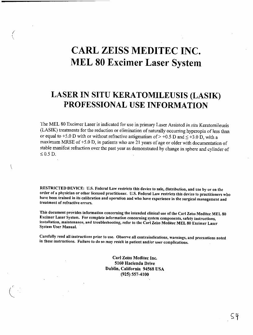
CARL ZEISS MEDITEC INC.MEL 80 Excimer Laser System
LASER IN SITU KERATOMILEUSIS (LASIK)PROFESSIONAL USE INFORMATION
The MEL 80 Excimer Laser is indicated for use in primary Laser Assisted in situ Keratomileusis(LASIK) treatments for the reduction or elimination of naturally occurring hyperopia of less thanor equal to +5.0 D with or without refractive astigmatism of > +0.5 D and S +3.0 D, with amaximum MRSE of +5.0 D, in patients who are 21 years of age or older with documentation ofstable manifest refraction over the past year as demonstrated by change in sphere and cylinder of
0.5 D.
RESTRICTED DEVICE: U.S. Federal Law restricts this device to sale, distribution, and use by or on theorder of a physician or other licensed practitioner. U.S. Federal Law restricts this device to practitioners whohave been trained in its calibration and operation and who have experience in the surgical management andtreatment of refractive errors.
This document provides information concerning the intended clinical use of the Carl Zeiss Meditec MEL 80Excimer Laser System. For complete information concerning system components, safety instructions,installation, maintenance, and troubleshooting, refer to the Carl Zeiss Meditec MEL 80 Excimer LaserSystem User Manual.
Carefully read all instructions prior to use. Observe all contraindications, warnings, and precautions notedin these instructions. Failure to do so may result in patient and/or user complications.
Carl Zeiss Meditec Inc.5160 Hacienda Drive
Dublin, California 94568 USA(925) 557-4100
CARL ZEISS MEDITEC INC. PROFESSIONAL USE INFORMATIONMEL 80 EXCIMER LASER SYSTEM PAGE 2
CARL ZEISS MEDITEC INC.MEL 80 EXCIMER LASER SYSTEM
PROFESSIONAL USE INFORMATION
TABLE OF CONTENTSPAGE
SECTION 1- SAFETY CONSIDERATIONS & GENERAL WARNINGS................... 7
SECTION 2 - DEVICE DESCRIPTION ................................................................................ 8
2 .1 L A SER S YS TEM ................................................................................................................ 8
2.1.1 FEATURES AND COMPONENTS OF THE MEL 80 EXCIMER LASER SYSTEM..................... 8
SECTION 3 - INDICATIONS, CONTRAINDICATIONS, WARNINGS, ANDPRECAUTIO NS ......................................................................................... 10
3.1 IN DICATION S FOR U SE ................................................................................................. 10
3.2 C ONTRA IN DICATION S.................................................................................................. 10
3 .3 W A R N IN G S..................................................................................................................... 11
3 .4 P RECA UTIO N S ................................................................................................................ 12
SECTION 4 - CLINICAL RESULTS .............................................................................. 14
4.1 STUDY O BJECTIVES.................................................................................................. 14
4.2 DATA ANALYSIS AND RESULTS.............................................................................. 14
4.2.1 DEMOGRAPHICS AND BASELINE PARAMETERS............................................................ 15
4.2.2 A CCOUNTABILITY ................................................. .......................... .................. 18
4.2.3 SAFETY OUTCOMES: CHANGE IN BSCVA, COMPLICATIONS,ADVERSE EVENTS, AND SUBJECTIVE SYMPTOMS........................................20
4.2.4 KEY EFFECTIVENESS PARAMETERS......................................................................... 28
4.2.4.1 KEY EFFECTIVENESS PARAMETERS BY PREOPERATIVE MRSE ............................... 31
4.2.4.2 STRATIFICATION OF KEY EFFICACY PARAMETERS BY OPTICAL ZONE..................... 334.2.4.3 MEAN MANIFEST REFRACTIVE SPHERICAL EQUIVALENT........................................ 34
4.2.4.4 STABILITY OF THE MANIFEST REFRACTION.............................................................35
4.2.4.5 CYLINDER CORRECTION/VECTOR ANALYSIS ........................................................... 39
4.2.4.6 POSTOPERATIVE UNCORRECTED VISUAL ACUITY COMPARED TOPREOPERATIVE BEST CORRECTED VISUAL AcuITY ........................ 41
4.2.4.7 PATIENT SYMPTOMS AND SATISFACTION ................................................................. 42
4.2.5 FACTORS ASSOCIATED WITH OUTCOMES ................................................................. 47
Cs
CARL ZEISS MEDITEC INC. PROFESSIONAL USE INFORMATIONMEL 80 EXCIMER LASER SYSTEM PAGE 3
TABLE OF CONTENTS
(CONTINUED)
SECTION 5 - SURGICAL PLANNING AND PROCEDURES....................................49
5 .1 IN TR O D U CTIO N ............................................................................................................... 4 9
5.2 PA TIEN T SELECTION ....................................................................................................... 49
5 .3 P R O C ED U R E ............................................................... .................................................. 50
5.4 PERI-O PERATIVE PROCEDURES.................................................................................. 51
5.4.1 A N ESTHESIA ................................................................. .......................................... 5 1
5.5 INTRA-O PERATIVE PROCEDURES.................................................................................51
5.5.1 CREATING THE LAMELLAR FLAP WITH THE MICROKERATOME.......................................51
5.5.2 PERFORMING THE LASER ABLATION .......................................................................... 51
5.6 POST-OPERATIVE PROCEDURES................................................................................... 51
5.6.1 PATCHING AND M EDICATIONS ............................................ ...................................... 51
5.6.2 ANALGESIA .................................... .................... ..... 52
5.6.3 HANDLING COMPLICATIONS............. ....................... ....... 6 ...... 525.7 POST-PROCEDURE ............................................ d .............. . 52
SECTION 6 - EXCIMER LASER STEP-BY-STEP SURGICAL PROCEDURE......... 536.1 PRIOR TO SURGERY.............. ............. ................................................ 53
6.2 PREPARING DEVICE AND PATIENT FOR TREATMENT ....................................... 536.3 M ICROKERATOME SURGERY.........................6 .................................................. 556.4 LASER TREATMENT......................................................55
SECTION 7 - EMERGENCY STOP ........................................................................... 57
(VC
CARL ZEISS MEDITEC INC. PROFESSIONAL USE INFORMATIONMEL 80 EXCIMER LASER SYSTEM PAGE 4
INDEX OF TABLESPAGE
TABLE 1 DEMOGRAPHICS, ALL TREATED EYES ......................................................... 15
TABLE 2.A PREOPERATIVE REFRACTION PARAMETERS STRATIFIED BY MRSEAND CYLINDER COMPONENTS, EYES TREATED FOR SPHERICALH YPEROPIA O NLY ........................................................................................ 16
TABLE 2.B PREOPERATIVE REFRACTION PARAMETERS STRATIFIED BY MRSEAND CYLINDER COMPONENTS, EYES TREATED FOR ASTIGMATICH YPEROPIA ......................................................................................... .......... 17
TABLE 3.A ACCOUNTABILITY, ALL TREATED EYES ..................................................... 18
TABLE 3.11 ACCOUNTABILITY, EFFECTIVENESS COHORT EYES....................................19
TABLE 4.A SUMMARY OF KEY SAFETY VARIABLES AT LAST AVAILABLE VISIT,ALL TREATED EYES STRATIFIED BY ALGORITHM AND TREATMENT.............20
TABLE 4.11 CHANGE IN BEST SPECTACLE-CORRECTED VISUAL ACUITY(BSCVA), ALL TREATED EYES .................................................................. 21
TABLE 4.C CHANGE IN BEST SPECTACLE-CORRECTED VISUAL ACUITY(BSCVA), STRATIFIED BY PREOPERATIVE MRSE, ALL TREATEDE YES ............................................................................................................. 22
TABLE 4.D CHANGE IN BEST SPECTACLE-CORRECTED VISUAL ACUITY(BSCVA), STRATIFIED BY TREATMENT, ALL TREATED EYES .................. 23
TABLE 5 COMPLICATIONS, ALL TREATED EYES ....................................................... 25
TABLE 6 ADVERSE EVENTS REPORTED AT ANY POSTOPERATIVE VISITS, ALLTREATED EYES STRATIFIED BY ALGORITHM AND TREATMENT.................26
TABLE 7.A CLINICALLY SIGNIFICANT PATIENT SYMPTOMS, ALL TREATEDE YES .................................................................................................................. 27
TABLE 7.13 COMPARISON OF SYMPTOMS BEFORE AND AFTER SURGERY AT 9M ONTHS, ALL TREATED EYES .................................................................... 28
TABLE 8.A SUMMARY OF KEY EFFECTIVENESS VARIABLES, EFFECTIVENESSC OHORT ........................................................................................................ 30
TABLE 8.11 SUMMARY OF KEY EFFECTIVENESS VARIABLES AT 9 MONTHS(POINT OF STABILITY), EFFECTIVENESS COHORT STRATIFIED BYTREATM ENT................................................................................................ 30
67
CARL ZEISS MEDITEC INC. PROFESSIONAL USE INFORMATIONMEL 80 EXCIMER LASER SYSTEM PAGE 5
TABLE 9.A SUMMARY OF KEY EFFECTIVENESS VARIABLES AT 9 MONTHS
(POINT OF STABILITY), STRATIFIED BY PREOPERATIVE MRSE,EFFECTIVENESS COHORT ............................................................................ 31
TABLE 9.B SUMMARY OF KEY EFFECTIVENESS VARIABLES AT 9 MONTHS(POINT OF STABILITY), STRATIFIED By PREOPERATIVE MRSE,EFFECTIVENESS COHORT TREATED FOR SPHERICAL HYPEROPIAONLY AND WITH ALGORITHM ADJUSTMENT ............................................. 32
TABLE 9.C SUMMARY OF KEY EFFECTIVENESS VARIABLES AT 9 MONTHS(POINT OF STABILITY), STRATIFIED BY PREOPERATIVE MRSE,EFFECTIVENESS COHORT TREATED FOR ASTIGMATIC HYPEROPIAWITH CYLINDER OF > 0.50 D AND WITH ALGORITHM ADJUSTMENT ............. 32
TABLE 10 SUMMARY OF KEY EFFECTIVENESS VARIABLES AT 9 MONTHS(POINT OF STABILITY), STRATIFIED By OPTICAL ZONE, SPHERICALEYES AND ASTIGMATIC EYES WITH CYLINDER OF > 0.50 D WITHALGORITHM ADJUSTMENT ......................................................... ......... 33
TABLE 11 MEAN MANIFEST REFRACTION SPHERICAL EQUIVALENT,EFFECTIVENESS COHORT ............................................................................ 34
TABLE 12.A STABILITY OF MANIFEST REFRACTION SPHERICAL EQUIVALENT(MRSE), EFFECTIVENESS COHORT ............................................................. 35
TABLE 12.B STABILITY OF MANIFEST REFRACTION SPHERICAL EQUIVALENT(MRSE), EFFECTIVENESS COHORT TREATED FOR SPHERICALHYPEROPIA ONLY AND WITH ALGORITHM ADJUSTMENT.......................... 36
TABLE 12.C STABILITY OF MANIFEST REFRACTION SPHERICAL EQUIVALENT(MRSE), EFFECTIVENESS COHORT TREATED FOR ASTIGMATICHYPEROPIA WITH CYLINDER OF > 0.50 D AND WITH ALGORITHMADJUSTMENT.................................................... ............................... 37
TABLE 12.D STABILITY OF MANIFEST REFRACTION CYLINDER (MRCYL),EFFECTIVENESS COHORT TREATED FOR ASTIGMATIC HYPEROPIAWITH CYLINDER OF > 0.50 D AND WITH ALGORITHM ADJUSTMENT.............38
TABLE 13 VECTOR MAGNITUDE ANALYSIS SUMMARY, EFFECTIVENESSCOHORT TREATED FOR ASTIGMATIC HYPEROPIA WITH CYLINDEROF > 0.50 D & WITH COMPLETE PREOPERATIVE ANDPOSTOPERATIVE REFRACTION WITH ABLATION ALGORITHMADJUSTMENT ................................................................... 39
TABLE 14 VECTOR ANALYSIS SUMMARY AT 9 MONTHS (POINT OF STABILITY),EFFECTIVENESS COHORT TREATED FOR ASTIGMATIC HYPEROPIAWITH CYLINDER OF > 0.50 D WITH ALGORITHM ADJUSTMENT.................40
CARL ZEISS MEDITEC INC. PROFESSIONAL USE INFORMATIONMEL 80 EXCIMER LASER SYSTEM PAGE 6
TABLE 15 POSTOPERATIVE UNCORRECTED VISUAL ACUITY (UCVA)COMPARED TO PREOPERATIVE BEST SPECTACLE-CORRECTEDVISUAL ACUITY (BSCVA), EFFECTIVENESS COHORT ............................. 41
TABLE 16.A PATIENT SYMPTOMS, ALL TREATED EYES ................................................. 43
TABLE 16.13 PATIENT SYMPTOMS CHANGE FROM BASELINE AT 9 MONTHS, ALLTREATED EYES ............................................................................................ 45
TABLE 17 PATIENT EVALUATION OF SATISFACTION AND VISION QUALITYIMPROVEMENT, EFFECTIVENESS COHORT SUBJECTS (SUBJECTB ASIS) ........................................................................................................... 46
CARL ZEISS MEDITEC INC. PROFESSIONAL USE INFORMATIONMEL 80 EXCIMER LASER SYSTEM PAGE 7
SECTION 1
SAFETY CONSIDERATIONS & GENERAL WARNINGS
Restricted Device: Federal (U.S.) law restricts these devices to sale by, or on the order of, aphysician.
Carefully read all instructions prior to use. Observe all contraindications, warnings, andprecautions. Failure to do so may result in patient and/or user complications.
Warning: Specific training from Carl Zeiss Meditec or an authorized representative ofCarl Zeiss Meditec is required before anyone is qualified to operate the MEL 80 ExcimerLaser. Read and understand the MEL 80 Excimer Laser User Manual before operatingthis system.
Refer to the MEL 80 Excimer Laser System User Manual for additional warningsregarding the use of this system.
High Pressure Gas CylindersIn the MEL 80 Excimer Laser, pressure vessels are used. Observe the relevant national andinternational regulations. If you notice a pungent smell (fluorine gas), open the windows, leavethe room, and call the Carl Zeiss Meditec Service Department.
Warning of High-Energy RadiationThe MEL 80 Excimer Laser is a medical laser device that emits high-intensity ultravioletradiation with energy levels of up to 2 mJ. These energy levels and short pulse durations result inextreme pulse powers and, if applied in an uncontrolled manner, may cause severe injury.
Warning for Reflective MaterialConsider that reflective material may deflect the laser beam in an uncontrollable manner.
CARL ZEISS MEDITEC INC. PROFESSIONAL USE INFORMATIONMEL 80 EXCIMER LASER SYSTEM PAGE 8
SECTION 2
DEVICE DESCRIPTION
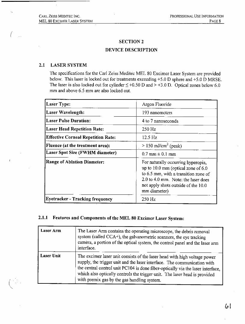
2.1 LASER SYSTEM
The specifications for the Carl Zeiss Meditec MEL 80 Excimer Laser System are providedbelow. This laser is locked out for treatments exceeding +5.0 D sphere and +5.0 D MRSE.The laser is also locked out for cylinder < +0.50 D and > +3.0 D. Optical zones below 6.0mm and above 6.5 mm are also locked out.
Laser Type: Argon Fluoride
Laser Wavelength: 193 nanometers
Laser Pulse Duration: 4 to 7 nanoseconds
Laser Head Repetition Rate: 250 Hz
Effective Corneal Repetition Rate: 12.5 Hz
Fluence (at the treatment area): > 150 mJ/cm 2 (peak)Laser Spot Size (FWHM diameter) 0.7 mn ± 0.1 mm
Range of Ablation Diameter: For naturally occurring hyperopia,up to 10.0 mm (optical zone of 6.0to 6.5 mm, with a transition zone of2.0 to 4.0 mm. Note: the laser doesnot apply shots outside of the 10.0mm diameter)
Eyetracker - Tracking frequency 250 Hz
2.1.1 Features and Components of the MEL 80 Excimer Laser System:
Laser Arm The Laser Arm contains the operating microscope, the debris removalsystem (called CCA+), the galvanometric scanners, the eye trackingcamera, a portion of the optical system, the control panel and the laser arminterface.
Laser Unit The excimer laser unit consists of the laser head with high voltage powersupply, the trigger unit and the laser interface. The communication withthe central control unit PC 104 is done fiber-optically via the laser interface,which also optically controls the trigger unit. The laser head is providedwith premix gas by the gas handling system.
CARL ZEISS MEDITEC INC. PROFESSIONAL USE INFORMATIONMEL 80 EXCIMER LASER SYSTEM PAGE 9
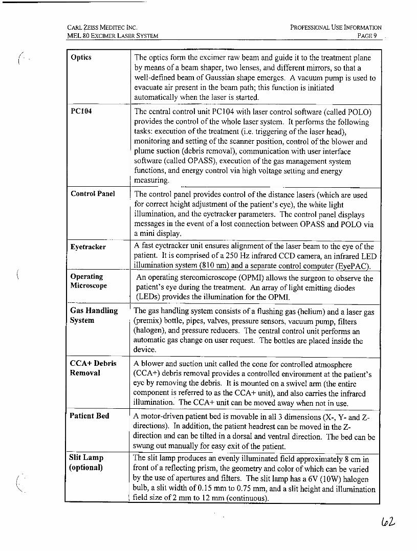
Optics The optics form the excimer raw beam and guide it to the treatment planeby means of a beam shaper, two lenses, and different mirrors, so that awell-defined beam of Gaussian shape emerges. A vacuum pump is used toevacuate air present in the beam path; this function is initiatedautomatically when the laser is started.
PC104 The central control unit PC] 04 with laser control software (called POLO)provides the control of the whole laser system. It performs the followingtasks: execution of the treatment (i.e. triggering of the laser head),monitoring and setting of the scanner position, control of the blower andplume suction (debris removal), communication with user interfacesoftware (called OPASS), execution of the gas management systemfunctions, and energy control via high voltage setting and energymeasuring.
Control Panel The control panel provides control of the distance lasers (which are usedfor correct height adjustment of the patient's eye), the white lightillumination, and the eyetracker parameters. The control panel displaysmessages in the event of a lost connection between OPASS and POLO viaa mini display.
Eyetracker A fast eyetracker unit ensures alignment of the laser beam to the eye of thepatient. It is comprised of a 250 Hz infrared CCD camera, an infrared LEDillumination system (810 in) and a separate control computer (EyePAC).
Operating An operating stereomicroscope (OPMI) allows the surgeon to observe theMicroscope patient's eye during the treatment. An array of light emitting diodes
(LEDs) provides the illumination for the OPMI.
Gas Handling The gas handling system consists of a flushing gas (helium) and a laser gasSystem (premix) bottle, pipes, valves, pressure sensors, vacuum pump, filters
(halogen), and pressure reducers. The central control unit performs anautomatic gas change on user request. The bottles are placed inside thedevice.
CCA+ Debris A blower and suction unit called the cone for controlled atmosphereRemoval (CCA+) debris removal provides a controlled environment at the patient's
eye by removing the debris. It is mounted on a swivel arm (the entirecomponent is referred to as the CCA+ unit), and also carries the infraredillumination. The CCA+ unit can be moved away when not in use.
Patient Bed A motor-driven patient bed is movable in all 3 dimensions (X-, Y- and Z-directions). In addition, the patient headrest can be moved in the Z-direction and can be tilted in a dorsal and ventral direction. The bed can beswung out manually for easy exit of the patient.
Slit Lamp The slit lamp produces an evenly illuminated field approximately 8 cm in(optional) front of a reflecting prism, the geometry and color of which can be varied
by the use of apertures and filters. The slit lamp has a 6V (lOW) halogenbulb, a slit width of 0.15 mm to 0.75 mm, and a slit height and illuminationfield size of2 mm to 12 mm (continuous).
CARL ZEISS MEDITEC INC. PROFESSIONAL USE INFORMATIONMEL 80 EXCIMER LASER SYSTEM PAGE 10
SECTION 3
INDICATIONS, CONTRAINDICATIONS, WARNINGS, AND PRECAUTIONS
3.1 INDICATIONS FOR USE
The MEL 80 Excimer Laser is indicated for use in primary Laser Assisted in situKeratomileusis (LASIK) treatments for the reduction or elimination of naturally occurringhyperopia of less than or equal to +5.0 D with or without refractive astigmatism of> +0.5 D and < +3.0 D, with a maximum MRSE of +5.0 D, in patients who are21 years of age or older with documentation of stable manifest refraction over the pastyear as demonstrated by change in sphere and cylinder of ! 0.5 D.
3.2 CONTRAINDICATIONS
Conditions under which risk of use outweighs possible benefit. LASIK surgery iscontraindicated in:
* Patients with severe dry eye;
* Patients with active corneal infection or inflammation;* Patients with glaucoma with marked optic nerve cupping, advanced visual field loss
or visual acuity loss, because of the risk of further loss of visual function related tomicrokeratome-induced pressure spikes;
* Patients with projected residual corneal stromal bed thickness after ablation of lessthan 250 microns, because this may lead to ectasia;
* Patients with active connective tissue diseases or autoimmune diseases which havebeen associated with corneal melting, such as rheumatoid arthritis, Wegener'sgranulomatosis, relapsing polychondritis, and polyarteritis nodosa;
* Pregnant or nursing women;* Patients with signs of ectatic disorders such as keratoconus or pellucid marginal
degeneration;* Patients with active, uncontrolled diabetes mellitus or visually significant diabetic
complications;* Patients with recent herpes keratitis (simplex or zoster) or significant corneal damage
(poor sensation, scarring, neovascularization) from prior herpes infection; and* Patients with immunodeficiency diseases, such as AIDS.
3.3 WARNINGS
Warnings are conditions for which there is reasonable evidence of serious risk with use ofthis device. There is reason to believe that there is increased risk for serious adverseconsequences if LASIK surgery is performed in the following patients:
CARL ZEISS MEDITEC INC. PROFESSIONAL USE INFORMATIONMEL 80 EXCIMER LASER SYSTEM PAGE II
* Patients who are taking isotretinoin (Accutane®). This increases risk of dry eye andmay cause increased complications after LASIK.
* Patients with significant dry eye. LASIK may increase dryness with accompanyingdiscomfort and visual problems. (See Contraindications for related information.)
* Patients with severe allergies. Patients who rub their eyes a lot may be at risk fordislodging the comeal flap, since the strength of the flap attachment to underlyingcorneal layers is significantly and permanently reduced after surgery. Additionally,LASIK may increase the dryness often associated with anti-allergy medication.
* Patients with well-controlled diabetes mellitus. Diabetes may be related to poorerhealing and a higher complication rate. (See Contraindications for relatedinformation.)
* Patients with well-controlled glaucoma, ocular hypertension, or are being followed forsuspicion of glaucoma, because of the risk of steroid response causing diseaseprogression, and the post-LASIK difficulty in accurately monitoring intraocularpressure due to changes in corneal thickness. (See Contraindications for relatedinformation.)
* Patients with a prior history of herpes simplex or herpes zoster keratitis, because it ispossible that LASIK may lead to reactivation of the virus. (See Contraindications forrelated information.)
* Patients with stable and well-controlled connective tissue diseases or autoimmunediseases, because they may be slower healing and have less predictable outcomes.Additionally, the stability and control of the disease (and risk, if any, of corneal melt)may be difficult to quantify. Because of this difficulty, increased caution should beused if LASIK is considered in patients with well controlled, stable disease. Thesurgeon should consider consulting with the doctor who is treating the underlyingdisease. The surgeon should consider unilateral surgery. The surgeon shouldthoroughly discuss the potential risks with the patient. (See Contraindications forrelated information)
* Patients with inactive and controlled immunocompromised conditions, because theymay be at increased risk of infection. (See Contraindications for related information.)
* Patients whose refractive error is not stable, because the correct refractive treatment isdifficult to determine.
* Patients with a history of blepharitis.
* Patients with a history of eye rubbing. The LASIK flap never completely heals, and itcan be dislodged by eye rubbing. Patients who are not sure they can refrain fromrubbing their eyes after LASIK may not be good LASIK candidates.
3.4 PRECAUTIONS
The safety and effectiveness of LASIK with the MEL 80 Excimer Laser System has NOTbeen established in the following patients:
6Pt
CARL ZEISS MEDITEC INC. PROFESSIONAL USE INFORMATIONMEL 80 ExCIMER LASER SYSTEM PAGE 12
* Patients taking amiodarone hydrochloride (e.g., Cordarone®). Use can causekeratopathy and might affect epithelial healing after LASIK.
* Patients taking the medication sumatriptan succinate (Imitrex®).* Patients with a family history of keratoconus, pellucid marginal degeneration or other
ectatic disorders. Such patients should be carefully checked for an undiagnosed mildcondition that may lead to post-LASIK ectasia.
* Patients with progressive hyperopia and/or astigmatism or ocular disease.* Patients with corneal abnormalities including, but not limited to, scars, irregular
astigmatism and corneal warpage.* Patients with a history of uveitis.
* Patients with previous corneal or intraocular surgery, or trauma.* Patients who have had prior LASJK treatment.* Patients under 21 years of age.
* Patients needing treatments of > +5.0 D of hyperopia, > +5.0 D MRSE, or with+0.5 D or > +3.0 D of astigmatism.
* Patients with large mesopic pupils, greater than optical zone size, as evaluated undermesopic illumination conditions. Such patients should be advised of the potential fornegative effects on vision after LASIK, such as glare, halos, and night time drivingdifficulty.
* Patients with a history of strabismus who are being treated for hyperopia may have anincreased risk of manifest exotropia after surgery.
* Patients with 0.75 D or more of latent hyperopia as determined by the differencebetween the preoperative MRSE and CRSE.
* Patients with media problems (corneal, lens, and/or vitreous opacities including, butnot limited to, cataract).
* Patients with iris problems including, but not limited to, coloboma and previous irissurgery compromising proper eyetracking.
* Patients taking medications likely to affect wound healing including, but not limitedto, antimetabolites.
* Patients with a history of keloid formation.* Patients taking hormone replacement therapy or antihistamines who may experience
delayed re-epithelialization of the cornea following surgery.* Patients undergoing retreatment with the MEL 80 Excimer Laser System. The risk
and accuracy of LASK retreatment, or LASIK after another surgery to correct vision,has not been evaluated.
* Over the long term (more than 24 months after surgery).
A LASIK flap diameter that is minimally larger (i.e., larger by < 2.2 mm) than the opticalzone size may result in decreased success rate.
CARL ZEISS MEDITEC INC. PROFESSIONAL USE INFORMATIONMEL 80 EXCIMER LASER SYSTEM PAGE 13
Pupil size should be evaluated under mesopic illumination conditions. Effects oftreatment on vision under poor illumination cannot be predicted prior to surgery. Somepatients may find it more difficult than usual to see in conditions such as very dim light,rain, snow, fog, and glare from bright lights at night. These symptoms could be worsenedin patients with large pupil sizes.
The optical zone should be (a) at least as large as the mesopic pupil and (b) small enoughto leave at least 250 microns of residual stromal thickness. Prospective patients whocannot satisfy both of these criteria should be disqualified for treatment.
Preoperative evaluation for dry eye should be performed. This may be particularlyimportant for patients who have been contact lens intolerant because of symptoms ofdryness. Patients should be advised of potential for worsening of symptoms associatedwith dry eye syndrome post-LASIK surgery.
The effects of LASIK on visual performance under poor lighting conditions have notbeen fully characterized. It is possible that following LASIK treatment, patients will findit more difficult to see in conditions such as very dim light, rain, snow, fog, or glare frombright lights at night. Visual performance possibly could be worsened in patients withlarge pupil sizes.
Intraocular pressure is more difficult to interpret after LASIK because of the resultingcorneal thinning. Patients should be informed that they should tell future eye careprofessionals that they have had LASIK.
Future intraocular lens calculations for cataract surgery may be affected by LASIK. Thesurgeon should give the patient a patient information card that has eye measurementsfrom before the LASIK surgery. Patients should be advised to keep this card and give itto their future cataract surgeon.
The risk/benefit ratio of the LASIK procedure may be significantly increased in patientswho have significantly reduced visual function in one eye (e.g., amblyopia).
Prior to surgery, prospective patients should be provided with a copy of the PatientInformation Brochure for this product and informed of the possible risks and benefitsassociated with its use.
CARL ZEISS MEDITEC INC. PROFESSIONAL USE INFORMATIONMEL 80 EXCIMER LASER SYSTEM PAGE 14
SECTION 4
CLINICAL RESULTS
4.1 STUDY OBJECTIVES
A prospective, non-randomized, multicenter clinical study of 369 eyes was conducted atsix (6) clinical sites. Patients with spherical hyperopia and astigmatic hyperopia weretreated with the MEL 80Tm Excimer Laser, and followed for a 24-month period. Thesafety and effectiveness of the MEL 80 " Excimer Laser was determined by evaluation ofthe UCVA outcomes (at least 85% of laser treated eyes with UCVA of 20/40 or better),predictability (at least 50% of treated eyes with MRSE within + 0.50 D deviation fromattempted correction, and at least 75% of treated eyes with MRSE within ± 1.00 Ddeviation from attempted correction), stability of the postoperative MRSE, subjectsatisfaction, preservation of BSCVA, induced astigmatism, incidence of adverseevents/complications, and patient symptoms.
Patients were screened for eligibility, and informed consent was obtained from those whomet screening criteria and were interested in participating in the study. Eligible patientswere examined preoperatively to obtain a medical history and to establish a baseline forocular condition. Baseline and postoperative measurements (taken at I day, 7 days,1 month, 3 months, 6 months, 9 months, 12 months, 18 months, and 24 months post-surgery) included manifest refraction, cycloplegic refraction, distance visual acuity (bestcorrected and uncorrected), slit-lamp examination, corneal topography (centralkeratometry via simulated K readings), pachymetry (baseline only), fundus examination,and intraocular pressure (IOP).
4.2 DATA ANALYSIS AND RESULTS
The safety summaries presented in this section were based on the entire PMA cohortwhile the effectiveness analyses were based on available data of treated eyes at each visitor available data for the treated subgroups. Due to aborted laser treatment, missing visitsor examinations, and different subgroups, the safety cohort and effectiveness cohorts forthe data analyses were different.
MEL 80 EXCIMER LASER SYSTEM
CARL ZEISS MEDITEC INC. PROFESSIONAL USE INFORMATIONMEL 80 ExcIMER LASER SYSTEM PAGE 15
4.2.1 Demographics and Baseline Parameters
Demographic characteristics of the study population are presented in Table 1. Thebaseline refraction parameters stratified by MRSE and cylinder components are presentedin Table 2.A for eyes treated for spherical hyperopia only and in Table 2.B for eyestreated for astigmatic hyperopia. The shaded columns indicate treatments outside of theapproved range, and are not part of the effectiveness cohort. For eyes treated forspherical hyperopia only (Table 2.A), the mean MRSE with algorithm adjustment for theapproved range was +2.468 D (SD 1.115).
For eyes treated for astigmatic hyperopia, the mean MRSE in the approved range was+2.965 D (SD 1.222) and the mean MRCYL was 0.970 D (SD 0.609); see Table 2.B.The intended correction was the full manifest refraction, with the goal of achieving aplano refraction after the surgery.
TABLE 1DEMOGRAPHICS
ALL TREATED EYES
-- VnW~ ~2 Nunibi&NUMBER OF EYES & 369 Eyes of 189 Enrolled SubjectsSUBJECTS'GENDER2
Male 54.5% 103Female 45.5% 86
RACE'White 94.7% 179Black 3.2% 6Asian 1.1% 2Other 1.1% 2
SURGICAL EYE'Right 50.1% 185
Left 49.9% 184AGE (in years)'
Mean (SD) 46.6 (9.3)Minimum, Maximum 22.0, 69.0
1 Two eyes had aborted procedures and were not included in theeffectiveness analyses.
2 Gender, Race, and Age were based on subjects, but SurgicalEye is based on eyes.
MEL 80 EXCIMER LASER SYSTEM
CARL ZEISS MEDITEC INC. PROFESSIONAL USE INFORMATIONMEL 80 EXCIMER LASER SYSTEM PAGE 16
TABLE 2.APREOPERATIVE REFRACTION PARAMETERS
STRATIFIED BY MRSE AND CYLINDER COMPONENTSEYES TREATED FOR SPHERICAL HYPEROPIA ONLY
MkSE Manifest CyHiader Totai,ffi ;tkt.W0D 0 25'DC
S >o i1Ng~> . ~% n/NWithout Algorithm Adjustment
MRSE: Mean: 2.468, SD: 1.270, Range: 1.25 to 5.88MRCYL:Mean: 0.065, SD: 0.112, Range: 0.00 to 0.25
0.00 to 1.00 D .0.0% (0/27) .0.0% (0/27)1 0.0% (0/27)1.01 to 2.00 D 44.4% (12/27) 14.8% (4/27) .59.3% (16/27)2.01 to 3.00 D IlI%' (3/27) 0.0% (0/27): 11.1% (3/27)3.01 to 4.00 D 18:5% (5/27) 0.0% (0/27) 18.5% (5/27)4.01 to 5.00 D .0.0% (0/27) 3.7% (1/27) 3.7% (1/27)5.01 to 6.00 D 0.0% (0/27) 7.4% (2/27) 7.4% (2/27)Total 74-J% (20/27) 25.9%, (7/27) 100.0% (27/27)
With Algorithm AdjustmentMRSE: Mean: 2.468, SD: 1.115, Range: 1.00 to 5.25
MRCYL:Mean: 0.033, SD: 0.086, Range: 0.00 to 0.250.00 to 1.00 D 3.3% (2/60) 0.0% (0/60) 3.3% (2/60)1.01 to 2.00 D 28.3% (17/60) 6.7% (4/60) 35.0% (21/60)2.01 to 3.00 D 31.7% (19/60) 3.3% (2/60) 35.0% (21/60)3.01 to 4.00 D 8.3% (5/60) 1.7% (1/60) 10.0% (6/60)4.01 to 5.00 D 11.7% (7/60) 1.7% (1/60) 13.3% (8/60)5.01 to 6.00 D 3.3% (2/60) 0.0% (0/60) 3.3% (2/60)Total 86.7% (52/60) 13.3% (8/60) 100.0% (60/60)The shaded cells were not included in the effectiveness cohort.
MEL 80 EXCIMER LASER SYSTEM
CARL ZEISS MEDITEC INC. PROFESSIONAL USE INFORMATION
MEL 80 EXCIMER LASER SYSTEM PAGE 17
TABLE 2.BPREOPERATIVE REFRACTION PARAMETERS
STRATIFIED BY MRSE AND CYLINDER COMPONENTSEYES TREATED FOR ASTIGMATIC HYPEROPIA
MRSE7H. ManiFt Clinder Total: 25to O.50.D .1 to 1.000 - J. .ito2ooD. 2.01 t0.00 D
%_ :% d/ift -%o :nN % n/N:
Without Algorithm AdjustmentMRSE: Mean: 2.965, SD: 1.253, Range: 1.00 to 6.38
MRCYL:Mean: 0.943, SD: 0.583, Range: 0.25 to 2.750.00 to 1.00 D .1.2% :(1/83). 1.2% (1/83) 0.0% (0/83)i 0.0% (0/83) 2.4% (2/83)1.01 to 2.00 D 13.3% (11/83) 9.6% (8/83) 3.6% (3/83) 2.4% (2/83) 28.9% (24/83)2.01 to 3.00 D 6.0% (5/83) 4.8% (4/83) 3.6% (3/83) 1.2% (1/83) 15.7% (13/83)3.01 to 4.00 D 7.2% (6/83) 16.9% (14/83) 8.4% (7/83) 0.0% (0/83) 32.5% (27/83)4.01 to 5.00 D 6.0% (5/83) 3.6% (3/83) 3.6% (3/83) 1.2% (1/83) 14.5% (12/83)5.01 to 6.00 D 2.4% (2/83) 1:2% . (1/83) 1.2% (1/83) 0.0% (0/83) 4.8% (4/83)6.01 to 7.00 D 0:0% ,(0/83) 0.0%' (0/83) . 1.2% (1/83) 0.0% (0/83) 1.2% (1/83),Total 136.1% (30/83) 37.3%' (31/83) 21.7% (18/83) 4.8% (4/83) 100.0% (83/83)
With Algorithm AdjustmentMRSE: Mean: 2.965, SD: 1.222, Range: 0.88 to 6.38
MRCYL:Mean: 0.970, SD: 0.609, Range: 0.50 to 3.000.00 to 1.00 D 2.5%, (5/199) 0.0% (0/199) 1.5% (3/199) 0.0% (0/199) 4.0% (8/199)1.01 to 2.00 D 10.6% (21/199) 7.5% (15/199) 4.0% (8/199) 2.0% (4/199) 24.1% (48/199)2.01 to 3.00 D 14.1% (28/199) 8.5% (17/199) 6.0% (12/199) 0.5% (1/199) 29.1% (58/199)3.01 to 4.00 D 11.6% (23/199) 5.5% (11/199) 4.5% (9/199) 3.5% (7/199) 25.1% (50/199)4.01 to 5.00 D 5.0% (10/199) 2.0% (4/199) 5.5% (11/199) 0.5% (1/199) 13.1% (26/199)5.01 to 6.00 D 1.5% (3/199) IL0% (2/199) 1.0% (2/199) 0.0% (0/199) 3.5% (7/199)6.01 to 7.00 D 0.0% (0/199)j 0.0% (0/199) 1.0% (2/199) 0.0% (0/199) 1.0% (2/199)Total 45.2% (90/199) 24.6% (49/199) 23.6% (47/199) 6.5% (13/199) 100.0% (199/199)Two eyes (with refraction of 3.50+1.00xl76, 3.00+.25x20) were reported with an aborted procedure and were nottreated later. These eyes were excluded from the effectiveness analyses.The shaded cells were not included in the effectiveness cohort. Additionally, I eye in the non-shaded cells was notincluded in the effectiveness cohort due to an aborted procedure.
MEL 80 EXCIMER LASER SYSTEM
CARL ZEISS MEDITEC INC. PROFESSIONAL USE INFORMATIONMEL 80 EXCIMER LASER SYSTEM PAGE 18
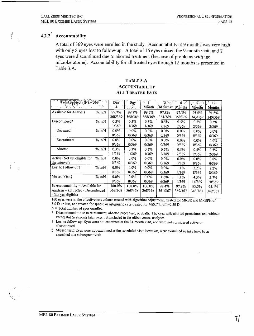
4.2.2 Accountability
A total of 369 eyes were enrolled in the study. Accountability at 9 months was very highwith only 8 eyes lost to follow-up. A total of 16 eyes missed the 9-month visit, and 2eyes were discontinued due to aborted treatment (because of problems with themicrokeratome). Accountability for all treated eyes through 12 months is presented inTable 3.A.
TABLE 3.AACCOUNTABILITY
ALL TREATED EYES
t6-a es -D 1 13- 6 1, 12,IlI7bMtnNh6 Moriths< Months Months MonthsAvailable for Analysis %, n/N 99.7% 99.7% 99.7% 97.8% 97.3% 93.0% 94.6%
368/369 368/369 368/369 361/369 359/369 343/369 349/369Discontinued* %, n/N 0.3% 0.3% 0.3% 0.5% 0.5% 0.5% 0.5%
1/369 1/369 1/369 2/369 2/369 2/369 2/369Deceased %, n/N 0.0% 0.0% 0.0% 0.0% 0.0% 0.0% 0.0%
0/369 0/369 0/369 0/369 0/369 0/369 0/369Retreatment %, n/N 0.0% 0.0% 0.0% 0.0% 0.0% 0.0% 0.0%
0/369 0/369 0/369 0/369 0/369 0/369 0/369Aborted %, n/N 0.3% 0.3% 0.3% 0.5% 0.5% 0.5% 0.5%
1/369 1/369 1/369 2/369 2/369 2/369 2/369Active (Not yet eligible for %, n/N 0.0% 0.0% 0.0% 0.0% 0.0% 0.0% 0.0%the interval) 0/369 0/369 0/369 0/369 0/369 0/369 0/369Lost to Follow-upt %, n/N 0.0% 0.0% 0.0% 0.0% 1.1% 2.2% 2.2%
0/369 0/369 0/369 0/369 4/369 8/369 8/369Missed Visit? %, n/N 0.0% 0.0% 0.0% 1.6% 1.1% 4.3% 2,7%
0/369 0/369 0/369 6/369 4/369 16/369 10/369%Accountability=Availablefor 100.0% 100.0% 100.0% 98.4% 97.8% 93.5% 95.1%Analysis+ (Enrolled - Discontinued 368/368 368/368 368/368 361/367 359/367 343/367 349/367- Not yet eligible) 1
160 eyes were in the effectiveness cohort: treated with algorithm adjustment, treated for MRSE and MRSPH of5.0 D or less, and treated for sphere or astigmatic eyes treated for MRCYL of > 0.50 D.N = Total number of eyes enrolled.* Discontinued = due to retreatment, aborted procedure, or death. The eyes with aborted procedures and without
successful treatments later were not included in the effectiveness analyses.t Lost to follow-up: Eyes were not examined at the 24-month visit, and were not considered active or
discontinued.Missed visit: Eyes were not examined at the scheduled visit, however, were examined or may have beenexamined at a subsequent visit.
MEL 80 EXCIMER LASER SYSTEM f
CARL ZEISS MEDITEC INC. PROFESSIONAL USE INFORMATIONMEL 80 EXCIMER LASER SYSTEM PAGE 19
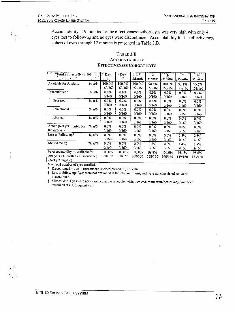
Accountability at 9 months for the effectiveness cohort eyes was very high with only 4eyes lost to follow-up and no eyes were discontinued. Accountability for the effectivenesscohort of eyes through 12 months is presented in Table 3.B.
TABLE 3.1ACCOUNTABILITY
EFFECTIVENESS COHORT EYES
Total'subjects (N) IN~ mD y Day Av~ 3 6.6. 7 Month Months Mimiths Months Months
Available for Analysis %, n/N 100.0% 100.0% 100.0% 98.8% 100.0% 93.1% 95.6%160/160 160/160 160/160 158/160 160/160 149/160 153/160
Discontinued* %, n/N 0.0% 0.0% 0.0% 0.0% 0.0% 0.0% 0.0%0/160 0/160 0/160 0/160 0/160 0/160 0/160
Deceased %, n/N 0.0% 0.0% 0.0% 0.0% 0.0% 0.0% 0.0%0/160 0/160 0/160 0/160 0/160 0/160 0/160
Retreatment %, n/N 0.0% 0.0% 0.0% 0.0% 0.0% 0.0% 0.0%0/160 0/60 0/160 0/160 0/160 0/160 0/160
Aborted %, n/N 0.0% 0.0% 0.0% 0.0% 0.0% 0.0% 0.0%0/160 0/160 0/160 0/160 0/160 0/160 0/160
Active (Not yet eligible for %, n/N 0.0% 0.0% 0.0% 0.0% 0.0% 0.0% 0.0%the interval) 0/160 0/160 0/160 0/160 0/160 0/160 0/160Lost to Follow-upt %, n/N 0.0% 0.0% 0.0% 0.0% 0.0% 2.5% 2.5%
0/160 0/160 0/160 0/160 0/160 4/160 4/160Missed Visitt %, n/N 0.0% 0.0% 0.0% 1.3% 0.0% 4.4% 1.9%
0/160 0/160 0/160 2/160 0/160 7/160 3/160%Accountability= Available for 100.0% 100.0% 100.0% 98.8% 100.0% 93.1% 95.6%Analysis+ (Enrolled - Discontinued 160/160 160/160 160/160 158/160 160/160 149/160 153/160- Not yet eligible)N = Total number of eyes enrolled.* Discontinued = due to retreatment, aborted procedure, or death.t Lost to follow-up: Eyes were not examined at the 24-month visit, and were not considered active or
discontinued.Missed visit: Eyes were not examined at the scheduled visit, however, were examined or may have beenexamined at a subsequent visit.
MEL 80 EXCIMER LASER SYSTEM
CARL ZEISS MEDITEC INC. PROFESSIONAL USE INFORMATIONMEL 80 EXCIMER LASER SYSTEM PAGE 20
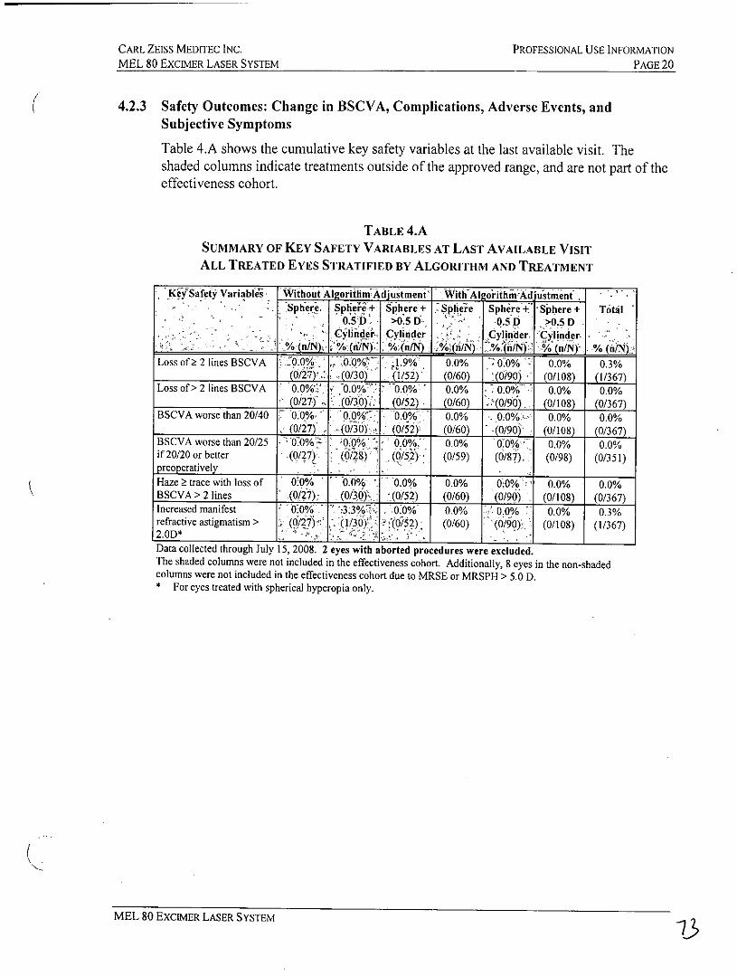
4.2.3 Safety Outcomes: Change in BSCVA, Complications, Adverse Events, andSubjective Symptoms
Table 4.A shows the cumulative key safety variables at the last available visit. Theshaded columns indicate treatments outside of the approved range, and are not part of theeffectiveness cohort.
TABLE 4.A
SUMMARY OF KEY SAFETY VARIABLES AT LAST AVAILABLE VISITALL TREATED EYES STRATIFIED BY ALGORITHM AND TREATMENT
KeYSafety Variables Without Algorithm Adjustment ith Al orithmndjustment .
Sphere. 0ier Sphere + he Sphere -& Sphere + Total0.5D >0.5 D- 0.5 D >0.5 D
Cylinder Cylinder Cylinder, C ylinder/%(n/N % ( )A / (n/N) W(n/N) . /N)J % (i/n/ % (i/)N
Loss of 2 2 lines BSCVA 0.0% 0.0% 1.9% 0.0% 0 60%t 0.0% 0.3%(0/27) (0/30) (1/52) (0/60) '(0/90) (0/108) (1/367)
Loss of> 2 lines BSCVA 0.0% / 000 0.0% 0.0% - 0:0%> 0.0% 0.0%(0/27) (0/30) (0/52) (0/60) .>(0/90). (0/108) (0/367)
BSCVA worse than 20/40 0.0% 00. 0 0% 0.0% 0.0%>- 0.0% 0.0%(0/27) (0/30) (0/52) (0/60) (0/90i (0/108) (0/367)
BSCVA worse than 20/25 ;0.0% 0.0% 0.0% 0.0% 6.0%- 0.0% 0.0%if 20/20 or better (0/27) (0/28) (0/52): (0/59) (0/87). (0/98) (0/351)preoperatively
Haze Z trace with loss of 0:0% 000/ 0.0% 0.0% 0.0% -, 0.0% 0.0%BSCVA > 2 lines (0/27) (0/30) (0/52) (0/60) (0/90) (0/108) (0/367)Increased manifest 0:0% 3.3 .0.9% 0.0% 0.0% 0.0% 0.3%refractive astigmatism > (0/7) () (0/5) (0/60) (0/90). (0/108) (1/367)2.0D*Data collected through July 15, 2008. 2 eyes with aborted procedures were excluded.The shaded columns were not included in the effectiveness cohort. Additionally, 8 eyes in the non-shadedcolumns were not included in the effectiveness cohort due to MRSE or MRSPH > 5.0 D.* For eyes treated with spherical hyperopia only.
MEL 80 EXCIMER LASER SYSTEM
CARL ZEISS MEDITEC INC. PROFESSIONAL USE INFORMATIONMEL 80 EXCIMER LASER SYSTEM PAGE 21
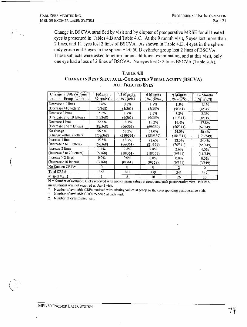
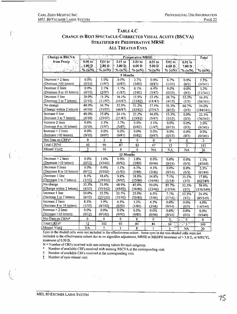
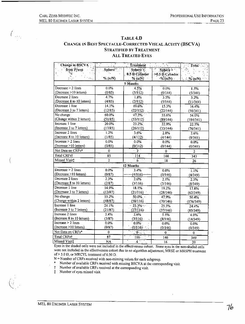
Change in BSCVA stratified by visit and by diopter of preoperative MRSE for all treatedeyes is presented in Tables 4.B and Table 4.C. At the 9 month visit, 5 eyes lost more than2 lines, and 11 eyes lost 2 lines of BSCVA. As shown in Table 4.D, 4 eyes in the sphereonly group and 5 eyes in the sphere + >0.50 D cylinder group lost 2 lines of BSCVA.These subjects were asked to return for an additional examination, and at this visit, onlyone eye had a loss of 2 lines of BSCVA. No eyes lost > 2 lines BSCVA (Table 4.A).
TABLE 4.11CHANGE IN BEST SPECTACLE-CORRECTED VISUAL ACUITY (BSCVA)
ALL TREATED EYES
Change in BSCVkfrom I o 6Mohs 9 M'6ths . - 12 Monthr,Pteo ~ .~ % (/Nf~ % -.n'N j %jnj f.J .P/o(n/N) .%(/N
Decrease > 2 lines 1.4% 0.8% 1.9% 1.5% 1.1%(Decrease>l0 letters) (5/368) (3/361) (7/359) (5/341) (4/349)Decrease 2 lines 2.7% 1.7% 2.5% 3.2% 2.3%(Decrease 8 to 10 letters) (10/368) (6/361) (9/359) (11/341) (8/349)Decrease I line 22.6% 18.3% 19.2% 16.4% 17.8%(Decrease 3 to 7 letters) (83/368) (66/361) (69/359) (56/341) (62/349)No change 56.5% 58.2% 51.0% 54.0% 50.4%(Change within 2 letters) (208/368) (210/361) (183/359) (184/341) (176/349)Increase I line 15.5% 18.3% 22.6% 22.3% 24.4%(Increase 3 to 7 letters) (57/368) (66/361) (81/359) (76/341) (85/349)Increase 2 lines 1.4% 2.8% 2.8% 2.6% 4.0%(Increase 8 to 10 letters) (5/368) (10/361) (10/359) (9/341) (14/349)Increase > 2 lines 0.0% 0.0% 0.0% 0.0% 0.0%(Increase >I0 letters) (0/368) (0/361) (0/359) (0/341) (0/349)No Data on CRFs* 0 0 0 2 0Total CRFst 368 361 359 343 349Missed Visit: 1 8 10 26 20N = Number of available CRFs received with non-missing values at preop and each postoperative visit. BSCVAmeasurement was not required at Day-I visit.* Number of available CRFs received with missing values at preop or the corresponding postoperative visit.t Number of available CRFs received at each visit.: Number of eyes missed visit.
MEL 80 EXCIMER LASER SYSTEM
CARL ZEISS MEDITEC INC. PROFESSIONAL USE INFORMATIONMEL 80 EXCEMER LASER SYSTEM PAGE 22
TABLE 4.CCHANGE IN BEST SPECTACLE-CORRECTED VISUAL ACUITY (BSCVA)
STRATIFIED BY PREOPERATIVE MRSEALL TREATED EYES
Change inBSCVA Preopertive'MRSE._ _ Totalfrom Preop 0.0 to 1E1 to'' 2.01 to 3.I to 4.011 to 01 to 6.01 to
2100D .00 D p 3.0D 7 4.00 D 5.00 D 6.00 D 7.00 D% __/N) % (n/N_ % (n/N) %(n/N) %.tn/N) % (n/N) n%(n/N) % (/N
9 Months
Decrease > 2 lines 0.0% 1.0% .0.0% 3.7% 0.0% 6.7% 0.0% 1.5%(Decrease>I0 letters) (0/10) (1/97) (0/87) (3/82) (0/47) (1/15) (0/3) (5/341)Decrease 2 lines 0.0% 2.1% 1.1% 6.1% 6.4% 0.0% 0.0% 3.2%(Decrease 8 to 10 letters) (0/10) (2/97) (1/87) (5/82) (3/47) (0/15) (0/3) (11/341)Decrease I line 20.0% 11.3% 16.1% 15.9% 23.4% 26.7% 33.3% 16.4%(Decrease 3 to 7 letters) (2/10) (11/97) (14/87) (13/82) (11/47) (4/15) (1/3) (56/341)No change 40.0% 56.7% 52.9% 51.2% 57.4% 53.3% 66.7% 54.0%(Change within 2 letters) (4/10) (55/97) (46/87) (42/82) (27/47) (8/15), (2/3) (184/341)Increase I line 40.0% 25.8% 24.1% 23.2% 10.6% 13.3% 0.0% 22.3%(Increase 3 to 7 letters) (4/10) (25/97) (21/87) (19/82) (5/47) (2/15) (0/3) (76/341)Increase 2 lines 0.0% 3.1% 5.7% 0.0% 2.1% 0.0% 0.0% 2.6%(Increase 8 to 10 letters) (0/10) (3/97) (5/87) (0/82) (1/47) (0/15) (0/3) (9/341)Increase > 2 lines 0.0% 0.0% 0.0% 0.0% 0.0% 0.0% 0.0% 0.0%(Increase >10 letters) (0/10) (0/97) (0/87) (0/82) (0/47) (0/15) (0/3) (0/341)Not Data on CRFs* . 0 2 0 0 0 0 0 2Total CRFst 10 99 87 82 47 15 3 343Missed Visit: 2 10 8 6 NA NA . NA 26
12 Months
Decrease > 2 lines 0.0% 1.0% 0.0% 3.8% 0.0% 0.0% 0.0% 1.1%(Decrease >10 letters) (0/12) (1/102) (0/92) (3/80) (0/46) (0/14) (0/3) (4/349)Decrease 2 lines 0.0% 0.0% 1.1% 6.3% 4.3% 0.0%: 010% 2.3%(Decrease 8 to 10 letters) (0/12) (0/102) (1/92) (5/80) (2/46) (0/14) (0/3) (8/349)Decrease I line 8.3% 18.6% 9.8% 18.8% 34.8% 71% 33.3% 17.8%(Decrease 3 to 7 letters) (1/12) (19/102) (9/92) (15/80) (16/46) (1/14) (1/3) (62/349)No change 33.3% 53.9% 48.9% 45.0% 50.0% 85.7% 33.3% 50.4%(Change within 2 letters) (4/12) (55/102) (45/92) (36/80) (23/46) (12/14) (1/3) (176/349)Increase I line 50.0% 22.5% 33.7% 25.0% 6.5% 7.1% 3313% 24.4%(Increase 3 to 7 letters) (6/12) (23/102) (31/92) (20/80) (3/46) (fMl4)s (1/3). (85/349)Increase 2 lines 8.3% 3.9% 6.5% 1.3% 4.3% 0.0%: 0.0% 4.0%(Increase 8 to 10 letters) (1/12) (4/102) (6/92) (1/80) (2/46) (0/14) (0/3) (14/349)Increase > 2 lines 0.0% 0.0% 0.0% 0.0% 0.0% 0.0% 0.0% 0.0%(Increase >10 letters) (0/12) (0/102) (0/92) (0/80) (0/46) (0/14) . (0/3) (0/349)Not Data on CRFs* 0 0 0 0 0 0. -Q 0 0Total CRFst 12 102 92 80 46 14 ,." 3 349Missed Visitt NA 7 3 8 1 . NA 20Eyes in the shaded cells were not included in the effectiveness cohort. Some eyes in the non-shaded cells were notincluded in the effectiveness cohort due to no algorithm adjustment, MRSE or MRSPH treatment of > 5.0 D, or MRCYLtreatment of 0.50 D.N = Number of CRFs received with non-missing values for each subgroup.* Number of available CRFs received with missing BSCVA at the corresponding visit.t Number of available CRFs received at the corresponding visit.: Number of eyes missed visit.
MEL 80 EXCIMER LASER SYSTEM 75
CARL ZEISS MEDITEC INC. PROFESSIONAL USE INFORMATIONMEL 80 EXCIMER LASER SYSTEM PAGE 23
TABLE 4.D
CHANGE IN BEST SPECTACLE-CORRECTED VISUAL ACUITY (BSCVA)STRATIFIED BY TREATMENT
ALL TREATED EYES
Change in BSCVA Treantt Totalfrom rrcop Spheri'" SpheE41 l>0i.e5 4 -,
0.5-D Cylinder D0. C inder
%L _ K .( n C%(n/N)%n/N).9 Months
Decrease > 2 lines 0.0% 4.5% 0.0% 1.5%(Decrease >10 letters) (0/85) (5/112) (0/144) (5/341)Decrease 2 lines 4.7% 1.8% 3.5% 3.2%(Decrease 8 to 10 letters) (4/85) . (2/112) (5/144) (11/341)Decrease I line 14.1% 19.6% 15.3% 16.4%(Decrease 3 to 7 letters) (12/85) ,. (22/112) (22/144) (56/341)No change 60.0% 47.3%> 55.6% 54.0%(Change within 2 letters) (51/85) (53/112) (80/144) (184/341)Increase I line 20.0% 23.2% 22.9% 22.3%(Increase 3 to 7 letters) (17/85) (26/112) (33/144) (76/341)Increase 2 lines 1.2% 3.6% 2.8% 2.6%(Increase 8 to 10 letters) (1/85) (4/112) (4/144) (9/341)Increase > 2 lines 0.0% 0.0% 0.0% 0.0%(Increase >10 letters) (0/85) (0/112) (0/144) (0/341)Not Data on CRFs* 0 2 0 2Total CRFst 85 114 144 343Missed Visit: 2 6 18 26
12 MonthsDecrease > 2 lines 0.0% 3.4% 0.0% 1.1%(Decrease >10 letters) (0/87) -- (4/1-16) (0/146) (4/349)Decrease 2 lines 2.3% 2.6% 2.1% 2.3%(Decrease 8 to 10 letters) (2/87) (3/116). (3/146) (8/349)Decrease I line 14.9% 18.10/ 19.2% 17.8%(Decrease 3 to 7 letters) (13/87) (21/116) (28/146) (62/349)No change 55.2% 50.0% 47.9% 50.4%(Change within 2 letters) (48/87) (58/116) (70/146) (176/349)Increase I line 24.1% 23.3% 25.3% 24.4%(Increase 3 to 7 letters) (21/87) (27/116) (37/146) (85/349)Increase 2 lines 3.4% 2.6% 5.5% 4.0%(Increase 8 to 10 letters) (3/87) (3/116) (8/146) (14/349)Increase > 2 lines 0.0% 0.0% 0.0% 0.0%(Increase >10 letters) (0/87) (0116) (0/146) (0/349)Not Data on CRFs* 0 0 0 0Total CRFst 87 116 146 349Missed Visitt NA 4 16 20Eyes in the shaded cells were not included in the effectiveness cohort. Some eyes in the non-shaded cellswere not included in the effectiveness cohort due to no algorithm adjustment, MRSE or MRSPH treatmentof > 5.0 D, or MRCYL treatment of 0.50 D.N = Number of CRFs received with non-missing values for each subgroup.* Number of available CRFs received with missing BSCVA at the corresponding visit.f Number of available CRFs received at the corresponding visit.: Number of eyes missed visit.
MEL 80 EXCIMER LASER SYSTEM 74
CARL ZEISS MEDITEC INC. PROFESSIONAL USE INFORMATIONMEL 80 EXCIMER LASER SYSTEM PAGE 24
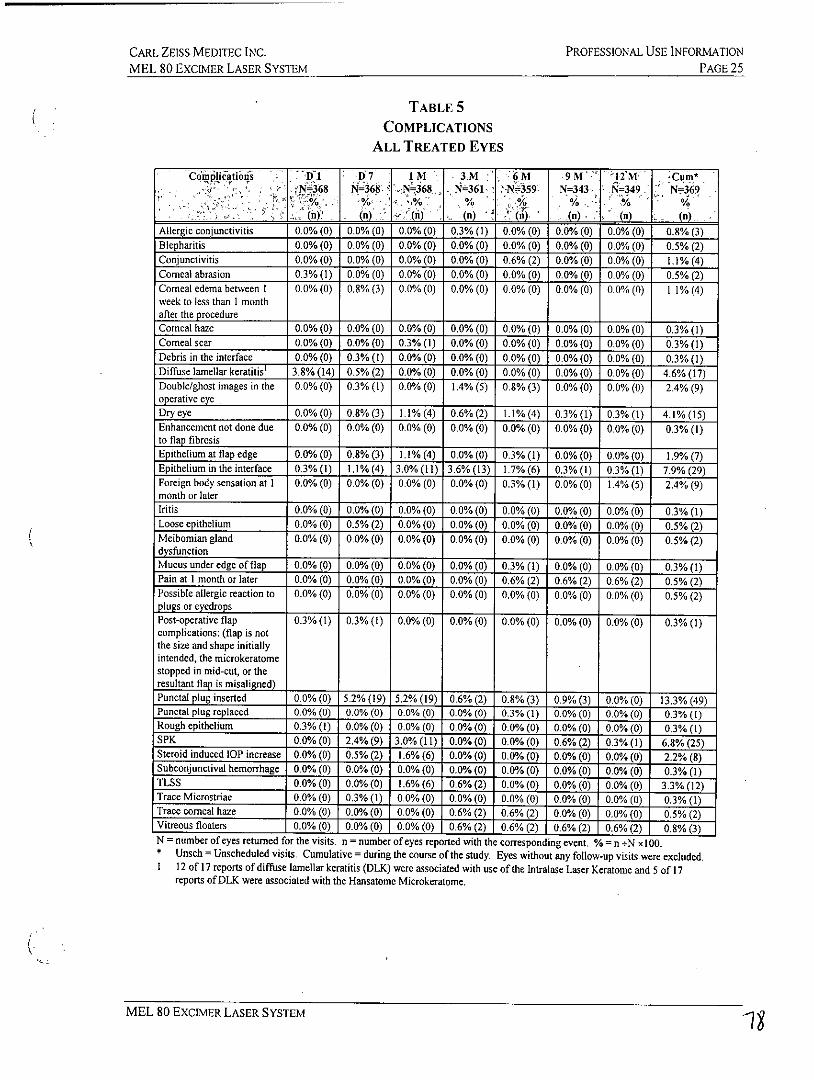
Table 5 presents a summary of all complications reported for all treated eyes. At the9-month visit, the complications included dry eye (0.3%), epithelium in the interface(0.3%), pain at I month or later (0.6%), punctal plug insertion (0.9%), superficialpunctate keratitis (SPK) (0.6%), and vitreous floaters (0.6%). Cumulative eventsreported through the course of the study at a frequency of >1% included conjunctivitis(1.1%), corneal edema between 1 week to less than I month after the procedure (1.1%),diffuse lamellar keratitis (DLK) (4.6%), ghost/double images (2.4%), dry eye (4.1%),epithelium at flap edge (1.9%), epithelium in the interface (7.9%), foreign body sensation(2.4%), punctal plug insertion (13.3%), superficial punctuate keratitis (SPK) (6.8%),steroid-induced LOP increase (2.2%), and transient light sensitivity syndrome, or TLSS(3.3%).
MEL 80 EXCIMER LASER SYSTEM
CARL ZEISS MEDITEC INC. PROFESSIONAL USE INFORMATION
MEL 80 EXCIMER LASER SYSTEM PAGE 25
TABLE 5COMPLICATIONS
ALL TREATED EYES
Cdiniciiatiods I 17 1M 3M K 6M 9M '12'M Cum*N-368 N=368 N-368 N=361 N-359 N=343 N=349 N= 369
% , % . % 9 . %r %(n), (n) (s) . ((n ) (n)(n (-)
Allergic conjunctivitis 0.0% (0) 0.0% (0) 0.0% (0) 0.3% (1) 0.0% (0) 0.0% (0) 0.0% (0) 0.8% (3)Blepharitis 0.0% (0) 0.0% (0) 0.0% (0) 0.0% (0) 0.0% (0) 0.0% (0) 0.0% (0) 0.5% (2)Conjunctivitis 0.0%(0) 0.0%(0) 0.0%(0) 0.0% (0) 0.6%(2) 0.0%(0) 0.0%(0) 1.1%(4)Comeal abrasion 0.3% (1) 0.0% (0) 0.0% (0) 0.0% (0) 0.0% (0) 0.0% (0) 0.0% (0) 0.5% (2)Comeal edema between I 0.0%(0) 0.8%(3) 0.0%(0) 0.0%(0) 0.0%(0) 0.0%(0) 0.0%(0) 1.1%(4)week to less than 1 monthafter the procedureComeal haze 0.0% (0) 0.0% (0) 0.0% (0) 0.0% (0) 0.0% (0) 0.0% (0) 0.0% (0) 0.3% (1)Cormeal scar 0.0% (0) 0.0% (0) 0.3% (1) 0.0% (0) 0.0% (0) 0.0% (0) 0.0% (0) 0.3% (1)Debris in the interface 0.0% (0) 0.3% (1) 0.0% (0) 0.0% (0) 0.0% (0) 0.0% (0) 0.0% (0) 0.3% (1)Diffuse lamellar keratitis' 3.8% (14) 0.5% (2) 0.0% (0) 0.0% (0) 0.0% (0) 0.0% (0) 0.0% (0) 4.6% (17)
Double/ghost images in the 0.0% (0) 0.3%(1) 0.0%(0) 1.4%(5) 0.8%(3) 0.0% (0) 0.0%(0) 2.4% (9)operative eyeDry eye 0.0%(0) 0.8%(3) 1.1%(4) 0.6%(2) 1.1%(4) 0.3%(1) 0.3% (1) 4.1%(15)Enhancement not done due 0.0% (0) 0.0% (0) 0.0% (0) 0.0% (0) 0.0% (0) 0.0% (0) 0.0% (0) 0.3% (1)to flap fibrosisEpithelium at flap edge 0.0% (0) 0.8% (3) 1.1% (4) 0.0% (0) 0.3% (1) 0.0% (0) 0.0% (0) 1.9% (7)Epithelium in the interface 0.3% (1) 1.1% (4) 3.0% (l1) 3.6% (13) 1.7%(6) 0.3% (1) 0.3%(1) 7.9% (29)Foreign body sensation at 1 0.0% (0) 0.0% (0) 0.0% (0) 0.0% (0) 0.3% (1) 0.0% (0) 1.4% (5) 2.4% (9)month or laterIritis 0.0% (0) 0.0% (0) 0.0% (0) 0.0% (0) 0.0% (0) 0.0% (0) 0.0% (0) 0.3% (1)Loose epithelium 0.0% (0) 0.5% (2) 0.0% (0) 0.0% (0) 0.0% (0) 0.0% (0) 0.0% (0) 0.5% (2)Meibomian gland 0.0% (0) 0.0% (0) 0.0% (0) 0.0% (0) 0.0% (0) 0.0% (0) 0.0% (0) 0.5% (2)dysfunctionMucus under edge of flap 0.0% (0) 0.0% (0) 0.0% (0) 0.0% (0) 0.3% (1) 0.0% (0) 0.0% (0) 0.3% (1)Pain at I month or later 0.0% (0) 0.0% (0) 0.0% (0) 0.0% (0) 0.6% (2) 0.6% (2) 0.6% (2) 0.5% (2)Possible allergic reaction to 0.0% (0) 0.0% (0) 0.0% (0) 0.0% (0) 0.0% (0) 0.0% (0) 0.0% (0) 0.5% (2)plugs or eyedropsPost-operative flap 0.3% (1) 0.3% (1) 0.0% (0) 0.0% (0) 0.0% (0) 0.0% (0) 0.0% (0) 0.3% (1)complications: (flap is notthe size and shape initiallyintended, the microkeratomestopped in mid-cut, or theresultant flap is misaligned)
Punctal plug inserted 0.0% (0) 5.2% (19) 5.2% (19) 0.6% (2) 0.8% (3) 0.9% (3) 0.0% (0) 13.3% (49)Punctal plug replaced 0.0% (0) 0.0% (0) 0.0% (0) 0.0% (0) 0.3% (1) 0.0% (0) 0.0% (0) 0.3% (1)Rough epithelium 0.3% (1) 0.0% (0) 0.0% (0) 0.0% (0) 0.0% (0) 0.0% (0) 0.0% (0) 0.3% (1)SPK 0.0%(0) 2.4%(9) 3.0%(11) 0.0%(0) 0.0%(0) 0.6% (2) 0.3%(1) 6.8%(25)Steroid induced IOP increase 0.0% (0) 0.5% (2) 1.6% (6) 0.0% (0) 0.0% (0) 0.0% (0) 0.0% (0) 2.2% (8)Subconjunctival hemorrhage 0.0% (0) 0.0% (0) 0.0% (0) 0.0% (0) 0.0% (0) 0.0% (0) 0.0% (0) 0.3% (1)TLSS 0.0% (0) 0.0% (0) 1.6% (6) 0.6% (2) 0.0% (0) 0.0% (0) 0.0%(0) 3.3% (12)Trace Microstriae 0.0% (0) 0.3% (I) 0.0% (0) 0.0% (0) 0.0% (0) 0.0% (0) 0.0% (0) 0.3% (1)Trace coreal haze 0.0% (0) 0.0% (0) 0.0% (0) 0.6% (2) 0.6% (2) 0.0% (0) 0.0% (0) 0.5% (2)Vitreous floaters 0.0% (0) 0.0% (0) 0.0% (0) 0.6% (2) 0.6% (2) 0.6% (2) 0.6% (2) 0.8% (3)N = number of eyes returned for the visits. n = number of eyes reported with the corresponding event. % = n +N xl00.* Unsch= Unscheduled visits. Cumulative = during the course of the study. Eyes without any follow-up visits were excluded.I 12 of 17 reports of diffuse larnellar keratitis (DLK) were associated with use of the Intralase Laser Keratome and 5 of 17
reports of DLK were associated with the Hansatome Microkeratome.
MEL 80 EXCIMER LASER SYSTEM 1
CARL ZEISS MEDITEC INC. PROFESSIONAL USE INFORMATIONMEL 80 EXCIMER LASER SYSTEM PAGE 26
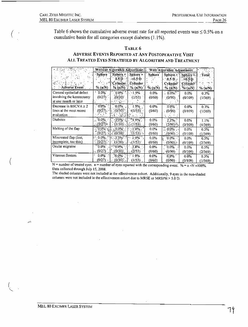
Table 6 shows the cumulative adverse event rate for all reported events was < 0.5% on acumulative basis for all categories except diabetes (1.1%).
TABLE 6ADVERSE EVENTs REPORTED AT ANY POSTOPERATIVE VISIT
ALL TREATED EYES STRATIFIED BY ALGORITHM AND TREATMENT
.Wikth hWlIgoitadj~siiit ~ 'Wiih'Aljo iifi<Ad iithihti -
9fr rb f? phiee S El re' S'[Cee Totjj.0.5SD >05 D - 0.5D 11 O>.5 D
- e( Cylie ylid er Cylidide > Clhi de -
AvereEven/ % (nN) A (n/N) % (n/N) % (buN) IO(n/N) % (n/N)Corneal epithelial defect 0 Oh 70Oho 1.9% 0.0% : ooW 0.0% 0.3%involving the keratectomy (0/27) .(0/30) (1/53) (0/60) (0/90) (0/109) (1/369)at one month or later .
Decrease in BSCVA 2 .'0%7.0/ 0 h0 1 9% 0.0% 0:0% 0 0% 0.3%lines at the most recent .(0/27 (0/30) (1/53) (0/60) (0/90) (0/109) (1/369)evaluationDiabetes .0/X 3 30 19% 0.0% 2:2% 0.0% 1.1%
(0/27) (130) (153) (0/60) (2/90) (0/109) (4/369)Melting of the flap 'O0% 00% 1 9%" 0.0% 0'0% 0.0% 0.3%
(0/27) ( ) (153) (0/60) (0/90) (0/109) (1/369)Miscreated flap (lost ' 0( O 3 3% 3W 9%' 0.0% 0:0% 7 00% 0.5%incomplete, too thin) (0/27) (1/30) (1/53) (0/60) (0/90) (0/109) 2/369)Ocular migraine 0.0O/0 0f0% 38% 0.0% 0:00 0/Oh 0.5%
(0/27) .(0/30) (2/53) (0/60) (0/90) (0/109) (2/369)Vitreous floaters 0 Oh 0 .0% 1.9% 0.0% 0:0% 0.0% 0.3%
(0/27) 0) (1/53) (0/60) (0/90) (0/109) 1/369N = number of treated eyes. n = number of eyes reported with the corresponding event. %= n -N xl00%.Data collected through July 15, 2008.The shaded columns were not included in the effectiveness cohort. Additionally, 9 eyes in the non-shadedcolumns were not included in the effectiveness cohort due to MRSE or MRSPH > 5.0 D.
MEL 80 EXCIMER LASER SYSTEM
CARL ZEISS MEDITEC INC. PROFESSIONAL USE INFORMATIONMEL 80 EXCIMER LASER SYSTEM PAGE 27
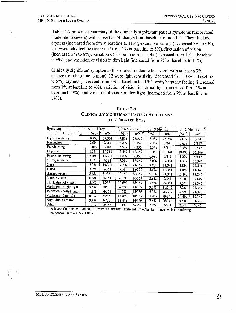
Table 7.A presents a summary of the clinically significant patient symptoms (those ratedmoderate to severe) with at least a 3% change from baseline to month 9. These includedryness (increased from 5% at baseline to 11%), excessive tearing (decreased 3% to 0%),gritty/scratchy feeling (increased from 1% at baseline to 5%), fluctuation of vision(increased 5% to 8%), variation of vision in normal light (increased from 1% at baselineto 6%), and variation of vision in dim light (increased from 7% at baseline to 11%).
Clinically significant symptoms (those rated moderate to severe) with at least a 3%change from baseline to month 12 were light sensitivity (decreased from 10% at baselineto 5%), dryness (increased from 5% at baseline to 10%), gritty/scratchy feeling (increasedfrom 1% at baseline to 4%), variation of vision in normal light (increased from 1% atbaseline to 7%), and variation of vision in dim light (increased from 7% at baseline to14%).
TABLE 7.ACLINICALLY SIGNIFICANT PATIENT SYMPTOMS*
ALL TREATED EYES
Symptom . 1 PMeo 6Months 9Monthi . 2IMonths- % - n/N %. %SI n/N /N %/ n/N '
Light sensitivity 10.2% 37/361 7.8% 28/357 8.2% 28/341 4.6% 16/347Headaches 2.5% 9/361 2.2% 8/357 2.3% 8/341 0.6% 2/347Pain/burning 0.8% 3/361 2.5% 9/356 2.3% 8/341 0.3% 1/347Dryness 5.3% 19/361 13.4% 48/357 11.4% 39/341 10.4% 36/346Excessive tearing 3.0% 11/361 0.8% 3/357 0.0% 0/340 1.2% 4/347Gritty, scratchy 1.1% 4/361 5.0% 18/357 5.0% 17/341 4.3% 15/347Glare 5.3% 19/361 5.9% 21/357 3.8% 13/341 3.8% 13/346Halos 2.2% 8/361 5.0% 18/357 3.5% 12/341 4.0% 14/347Blurred vision 8.6% 31/361 10.1% 36/357 9.7% 33/341 10.4% 36/347Double vision 0.6% 2/361 4.5% 16/357 2.6% 9/341 2.3% 8/346Fluctuation of vision 5.0% 18/361 10.6% 38/357 7.9% 27/341 7.5% 26/347Variation - bright light 5.5% 20/361 6.4% 23/357 3.2% 11/341 7.2% 25/347Variation - normal light 1.1% 4/361 4.2% 15/356 5.9% 20/339 6.6% 23/347Variation - dim light 6.9% 25/361 13.4% 48/357 11,4% 50/347Night driving vision 9.4% 34/361 12.4% 44/356 7.6% 26/341 9.5% 33/347Other 1.4% 5/361 1.4% 5/356 2.1% 7/341 2.0% 7/347* A level of moderate, marked, or severe is clinically significant. N = Number of eyes with non-missing
responses. % = n + N x 100%.
MEL 80 EXCIMER LASER SYSTEM 6
CARL ZEISS MEDITEC INC. PROFESSIONAL USE INFORMATIONMEL 80 EXCIMER LASER SYSTEM PAGE 28
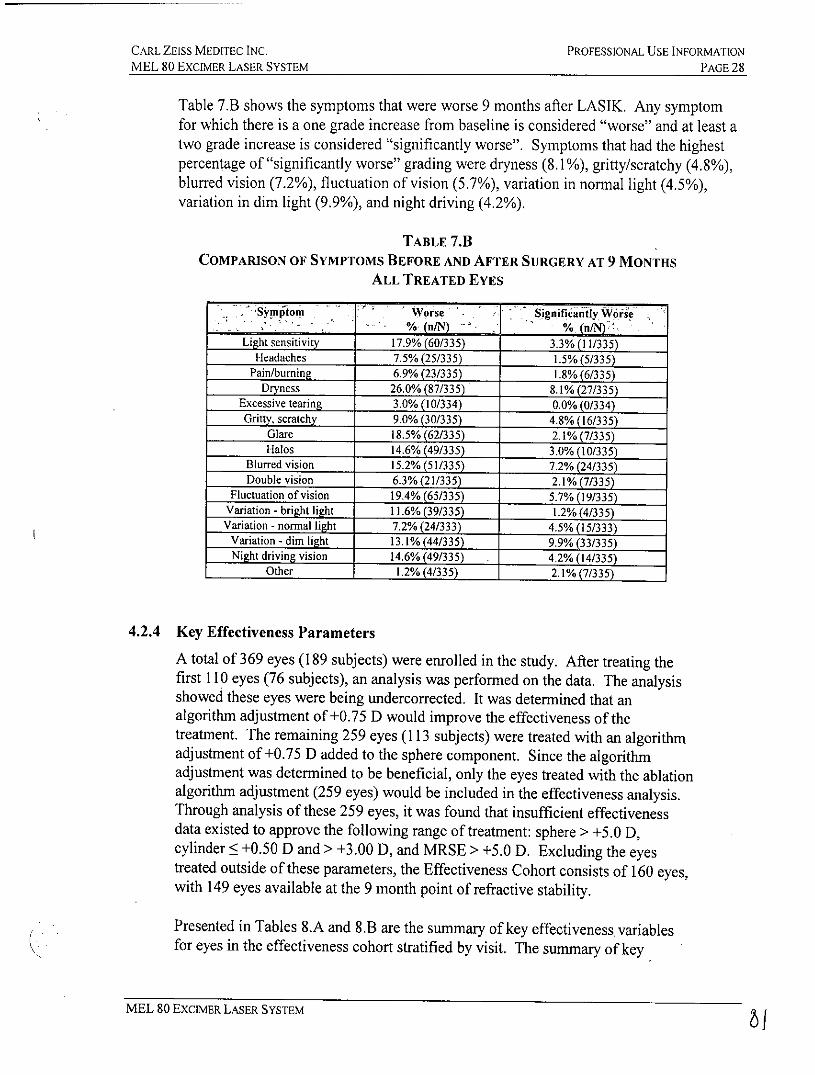
Table 7.B shows the symptoms that were worse 9 months after LASIK. Any symptomfor which there is a one grade increase from baseline is considered "worse" and at least atwo grade increase is considered "significantly worse". Symptoms that had the highestpercentage of "significantly worse" grading were dryness (8.1%), gritty/scratchy (4.8%),blurred vision (7.2%), fluctuation of vision (5.7%), variation in normal light (4.5%),variation in dim light (9.9%), and night driving (4.2%).
TABLE 7.BCOMPARISON OF SYMPTOMS BEFORE AND AFTER SURGERY AT 9 MONTHS
ALL TREATED EYES
Synitom Worse Significaty Wore- - .% (n/N) %. (n/N)
Light sensitivity 17.9% (60/335) 3.3% (11/335)Headaches 7.5% (25/335) 1.5%(5/335)
Pain/burning 6.9% (23/335) 1.8% (6/335)Dryness 26.0% (87/335) 8.1% (27/335)
Excessive tearing 3.0% (10/334) 0.0% (0/334)Gritty, scratchy 9.0% (30/335) 4.8% (16/335)
Glare 18.5% (62/335) 2.1% (7/335)Halos 14.6% (49/335) 3.0% (10/335)
Blurred vision 15.2% (51/335) 7.2% (24/335)Double vision 6.3% (21/335) 2.1% (7/335)
Fluctuation of vision 19.4% (65/335) 5.7% (19/335)Variation - bright light I 1.6% (39/335) 1.2% (4/335)Variation - normal light 7.2% (24/333) 4.5% (15/333)
Variation - dim light 13.1% (44/335) 9.9%(33/335)Night driving vision 14.6% (49/335) 4.2% (14/335)
Other 1.2% (4/335) 2.1% (7/335)
4.2.4 Key Effectiveness Parameters
A total of 369 eyes (189 subjects) were enrolled in the study. After treating thefirst 110 eyes (76 subjects), an analysis was performed on the data. The analysisshowed these eyes were being undercorrected. It was determined that analgorithm adjustment of +0.75 D would improve the effectiveness of thetreatment. The remaining 259 eyes (113 subjects) were treated with an algorithmadjustment of +0.75 D added to the sphere component. Since the algorithmadjustment was determined to be beneficial, only the eyes treated with the ablationalgorithm adjustment (259 eyes) would be included in the effectiveness analysis.Through analysis of these 259 eyes, it was found that insufficient effectivenessdata existed to approve the following range of treatment: sphere > +5.0 D,cylinder < +0.50 D and > +3.00 D, and MRSE > +5.0 D. Excluding the eyestreated outside of these parameters, the Effectiveness Cohort consists of 160 eyes,with 149 eyes available at the 9 month point of refractive stability.
Presented in Tables 8.A and 8.B are the summary of key effectiveness variablesfor eyes in the effectiveness cohort stratified by visit. The summary of key
MEL 80 EXCIMER LASER SYSTEM 7
CARL ZEISS MEDITEC INC. PROFESSIONAL USE INFORMATIONMEL 80 EXCIMER LASER SYSTEM PAGE 29
effectiveness variables at 9 months for eyes in the effectiveness cohort stratifiedby treatment is also shown.
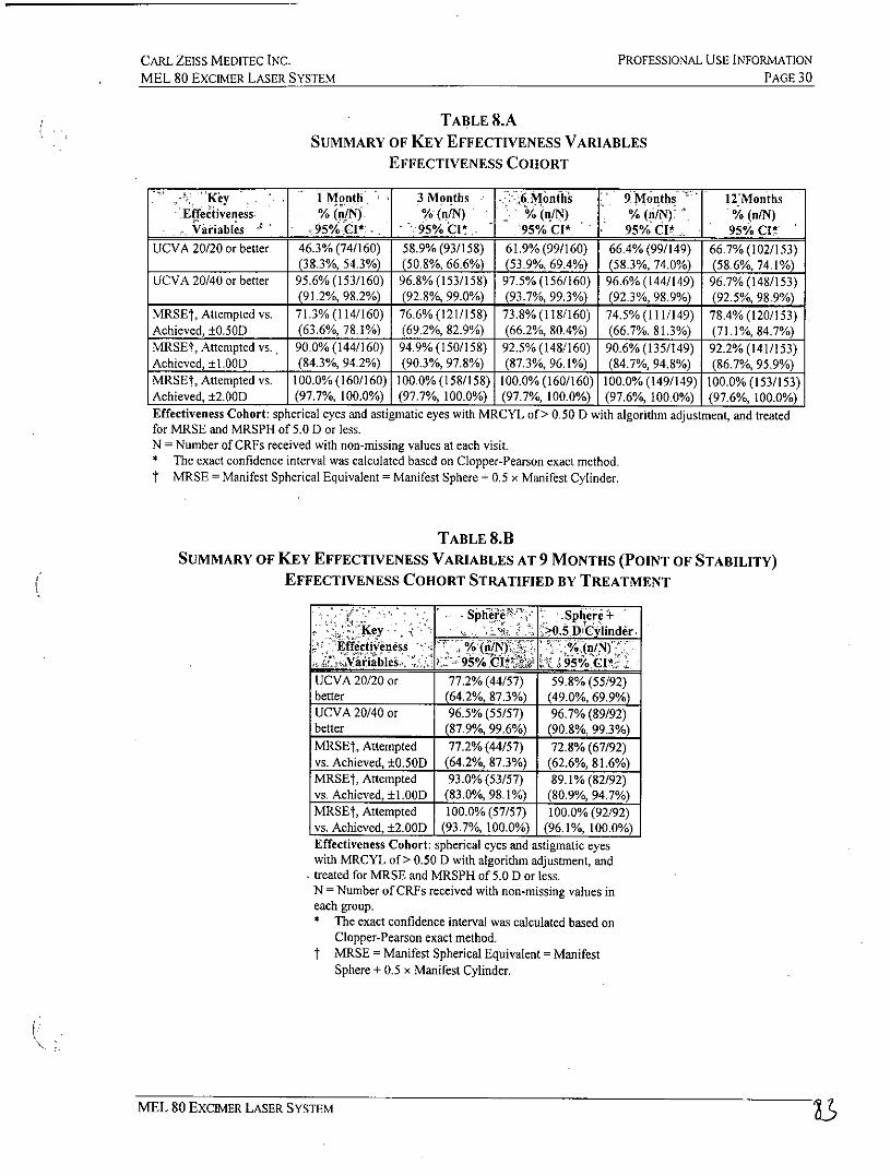
As shown in Table 8.A for the effectiveness cohort of eyes, the three primaryoutcomes for percent of eyes with 20/40 or better uncorrected visual acuity andpercent of eyes within ± 0.50 D and ± 1.00 D of attempted correction exceedtarget values established in the study protocol consistent with FDA guidance. At9 months, 66.4% of eyes had UCVA 20/20 or better, and 96.6% of eyes hadUCVA 20/40 or better. At 12 months, 66.7% of eyes had UCVA 20/20 or better,and 96.7% of eyes had UCVA 20/40 or better. In Table 8.B, the sphere only andthe sphere + > 0.5 D cylinder treatment groups showed similar results to the eyesin Table 7.A. That is, at 9 months, for the sphere only treated eyes, 77.2% of eyeshad UCVA 20/20 or better, and 96.5% of eyes had UCVA 20/40 or better. At 9months, for the sphere + >0.5 D cylinder eyes, 59.8% of eyes had UCVA 20/20 orbetter, and 96.7% of eyes had UCVA 20/40 or better.
MEL 80 EXCIMER LASER SYSTEM 62-
CARL ZEISS MEDITEC INC. PROFESSIONAL USE INFORMATIONMEL 80 EXCIMER LASER SYSTEM PAGE 30
TABLE 8.ASUMMARY OF KEY EFFECTIVENESS VARIABLES
EFFECTIVENESS COHORT
. Key . iMonth 3 Months 6 Months 9 Months - 12MonthsEffectiveness % (n/N) % (nlN) % (n/N) % (n/N). % (n/N)
riables95% CI*. ClI 95% CI* 95% CI* 95% CIUCVA 20/20 or better 46.3% (74/160) 58.9% (93/158) 61.9% (99/160) 66.4% (99/149) 66.7% (102/153)
(38.3%, 54.3%) (50.8%, 66.6%) (53.9%, 69.4%) (58.3%, 74.0%) (58.6%, 74.1%)UCVA 20/40 or better 95.6% (153/160) 96.8% (153/158) 97.5% (156/160) 96.6% (144/149) 96.7% (148/153)
(91.2%, 98.2%) (92.8%, 99.0%) (93.7%, 99.3%) (92.3%, 98.9%) (92.5%, 98.9%)MRSEt, Attempted vs. 71.3% (114/160) 76.6% (121/158) 73.8% (118/160) 74.5% (111/149) 78.4% (120/153)Achieved, ±0.50D (63.6%, 78.1%) (69.2%, 82.9%) (66.2%, 80.4%) (66.7%, 81.3%) (71.1%, 84.7%)MRSEt, Attempted vs., 90.0% (144/160) 94.9% (150/158) 92.5% (148/160) 90.6% (135/149) 92.2% (141/153)Achieved, ±1.00D (84.3%, 94.2%) (90.3%, 97.8%) (87.3%, 96.1%) (84.7%, 94.8%) (86.7%, 95.9%)MRSEt, Attempted vs. 100.0% (160/160) 100.0% (158/158) 100.0% (160/160) 100.0% (149/149) 100.0% (153/153)Achieved, ±2.OOD (97.7%, 100.0%) (97.7%, 100.0%) (97.7%, 100.0%) (97.6%, 100.0%) (97.6%, 100.0%)Effectiveness Cohort: spherical eyes and astigmatic eyes with MRCYL of> 0.50 D with algorithm adjustment, and treatedfor MRSE and MRSPH of 5.0 D or less.N = Number of CRFs received with non-missing values at each visit.* The exact confidence interval was calculated based on Clopper-Pearson exact method.t MRSE = Manifest Spherical Equivalent = Manifest Sphere + 0.5 x Manifest Cylinder.
TABLE 8.1SUMMARY OF KEY EFFECTIVENESS VARIABLES AT 9 MONTHS (POINT OF STABILITY)
EFFECTIVENESS COHORT STRATIFIED BY TREATMENT
Key , Sjhii2:' .Sphere+Key. . 0.5 D ulinder.
Effectiveness - 0/. (n/N)4i<",Vaiables,. 1195% I 95% CI* * :
UCVA 20/20 or 77.2% (44/57) 59.8% (55/92)better (64.2%, 87.3%) (49.0%, 69.9%)UCVA 20/40 or 96.5% (55/57) 96.7% (89/92)better (87.9%, 99.6%) (90.8%, 99.3%)MRSE , Attempted 77.2% (44/57) 72.8% (67/92)vs. Achieved, ±0.50D (64.2%, 87.3%) (62.6%, 81.6%)MRSEt, Attempted 93.0% (53/57) 89.1% (82/92)vs. Achieved, ±1.00D (83.0%, 98.1%) (80.9%, 94.7%)MRSEt, Attempted 100.0% (57/57) 100.0% (92/92)vs. Achieved, ±2.00D (93.7%, 100.0%) (96.1%, 100.0%)Effectiveness Cohort: spherical eyes and astigmatic eyeswith MRCYL of > 0.50 D with algorithm adjustment, andtreated for MRSE and MRSPH of 5.0 D or less.N = Number of CRFs received with non-missing values ineach group.* The exact confidence interval was calculated based on
Clopper-Pearson exact method.t MRSE = Manifest Spherical Equivalent = Manifest
Sphere + 0.5 x Manifest Cylinder.
MEL 80 EXCIMER LASER SYSTEM
CARL ZEISS MEDITEC INC. PROFESSIONAL USE INFORMATIONMEL 80 EXCIMER LASER SYSTEM PAGE 31
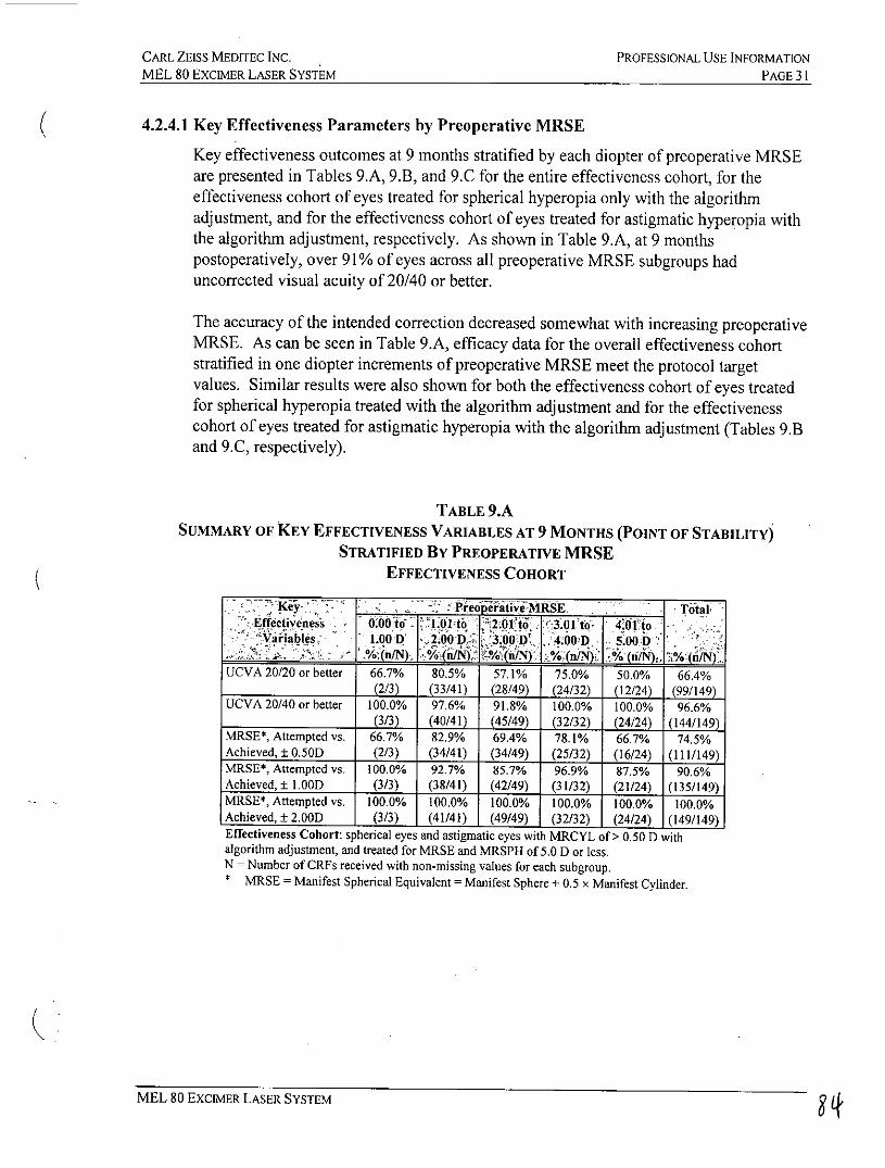
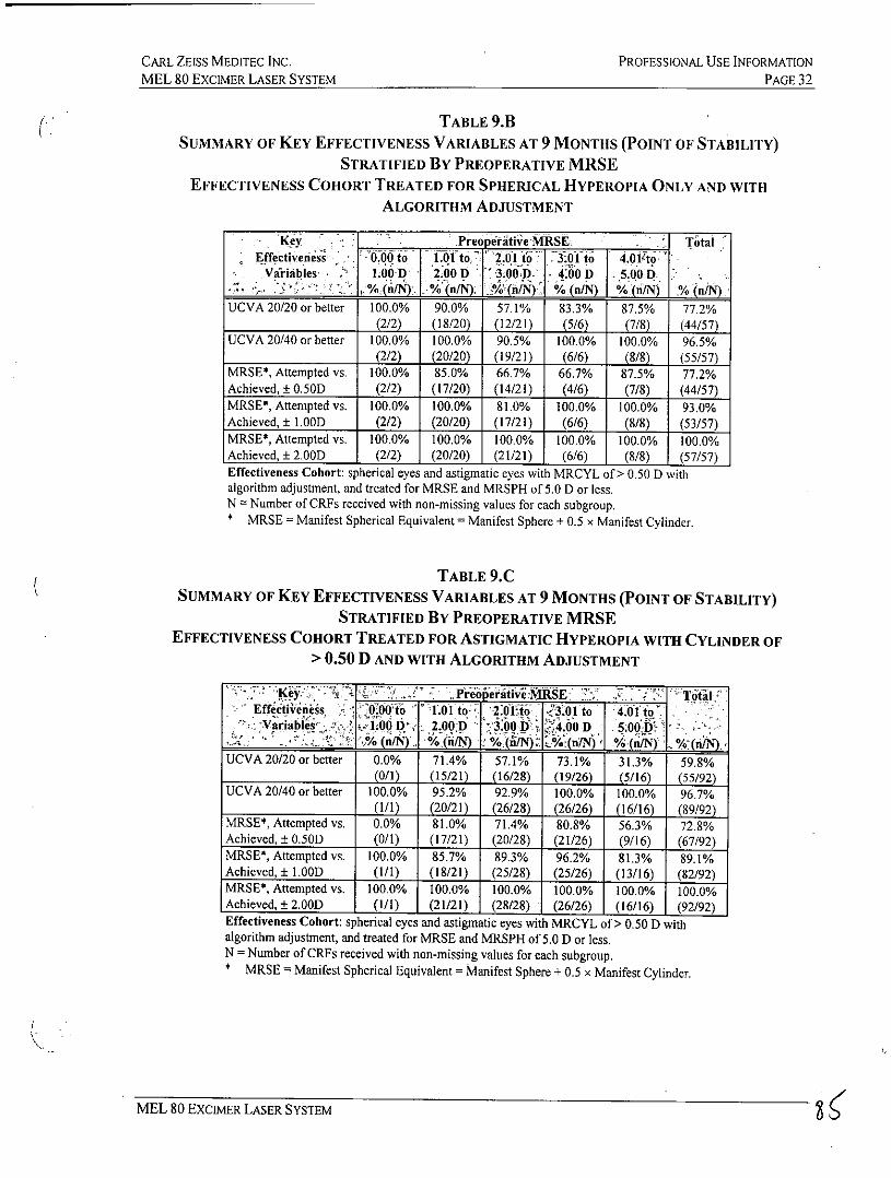
4.2.4.1 Key Effectiveness Parameters by Preoperative MRSE
Key effectiveness outcomes at 9 months stratified by each diopter of preoperative MRSEare presented in Tables 9.A, 9.B, and 9.C for the entire effectiveness cohort, for theeffectiveness cohort of eyes treated for spherical hyperopia only with the algorithmadjustment, and for the effectiveness cohort of eyes treated for astigmatic hyperopia withthe algorithm adjustment, respectively. As shown in Table 9.A, at 9 monthspostoperatively, over 91% of eyes across all preoperative MRSE subgroups haduncorrected visual acuity of 20/40 or better.
The accuracy of the intended correction decreased somewhat with increasing preoperativeMRSE. As can be seen in Table 9.A, efficacy data for the overall effectiveness cohortstratified in one diopter increments of preoperative MRSE meet the protocol targetvalues. Similar results were also shown for both the effectiveness cohort of eyes treatedfor spherical hyperopia treated with the algorithm adjustment and for the effectivenesscohort of eyes treated for astigmatic hyperopia with the algorithm adjustment (Tables 9.Band 9.C, respectively).
TABLE 9.ASUMMARY OF KEY EFFECTIVENESS VARIABLES AT 9 MONTHS (POINT OF STABILITY)
STRATIFIED BY PREOPERATIVE MRSEEFFECTIVENESS COHORT
K-y PeoieitiveMRSE. _ TotalEffectiveness 0 todi0126tb .11 [3.01 to Of to
-iables 1.00 D y2;00D 3.00D' 40D i500D.:Kh . %~n/N. j ~ (n/NODj %.(n/N n
UCVA 20/20 or better 66.7% 80.5% 57.1% 75.0% 50.0% 66.4%(2/3) (33/41) (28/49) (24/32) (12/24) (99/149)
UCVA 20/40 or better 100.0% 97.6% 91.8% 100.0% 100.0% 96.6%(3/3) (40/41) (45/49) (32/32) (24/24) (144/149)
MRSE*, Attempted vs. 66.7% 82.9% 69.4% 78.1% 66.7% 74.5%Achieved, ± 0.50D (2/3) (34/41) (34/49) (25/32) (16/24) (111/149)MRSE*, Attempted vs. 100.0% 92.7% 85.7% 96.9% 87.5% 90.6%Achieved, ± 1.00D (3/3) (38/41) (42/49) (31/32) (21/24) (135/149)MRSE*, Attempted vs. 100.0% 100.0% 100.0% 100.0% 100.0% 100.0%Achieved, ± 2.OOD (3/3) (41/41) (49/49) (32/32) (24/24) (149/149)Effectiveness Cohort: spherical eyes and astigmatic eyes with MRCYL of > 0.50 D withalgorithm adjustment, and treated for MRSE and MRSPH of 5.0 D or less.N = Number of CRFs received with non-missing values for each subgroup.
MRSE = Manifest Spherical Equivalent = Manifest Sphere + 0.5 x Manifest Cylinder.
MEL 80 EXCIMER LASER SYSTEM 3
CARL ZEISS MEDITEC INC. PROFESSIONAL USE INFORMATIONMEL 80 EXCIMER LASER SYSTEM PAGE 32
TABLE 9.BSUMMARY OF KEY EFFECTIVENESS VARIABLES AT 9 MONTHS (POINT OF STABILITY)
STRATIFIED By PREOPERATIVE MRSEEFFECTIVENESS COHORT TREATED FOR SPHERICAL HYPEROPIA ONLY AND WITH
ALGORITHM ADJUSTMENT
Key Preop eitive MRSE. . Total
Effectiveness 0.0I to I01to 01th 3:1 o 4to toVariables 1O00D 2.00 D 3.00.D- 4:00 D 5.00 D
% IM %n/N) J (nN), %i (n/N) % (niiN) _% in/N)UCVA 20/20 or better 100.0% 90.0% 57.1% 83.3% 87.5% 77.2%
(2/2) (18/20) (12/21) (5/6) (7/8) (44/57)UCVA 20/40 or better 100.0% 100.0% 90.5% 100.0% 100.0% 96.5%
(2/2) (20/20) (19/21) (6/6) (8/8) (55/57)MRSE*, Attempted vs. 100.0% 85.0% 66.7% 66.7% 87.5% 77.2%Achieved, ± 0.50D (2/2) (17/20) (14/21) (4/6) (7/8) (44/57)MRSE*, Attempted vs. 100.0% 100.0% 81.0% 100.0% 100.0% 93.0%Achieved, ± 1.00D (2/2) (20/20) (17/21) (6/6) (8/8) (53/57)MRSE*, Attempted vs. 100.0% 100.0% 100.0% 100.0% 100.0% 100.0%Achieved, ± 2.OOD (2/2) (20/20) (21/21) (6/6) (8/8) (57/57)Effectiveness Cohort: spherical eyes and astigmatic eyes with MRCYL of > 0.50 D withalgorithm adjustment, and treated for MRSE and MRSPH of 5.0 D or less.N = Number of CRFs received with non-missing values for each subgroup.* MRSE = Manifest Spherical Equivalent = Manifest Sphere + 0.5 x Manifest Cylinder.
TABLE 9.CSUMMARY OF KEY EFFECTIVENESS VARIABLES AT 9 MONTHS (POINT OF STABILITY)
STRATIFIED BY PREOPERATIVE MRSEEFFECTIVENESS COHORT TREATED FOR ASTIGMATIC HYPEROPIA WITH CYLINDER OF
> 0.50 D AND WITH ALGORITHM ADJUSTMENT
Key-A. k~~. Preoperativ&~Se: ?t E otlEf c tiveness 000% 1.01 to 2ot1,io - .01 to 14.1 toKVariabl& 2 00 D, 2.09:D 3:0f4. 4h0 D 5.00
......... ... (n/N) % (nN) %(ii/N (n/N) (a) _o (n/N).UCVA 20/20 or better 0.0% 71.4% 57.1% 73.1% 31.3% 59.8%
(0/1) (15/21) (16/28) (19/26) (5/16) (55/92)UCVA 20/40 or better 100.0% 95.2% 92.9% 100.0% 100.0% 96.7%
(1/1) (20/21) (26/28) (26/26) (16/16) (89/92)MRSE*, Attempted vs. 0.0% 81.0% 71.4% 80.8% 56.3% 72.8%Achieved, ± 0.50D (0/1) (17/21) (20/28) (21/26) (9/16) (67/92)MRSE*, Attempted vs. 100.0% 85.7% 89.3% 96.2% 81.3% 89.1%Achieved, ± 1.00D (1/1) (18/21) (25/28) (25/26) (13/16) (82/92)MRSE*, Attempted vs. 100.0% 100.0% 100.0% 100.0% 100.0% 100.0%Achieved,± 2.0D (1/1) (21/21) (28/28) (26/26) (16/16) (92/92)Effectiveness Cohort: spherical eyes and astigmatic eyes with MRCYL of > 0.50 D withalgorithm adjustment, and treated for MRSE and MRSPH of 5.0 D or less.N = Number of CRFs received with non-missing values for each subgroup.* MRSE = Manifest Spherical Equivalent = Manifest Sphere + 0.5 x Manifest Cylinder.
MEL 80 EXCIMER LASER SYSTEM
CARL ZEISS MEDITEC INC. PROFESSIONAL USE INFORMATION
MEL 80 EXCIMER LASER SYSTEM PAGE 33
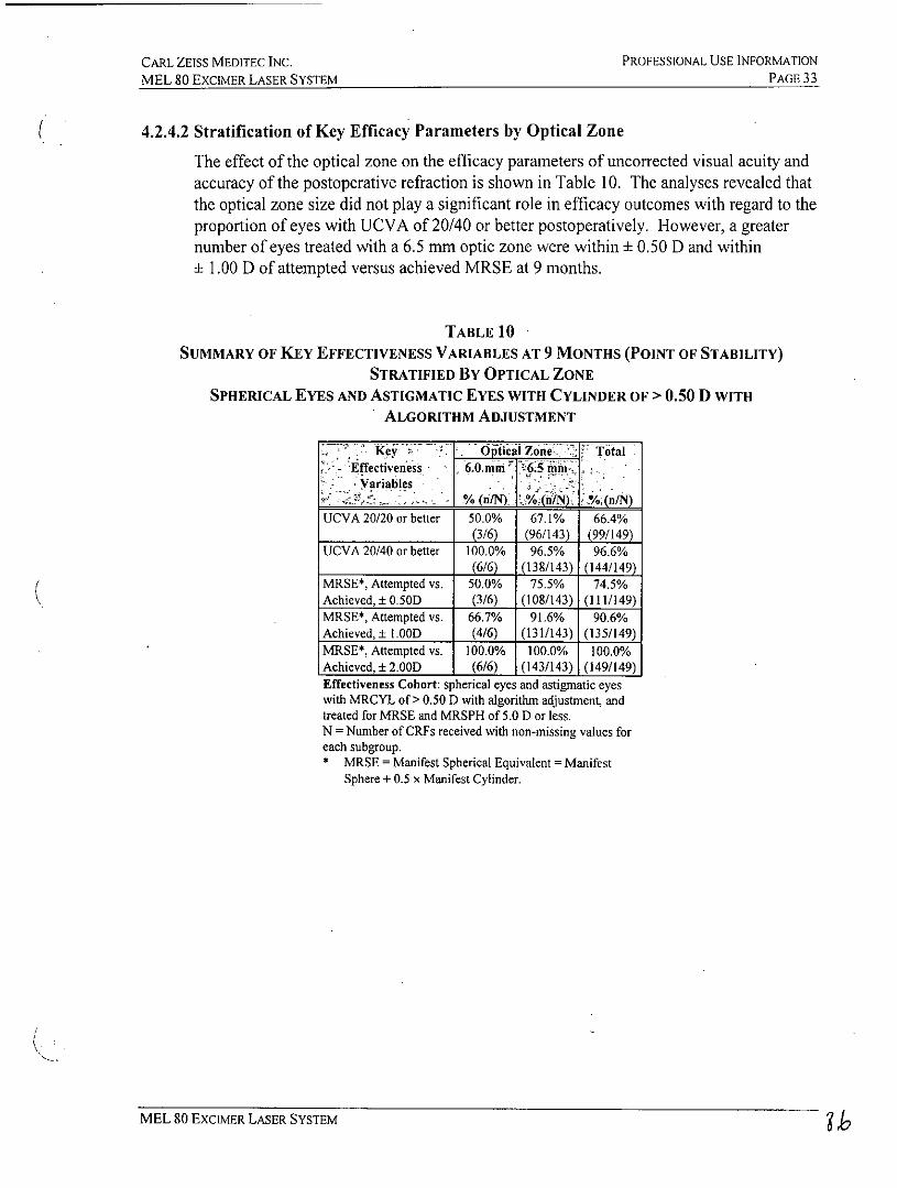
4.2.4.2 Stratification of Key Efficacy Parameters by Optical Zone
The effect of the optical zone on the efficacy parameters of uncorrected visual acuity andaccuracy of the postoperative refraction is shown in Table 10. The analyses revealed thatthe optical zone size did not play a significant role in efficacy outcomes with regard to theproportion of eyes with UCVA of 20/40 or better postoperatively. However, a greaternumber of eyes treated with a 6.5 mm optic zone were within ± 0.50 D and within
1.00 D of attempted versus achieved MRSE at 9 months.
TABLE 10SUMMARY OF KEY EFFECTIVENESS VARIABLES AT 9 MONTHS (POINT OF STABILITY)
STRATIFIED By OPTICAL ZONESPHERICAL EYES AND ASTIGMATIC EYES WITH CYLINDER OF > 0.50 D WITH
ALGORITHM ADJUSTMENT
Key - OpicaiZone. Total- Effectiveness 6.0.nim 56
Variables.%3i/N). %./N) %(n/)
UCVA 20/20 or better 50.0% 67.1% 66.4%(3/6) (96/143) (99/149)
UCVA 20/40 or better 100.0% 96.5% 96.6%(6/6) (138/143) (144/149)
MRSE*, Attempted vs. 50.0% 75.5% 74.5%Achieved, ± 0.50D (3/6) (108/143) (111/149)MRSE*, Attempted vs. 66.7% 91.6% 90.6%Achieved, ± 1.00D (4/6) (131/143) (135/149)MRSE*, Attempted vs. 100.0% 100.0% 100.0%Achieved, ± 2.00D (6/6) (143/143) (149/149)Effectiveness Cohort: spherical eyes and astigmatic eyeswith MRCYL of > 0.50 D with algorithm adjustment, andtreated for MRSE and MRSPH of 5.0 D or less.N = Number of CRFs received with non-missing values foreach subgroup.* MRSE = Manifest Spherical Equivalent = Manifest
Sphere + 0.5 x Manifest Cylinder.
MEL 80 EXCIMER LASER SYSTEM
CARL ZEISS MEDITEC INC. PROFESSIONAL USE INFORMATIONMEL 80 EXCIMER LASER SYSTEM PAGE 34
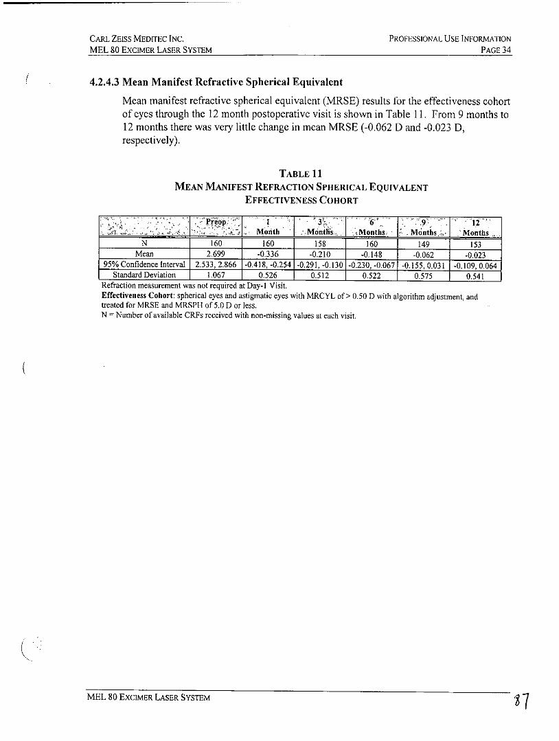
4.2.4.3 Mean Manifest Refractive Spherical Equivalent
Mean manifest refractive spherical equivalent (MRSE) results for the effectiveness cohortof eyes through the 12 month postoperative visit is shown in Table 11. From 9 months to12 months there was very little change in mean MRSE (-0.062 D and -0.023 D,respectively).
TABLE 11MEAN MANIFEST REFRACTION SPHERICAL EQUIVALENT
EFFECTIVENESS COHORT
* - i1eo, 16 -12,,
- Month .Months , I Months. Montho,;h . Months
N 160 160 158 160 149 153Mean 2.699 -0,336 -0.210 -0.148 -0.062 -0.023
95% Confidence Interval 2.533, 2.866 1-0.418, -0.254 -0.291, -0.130 1-0.230,-0.067 -0.155, 0.031 -0.109, 0.064Standard Deviation 1.067 0.526 0.512 0.522 0.575 0.541
Refraction measurement was not required at Day-I Visit.Effectiveness Cohort: spherical eyes and astigmatic eyes with MRCYL of> 0.50 D with algorithm adjustment, andtreated for MRSE and MRSPH of 5.0 D or less.N = Number of available CRFs received with non-missing values at each visit.
MEL 80 ExcIMER LASER SYSTEM 7
CARL ZEISS MEDITEC INC. PROFESSIONAL USE INFORMATIONMEL 80 EXCIMER LASER SYSTEM PAGE 35
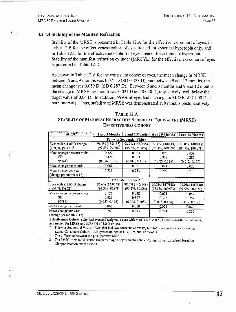
4.2.4.4 Stability of the Manifest Refraction
Stability of the MRSE is presented in Table 12.A for the effectiveness cohort of eyes, inTable 12.B for the effectiveness cohort of eyes treated for spherical hyperopia only, andin Table 12.C for the effectiveness cohort of eyes treated for astigmatic hyperopia.Stability of the manifest refraction cylinder (MRCYL) for the effectiveness cohort of eyesis presented in Table 12.D.
As shown in Table 12.A for the consistent cohort of eyes, the mean change in MRSEbetween 6 and 9 months was 0.071 D (SD 0.328 D), and between 9 and 12 months, themean change was 0.059 D, (SD 0.287 D). Between 6 and 9 months and 9 and 12 months,the change in MRSE per month was 0.024 D and 0.020 D, respectively, well below thetarget value of 0.04 D. In addition, >99% of eyes had a change in MRSE of ; 1.00 D atboth intervals. Thus, stability of MRSE was demonstrated at 9 months postoperatively.
TABLE 12.ASTABILITY OF MANIFEST REFRACTION SPHERICAL EQUIVALENT (MRSE)
EFFECTIVENESS COHORT
MRSE'' :1 1 and 3 Months 3 ahd,6 Montlis 6 and 9Mokhsl Tand 12MointhsPairwise Sequential Visits*
Eyes with 1.00 D change 96.8% (153/158) 98.7% (156/158) 99.3% (148/149) 100.0% (148/148)(n/N, %, [% CI]) 2 (92.8%, 99.0%) (95.5%, 99.8%) (96.3%, 100.0%) (97.5%, 100.0%)Mean change between visits 0.122 0.063 0.073 0.059
SD 0.421 0.303 0.328 0.28795% CI (0.056, 0.188) (0.016,0.111) (0.020, 0.126) (0.012, 0.106)
Mean change per month 0.061 0.021 0.024 0.020Mean change per year 0.731 0.253 0.292 0.236(change per month x 12)
Consistent CohortEyes with 1.00 D change 96.6%(143/148) 98.6%(146/148) 99.3%(147/148) 100.0% (148/148)(n/N, 0/o, [% Cl) 2 (92.3%, 98.9%) (95.2%, 99.8%) (96.3%, 100.0%) (97.5%, 100.0%)Mean change between visits 0.127 0.058 0.071 0.059
SD 0.426 0.307 0.328 0.28795% CI (0.057, 0.196) (0.008, 0.108) (0.018, 0.124) (0.012, 0.106)
Mean change per month 0.063 0.019 0.024 0.020Mean change per year 0.760 0.233 0.284 0.236(change per month x 12) 1
Effectiveness Cohort: spherical eyes and astigmatic eyes with MRCYL of > 0.50 D with algorithm adjustment,and treated for MRSE and MRSPH of 5.0 D or less.* Pairwise Sequential Visits = Eyes that had two consecutive exams, but not necessarily every follow-up
exam. Consistent Cohort = All eyes examined at 1, 3, 6, 9, and 12 months.I The difference between the postoperative MRSE.2 The 95%Cl = 95% C1 around the percentage of eyes meeting the criterion. It was calculated based on
Clopper-Pearson exact method.
MEL 80 EXCIMER LASER SYSTEM
CARL ZEISS MEDITEC INC. PROFESSIONAL USE INFORMATIONMEL 80 EXCIMER LASER SYSTEM PAGE 36
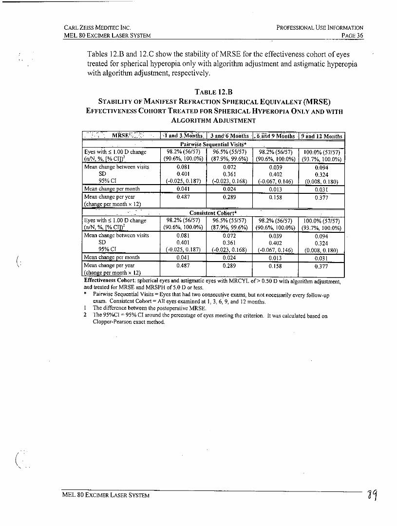
Tables 12.3 and 12.C show the stability of MRSE for the effectiveness cohort of eyestreated for spherical hyperopia only with algorithm adjustment and astigmatic hyperopiawith algorithm adjustment, respectively.
TABLE 12.BSTABILITY OF MANIFEST REFRACTION SPHERICAL EQUIVALENT (MRSE)
EFFECTIVENESS COHORT TREATED FOR SPHERICAL HYPEROPIA ONLY AND WITHALGORITHM ADJUSTMENT
MMRSE't> *I and 3.Mnths, 3 and6 Months 6'and 9 Months 9 and 12 MonthsPairwise Sequential Visits*
Eyes with 1.00 D change 98.2% (56/57) 96.5% (55/57) 98.2% (56/57) 100.0%(57/57)(n/N, %, [% CI]) 2 (90.6%, 100.0%) (87.9%, 99.6%) (90.6%, 100.0%) (93.7%, 100.0%)Mean change between visits 0.081 0.072 0.039 0.094
SD 0.401 0.361 0.402 0.32495% C1 (-0.025, 0.187) (-0.023, 0.168) (-0.067, 0.146) (0.008, 0.180)
Mean change per month 0.041 0.024 0.013 0.031Mean change per year 0.487 0.289 0.158 0.377(change per month x 12)
Consistent Coliort*
Eyes with 1.00 D change 98.2% (56/57) 96.5% (55/57) 98.2% (56/57) 100.0% (57/57)(n/N, %, [% Cl]) 2 (90.6%, 100.0%) (87.9%, 99.6%) (90.6%, 100.0%) (93.7%, 100.0%)Mean change between visits 0.081 0.072 0.039 0.094
SD 0.401 0.361 0.402 0.32495% CI (-0.025, 0.187) (-0.023, 0.168) (-0.067, 0.146) (0.008, 0.180)
Mean change per month 0.041 0.024 0.013 0.031Mean change per year 0.487 0.289 0.158 0.377(change per month x 12) 1
Effectiveness Cohort: spherical eyes and astigmatic eyes with MRCYL of> 0.50 D with algorithm adjustment,and treated for MRSE and MRSPH of 5.0 D or less.* Pairwise Sequential Visits = Eyes that had two consecutive exams, but not necessarily every follow-up
exam. Consistent Cohort = All eyes examined at 1, 3, 6, 9, and 12 months.I The difference between the postoperative MRSE.2 The 95%CI= 95% C1 around the percentage of eyes meeting the criterion. It was calculated based on
Clopper-Pearson exact method.
MEL 80 EXCIMER LASER SYSTEM
CARL ZEISS MEDITEC INC. PROFESSIONAL USE INFORMATIONMEL 80 EXCIME-R LASER SYSTEM PAGE37
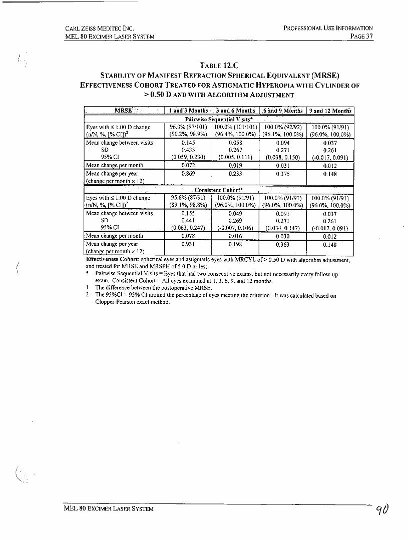
TABLE 12.CSTABILITY OF MANIFEST REFRACTION SPHERICAL EQUIVALENT (MRSE)
EFFECTIVENESS COHORT TREATED FOR ASTIGMATIC HYPEROPIA WITH CYLINDER OF> 0.50 D AND WITH ALGORITHM ADJUSTMENT
M MRSE' I and 3 Months 3 and 6 Months 6 hind 9Miiths. 9 and 12 MonthsPairwise Sequential Visits*
Eyes with 1.00 D change 96.0% (97/101) 100.0%(101/101) 100.0% (92/92) 100.0% (91/91)(n/N, %, [% CI])' (90.2%, 98.9%) (96.4%, 100.0%) (96.1%, 100.0%) (96.0%, 100.0%)Mean change between visits 0.145 0.058 0.094 0.037
SD 0.433 0.267 0.271 0.26195% C1 (0.059, 0.230) (0.005,0.111) (0.038,0.150) (-0.017, 0.091)
Mean change per month 0.072 0.019 0.031 0.012Mean change per year 0.869 0.233 0.375 0.148(change per month x 12)
. Consistent Cohort*
Eyes with 5 1.00 D change 95.6% (87/91) 100.0% (91/91) 100.0% (91/91) 100.0% (91/91)(n/N, %, [% C,])2 (89.1%, 98.8%) (96.0%, 100.0%) (96.0%, 100.0%) (96.0%, 100.0%)Mean change between visits 0.155 0.049 0.091 0.037
SD 0.441 0.269 0.271 0.26195% CI (0.063, 0.247) (-0.007, 0.106) (0.034, 0.147) (-0.017, 0.091)
Mean change per month 0.078 0.016 0.030 0.012Mean change per year 0.931 0.198 0.363 0.148(change per month x 12) 1 1
Effectiveness Cohort: spherical eyes and astigmatic eyes with MRCYL of > 0.50 D with algorithm adjustment,and treated for MRSE and MRSPH of 5.0 D or less.* Pairwise Sequential Visits = Eyes that had two consecutive exams, but not necessarily every follow-up
exam. Consistent Cohort = All eyes examined at 1, 3, 6, 9, and 12 months.I The difference between the postoperative MRSE.2 The 95%CI = 95% CI around the percentage of eyes meeting the criterion. It was calculated based on
Clopper-Pearson exact method.
MEL 80 EXCIMER LASER SYSTEM 96
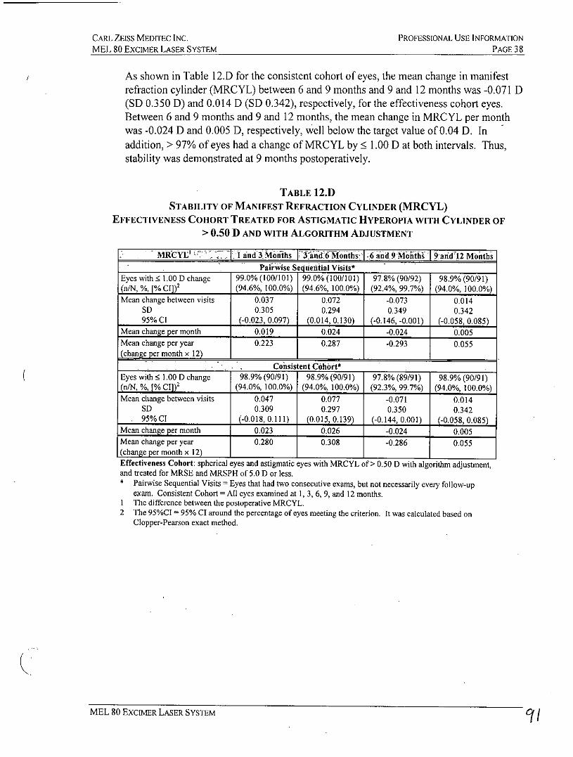
CARL ZEISS MEDITEC INC. PROFESSIONAL USE INFORMATIONMEL 80 ExcIMER LASER SYSTEM PAGE 38
As shown in Table 12.D for the consistent cohort of eyes, the mean change in manifestrefraction cylinder (MRCYL) between 6 and 9 months and 9 and 12 months was -0.071 D(SD 0.350 D) and 0.014 D (SD 0.342), respectively, for the effectiveness cohort eyes.Between 6 and 9 months and 9 and 12 months, the mean change in MRCYL per monthwas -0.024 D and 0.005 D, respectively, well below the target value of 0.04 D. Inaddition, > 97% of eyes had a change of MRCYL by : 1.00 D at both intervals. Thus,stability was demonstrated at 9 months postoperatively.
TABLE 12.DSTABILITY OF MANIFEST REFRACTION CYLINDER (MRCYL)
EFFECTIVENESS COHORT TREATED FOR ASTIGMATIC HYPEROPIA WITH CYLINDER OF> 0.50 D AND WITH ALGORITHM ADJUSTMENT
- MRCYL'I"7 1 1 1 and 3.iMonths -ind 6Montlis .6 a1d 9 MohII's and'I2 MonthsPaih&ise Sequential Visits*
Eyes with 1.00 D change 99.0% (100/101) 99.0% (100/101) 97.8% (90/92) 98.9% (90/91)(n/N, %, [% Cl])2 (94.6%, 100.0%) (94.6%, 100.0%) (92.4%, 99.7%) (94.0%, 100.0%)Mean change between visits 0.037 0.072 -0.073 0.014
SD 0.305 0.294 0.349 0.34295% CI (-0.023, 0.097) (0.014, 0.130) (-0.146, -0.001) (-0.058, 0.085)
Mean change per month 0.019 0.024 -0.024 0.005Mean change per year 0.223 0.287 -0.293 0.055(change per month x 12)
Consistent Cohort*
Eyes with 1.00 D change 98.9% (90/91) 98.9% (90/91) 97.8% (89/91) 98.9% (90/91)(n/N, %, [% CI]) 2 (94.0%, 100.0%) (94.0%, 100.0%) (92.3%, 99.7%) (94.0%, 100.0%)Mean change between visits 0.047 0.077 -0.071 0.014
SD 0.309 0.297 0.350 0.34295% CI (-0.018,0.111) (0.015, 0.139) (-0.144, 0.001) (-0.058, 0.085)
Mean change per month 0.023 0.026 -0.024 0.005Mean change per year 0.280 0.308 -0.286 0.055(change per month x 12) 1 1 1
Effectiveness Cohort: spherical eyes and astigmatic eyes with MRCYL of > 0.50 D with algorithm adjustment,and treated for MRSE and MRSPH of 5.0 D or less.* Pairwise Sequential Visits = Eyes that had two consecutive exams, but not necessarily every follow-up
exam. Consistent Cohort= All eyes examined at 1, 3, 6, 9, and 12 months.I The difference between the postoperative MRCYL.2 The 95%CI = 95% CI around the percentage of eyes meeting the criterion. It was calculated based on
Clopper-Pearson exact method.
MEL 80 EXCIMER LASER SYSTEM C/
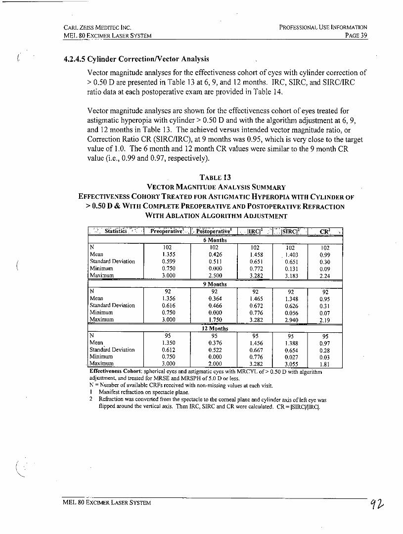
CARL ZEISS MEDITEC INC. PROFESSIONAL USE INFORMATIONMEL 80 EXCMER LASER SYSTEM PAGE 39
4.2.4.5 Cylinder Correction/Vector Analysis
Vector magnitude analyses for the effectiveness cohort of eyes with cylinder correction of> 0.50 D are presented in Table 13 at 6, 9, and 12 months. IRC, SIRC, and SIRC/LRCratio data at each postoperative exam are provided in Table 14.
Vector magnitude analyses are shown for the effectiveness cohort of eyes treated forastigmatic hyperopia with cylinder > 0.50 D and with the algorithm adjustment at 6, 9,and 12 months in Table 13. The achieved versus intended vector magnitude ratio, orCorrection Ratio CR (SIRC/IRC), at 9 months was 0.95, which is very close to the targetvalue of 1.0. The 6 month and 12 month CR values were similar to the 9 month CRvalue (i.e., 0.99 and 0.97, respectively).
TABLE 13VECTOR MAGNITUDE ANALYSIS SUMMARY
EFFECTIVENESS COHORT TREATED FOR ASTIGMATIC HYPEROPIA WITH CYLINDER OF> 0.50 D & WITH COMPLETE PREOPERATIVE AND POSTOPERATIVE REFRACTION
WITH ABLATION ALGORITHM ADJUSTMENT
Statistia Preo erative' I Potoperative' I IIRC12 ISIRC 2' CR
6 Months
N 102 102 102 102 102Mean 1.355 0.426 1.458 1.403 0.99Standard Deviation 0.599 0.511 0.651 0.651 0.30Minimum 0.750 0.000 0.772 0.131 0.09Maximum 3.000 2.500 3.282 3.183 2.24
9 Months
N 92 92 92 92 92Mean 1.356 0.364 1.465 1.348 0.95Standard Deviation 0.616 0.466 0.672 0.626 0.31Minimum 0.750 0.000 0.776 0.056 0.07Maximum 3.000 1.750 3.282 2.940 2.19
12 Months
N 95 95 95 95 95Mean 1.350 0.376 1.456 1.388 0.97Standard Deviation 0.612 0.522 0.667 0.654 0.28Minimum 0.750 0.000 0.776 0.027 0.03.Maximum 3.000 2,000 3.282 3.055 1.81Effectiveness Cohort: spherical eyes and astigmatic eyes with MRCYL of > 0.50 D with algorithmadjustment, and treated for MRSE and MRSPH of 5.0 D or less.N = Number of available CRFs received with non-missing values at each visit.I Manifest refraction on spectacle plane.
2 Refraction was converted from the spectacle to the corneal plane and cylinder axis of left eye wasflipped around the vertical axis. Then IRC, SIRC and CR were calculated. CR = ISIRCV1IRCI.
MEL 80 EXCIMER LASER SYSTEM C7
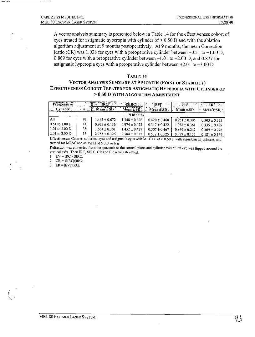
CARL ZEISS MEDITEC INC. PROFESSIONAL USE INFORMATIONMEL 80 EXCIMER LASER SYSTEM PAGE 40
A vector analysis summary is presented below in Table 14 for the effectiveness cohort ofeyes treated for astigmatic hyperopia with cylinder of > 0.50 D and with the ablationalgorithm adjustment at 9 months postoperatively. At 9 months, the mean CorrectionRatio (CR) was 1.038 for eyes with a preoperative cylinder between +0.51 to +1.00 D,0.869 for eyes with a preoperative cylinder between +1.01 to +2.00 D, and 0.877 forastigmatic hyperopia eyes with a preoperative cylinder between +2.01 to +3.00 D.
TABLE 14VECTOR ANALYSIS SUMMARY AT 9 MONTHS (POINT OF STABILITY)
EFFECTIVENESS COHORT TREATED FOR ASTIGMATIC HYPEROPIA WITH CYLINDER OF> 0.50 D WITH ALGORITHM ADJUSTMENT
-. Cylinder ni MeJii±SD Mean:B .± ISD. Mean £SYI M AW±S M-
9 MonthsAll 92 1.465 ± 0.672 1.348 ± 0.626 0.420 ± 0.460 0.951 ± 0.306 0.303 ± 0,3550.51 to 1.00 D 44 0.925 ± 0.136 0.974±0.422 0.317±0.422 1.038±0.361 0.335 ± 0.4391.01 to 2.00 D 35 1.664 ± 0.301 1.432 ± 0.429 0.507 ± 0.467 0.869 ± 0.242 0.309 ± 0.2782.01 to 3.00 D 13 2.755 ± 0.326 2.384 ± 0.332 0.532 ± 0.525 0.877 ± 0.155 0.181± 0.169Effectiveness Cohort: spherical eyes and astigmatic eyes with MRCYL of > 0.50 D with algorithm adjustment, andtreated for MRSE and MRSPH of 5.0 D or less.Refraction was converted from the spectacle to the corneal plane and cylinder axis of left eye was flipped around thevertical axis. Then IRC, SIRC, CR and ER were calculated.I EV = IRC - SIRC.2 CR =ISIRC|1IRCI.3 ER = IEV|/|RC.
MEL 80 EXCIMER LASER SYSTEM
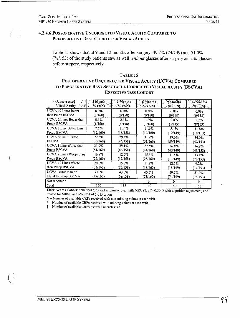
CARL ZEISS MEDITEC INC. PROFESSIONAL USE INFORMATIONMEL 80 EXCIMER LASER SYSTEM PAGE 41
4.2.4.6 POSTOPERATIVE UNCORRECTED VISUAL ACUITY COMPARED TOPREOPERATIVE BEST CORRECTED VISUAL ACUITY
Table 15 shows that at 9 and 12 months after surgery, 49.7% (74/149) and 51.0%(78/153) of the study patients saw as well without glasses after surgery as with glassesbefore surgery, respectively.
TABLE 15POSTOPERATIVE UNCORRECTED VISUAL ACUITY (UCVA) COMPARED
TO PREOPERATIVE BEST SPECTACLE CORRECTED VISUAL ACUITY (BSCVA)EFFECTIVENESS COHORT
Uncoirecfed J Month 3.Months - 6 Motks 9'Months 1j ,2MoithsVisual Acuity Wt. % (n/N) o%(hN) % n/N) % (n/N) %(j.
UCVA >2 Lines Better 0.0% 0.0% 0.0% 0.0% 0.0%than Preop BSCVA (0/160) (0/158) (0/160) (0/149) (0/153)UCVA 2 Lines Better than 0.6% 2.5% 1.9% 2.0% 5.2%Preop BSCVA (1/160) (4/158) (3/160) (3/149) (8/153)UCVA I Line Better than 7.5% 11.4% 11.9% 8.1% 11.8%Preop BSCVA (12/160) (18/158) (19/160) (12/149) (18/153)UCVA Equal to Preop 22.5% 29.1% 31.9% 39.6% 34.0%BSCVA (36/160) (46/158) (51/160) (59/149) (52/153)UCVA I Line Worse than 31.9% 29.1% 27.5% 26.8% 26.8%Preop BSCVA (51/160) (46/158) (44/160) (40/149) (41/153)UCVA 2 Lines Worse than 16.9% 12.0% 15.6% 11.4% 13.1%Preop BSCVA (27/160) (19/158) (25/160) (17/149) (20/153)UCVA >2 Lines Worse 20.6% 15.8% 11.3% 12.1% 9.2%than Preop BSCVA (33/160) (25/158) (18/160) (18/149) (14/153)UCVA Better than or 30.6% 43.0% 45.6% 49.7% 51.0%Equal to Preop BSCVA (49/160) (68/158) (73/160) (74/149) (78/153)Not reported* 0 0 0 0 0Totalf 160 158 160 149 153Effectiveness Cohort: spherical eyes and astigmatic eyes with MRCYL of> 0.50 D with algorithm adjustment, andtreated for MRSE and MRSPH of 5.0 D or less.N = Number of available CRFs received with non-missing values at each visit.* Number of available CRFs received with missing values at each visit.t Number of available CRFs received at each visit.
MEL 80 EXCIMER LASER SYSTEM
CARL ZEISS MEDITEC INC. PROFESSIONAL USE INFORMATIONMEL 80 EXCIMER LASER SYSTEM PAGE 42
4.2.4.7 Patient Symptoms and Satisfaction
Subjects filled out a subject questionnaire at the preoperative visit and at all follow-upvisits starting at 3 months. They graded their symptoms according to severity as eithernone, mild, moderate, marked, or severe (see Table 16.A). Table 16.B presents thepatient symptoms change from baseline to 9 months postoperatively. Any symptom forwhich there is at least a one grade increase from baseline is considered "worse" and atleast a one grade decrease is considered "better".
Table 16.A provides all patient symptoms for all treated eyes both preoperatively and at6, 9, and 12 months. Symptoms are grouped by severity level into absent, mild,moderate, marked, and severe. Symptoms in the mild category are not considered to beclinically significant. It can be seen that those symptoms reported at 9 and 12 months fallpredominantly into the "mild" category. Symptoms (those rated moderate to severe)reported with an incidence of at least 5% occurrence at month 9 include light sensitivity(moderate, 7.0%), dryness (moderate, 8.5%), blurred vision (moderate, 7.9%), fluctuationof vision (moderate, 5.6%), variation of vision in normal light (moderate, 5.6%), andvariation of vision in dim light (moderate, 7.0%). At 9 months, no symptom was reportedas marked or severe with an incidence of 5% or greater.
MEL 80 EXCIMER LASER SYSTEM
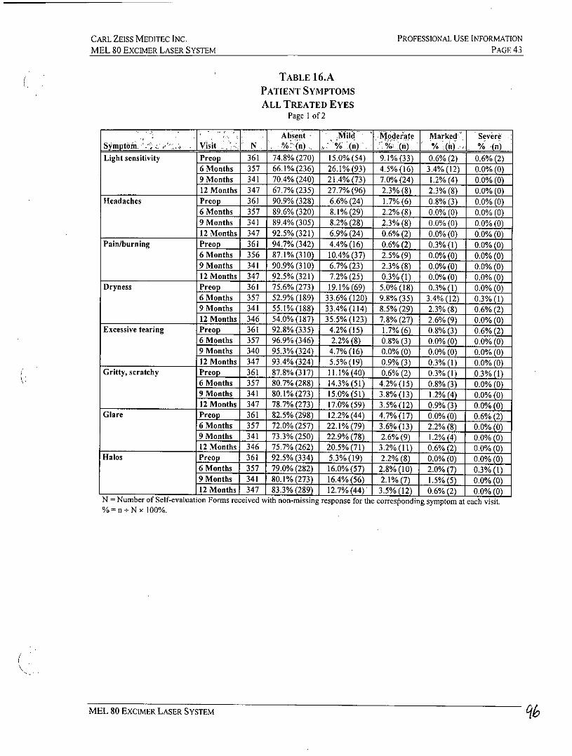
CARL ZEISS MEDITEC INC. PROFESSIONAL USE INFORMATIONMEL 80 EXCimER LASER SYSTEM PAGE 43
TABLE 16.APATIENT SYMPTOMS
ALL TREATED EYESPage 1 of 2
Absent - Mild Moderate Marked SevereSypthi V Visit N %jn) . , % ) /% (n) % L. % ._nLight sensitivity Preop 361 74.8%(270) 15.0%(54) 9.1%(33) 0.6%(2) 0.6%(2)
6 Months 357 66.1% (236) 26.1%(93) 4.5%(16) 3.4% (12) 0.0%(0)9 Months 341 70.4% (240) 21.4% (73) 7.0% (24) 1.2% (4) 0.0% (0)12 Months 347 67.7% (235) 27.7% (96) 2.3% (8) 2.3% (8) 0.0% (0)
Headaches Preop 361 90.9% (328) 6.6% (24) 1.7% (6) 0.8% (3) 0.0% (0)6 Months 357 89.6% (320) 8.1% (29) 2.2% (8) 0.0% (0) 0.0% (0)9 Months 341 89.4% (305) 8.2% (28) 2.3% (8) 0.0% (0) 0.0% (0)12 Months 347 92.5% (321) 6.9% (24) 0.6%(2) 0.0%(0) 0.0%(0)
Pain/burning Preop 361 94.7% (342) 4.4% (16) 0.6%(2) 0.3%(1) 0.0%(0)6 Months 356 87.1% (310) 10.4%(37) 2.5%(9) 0.0%(0) 0.0%(0)9 Months 341 90.9% (310) 6.7% (23) 2.3%(8) 0.0%(0) 0.0%(0)12 Months 347 92.5% (321) 7.2% (25) 0.3% (1) 0.0% (0) 0.0% (0)
Dryness Preop 361 75.6% (273) 19.1% (69) 5.0% (18) 0.3% (1) 0.0% (0)6 Months 357 52.9% (189) 33.6% (120) 9.8% (35) 3.4% (12) 0.3% (1)9 Months 341 55.1% (188) 33.4% (114) 8.5% (29) 2.3%(8) 0.6%(2)12 Months 346 54.0%(187) 35.5%(123) 7.8% (27) 2.6%(9) 0.0%(0)
Excessive tearing Preop 361 92.8% (335) 4.2% (15) 1.7%(6) 0.8%(3) 0.6%(2)6 Months 357 96.9% (346) 2.2% (8) 0.8% (3) 0.0% (0) 0.0% (0)9 Months 340 95.3% (324) 4.7% (16) 0.0% (0) 0.0% (0) 0.0% (0)12 Months 347 93.4% (324) 5.5% (19) 0.9% (3) 0.3% (1) 0.0% (0)
Gritty, scratchy Preop 361 87.8% (317) 11.1% (40) 0.6%(2) 0.3%(1) 0.3%(1)6 Months 357 80.7% (288) 14.3% (51) 4.2% (15) 0.8% (3) 0.0%(0)9 Months 341 80.1% (273) 15.0% (51) 3.8% (13) 1.2%(4) 0.0%(0)12 Months 347 78.7% (273) 17.0% (59) 3.5% (12) 0.9%(3) 0.0% (0)
Glare Preop 361 82.5%(298) 12.2% (44) 4.7%(17) 0.0%(0) 0.6% (2)6 Months 357 72.0% (257) 22.1% (79) 3.6% (13) 2.2%(8) 0.0%(0)9 Months 341 73.3% (250) 22.9% (78) 2.6% (9) 1.2% (4) 0.0% (0)12 Months 346 75.7% (262) 20.5% (71) 3.2%(I1) 0.6%(2) 0.0%(0)
Halos Preop 36 92.5%(334) 5.3%(19) 2.2%(8) 0.0%(0) 0.0%(0)6 Months 357 79.0% (282) 16.0% (57) 2.8% (10) 2.0%(7) 0.3%(1)9Months 341 80.1%(273) 16.4%(56) 2.1%(7) 1.5% (5) 0.0%(0)12 Months 347 83.3%(289) 12.7%(44) 3.5%(12) 0.6% (2) 0.0% (0)
N = Number of Self-evaluation Forms received with non-missing response for the corresponding symptom at each visit.%= n - Nx 100%.
MEL 80 EXCIMER LASER SYSTEM 96
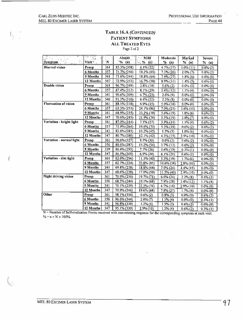
CARL ZEISS MEDITEC INC. PROFESSIONAL USE INFORMATIONMEL 80 ExcIMER LASER SYSTEM PAGE 44
TABLE 16.A (CONTINUED)
PATIENT SYMPTOMS
ALL TREATED EYESPage 2 of 2
Nb se t Mo er te M ark d S ev ereVisit 'N % (n) % ()n) % (n)
Blurred vision Preop 361 85.3% (308) 6.1% (22) 4.7% (17) 3.0%(11) 0.8%(3)6 Months 357 71.7% (256) 18.2% (65) 7.3% (26) 2.0% (7) 0.8% (3)9 Months 341 71.6% (244) 18.8% (64) 7.9% (27) 1.8%(6) 0.0%(0)12 Months 347 72.9% (253) 16.7% (58) 8.9% (31) 1.4%(5) 0.0%(0)
Double vision Preop 361 96.7% (349) 2.8% (10) 0.6% (2) 0.0% (0) 0.0% (0)6 Months 357 87.4% (312) 8.1% (29) 3.4%(12) 1.1%(4) 0.0%(0)9 Months 341 90.6% (309) 6.7% (23) 2.6% (9) 0.0% (0) 0.0% (0)12 Months 346 91.3% (316) 6.4% (22) 2.3%(8) 0.0%(0) 0.0%(0)
Fluctuation of vision -PrEop- 361 88.1% (318) 6.9% (25) 5.0% (18) 0.0% (0) 0.0%(0)6 Months 357 65.3% (233) 24.1% (86) 7.0% (25) 3.6% (13) 0.0% (0)9 Months 341 68.9% (235) 23.2% (79) 5.6% (19) 1.8%(6) 0.6%(2)12 Months 347 70.6%(245) 21.9%(76) 5.5%(19) 2.0%(7) 0.0%(0)
Variation - bright light Preop 361 87.0% (314) 7.5%(27) 3.9%(14) 1.1%(4) 0.6%(2)6 Months 357 73.9% (264) 19.6% (70) 5.3% (19) 0.6% (2) 0.6% (2)9 Months 341 83.6% (285) 13.2% (45) 1.5% (5) 1.8% (6) 0.0% (0)12 Months 347 80.7% (280) 12.1%(42) 4.3% (15) 2.9% (10) 0.0%(0)
Variation - normal light Preop 361 90.6% (327) 8.3% (30) 0.6% (2) 0.6% (2) 0.0% (0)6 Months 356 80.6% (287) 15.2% (54) 3.7% (13) 0.6% (2) 0.0% (0)9 Months 339 86.4% (293) 7.7% (26) 5.6% (19) 0.3% (1) 0.0% (0)12 Months 347 86.5% (300) 6.9% (24) 6.1% (21) 0.6% (2) 0.0% (0)
Variation - dim light Preop 361 82.0% (296) 11.1% (40) 5.3%(19) 1.7%(6) 0.0% (0)6 Months 357 62.7% (224) 23.8% (85) 10.6%(38) 2.8% (10) 0.0%(0)9 Months 341 69.8%(238) 18.8%(64) 7.0%(24) 4.4%(15) 0.0%(0)12 Months 347 68.6% (238) 17.0% (59) 11.5% (40) 2.9% (10) 0.0% (0)
Night driving vision Preop 361 70.9% (256) 19.7% (71) 6.6% (24) 2.2% (8) 0.6% (2)6 Months 356 68.5% (244) 19.1% (68) 7.9% (28) 3.4% (12) 1.1%(4)9 Months 341 70.1%(239) 22.3%(76) 4.7% (16) 2.9%(10) 0.0%(0)12 Months 347 70.9% (246) 19.6% (68) 7.8% (27) 1.7%(6) 0.0%(0)
Other Preop 361 98.1% (354) 0.6% (2) 0.8% (3) 0.0% (0) 0.6% (2)6Months 356 96.6%(344) 2.0% (7) l.1% (4) 0.0%(0) 0.3% (1)9 Months 341 96.8% (330) 1.2% (4) 1,5% (5) 0.6% (2) 0.0% (0)12 Months 347 95.1%(330) 2.9%(10) 1.2%(4) 0.6%(2) 0.3%(1)
N = Number of Self-evaluation Forms received with non-missing response for the corresponding symptom at each visit.% - n - Nx 100%.
MEL 80 ExciMER LASER SYSTEM 7
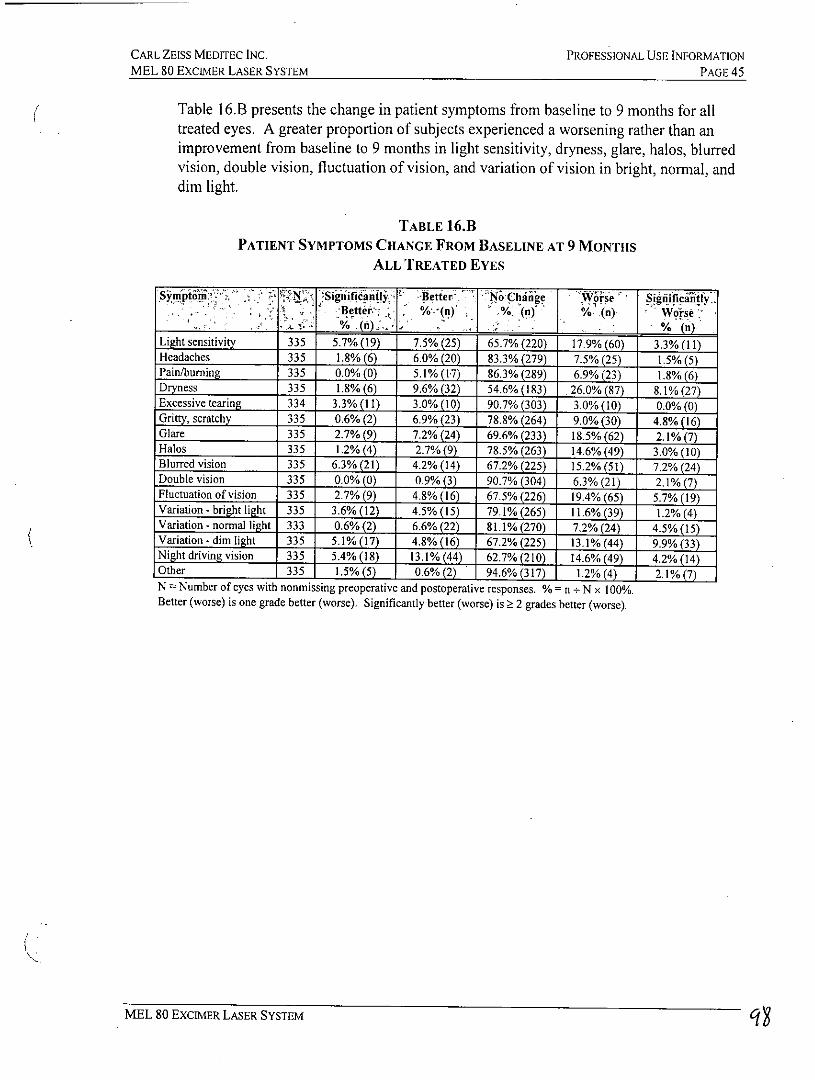
CARL ZEISS MEDITEC INC. PROFESSIONAL USE INFORMATIONMEL 80 EXCIMER LASER SYSTEM PAGE 45
Table 16.B presents the change in patient symptoms from baseline to 9 months for alltreated eyes. A greater proportion of subjects experienced a worsening rather than animprovement from baseline to 9 months in light sensitivity, dryness, glare, halos, blurredvision, double vision, fluctuation of vision, and variation of vision in bright, normal, anddim light.
TABLE 16.1PATIENT SYMPTOMS CHANGE FROM BASELINE AT 9 MONTHS
ALL TREATED EYES
S mt(1Significantl Better' N; Chjng V&ise Signiific "lifyBete n % (n -(a) Worse
% (n1 J . (nLight sensitivity 335 5.7%(19) 7.5%(25) 65.7%(220) 17.9%(60) 3.3%(l)Headaches 335 1.8%(6) 6.0% (20) 83.3% (279) 7.5% (25) 1.5%(5)Pain/burning 335 0.0% (0) 5.1% (17) 86.3% (289) 6.9% (23) 1.8%(6)Dryness 335 1.8% (6) 9.6% (32) 54.6% (183) .26.0% (87) 8.1% (27)Excessive tearing 334 3.3%(11) 3.0%(10) 90.7%(303) 3.0%(10) 0.0%(0)Gritty, scratchy 335 0.6% (2) 6.9% (23) 78.8% (264) 9.0% (30) 4.8% (16)Glare 335 2.7%(9) 7.2%(24) 69.6%(233) 18.5%(62) 2.1%(7)Halos 335 1.2%(4) 2.7%(9) 78.5%(263) 14.6%(49) 3.0%(10)Blurred vision 335 6.3% (21) 4.2% (14) 67.2% (225) 15.2% (51) 7.2% (24)Double vision 335 0.0%(0) 0.9% (3) 90.7% (304) 6.3% (21) 2.1%(7)Fluctuation of vision 335 2.7% (9) 4.8% (16) 67.5% (226) 19.4% (65) 5.7% (19)Variation - bright light 335 3.6%(12) 4.5%(15) 79.1%(265) l.6% (39) 1.2%(4)Variation - normal light 333 0.6%(2) 6.6%(22) 81.1% (270) 7.2%(24) 4.5% (15)Variation - dim light 335 5.1% (17) 4.8% (16) 67.2% (225) 13.1% (44) 9.9% (3)Night driving vision 335 5.4% (18) 13.1% (44) 62.7% (210) 14.6% (49) 4.2% (14)Other 335 1.5%(5) 0.6%(2) 94.6%(317) 1.2%(4) 2.1%(7)N= Number of eyes with nonmissing preoperative and postoperative responses. %= n +Nx 100%.Better (worse) is one grade better (worse). Significantly better (worse) is 2 grades better (worse).
MEL 80 EXCIMER LASER SYSTEM
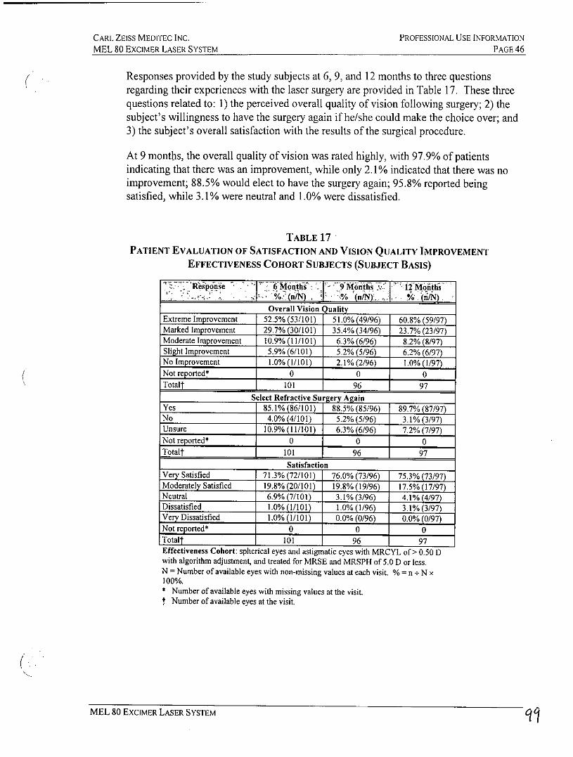
CARL ZEISS MEODTEC INC. PROFESSIONAL USE INFORMATIONMEL 80 EXCIMER LASER SYSTEM PAGE 46
Responses provided by the study subjects at 6, 9, and 12 months to three questionsregarding their experiences with the laser surgery are provided in Table 17. These threequestions related to: 1) the perceived overall quality of vision following surgery; 2) thesubject's willingness to have the surgery again if he/she could make the choice over; and3) the subject's overall satisfaction with the results of the surgical procedure.
At 9 months, the overall quality of vision was rated highly, with 97.9% of patientsindicating that there was an improvement, while only 2.1% indicated that there was noimprovement; 88.5% would elect to have the surgery again; 95.8% reported beingsatisfied, while 3.1% were neutral and 1.0% were dissatisfied.
TABLE 17PATIENT EVALUATION OF SATISFACTION AND VISION QUALITY IMPROVEMENT
EFFECTIVENESS COHORT SUBJECTS (SUBJECT BASIS)
R'esp'pnse -- 6Mnts 9Months l2'Moitfsno % (n/N,
Overall Vision QualityExtreme Improvement 52.5% (53/101) 51.0% (49/96) 60.8% (59/97)Marked Improvement 29.7% (30/101) 35.4% (34/96) 23.7% (23/97)Moderate Improvement 10.9% (11/101) 6.3% (6/96) 8.2% (8/97)Slight Improvement 5.9% (6/101) 5.2% (5/96) 6.2% (6/97)No Improvement 1.0% (1/101) 2.1%(2/96) 1.0%(1/97)Not reported* 0 0 0Totalt 101 96 97
Select Refractive Surgery AgainYes 85.1% (86/101) 88.5% (85/96) 89.7% (87/97)No 4.0%(4/101) 5.2%(5/96) 3.1%(3/97)Unsure 10.9% (11/101) 6.3%(6/96) 7.2% (7/97)Not reported* 0 0 0Totalt 101 96 97
SatisfactionVery Satisfied 71.3% (72/101) 76.0% (73/96) 75.3% (73/97)Moderately Satisfied 19.8% (20/101) 19.8% (19/96) 17.5% (17/97)Neutral 6.9% (7/101) 3.1%(3/96) 4.1%(4/97)Dissatisfied 1.0% (1/101) 1.0%(1/96) 3.1%(3/97)Very Dissatisfied 1.0% (1/101) 0.0% (0/96) 0.0% (0/97)Notreorted* 0 0 0Totalt 101 96 97Effectiveness Cohort: spherical eyes and astigmatic eyes with MRCYL of > 0.50 Dwith algorithm adjustment, and treated for MRSE and MRSPH of 5.0 D or less.N = Number of available eyes with non-missing values at each visit. %= n + N x100%.* Number of available eyes with missing values at the visit.t Number of available eyes at the visit.
MEL 80 EXCIMER LASER SYSTEM O
CARL ZEISS MEDITEC INC. PROFESSIONAL USE INFORMATIONMEL 80 ExcIMER LASER SYSTEM PAGE 47
4.2.5 Factors Associated with Outcomes
Gender, preoperative refraction, age, baseline MRSE, primary vs. fellow eye, study site,and use of an ablation algorithm adjustment were evaluated as statistically significantpredictors of the UCVA and refractive outcome for the LASIK procedure. Theseanalyses identified a site effect, an ablation algorithm adjustment effect, an effect ofbaseline MRSE, a small cylinder cyclotorsional effect, and an effect of keratome use.
Statistical analysis of the study data by site revealed that the percentage of eyes reportedwith a MRSE within ± 0.50 D of the attempted correction was significantly differentamong the six investigational sites at 9 months. At 9 months, 93% of eyes were within0.50 D of intended MRSE at site #5, compared with 81%, 67%, 63%, 62%, and 65% atthe other five study sites. There were no statistically significant differences observedbetween the study sites with respect to attempted versus achieved MRSE within ± 1.00 Dof the intended correction at 9 months.
Statistical analysis of the study data by site revealed that the percentage of eyes reportedwith UCVA outcomes of 20/40 or better and 20/20 or better were not significantlydifferent among the six investigational sites at 9 months. At 9 months, 100% of eyes had20/32 or better UCVA at site #1 and site #2, compared with 99%, 92%, 96%, and 86% atthe other four study sites. Similar findings were observed for UCVA outcomes of 20/25or better, 20/16 or better, and 20/12.5 or better. The clinical significance, if any, of thisdifference is uncertain.
Analyses of the effect of the use of an ablation algorithm adjustment revealed that eyestreated with the ablation algorithm adjustment had significantly better predictabilityoutcomes for the proportion of eyes within ± 0.50 D of attempted versus achieved MRSEat 9 months (p= 0.0006). Other key effectiveness endpoints were shown to besignificantly better as a result of the algorithm adjustment.
Eyes with preoperative MRSE > 5.0 D were significantly less likely to achieve refractivepredictability within + 0.50 D of the intended outcome at 9 months. In contrast, at 9months, eyes with baseline MRSE up to +5.00 D had significantly better MRSE accuracyoutcomes (> 61% within + 0.50 D of intended MRSE) than eyes with baseline MRSEgreater than +5.00 D (ranging from 33% to 40% for both groups within 0.50 D ofintended MRSE). Baseline MRSE did not have a significant association with UCVAoutcomes of 20/40 or better at 9 months. However, a greater proportion of eyes withbaseline MRSE 5 +5.00 D achieved UCVA better than 20/40 (i.e., 20/16 to 20/25 at 9months) than eyes with baseline MRSE higher than +5.00 D.
Analysis of the cylinder treatment revealed a small amount of cyclorotation. This mayhave been the result of cyclotorsional movement of the patient's eye that occurs whenmoving from the sitting position to the position under the laser. It is recommended that amark be made on the patient's corneal limbus using a sterile single use marker foralignment with the reticle of the laser surgical microscope to be certain that no
MEL 80 EXCIMER LASER SYSTEM loo
CARL ZEISS MEDITEC INC. PROFESSIONAL USE INFORMATION
MEL 80 EXCIMER LASER SYSTEM PAGE 48
cyclorotation is present. The marking should be made with the patient in the sittingposition behind a slit lamp. Once the patient is lying down, if necessary, the patient'shead can be repositioned to properly align the reticle with the mark(s) during thetreatment to reduce or eliminate any rotational misalignment.
An analysis of the keratome used in the study procedures revealed a possible effect onUCVA outcomes and MRSE predictability. Site 5, which used the Moria keratome,showed better efficacy outcomes with regard to the proportion of eyes with deviationfrom the intended correction within ± 0.50 D at 9 months postoperatively (92.9%,p<0.0001). In addition, the IntraLase keratome and the Hansatome keratome wereassociated with a significantly lower proportion of eyes achieving UCVA of 20/20 orbetter (62.1% and 62.9%, respectively) as compared to the other keratomes. However,this analysis of outcomes by keratome type is confounded by other factors that may havecontributed to this difference in outcomes, including site and introduction of thealgorithm adjustment. Since the key effectiveness outcomes for the total cohort of eyeswere not affected by the type of keratome used, the differences observed with differentkeratomes for the sphere only and the astigmatic hyperopia eyes in the study may reflectother, non-keratome factors.
In addition, the complications were reported with a significantly higher proportion of eyesfor the IntraLase keratome as compared to the various mechanical microkeratomesincluded diffuse lamellar keratitis (8.3%), dry eye (8.3%), epithelium at flap edge (4.9%),epithelium in the interface (13.9%), foreign body sensation (5.6%), punctal plug insertion(30.6%), superficial punctate keratitis (16.0%), steroid induced IOP increase (5.6%), andtransient light sensitivity syndrome (8.3%).
MEL 80 EXCIMER LASER SYSTEM
CARL ZEISS MEDITEC INC. PROFESSIONAL USE INFORMATIONMEL 80 EXCIMER LASER SYSTEM PAGE 49
SECTION 5
SURGICAL PLANNING AND PROCEDURES
5.1 INTRODUCTION
LASIK is a procedure that combines the use of a microkeratome to create a lamellarcorneal flap and the energy of the excimer laser to create a keratectomy in the cornealstroma of a shape designed to correct or reduce a specific refractive error. The intent is toproperly focus visible light entering the eye to provide improved vision. It is essentialthat the refractive information upon which this surgical procedure is based is accurate andcorrectly transmitted to the laser. It is the sole responsibility of the surgeon to ensure thatthe information for each individual patient is accurate.
5.2 PATIENT SELECTION
Consideration should be given to the following in determining the appropriate patients forLASIK:
* Complete examination, including, but not limited to, manifest and cycloplegicrefraction evaluation, must be performed. The lens must be evaluated, especially inthe older patient, to assure that nuclear sclerosis or any other lens opacity is notpresent prior to laser surgery. Indirect ophthalmoscopy through a dilated pupil and aclear crystalline lens is essential.
* To obtain accurate refractive information, contact lens wearers must be examinedafter a period of not wearing contact lenses for at least 2 weeks for soft lenses and atleast 3 weeks for hard (PMMA) and gas-permeable lenses. Prior to treatment andafter at least 3 weeks of not wearing contact lenses, patients who wear rigid gaspermeable or hard lenses must have 3 central keratometry readings and manifestrefraction taken at one-week intervals, the last 2 of which must not differ by morethan 0.50 diopter in either meridian. All mires must be regular.
* Glaucoma is more common in hyperopic patients than in the general population.Evaluation of the optic nerve and measurement of intraocular pressure are necessary.If there are any concerns regarding the appearance of the optic nerve, a threshold testof the visual field should be performed. If elevated intraocular pressure and/orevidence of glaucomatous damage are found, topical steroids should only be usedwith careful medical supervision or the patient should not undergo LASIK surgery.
MEL 80 EXCIMER LASER SYSTEM t02,
CARL ZEISS MEDITEC INC. PROFESSIONAL USE INFORMATIONMEL 80 EXCIMER LASER SYSTEM PAGE 50
* Pachymetry must be performed to obtain a baseline central corneal thicknessmeasurement to assure that the combination of the planned corneal flap thickness andthe planned laser ablation will not approach closer than 250 microns to the cornealendothelium.
* Baseline evaluation of patients requesting refractive surgery should be performedwithin 30 days of the LASIK surgery.
* The patient should have the ability to lie flat without difficulty.
* The patient should be able to fixate steadily and accurately for the duration of theLASIK procedure.
* The patient must be able to understand the surgery and give informed consent.
* The patient must be able to tolerate eye drops to numb the eye.
* The patient should be clearly informed of all alternatives for the correction of his/herhyperopia including, but not limited to, spectacles, contact lenses, and other refractivesurgeries.
* Due to the importance of managing a patient's expectations in elective refractivesurgery, it is recommended that the physician also complete an assessment of thepatient's expectations and psychological state, which would include:
* conveying realistic expectations to the prospective patient;
* attempting to ensure patient comprehension of the risks and benefits at the ofthe informed consent process;
* providing a patient information card that has eye measurements from beforethe LASIK surgery. Patients can keep this card to help their doctor calculatethe lens implant power should they need to have future cataract surgery; aform for the necessary information is available on the internet at:http://www.getevesmart.org/eyesmart/upload/kcard.pdf
* discussing with patients how having LASIK may affect the futureinterpretation of intraocular pressure measurements; patients should beinstructed to inform future eye care providers that they have had LASIK.
5.3 PROCEDURE
The output of the laser is potentially hazardous only to the skin and the surface layers ofthe cornea. This radiation has not been shown to pose a threat to retinal structures or tothe crystalline lens. The area of potential hazard (Nominal Hazard Zone) for theproduction of a photochemical keratitis has been determined to be less than 40 cm fromthe primary beam. All healthcare personnel should avoid direct exposure to the skin oreye by the primary beam. While no hazard may exist further than 40 cm from the beam,the use of protective eyewear is recommended if the possibility exists that healthcarepersonnel will approach closer than this distance to the primary beam.
MEL 80 EXCIMER LASER SYSTEM 1Q3
CARL ZEISS MEDITEC INC. PROFESSIONAL USE INFORMATIONMEL 80 EXCIMER LASER SYSTEM PAGE 51
Prior to initiating the lamellar keratectomy portion of the surgery with the microkeratome,the physician should perform the fluence test to ensure that the laser is ready to deliverlaser energy.
5.4 PERI-OPERATIVE PROCEDURES
5.4.1 Anesthesia
Extensive clinical experience has shown that LASIK excimer surgery is welltolerated and rarely causes significant pain. For this reason, systemic sedativesand injected local anesthetics are not required. Topical anesthesia applied justbefore insertion of the lid speculum should provide adequate control of painduring surgery. For those patients with a high degree of anxiety, appropriatemedication may be given preoperatively.
5.5 INTRA-OPERATIVE PROCEDURES
5.5.1 Creating the Lamellar Flap with the Microkeratome
The LASIK procedure requires the creation of a hinged comeal flap using amicrokeratome prior to the laser ablation procedure. The microkeratome used toperform the LASIK procedure should be a legally marketed device in the UnitedStates. The physician should follow the specific procedures recommended in theOperator's Manual supplied with the particular brand of microkeratome to beused. Once the corneal flap has been created, the laser ablation step may beperformed.
5.5.2 Performing the Laser Ablation
Following creation of the corneal flap, the laser ablation is then performed. Thephysician should refer to the User Manual supplied with the MEL 80 ExcimerLaser System for proper operation and maintenance instructions. The physicianalso must have completed the appropriate technical and medical in-servicetraining provided by the manufacturer prior to using the laser for actual surgery.
5.6 POST OPERATIVE PROCEDURES
5.6.1 Patching and Medications
Following completion of the excimer laser surgery, appropriate topicalmedications and a corneal shield or firm patch should be applied to the eye. Acombination steroid-antibiotic medication should be included at the time ofpatching. Some physicians may wish to omit steroids until the edge of thelamellar keratectomy has healed completely. The patient should be seen one daypostoperatively to ensure that the comeal flap is properly in place.
MEL 80 EXCIMER LASER SYSTEM
CARL ZEISS MEDITEC INC. PROFESSIONAL USE INFORMATIONMEL 80 EXCIMER LASER SYSTEM PAGE 52
5.6.2 Analgesia
The physician may wish to administer appropriate postoperative medications forthe management of ocular pain. These may include the use of topical ophthalmicnon-steroidal anti-inflammatory drugs (NSAIDs), as well as systemicallyadministered medications for pain management.
5.6.3 Handling Complications
Following the LASIK procedure, the physician should carefully monitor thecondition of the patient's cornea on a periodic basis with regard to the conditionof the corneal flap and its location. Special attention should be given to thepresence of any debris or epithelial cells in the interface between the flap and theunderlying corneal stroma. The presence of such foreign material may requirelifting of the flap to remove such debris and/or cells using appropriate surgicaltechniques. The use of topical ophthalmic steroid medications may be required tosuppress any associated inflammation caused by debris or cells in the interface.
5.7 POST PROCEDURE
A slit-lamp examination should be performed on postoperative day one and as neededthereafter to ensure that healing of the cornea is complete. After the one-dayexamination, the following examinations are recommended at a schedule of at least 1, 3,6, and 9 months:
* Uncorrected visual acuity (UCVA or VA-sc)* Manifest refraction with best spectacle-corrected visual acuity (BSCVA
or VA-cc)* Intraocular pressure (1OP)
* Slip-lamp examination, including evaluation of corneal clarity and the conditionof the flap.
MEL 80 EXCIMER LASER SYSTEM tK
CARL ZEISS MEDITEC INC. PROFESSIONAL USE INFORMATIONMEL 80 EXCIMER LASER SYSTEM PACE 53
SECTION 6
CARL ZEISS MEDITEC MEL 80 EXCIMER LASER SURGICAL PROCEDURE STEPBY-STEP PROCEDURE
6.1 Prior to Surgery
I. Refer to the User Manual for the complete step-by-step procedure to be followedprior to commencement of the actual surgical procedure (laser set-up, fluence test,etc.).
2. Provide instructions to the patient explaining what to expect during (i.e. where tolook during the procedure) and after the procedure (i.e. the eyes will feel irritatedand the vision will be blurry). In addition, provide patient with the device labeling"Patient Information Booklet" for additional information.
6.2 Preparing Device and Patient for Treatment
1. Fully examine the patient's eye(s) including the retina (inquire also about thepatient's working conditions, e.g. night driving).
2. Two weeks before surgery, the patient should stop wearing hard contact lenses. Softlenses should be removed one week before surgery.
3. Check refraction yourself or have it done by an appropriately qualified person. Usesubjective manifest refraction.
4. Check pupil size in the dark (e.g. with a Colvard pupillometer or WASCAAnalyzer).
5. Determine corneal thickness at the thinnest point.
6. Calculate the ablation depth with the adequate treatment diameter (0.5 mm >mesopic pupil size) and check for a sufficient residual stroma (250 gm).
7. Never perform LASIK in a patient who is presumed to have a residual stromathickness of less than 250 pm. Always consider the inaccuracy of themicrokeratome.
8. Check the national legislation and all contraindications specified in relevantliterature (e.g. keratoconus, irregular topography, etc.), before you decide on thesurgery.
9. Recheck refraction and topography (3 pictures minimum) immediately beforesurgery (possibly the topography has changed by contact lens effects since the firstmeasurement).
10. The clinical study showed small amounts of cylcorotation present. It isrecommended to mark the cylinder axis on the corneal limbus using a sterile singleuse marker behind the slit lamp prior to entering the operating room. The cylinder
MEL 80 EXCIMER LASER SYSTEM Je
CARL ZEISS MEDITEC INC. PROFESSIONAL USE INFORMATIONMEL 80 EXCIMER LASER SYSTEM PAGE 54
marking can be used to align the reticle during the treatment to reduce or eliminateany rotational misalignment.
11. Check room conditions (see technical data, ambient conditions for intended use inthe MEL 80 User Manual).
12. Starting 30 minutes before surgery, it is advisable to put a drop of a non-toxicantibiotic in the eye every 10 minutes.
13. Prepare the microkeratome.
14. Enter and verify patient data.
15. Microscope setting: magnification I.Ox.
16. Run the fluence test and confirm it with <Energy Ok>.
17. Switch ff the aiming beam to avoid coverage of the fixation light. Move joystickaside, if necessary.
18. Bring patient in.
19. Check patient data and the eye to be operated. If you use OPASS, you may correctpatient data, if necessary. If patient data is correct, click on <Calculate> to have thecorrection program calculated.
20. Position the patient on the bed so that the patient's eye is effortless in the positionrequired.
21. It may be favorable to wipe the inner side of the lids with a triangular swab soakedwith anesthetics (1% Xylocaine, no preservatives) in order to reduce irritation andthus avoid increased production of tear fluid.
22. Cover the operation area with a fenestrated adhesive drape and arrange it in such away that the lashes are folded back over the margin of the eyelid.
23. Microscope setting: magnification 0.6x.
24. Coarsely adjust the patient bed using the focusing beams.
25. Use an eyelid retractor.
26. Apply a drop of an anesthetic (e.g. Oxybuprocain-HCI) (not yet in the other eye ifyou plan bilateral treatment).
27. Open the lid retractor as wide as the patient can just tolerate. Fine align the patientbed so that the iris is in the center of the palpebral fissure.
28. Using gentian violet, mark the cornea non-symmetrically.
29. Rinse excessive dye away.
30. Perform the keratome cut following the instructions for use of the keratome. Do notopen the flap yet.
31. Microscope setting: magnification I.Ox.
32. Again, accurately position the patient's eye with the patient bed.
MEL 80 EXCIMER LASER SYSTEM 107
CARL ZEISS MEDITEC INC. PROFESSIONAL USE INFORMATIONMEL 80 EXCIMER LASER SYSTEM PAGE 55
33. Swing in the CCA+ unit taking care that the position of the patient's eye remainsstable.
34. Have the patient look at the green, blinking LED (Note: The patient's eye must be inthe center of the field of view; microscope setting: magnification I.Ox.) Tell thepatient that the aiming beam will outshine the LED.
6.3 Microkeratome Surgery
The physician should perform the lamellar keratectomy to create the corneal flapaccording to the instructions provided with the microkeratome. Once the corneal flap hasbeen created, the laser ablation portion of the procedure may be started.
6.4 Laser Treatment
1. Activate the eyetracker.
2. The eyetracker automatically sets the aiming beam to the center of the entrancepupil ("line of sight"). To choose another centration point, use the offset keys. Turnon the satellite illumination, if necessary.
Note: Since the fixation light is central, it is advisable to center the patient's righteye with the surgeon's left eye and vice versa to avoid parallax errors.
3. Open the flap and settle it on a sterile LASIK shield soaked in BSS; the inside of theflap must not be touched throughout the treatment.
4. Click on the <Ready> button.
5. Start ablation by depressing the footswitch.
6. Stop every 15 seconds to verify proper centration.
7. After the last shot, move the CCA+ unit out of the surgical field.
Note: The treatment progress is indicated by a progress display. By a click on the<Cancel> button, you can abort the treatment after releasing the footswitch.
The control computer will automatically stop lasing after the correctionprogram is finished. If you aborted the treatment by releasing the footswitch,you can continue it by depressing the footswitch again.
Note: If the eyetracker loses track of the pupil during the treatment or the pupilleaves the hot zone, the control computer will interrupt the treatment.
You can then continue the treatment with the <Continue> button, when the pupilhas been detected again or the eyetracker has been deactivated.
8. Immediately after ablation, clean the stromal bed very carefully with a new moisttriangular swab moved clockwise and with another new moist swab moved
MEL 80 EXCIMER LASER SYSTEM t O
CARL ZEISS MEDITEC INC. PROFESSIONAL USE INFORMATIONMEL 80 EXCIMER LASER SYSTEM PAGE 56
counterclockwise to remove debris and epithelial cells. Continue until the bed looksdry.
9. Fold back the flap by means of a bent cannula. If folds have formed, use the cannulato unfold them. Then, rinse with BSS to clean the site and remove any wrinkles.
Note: Rinse briefly at high pressure to avoid hydration of the flap and thuscanalization -greater ingrowth of epithelium.
10. Make sure the flap is properly positioned (marks, symmetry of the duct). If not, usethe cannula again. Then, using a wet triangular swab, wipe the flap away from thehinge to fix its position.
Note: Use the segmenting facility of the ring lamp, if necessary, to check the correctposition of the flap.
11. Apply a few drops of an antibiotic.
12. Wait one minute while keeping the central epithelium moist.
13. Carefully remove the eyelid retractor and the drapes. Ask the patient to blinkrepeatedly and observe the behavior of the flap.
14. Microscope setting: Magnification 0.6x.
15. Contact lenses may be inserted only if the epithelium is injured.
16. Tell the patient to keep the eye closed until the next morning. For one week, havehim or her wear an eyepatch at night and sunglasses during the day when being inthe open air.
17. When checking the position of the flap on the first postoperative day, you canreposition the flap, if necessary.
18. The treatment with antibiotics and anti-inflammatory drops should be continued forfour days.
MEL 80 EXCIMER LASER SYSTEM
CARL ZEISS MEDITEC INC. PROFESSIONAL USE INFORMATIONMEL 80 EXCIMER LASER SYSTEM PAGE 57
SECTION 7
EMERGENCY STOP
If a system emergency situation arises, press the Emergency Stop button. This switch turns offthe complete laser system. It is located on the front side below the computer monitor in the CarlZeiss Meditec MEL 80 Excimer Laser. Pressing the Emergency Stop button is the fastestpossible way to completely switch off the laser system.
MEL 80 EXCIMER LASER SYSTEM
FACTS YOU NEED TO KNOW ABOUT LASER IN SITU KERATOMILEUSIS (LASIK)
A SURGERY TO REDUCE OR ELIMINATE NATURALLY OCCURING HYPEROPIAWITH OR WITHOUT ASTIGMATISM USING THE
CARL ZEISS MEDITEC MEL 80 EXCIMER LASER SYSTEM
PATIENT INFORMATION BOOKLET
Please read this entire booklet. If you have any questions about it, discuss them withyour doctor before you agree to the surgery.
The MEL 80 Excimer Laser is indicated for use in primary Laser Assisted in situ Keratomileusis(LASIK) treatments for the reduction or elimination of naturally occurring hyperopia of less thanor equal to +5.0 D with or without refractive astigmatism of > +0.5 D and < +3.0 D, with amaximum MRSE of +5.0 D, in patients who are 21 years of age or older with documentation ofstable manifest refraction over the past year as demonstrated by change in sphere and cylinder of
0.5 D.
Carl Zeiss Meditec, Inc.5160 Hacienda Drive
Dublin, California 94568 USA(925) 557-4100
Hyperopia Patient Information Booklet 1-31-2011
CARL ZEISS MEDITEC, INC. PATIENT INFORMATION BOOKLET
MEL 80 EXCIMER LASER SYSTEM PAGE 2
CARL ZEISS MEDITEC, INC.MEL 80 EXCIMER LASER SYSTEM
PATIENT INFORMATION BOOKLET
TABLE OF CONTENTS -
PAGE
GLOSSARY OF TERM S ....................................................................................................... 4
IN TR O D U C TION .......................................................................................................................... 6
HOW THE EYE FUNCTIONS .............................................................................................. 6
FOCUSING W ITH YOUR EYE ....... ............................................................. 6
CHECKING YOUR FOCUS ............................................. 7
THE FARSIGHTED EYE ............................................... 7
WHAT IS LASIK? ........................... ..................................... 8INDICATIONS FOR USE ............................................. 8
CONTRAINDICATIONS - WHEN YOU SHOULD NOT HAVE LASIK........................8
WARNINGS.....................................................................9
PRECAUTIONS ............................ .. ............................ 11
WHAT ARE THE RISKS OF LASIK SURGERY? ........................................................... 13
WHAT ARE THE BENEFITS OF LASIK SURGERY ? ..................................................... 16
ARE YOU A GOOD CANDIDATE FOR LASIK ? ............................................................ 16
WHAT YOU NEED TO KNOW ABOUT THE SURGERY .............................................. 17
QUESTIONS TO ASK YOUR DOCTOR ................................................... 19
SUMMARY OF IMPORTANT INFORMATION .................................... 20
(I
CARL ZEISS MEDITEC, INC. PATIENT INFORMATION BOOKLET
MEL 80 EXCIMER LASER SYSTEM PAGE 3
CLINICAL STUDY TO EVALUATE RISKS AND BENEFITS ....................................... 21
D E M O G R A PH IC S .......................................... ................................................. 2 1
SAFETY OUTCOMES: ADVERSE EVENTS, COMPLICATIONS, AND MODERATE-SEV ER E SY M PT O M S .............................................................................................. 21
1. POSTOPERATIVE BEST-CORRECTED VISION WITH GLASSES COMPARED TOPREOPERATIVE BEST-CORRECTED VISION WITH GLASSES....................................21
2. A DVERSE E VENTS................................. ....................................................... 223 . C O M PLIC A T IO N S ........................................................................................................... 2 24. M ODERATE TO SEVERE PATIENT SYMPTOMS................................................................23
EFFECTIVEN ESS OUTCOM ES ..................................................................................... 251. UNCORRECTED VISUAL ACUITY (UCVA) ................................................................ 252. UNCORRECTED VISUAL ACUITY (VISION WITHOUT GLASSES) AFTER SURGERY
COMPARED TO BEST-CORRECTED VISION (VISION WITH GLASSES) BEFORES U R G E R Y ................................................................................................................... 2 5
3. PATIENT SELF-EVALUATION BEFORE AND AFTER LASIK ....................................... 27
PATIENT ASSISTANCE INFORMATION ................................................. 28
INDEX............................................................................. 29
CARL ZEISS MEDITEC, INC. PATIENT INFORMATION BOOKLETMEL 80 EXCIMER LASER SYSTEM PAGE 4
GLOSSARY OF TERMS
This section explains important terms in this booklet. Please discuss any related questionswith your doctor.
Astigmatism: A type of refractive error in which light rays from horizontal and verticallines cannot both be in focus on the retina at the same time.
Cataract: Cloudiness of the natural lens of the eye.
Cornea: Transparent front portion of the eye. It covers the iris and the pupil. Itprovides most of the eye's focusing power.
Device Treatment: The pattern of laser pulses which the laser applies to the cornea to treatyour hyperopia.
Diopter: Unit of measure for refractive power of lenses.
Ectasia: Bulging region of the cornea due to a thinning of the comeal tissue.
Excimer Laser: A medical device used to remove tissue from the cornea with short pulsesof ultraviolet light. It re-shapes the cornea to correct refractive errors. Thisallows light rays to better focus on the retina.
Farsightedness or Hyperopia: A type of refractive error in which both distant and nearobjects appear blurred because the light rays are focused behind the retina.
Glare: A light within the field of vision that is bright enough to interfere with theability to see other lights or objects.
Glaucoma: Group of diseases marked by increased pressure in the eye. It results indamage to the optic nerve and retinal nerve fibers.
Halo: Hazy ring around a bright light. Some patients with refractive errors oroptical defects (e.g., cataracts or corneal swelling) see halos.
Keratoconus: An inherited disease of the cornea. It is progressive. It is marked by thinningand cone-shaped bulging of the central cornea.
LASIK: An acronym for "Laser in situ Keratomileusis." LASIK is a type of surgeryto treat nearsightedness, farsightedness, and astigmatism. A device called amicrokeratome, which is like a carpenter's plane, cuts a thin flap of tissuefrom the front of the cornea (clear part on the front of the eye). The doctorthen folds the flap out of the way. Next, an excimer laser removes tissue
CARL ZEISS MEDITEC, INC. PATIENT INFORMATION BOOKLETMEL 80 EXCIMER LASER SYSTEM PAGE 5
from the front surface of the cornea to make it more curved in hyperopictreatments. After the laser treatment, the doctor replaces the corneal flap.
Lens: A clear structure located behind the iris of the eye. It adds optical power tothe eye to sharpen the image on the retina.
MRSE: Abbreviation for Manifest Refraction Spherical Equivalent, which is theaverage refractive power in diopters needed to correct the refractive error ofthe eye.
Nearsightedness or Myopia: A type of refractive error in which distant objects appearblurred because light rays from them are focused in front of the retina.
Ocular Hypertension: Pressure inside the eye of more than 21 mm Hg with no other signsof glaucoma.
Pellucid Marginal Degeneration: Thinning of the lower outer edge of your cornea, whereadjacent cornea may protrude
Pupil: The dark circular opening in the center of the iris that allows light into theeye. It shrinks in bright light and enlarges in dim light. (It is like a cameraaperture.)
PRK: An acronym for "photorefractive keratectomy." In this surgery, an excimerlaser removes tissue from the front part of the cornea. The goal is to re-shapethe cornea to correct refractive errors of the eye.
Refractive Surgery: Surgery to change how the eye bends light. The goal is to repair theeye's focusing errors.
Retina: The thin layer of nerve cells, or "film," at the back of the eye. It convertslight images into nerve signals sent to the brain.
RK: An acronym for "radial keratotomy." In this surgery, radial cuts are madenear the edge of the cornea. This flattens the central cornea to reducenearsightedness.
Vitreous: A clear, jelly-like substance that fills the middle of the eye.
CARL ZEISS MEDITEC, INC. PATIENT INFORMATION BOOKLETMEL 80 EXCIMER LASER SYSTEM PAGE 6
INTRODUCTION
This booklet is written to help you decide whether to have LASIK surgery to correct yourfarsightedness (LASIK stands for Laser in situ Keratomileusis). Glasses and contact lensesalso correct farsightedness, as do the surgeries known as PRK (PRK stands forphotorefractive keratectomy). This booklet refers to LASIK using the Carl Zeiss MeditecMEL 80 Excimer Laser System. It is somewhat similar to PRK.
If you are farsighted in both eyes, you may want to treat both eyes with LASIK.Sometimes, it is better to treat only one eye with LASIK. Talk with your doctor aboutwhether it would be better to treat one eye or both eyes.
Please read this whole booklet. Discuss your questions with your doctor. Your doctor candetermine whether or not you are medically suitable for LASIK, but only you can decidewhether the expected benefits are worth the risks. Some jobs have vision requirements thatRK (Radial Keratotomy), PRK, or LASIK do not meet (for example, military pilots).
Please note that words in bold print indicate that they can be found in the glossary.
HOW THE EYE FUNCTIONS
Your eye focuses light to form images or "pictures" much like a camera. Your eye changesthe images into nerve signals. Then it sends them to the brain. If your eye is out of focus,what you see is blurred.
The cornea (clear front portion of the eye) bends the light toward your retina. The cleartissue of the cornea provides two-thirds of the focusing power of the eye. The lens (a clearorgan found in the front third of the eyeball, and it adds optical power to the eye) finishesthe job of focusing the light onto your retina.
FOCUSING WITH YOUR EYE
The eye focuses light by bending all light rays to meet at a single point. If it worksperfectly, a sharp image of the object you look at will be focused on the retina (Fig. 1). Youwill see a clear image. But, if the light focuses in front of or behind the retina, the imageyou see will be blurred. Whether you are farsighted or astigmatic (Fig.2, Fig.3) depends onwhere the image focuses.
The shape of the cornea determines the focusing power of the eye. The more curved thecornea, the more the light rays are bent. If the cornea is too flat, the image focuses behindthe retina. The eye then is farsighted (Fig. 2). If the cornea shape is irregular (like afootball rather than a basketball), it is called astigmatic (Fig. 3).
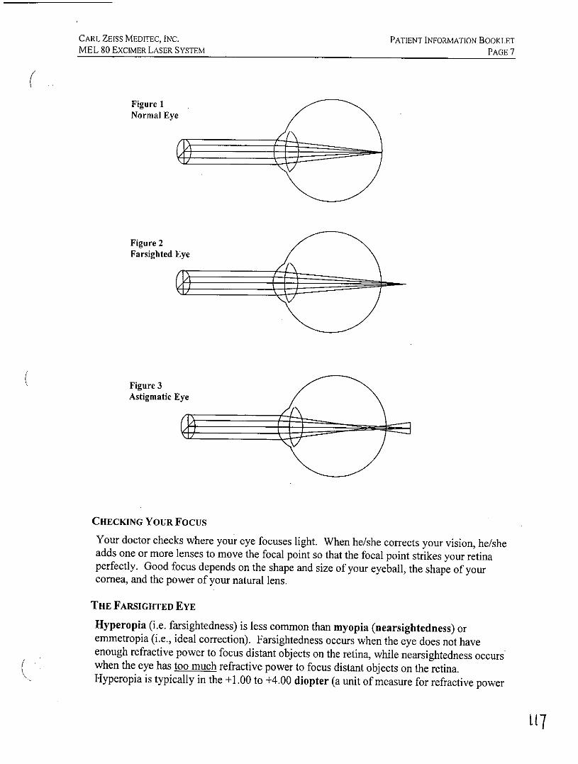
CARL ZEISS MEDITEC, INC. PATIENT INFORMATION BOOKLETMEL 80 ExcIMER LASER SYSTEM PAGE 7
Figure INormal Eye
Figure 2Farsighted Eye
Figure 3Astigmatic Eye
CHECKING YOUR Focus
Your doctor checks where your eye focuses light. When he/she corrects your vision, he/sheadds one or more lenses to move the focal point so that the focal point strikes your retinaperfectly. Good focus depends on the shape and size of your eyeball, the shape of yourcornea, and the power of your natural lens.
THE FARSIGHTED EYE
Hyperopia (i.e. farsightedness) is less common than myopia (nearsightedness) oremmetropia (i.e., ideal correction). Farsightedness occurs when the eye does not haveenough refractive power to focus distant objects on the retina, while nearsightedness occurswhen the eye has too much refractive power to focus distant objects on the retina.Hyperopia is typically in the +1.00 to +4.00 diopter (a unit of measure for refractive power
[17
CARL ZEISS MEDITEC, INC. PATIENT INFORMATION BOOKLETMEL 80 EXCIMER LASER SYSTEM PAGE 8
of lenses; abbreviated D) range, and rarely can be as high as +8.00 D. In contrast to myopia,hyperopia occurs when the eye is too short for the power of its optical components. Inhyperopia, the cornea is not steep enough and light rays hit the retina before they come intofocus. Distant objects appear blurred, and nearby objects are even more fuzzy. Mostfarsighted individuals need corrective eyewear to see clearly at all distances. Glasses,contact lenses or refractive surgery (surgery to change how the eye bends light) can correctfarsightedness.
If your vision changes over time, you can change glasses or contact lenses. Changes due torefractive surgery cannot be reversed. Sometimes the first surgery doesn't correct yourvision enough or corrects it too much. Sometimes your vision changes as time passes. Ineither case, your doctor can try to improve it with added treatments.
WHAT is LASIK?
LASIK is a surgical treatment for farsightedness. A device called a microkeratome, whichis like a carpenter's plane, cuts a thin flap of tissue from the front of the cornea. The doctorthen folds the flap out of the way. Next, an excimer laser (a medical device used to removetissue from the cornea) removes some tissue from the front surface of the cornea to make itmore curved. After the laser treatment, the doctor replaces the corneal flap back in place.
An excimer laser is a laser that aims a strong beam of UV (ultraviolet) light at your eye.The laser creates a brief, intense pulse that lasts just a few billionths of a second. Each pulseremoves a tiny amount of tissue from the surface of the cornea. It makes little heat and doesnot change the tissue beneath.
Doctors perform LASIK surgery on one eye at a time. If all goes well with the first eye,he/she can treat the second eye. Often the doctor does the second eye on the same day. Thedoctor can also do it later; it depends on his/her judgment of your particular case.
LASIK corrects your vision so you can see distant objects better. It does not take away theneed for reading glasses. You may need reading glasses after laser surgery even if you didNOT wear them before.
INDICATIONS FOR USE
The MEL 80 Excimer Laser is indicated for use in primary LASIK treatments for thereduction or elimination of naturally occurring hyperopia of less than or equal to +5.0 Dwith or without refractive astigmatism of > 0.5 D and < 3.0 D, with a maximum MRSE of+5.0 D, in patients who are 21 years of age or older with documentation of stable manifestrefraction over the past year as demonstrated by change in sphere and cylinder of 0.5 D.
CONTRAINDICATIONS - WHEN YOU SHOULD NOT HAVE LASIK
You should NOT have LASIK if any of the things below apply. In these cases, the risk isgreater than the benefit.
CARL ZEISS MEDITEC, INC. PATIENT INFORMATION BOOKLETMEL 80 ExcIMER LASER SYSTEM PAGE 9
* You have severe dry eye; this problem may become worse after LASIK and may put youat risk for infection.
* You have an active infection or inflammation of the eye. These may increase the risk ofcomplications and should be cleared up before LASIK treatment.
* You have advanced glaucoma, because the increased eye pressure associated with cuttingthe LASIK flap might cause further damage to your vision, as might increased eyepressure associated with response to steroid eye drops in some patients.
* Your cornea is too thin to perform LASIK without increasing the risk of causing abulging cornea (ectasia).
* You have an active connective tissue disease or autoimmune disease which may beassociated with comeal melting, such as rheumatoid arthritis, Wegener's granulomatosis,relapsing polychondritis, systemic lupus erythematosus, or polyarteritis nodosa. If youhave LASIK, there is risk of severe damage to your cornea.
* You have a significantly weakened immune system due to medications (such as highdose steroids) or disease (such as AIDS). These conditions make you more prone toinfection after surgery.
* You are pregnant or nursing, which can cause short-term changes in your cornea. Suchchanges cannot be predicted. In such cases, LASIK might improperly change the shapeof your cornea in a way that would harm your vision.
* You have a history of, or show any signs of, keratoconus (thinning and cone-shapedbulging of your cornea), pellucid marginal degeneration (thinning of the lower outer edgeof your cornea, where adjacent cornea may protrude), or any other condition that causesthinning or bulging of your cornea. These conditions can lead to further distortion andthinning of the cornea, and permanently reduced vision after LASIK surgery. This mayresult in the need for additional surgery (such as a comeal transplant) after LASIK.
* You have recently had or now have a herpes eye infection (simplex or zoster), or havesignificant corneal damage (poor sensation, scarring, blood vessels growing into thecornea) from a past herpes infection. These may put you at high risk for further comealdamage.
* You have uncontrolled diabetes or you have visually significant diabetic complications. Ifyour blood sugar is uncontrolled, your spectacle prescription will be changing and yourdoctor will not be able to accurately determine what treatment is appropriate. If you havevision problems related to diabetes, the LASIK procedure may make them worse.
WARNINGS
If you have any of the following conditions, you should talk to your doctor before you decide tohave LASIK, because you may be at greater risk for poor outcomes or injury related to LASIK.You should discuss your level of risk with your doctor. You and your doctor should determinewhether the benefits of LASIK outweigh the risks for you, based on the nature and severity of
CARL ZEISS MEDITEC, INC. PATIENT INFORMATION BOOKLETMEL 80 EXCIMER LASER SYSTEM PAGE 10
your condition. There is risk of serious adverse outcomes if LASIK surgery is performed inpatients with the following conditions:
* Remote history of herpes (simplex or zoster) infection that affected your eyes. It is possiblethat LASIK surgery might lead to reactivation of the dormant herpes virus. (SeeCONTRAINDICATIONS: Conditions When You Should Not Have LASIK for relatedinformation.)
* Symptoms of dry eye. If you have dry eyes, LASIK may increase the dryness, withaccompanying discomfort and visual problems. This may or may not go away. (SeeCONTRAINDICATIONS: Conditions When You Should Not Have LASIK for relatedinformation.)
* You are taking isotretinoin (Accutane®)l, for acne treatment. This medication may causeincreased dryness and result in increased complications after LASIK.
* Diabetes. Even if you have well controlled diabetes, LASIK may be risky for you becauseyour diabetes may interfere with the healing of your eyes. (See CONTRAINDICATIONS:Conditions When You Should Not Have LASIK for related information.)
* Glaucoma, elevated eye pressure (ocular hypertension), or being followed for possibleglaucoma (glaucoma suspect). These conditions may be associated with increased risk withLASIK. There is greater difficulty in accurately monitoring eye pressure after LASIK. Youmay also be at greater risk for damage to your vision associated with increased eye pressurewhen cutting the LASIK flap. You may also be at greater risk for increased eye pressurerelated to medication given after surgery (steroid eye drops). (See CONTRAINDICATIONS:Conditions When You Should Not Have LASIK for related information.)
* Severe allergies. If you rub your eyes a lot after LASIK, you may be at risk for dislodging thecomeal "flap" that was cut for the procedure. The strength of the flap attachment to theunderlying cornea is significantly and permanently reduced after surgery. Additionally, ifyou have severe allergies and take medicines for them, LASIK may be more risky for you,because symptoms of dryness, often associated with anti-allergy medication, may increaseafter LASIK.
* Connective tissue diseases or autoimmune diseases (such as rheumatoid arthritis, lupuserythematosus), even if well controlled and stable. You may have slower healing and lesspredictable outcomes. Depending upon your disease, its severity, and the medication you aretaking, there may be additional risks. These may include an increased risk of severe dry eye,infection, inflammation, or corneal melting. Discuss your additional risks with your surgeon,after he or she has consulted with the other doctors who are treating you. (SeeCONTRAINDICATIONS: Conditions When You Should Not Have LASIK for relatedinformation.)
* A condition (such as HIV) or a medication (such as steroids) that affects your immunesystem. You may be at increased risk for infection. (See CONTRAINDICATIONS:Conditions When You Should Not Have LASIK for related information.)
I Accutane (isotretinoin) is the registered trademark of Hoffman La Roche Inc.
CARL ZEISS MEDITEC, INC. PATIENT INFORMATION BOOKLETMEL 80 EXCIMER LASER SYSTEM PAGE II
* Glasses or contact lens prescription has changed in the last 12 months. If your prescription isnot stable, the right amount of treatment may not be determined accurately enough. This mayresult in poor vision after LASIK.
* Blepharitis, or prior history of blepharitis. Blepharitis is redness on the line of the eyelash. Itincludes crusting of the lashes, often with burning, itching and irritation of the eyes. It canincrease the risk of infection of the flap after LASIK. It also can increase the risk ofinflammation of the flap.
* You have a history of rubbing your eyes. The LASIK flap never completely heals, and it canbe dislodged by eye rubbing. If you are not sure you can keep from rubbing your eyes afterLASIK, you may not be a good LASIK candidate.
PRECAUTIONS
It is not known whether LASIK is safe and effective for the following conditions orsituations. If you have any of the conditions below, tell your doctor. You should discusspossible risks with your doctor before you decide whether to have LASIK.
* You have 0.75 D or more of difference between refractions of your eye taken with andwithout eye drops that prevent your lens from adjusting.
* Your cornea, lens, or the vitreous (a clear, jelly-like substance that fills the middle ofthe eye) are not normal due to disease or other factors (such as a scar or cataract).Things like corneal scars may affect the accuracy of LASIK or the way your eye heals.This may result in poor vision after LASIK.
* You have or had uveitis/iritis (inflammation of the iris or other structures within theeye). In such cases, it is not known whether LASIK is safe and effective. Suchdiseases are often treated with steroids, which can affect wound healing. While thesediseases are active or resolving, they could affect the accuracy of LASIK. They alsocould affect the healing process.
* You have had prior eye surgery (including refractive surgery such as RK, PRK,LASIK, or other surgery) or eye injury. You should tell your doctor about any suchhistory, as it may affect your risk with LASIK.
* You are less than 21 years of age. It is not known whether LASIK is safe and effectivefor you. The safety and effectiveness of LASIK has not been evaluated in people in thisage group.
* You take medicines that may affect wound healing. One such medicine is Imitrex2,used for migraine headaches, and others which include hormone replacement therapyand antihistamines. It is not known whether LASIK is safe and effective for you.
* You are taking any medicines. Tell your doctor all medicines you are taking, with orwithout a prescription. It is possible that medications you are taking might have someeffect on surgical outcome or healing. Tell your doctor if you are taking any
2 Imitrex (sumatriptan succinate) is a registered trademaik ofGlaxo Group Limited
12,1
CARL ZEISS MEDITEC, INC. PATIENT INFORMATION BOOKLETMEL 80 EXCIMER LASER SYSTEM PAGE 12
prescription medicines, any medications you bought without a prescription, or anyvitamins or supplements.
* You have a history of keloid formation.
* You have a MRSE of > +5.0 D, sphere > +5.0 D, cylinder +0.5 D or less, or cylindergreater than +3.0 D, as insufficient safety and effectiveness data are available for eyesin this range.
* You are older than 50 years of age. Compared to younger patients, it is harder topredict outcomes for those 50 years and older.
* You are taking amiodarone hydrochloride (e.g., Cordarone®3), used to treat irregularheartbeats (ventricular arrhythmias). This medication can cause opacities in the corneaand might cause problems with healing after LASIK.
* You have a close family member with keratoconus, pellucid marginal degeneration oranother disorder which may cause thinning or bulging of the cornea. These conditionsmay run in families. If you have one of these conditions and it has not been diagnosed,LASIK may cause more rapid progression of the disease. You should tell your doctorabout any family history of these problems. (See CONTRAINDICATIONS:Conditions When You Should Not Have LASIK for related information.)
* You have a history of any eye disease or abnormality. If you are aware of any eyedisease or abnormality, you should discuss this with your doctor, as it might increaserisks associated with LASIK.
* You have large pupils. You should discuss with your doctor whether your pupil sizemay have negative effects on your vision after LASIK, such as glare, halos, and nightdriving difficulty. Regardless of how large your pupils appear to you, your doctorshould measure your pupil size under dim lighting conditions before surgery.
* You are having LASIK for farsightedness and have a history of "crossed eyes". Youshould tell your doctor if you have this condition as you might have an increased risk ofincreased problems with "misaligned eyes" after surgery.
* You have any other medical conditions (other than those already mentioned). It ispossible that your medical condition might have some effect on surgical outcomes orhealing. Let your doctor know all your medical conditions.
For the following additional conditions, it is not known whether LASIK is safe andeffective:
* Over the long term, more than 24 months after LASIK, it is not known whether LASIKis safe and effective.
* LASIK retreatment with the MEL 80. The clinical study provided insufficient data todetermine the safety or effectiveness of LASIK retreatment with the MEL 80 laser. The
3 Cordarone (amodarone hydrochloride) is the registered trademark ofSanofi-Synthlabo.
CARL ZEISS MEDITEC, INC. PATIENT INFORMATION BOOKLET'MEL 80 EXCNER LASER SYSTEM PAGE 13
risk and accuracy of LASIK retreatment, or LASIK with another surgery to correctvision, has not been evaluated.
* Under conditions of dim lighting, rain, snow, fog or bright glare. Under theseconditions, you might have problems seeing after LASIK. Whether you may have poorvision under these conditions is hard to predict because it has been studied so little. Youshould discuss with your doctor the effects of LASIK on vision in dim lighting, rain,snow, fog, or bright glare, since these conditions have not been fully evaluated.
* It is harder to predict outcomes for those 50 years and older.
Your doctor should evaluate you for dry eyes before surgery. Be sure you tell your eyedoctor if you have ever had symptoms of eye dryness or difficulty wearing contact lensesbecause of dryness. You may have dry eyes after LASIK surgery even if you did not havedry eyes before surgery.
You should be aware that the thinning of the cornea due to LASIK surgery may affect theaccuracy of eye pressure measurements after LASIK, for instance when evaluating your eyesfor glaucoma, and make them more difficult to interpret. For this reason, you should informfuture eye care providers that you have had LASIK, so that they may be better able tointerpret your eye pressure measurements.
LASIK surgery may make it more difficult for a surgeon to determine the correct artificiallens power for cataract surgery implantation. For this reason, you should ask for a patientinformation card that has your eye measurements from before the LASIK surgery. Youshould keep this card to help the cataract surgeon accurately calculate the lens implant powershould you need to have cataract surgery in the future.
If you have one eye that cannot see clearly even with glasses (e.g., if you have amblyopia or"lazy eye", or bad vision from injury or disease), you should discuss this with your doctor.The risk of LASIK-related vision loss in your "good eye" might mean a lot more to you,since you cannot see well with your other eye.
WHAT ARE THE RISKS OF LASIK SURGERY?
* Sometimes LASIK does not give you the best vision the first time. To get the best visionyou can, you may need to have LASIK surgery again.
* LASIK can sometimes leave your vision worse than before, even with glasses orcontacts.
* After LASIK, you may need to wear glasses or contacts to see clearly up close, even ifyou did not need to wear them before. Starting around 40 years of age, people usuallyneed glasses for close work such as reading, even if they did not need them beforesurgery.
* LASIK can give you vision problems or symptoms that you did not have before. It canalso cause vision problems or symptoms that you had before to become worse (for
CARL ZEISS MEDITEC, INC. PATIENT INFORMATION BOOKLETMEL 80 EXCIMER LASER SYSTEM PAGE 14
example, dry eye, halos, glare, difficulty with night driving, ghost images, and fluctuatingvision) with symptoms that may range from mild and annoying to severe and affectingability to perform tasks.
* Permanent loss of vision; some complications may lead to vision loss that cannot becorrected by glasses, contact lenses, or further surgical treatment.
Some associated risks of the LASIK procedure are listed below:
* The cornea or other parts of the eye can become infected because the cornea has been cutand tissue has been removed from it.
* The blade that is used to make the corneal flap can cut all the way through the front partof the eye. This is called perforation of the eye, which can lead to loss of fluid frominside the eye, cataract (cloudiness of the natural lens of the eye) formation, andinfection of the eye.
Other flap complications include:
* The corneal flap can come loose, or can be torn or cut off the eye completely and be lost,making your vision much worse.
The following possibilities are also considered risk factors:
* Retreatment with the MEL 80. It is not known whether LASIK is safe and effective torepeat on the same eye.
* Undiagnosed dry eyes. Your doctor should test you for dry eyes before you have LASIK.LASIK can make dry eyes worse, and it can give you dry eyes even if you did not havethem before. Symptoms may include burning, feeling as though something is in the eye,excessive tearing, pain, redness, and blurred vision. In some cases, the symptomsinterfere with the ability to do daily tasks.
* Allergic reactions. Your eye may have an allergic reaction such as itching, swelling,irritation, tearing or redness.
* Large pupils. Before LASIK, your doctor should measure your pupil (the dark circularopening in the center of the iris that allows light into the eye; it shrinks in bright light andenlarges in dim light) size under dim light. Pupils too large under dim light can lead tobad effects from LASIK. "Too large" means more than 7 mm. These effects includeglare (a light within the field of vision that is bright enough to interfere with the ability tosee other lights or objects), halo (hazy ring around a bright light), and problems drivingat night. If your pupils are too large, talk with your doctor about the risks.
* Ectasia- Bulging of the cornea due to remaining tissue being too thin after LASIK.
* Inflammation- Swelling of the eye often associated with redness and pain. Sometimesswelling can occur without any associated pain, but vision is often blurred. Inflammation
t29
CARL ZEISS MEDITEC, INC. PATIENT INFORMATION BOOKLETMEL 80 EXCIMER LASER SYSTEM PAGE 15
is the body's response to harmful stimuli or injury, achieved by movement of white bloodcells into the injured tissues. Prolonged inflammation can lead to destruction of the tissue.Some inflammation of the cornea after LASIK surgery is normal. But if it is uncontrolled,it can interfere with healing and cause vision loss. It often responds to therapies such asantibiotics and topical steroids, but the flap also might need to be lifted and rinsed forremoval of inflammatory cells and to prevent tissue damage.
During the First Week Following Surgery
* You may have pain and discomfort for up to 7 days after surgery.
* You can expect to have blurred vision and tearing as the cornea heals.
* You may be sensitive to bright lights, or you may have a feeling of a foreign bodysensation on the surface of your eye.
* You may have short-term swelling of the front surface of your eye or eyelids.
* The pressure in your eye may increase. This is often due to the use of eye drops tocontrol inflammation. To control this, your doctor may prescribe another kind of eyedrop, or stop giving you such drops. An increase in eye pressure does not usually causeany symptoms. But to make sure, you must see your doctor as directed to check for anincrease in eye pressure. A severe increase in eye pressure could cause eye pain ornausea. If you have these symptoms, you should contact your doctor.
* You must never rub your eyes. This can cause a shift of the flap, which leads to badeffects. These can include blurred vision, risk of infection, inflammation, edema, loss ofcornea cells or cells to start growing under the LASIK flap, which can cause bumps in theflap.
During the First Week to One Month Following Surgery
* The pressure in your eye may increase. This is often due to the use of eye drops tocontrol inflammation. When you stop the drug therapy, the pressure goes back to normal.
* Your cornea may become so hazy or cloudy that it affects your vision. This hazedisappears over time, and may be related to cells being introduced between the cornealflap and underlying tissue. Some patients see haze up to 6 months after LASIK.
3 months or Longer After Surgery
* You may have periodic loss of the outer layer of the cornea. This can happen followingPRK, and does not usually occur following LASIK. Treatment for loss of the cells mayinclude medication or surgical intervention.
* You may have a wrinkle in the inner layer of the cornea. This layer is made up of strongcollagen fibers and helps the cornea maintain its shape. A wrinkle in the flap may be
CARL ZEISS MEDITEC, INC. PATIENT INFORMATION BOOKLETMEL 80 EXCIMER LASER SYSTEM PAGE 16
caused by external pressure after surgery (rubbing the eye, eye squeezing) or due to ahigh amount of tissue removal from the refractive procedure. A procedure of re-liftingthe LASIK flap is performed to remove the wrinkle. Re-lifting of the flap may cause adecrease in best corrected visual acuity, which should return as the eye heals.
* Some patients have vision complaints and experience a loss of best corrected vision (withglasses).
Research has not shown what effects LASIK has on vision performance in poor lighting.After LASIK, some patients may find it harder than before to see in dim light, rain, snow,fog, or glare from lights at night. Vision performance could be worsened by large pupilsize.
Speak with your doctor about the risk that LASIK may cause bad effects on your vision.These include glare, halos, and problems driving at night.
Your doctor should test you for dry eyes before LASIK. LASIK may make dry eyes worse.You may have dry eyes after LASIK even if you did not before.
Compared to younger patients, it is harder to predict outcomes for those 50 years and older.
WHAT ARE THE BENEFITS OF LASIK SURGERY?
The MEL 80 is intended to reduce or eliminate your farsightedness. It may reduce or endyour need to use contact lenses or glasses. Doctors can use it on farsighted patients up tothese limits:
Patients who are farsighted up to +5.0 D, with astigmatism > +0.50 and < +3.0 D, witha maximum manifest refraction spherical equivalent (MRSE) of +5.0 D.
The clinical study described at the end of this booklet found that LASIK surgery with theMEL 80 Excimer Laser System is a reasonably safe and effective way to correctfarsightedness.
ARE YOU A GOOD CANDIDATE FOR LASIK?
To have LASIK, you must:
* Be 21 years of age or older.
* Have healthy eyes free from retinal problems, corneal scars, and any eye disease.
* Have farsightedness within the range of treatment. The range is up to +5.0 D of spherewith > +0.50 and < +3.0 D of astigmatism. (The maximum manifest refraction sphericalequivalent (MRSE) is +5.0 D.)
* Have proof your vision has not changed more than one half diopter for at least one yearbefore your pre-surgery exam.
CARL ZEISS MEDITEC, INC. PATIENT INFORMATION BOOKLETMEL 80 EXCIMER LASER SYSTEM PAGE 17
* Be fully informed about the risks and benefits of LASIK as compared to other treatmentsfor farsightedness.
* Be able to lie flat without difficulty.
* Be able to keep your eye on the blinking fixation light during the whole LASIK process.
* Be willing to sign an Informed Consent Form provided by your doctor.
* Be able to tolerate eye drops to numb your eye.
WHAT You NEED TO KNow ABOUT THE SURGERY
Unrealistic Expectations about Surgery
Before LASIK, talk to your doctor about your expectations for LASIK surgery.Unrealistic expectations may lead you to be disappointed or cause you to make the wrongdecision about whether to have surgery. You should discuss with your doctor whetheryour expectations are realistic, particularly about how LASIK will change your quality oflife.
If you expect "perfect vision" and believe that you will never need to wear glasses again,then this is an unrealistic expectation. It is realistic for most patients to expect to be ableto perform most distance tasks without glasses. However, a significant number of peoplewill not achieve 20/20 vision without glasses. Patients should consider whether they willbe happy with less than "perfect vision" or will be able to do their work. Patients shouldconsider whether they would be willing to wear glasses for certain activities such asdriving at night, or for reading.
As people age, everyone loses the ability to focus their eyes from far to near. Thisusually becomes a significant problem by the time a person is in his or her forties. Ifpatients have their eyes fully corrected for distance vision, they will need reading glassesin middle age. This is true, even if before surgery they could read clearly withoutglasses. In some cases, the LASIK surgery may cause the need for reading glassessooner than if they had not had the surgery.
Before the Surgery
If you think you want LASIK, you will need an exam first to assess your eyes. This is tomake sure your eyes are healthy and suitable for LASIK. This would include a medicalexam and eye history. Both eyes will be checked.
WARNING: If you wear contact lenses, the doctor will ask you to stop wearingthem before your exam. You must stop two weeks before for hard contact lenses, orone week before for soft contact lenses. This is so the doctor can get a stable eyemeasurement. Failure to do this may lead to poor results from LASIK.
CARL ZEISS MEDITEC, INC. PATIENT INFORMATION BOOKLETMEL 80 EXCIMER LASER SYSTEM PAGE 18
Before LASIK, talk to your doctor if you take any medications or if you have anyallergies. These may cause healing problems. Also discuss whether you should eat anddrink just before surgery. You should arrange to have someone drive you home aftersurgery. Also have them drive you to your next doctor's visit. You should not drive aftersurgery until your doctor gives you permission.
The Day of Surgery
Before the day of surgery, you will be given the chance to hear the sounds the lasermakes. Thus you will be prepared for the noise. On the actual day of surgery, you willbe given some numbing drops in the eye that will be treated. If necessary, the surgeonmay also place a reference mark on your cornea to aid in alignment prior to the surgicalprocedure. This is a temporary dot made on your cornea with an instrument similar to afelt tipped marker. You will not feel any pain with this additional procedure. You willbe shown into the surgery room. There you will see a large machine with a computerscreen, a surgeon's chair and a patient bed. You will be asked to lie down on the bed.You will lay face up toward the laser's microscope and the ceiling. Your eye may benumbed with more drops. The eye not having surgery may be covered with a temporaryshield.
The surgery takes 10 to 20 minutes in total, but the laser is used only about 30 to 60seconds. The doctor will place a small spring-like device between your eyelids to holdthem open.
When the surgery begins, the doctor will put a suction ring onto your eye. This will serveas a track for a small device called a microkeratome. This device will create a thin flapof corneal tissue. While this is done, your vision will become blurry. This is due to thesuction ring, which can cause extreme, temporary increase in eye pressure. After the flapis made, the suction will be switched off. Then the doctor will fold the flap away from thecornea. The doctor will then reposition your head under the microscope. You will beasked to look directly at the blinking light. Try to keep both eyes open without squinting.Try even though a drape or a patch may cover the eye not having LASIK. This makes iteasier to keep looking at the blinking light. You will then hear the noise the laser makeswhen it does its work.
WARNING: It is vital to keep looking right at the blinking light, even if the lightfades or dims. Your results depend on how well you keep focus on this blinkinglight throughout the treatment.
Immediately After the Surgery
After it's done, your doctor will put medicine drops or ointment into your eye. Yourdoctor may apply a patch or shield to your eye for protection and comfort.
Numbing drops make the surgery painless. When these drops wear off, your eye willprobably hurt for a day or two. Most patients describe the pain as moderate to severe.
CARL ZEISS MEDITEC, INC. PATIENT INFORMATION BOOKLETMEL 80 EXCIMER LASER SYSTEM PAGE 19
Your doctor may prescribe pain medicine to make you more comfortable. Do not removethe patch or shield until instructed to do so.
WARNING: You should never rub or touch your treated eye after surgery. Thiscan cause a shift of the flap, which leads to bad effects. These can include blurredvision, risk of infection, inflammation, edema or epithelial in-growth (a conditionwhere epithelial cells grow in between the undersurface of the flap and the frontsurface of the cornea, which can decrease the quality of vision).
First Days After Surgery
The patch or shield is usually removed the next day. You may be mildly sensitive to lightand glare. Wear sunglasses to ease your discomfort. You may also have the feeling thatsomething is in your eye. This happens while the outer layer of your cornea is healing.
Your vision should stabilize within a few weeks. Some patients report small changes invision. These could be better or worse. These may occur up to six months or more afterLASIK.
You may see a haze or cloudiness in the cornea after LASIK. It usually will not affectyour vision. This haze tends to decrease over time. Most often it ends by 12 to 24months after LASIK. However, some patients continue to see haze.
Use as directed any drops and lubricants your doctor prescribes. Your results depend onyou following your doctor's orders. If you use topical steroids, your doctor should watchyou for side effects of long-term use. One side effect may be increased eye pressure orocular hypertension (pressure inside the eye of more than 21 mm Hg with no other signsof glaucoma). This condition is often linked to bad effects. One such effect isglaucoma, which is a group of diseases often marked by increased pressure in the eye.. Itis marked by damage to the nerve fiber layer. It can cause loss of vision. Another badeffect of high eye pressure could be cataract formation. It also can cause a loss of vision.
WARNING: You should contact your doctor if you notice any pain or change orloss of vision in the eye. These may be signs of a serious medical condition.
QUESTIONS TO ASK YOUR DOCTOR
You may want to ask the questions below to help you decide if LASIK with the MEL 80Excimer Laser is right for you.
* What are the other options to correct farsightedness?
* Will I have to limit what I do after LASIK? If yes, for how long?
* What are the benefits of LASIK for my level of farsightedness?
* What vision can I expect in the first few months after LASIK?
1 71
CARL ZEISS MEDITEC, INC. PATIENT INFORMATION BOOKLETMEL 80 ExcIMER LASER SYSTEM PAGE 20
* If LASIK does not correct my vision, could my vision be worse than before? Could myneed for glasses increase over time?
* Will I be able to wear contact lenses if I still need them after LASIK?
* How is LASIK likely to affect my need to use glasses or contact lenses as I get older?
* Will my cornea heal differently if I injure it after LASIK?
* Should I have LASIK surgery on my other eye?
* How long will I have to wait before I can have LASIK surgery on my other eye?
* What vision problems will I have if I have LASIK in only one eye?
Discuss the cost of surgery and follow-up care with your doctor. Most health insurance doesnot cover LASIK for vision correction.
SUMMARY OF IMPORTANT INFORMATION
* LASIK is permanent. Once done, it cannot be reversed.
* LASIK does NOT end the need for reading glasses, even if you have never worn them.
* LASIK is used to treat farsightedness.
* Your vision must be stable for at least one year before LASIK. You will need writtenproof that it has not changed by more than 0.50 D.
* Pregnant and nursing women should wait to have LASIK. For these women, it maycause over- or under-correction as well as regression (reduction or loss of the correctionover time).
* You should not have LASIK if you have a medical condition that impairs wound healing.(For example, corneal scars, uveitis, diabetes, dry eyes etc.)
* The LASIK treatment may cause you discomfort.
* LASIK is not risk-free. Please read this whole booklet before you agree to the treatment.Pay special attention to the sections on Risks and Benefits.
* LASIK is not a laser version of RK. (RK stands for radial keratotomy.) LASIK and RKare completely different from each other.
* Some alternatives to LASIK are glasses, contact lenses, RK and PRK. (PRK stands forphotorefractive keratectomy.)
* Some jobs have vision requirements that RK, PRK, and LASIK do not meet (forexample, military pilots).
* Before you decide to have LASIK you should do as follows:
130
CARL ZEISS MEDITEC, INC. PATIENT INFORMATION BOOKLETMEL 80 EXCIMER LASER SYSTEM PAGE 21
* Have a complete eye exam.
* Talk with one or more doctors about LASIK. Discuss its benefits,complications, risks and time required to heal.
CLINICAL STUDY TO EVALUATE RISKS AND BENEFITS
Researchers did a study to evaluate the safety and effectiveness of LASIK with the MEL 80Excimer Laser. The safety summaries presented in this section were based on the entirestudy eye population while the effectiveness analyses were based on study subjects treated inthe refractive range and with the device treatment consistent with the marketed indicationfor use. The study included 369 eyes.
DEMOGRAPHICS
* 94.7% of study subjects were Caucasian* Subjects ages ranged from 22 to 69 years
SAFETY OUTCOMES: ADVERSE EVENTS, COMPLICATIONS, AND MODERATE-SEVERESYMPTOMS
The study also reports adverse events after LASIK. This is to assess risks, includingworse vision. Some of the more. common adverse events and complications are alsodescribed in the previous section of this document titled "What Are The Risks OfLASIK Surgery?"
1. POSTOPERATIVE BEST-CORRECTED VISION WITH GLASSES COMPARED TOPREOPERATIVE BEST-CORRECTED VISION WITH GLASSES
Some people still needed glasses or contact lenses after LASIK. The study measuredvision with glasses at 6, 9, and 12 months after LASIK. (See Table 1 on thefollowing page)
* At 9 months, the vision with glasses did not change or was improved in 79% ofeyes compared with best vision with glasses before LASIK.
* At 9 months, 16.4% of eyes lost 1 line, and 4.7% lost 2 or more lines of vision;however, only 0.3% of eyes were reported with a decrease of 2 lines or more at thelast available visit.
* At 9 months, 0.3% of eyes had best vision with glasses worse than 20/40.* At 9 months, 0.3% of eyes had best vision with glasses worse than 20/25 when
they were 20/20 or better with glasses before surgery
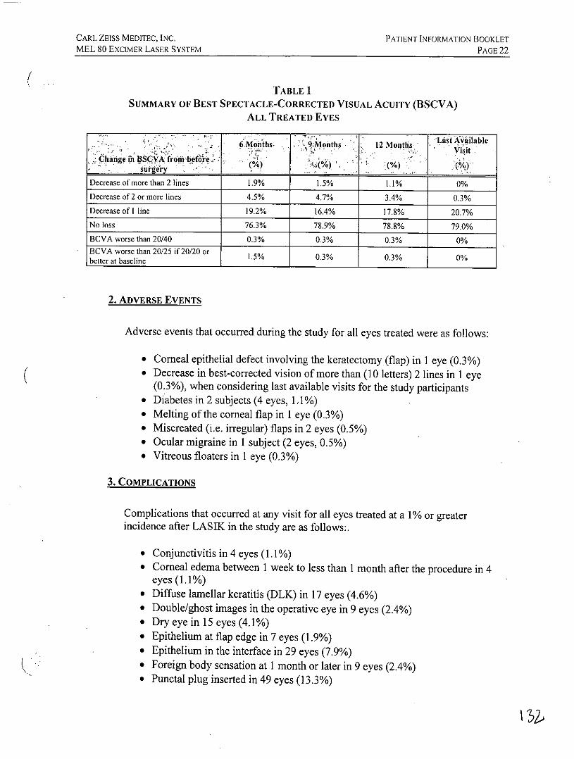
CARL ZEISS MEDITEC, INc, PATIENT INFORMATION BOOKLETMEL 80 EXCIMER LASER SYSTEM PAGE 22
TABLE 1SUMMARY OF BEST SPECTACLE-CORRECTED VISUAL ACUITY (BSCVA)
ALL TREATED EVES
Months 9Months 12 Months
Change in BSCVA from oeforesuirgery1-
Decrease of more than 2 lines 1.9% 1.5% 1.1% 0%
Decrease of 2 or more lines 4.5% 4.7% 3.4% 0.3%
Decrease of I line 19.2% 16.4% 17.8% 20.7%No loss 76.3% 78.9% 78.8% 79.0%
BCVA worse than 20/40 0.3% 0.3% 0.3% 0%BCVA worse than 20/25 if 20/20 orbetter at baseline 1.5% 0.3% 0.3% 0%
2. ADVERSE EVENTS
Adverse events that occurred during the study for all eyes treated were as follows:
* Corneal epithelial defect involving the keratectomy (flap) in 1 eye (0.3%)* Decrease in best-corrected vision of more than (10 letters) 2 lines in 1 eye
(0.3%), when considering last available visits for the study participants* Diabetes in 2 subjects (4 eyes, 1. 1%)* Melting of the corneal flap in I eye (0.3%)* Miscreated (i.e. irregular) flaps in 2 eyes (0.5%)* Ocular migraine in 1 subject (2 eyes, 0.5%)* Vitreous floaters in I eye (0.3%)
3. COMPLICATIONS
Complications that occurred at any visit for all eyes treated at a 1% or greaterincidence after LASIK in the study are as follows:.
* Conjunctivitis in 4 eyes (1.1%)* Corneal edema between 1 week to less than 1 month after the procedure in 4
eyes (1.1%)* Diffuse lamellar keratitis (DLK) in 17 eyes (4.6%)* Double/ghost images in the operative eye in 9 eyes (2.4%)* Dry eye in 15 eyes (4.1%)* Epithelium at flap edge in 7 eyes (1.9%)* Epithelium in the interface in 29 eyes (7.9%)* Foreign body sensation at 1 month or later in 9 eyes (2.4%)* Punctal plug inserted in 49 eyes (13.3%)
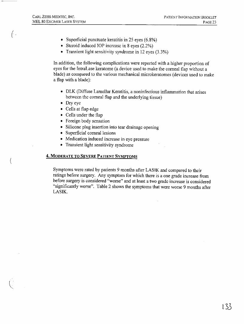
CARL ZEISS MEDITEC, INC. PATIENT INFORMATION BOOKLETMEL 80 EXCIMER LASER SYSTEM PAGE 23
* Superficial punctuate keratitis in 25 eyes (6.8%)* Steroid induced IOP increase in 8 eyes (2.2%)* Transient light sensitivity syndrome in 12 eyes (3.3%)
In addition, the following complications were reported with a higher proportion ofeyes for the IntraLase keratome (a device used to make the corneal flap without ablade) as compared to the various mechanical microkeratomes (devices used to makea flap with a blade):
* DLK (Diffuse Lamellar Keratitis, a noninfectious inflammation that arisesbetween the corneal flap and the underlying tissue)
* Dry eye* Cells at flap edge* Cells under the flap* Foreign body sensation* Silicone plug insertion into tear drainage opening* Superficial corneal lesions* Medication induced increase in eye pressure* Transient light sensitivity syndrome
4. MODERATE TO SEVERE PATIENT SYMPTOMS
Symptoms were rated by patients 9 months after LASIK and compared to theirratings before surgery. Any symptom for which there is a one grade increase frombefore surgery is considered "worse" and at least a two grade increase is considered"significantly worse". Table 2 shows the symptoms that were worse 9 months afterLASIK.
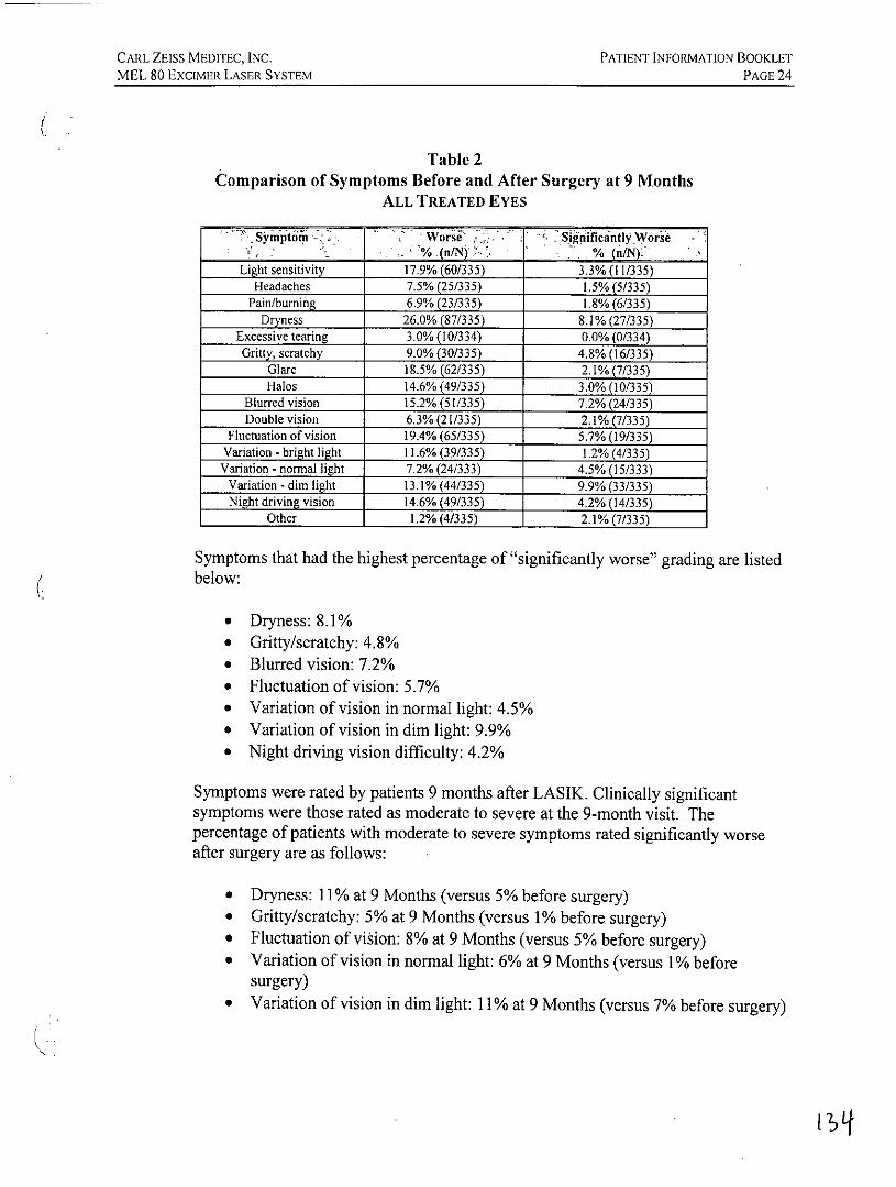
CARL ZEISS MEDITEC, INC. PATIENT INFORMATION BOOKLETMEL 80 EXCIMER LASER SYSTEM PAGE 24
Table 2Comparison of Symptoms Before and After Surgery at 9 Months
ALL TREATED EYES
Sy'injtoAk 7 Worse. Significatly Worie% .(n/N)! % (n/NY.
Light sensitivity 17.9% (60/335) 3.3% (11/335)Headaches 7.5% (25/335) 1.5% (5/335)
Pain/burning 6.9% (23/335) 1.8% (6/335)Dryness 26.0% (87/335) 8.1% (27/335)
Excessive tearing 3.0% (10/334) 0.0% (0/334)Gritty, scratchy 9.0% (30/335) 4.8% (16/335)
Glare 18.5% (62/335) 2.1% (7/335)Halos 14.6% (49/335) 3.0% (10/335)
Blurred vision 15.2% (51/335) 7.2% (24/335)Double vision 6.3% (21/335) 2.1% (7/335)
Fluctuation of vision 19.4% (65/335) 5.7% (19/335)Variation - bright light 11.6% (39/335) 1.2% (4/335)Variation - normal light 7.2% (24/333) 4.5% (15/333)
Variation - dim light 13.1% (44/335) 9.9% (33/335)Night driving vision 14.6% (49/335) 4.2% (14/335)
Other 1.2% (4/335) 2.1% (7/335)
Symptoms that had the highest percentage of "significantly worse" grading are listedbelow:
* Dryness: 8.1%* Gritty/scratchy: 4.8%* Blurred vision: 7.2%* Fluctuation of vision: 5.7%* Variation of vision in normal light: 4.5%* Variation of vision in dim light: 9.9%* Night driving vision difficulty: 4.2%
Symptoms were rated by patients 9 months after LASIK. Clinically significantsymptoms were those rated as moderate to severe at the 9-month visit. Thepercentage of patients with moderate to severe symptoms rated significantly worseafter surgery are as follows:
* Dryness: 11% at 9 Months (versus 5% before surgery)* Gritty/scratchy: 5% at 9 Months (versus 1% before surgery)* Fluctuation of vision: 8% at 9 Months (versus 5% before surgery)* Variation of vision in normal light: 6% at 9 Months (versus 1% before
surgery)* Variation of vision in dim light: 11% at 9 Months (versus 7% before surgery)
CARL ZEISS MEDITEC, INC. PATIENT INFORMATION BOOKLETMEL 80 EXCIMER LASER SYSTEM PAGE 25
EFFECTIVENESS OUTCOMES
The research study included 369 eyes, of which 259 eyes were treated with analgorithm adjustment of +0.75 D added to the sphere component. However, afteranalysis of these 259 eyes, it was found that insufficient effectiveness data existed toapprove the following range of treatment: sphere > +5.0 D, cylinder < +0.50 D and> +3.00 D, and MRSE > +5.0 D. Thus, after excluding eyes outside of theseparameters, the effectiveness cohort consisted of a total of 160 eyes, of which 149eyes were available at the 9 month point of stability and 153 eyes were available at 12months for the effectiveness analysis.
1. UNCORRECTED VISUAL ACUITY (UCVA)
In the study, doctors gave each eye one LASIK treatment with the Carl Zeiss MeditecMEL 80 Excimer Laser. Then the doctors measured the vision of each eye withoutglasses after 1, 3, 6, 9, and 12 months. After 3, 6, 9, and 12 months, more than 59%of eyes had 20/20 vision or better without correction. More than 96% had 20/40vision or better without correction after 3, 6, 9, and 12 months. Most states requireyour vision to be 20/40 or better for you to drive without correction (such as glassesor contacts). In the study on LASIK using the Carl Zeiss MEL 80 Excimer LaserSystem, vision without glasses improved for all eyes.
* At 9 months, 66% of eyes treated for hyperopia with a laser setting adjustment inthe approved range had 20/20 or better vision without glasses.
* 77.2% of the eyes treated for sphere only had 20/20 or better vision. without glasses
* 59.8% of the eyes treated for sphere + cylinder had 20/20 or bettervision without glasses
* After 3, 6, 9, and 12 months, more than 59% of all eyes had 20/20 vision or betterwithout correction.
2. UNCORRECTED VISUAL ACUITY (VISION WITHOUT GLASSES) AFTER SURGERYCOMPARED TO BEST-CORRECTED VISION (VISION WITH GLASSES) BEFORESURGERY
The study compared the vision of each eye treated with a laser setting adjustment inthe approved range with glasses before LASIK and without glasses 9 and 12 monthsafter LASIK. At 9 months, 49.7% saw as well without glasses as they did beforewith glasses. At 12 months, it was 51.3%. This was measured in terms of a gain oflines on the eye chart (the eye chart is the visual acuity chart). A gain of lines meansa patient could read lines of smaller letters after LASIK than they could read before.
* At 9 months, 49.7% saw as well without glasses as they did before surgery withtheir glasses.
CARL ZEISS MEDITEC, INC. PATIENT INFORMATION BOOKLETMEL 80 EXCIMER LASER SYSTEM PAGE 26
* At 12 months, 51.3% saw as well without glasses as they did before surgery withtheir glasses.
CARL ZEISS MEDITEC, INC. PATIENT INFORMATION BOOKLETMEL 80 EXCIMER LASER SYSTEM PAGE 27
3. PATIENT SELF-EVALUATION BEFORE AND AFTER LASIK
At each scheduled visit after LASIK, patients were asked to complete a survey.Through it they could report on their vision and eye comfort in each eye. Patientswere asked to grade their symptoms compared to the same symptoms before LASIKaccording to their severity as either none, mild, moderate, marked, or severe. Patientsgraded symptoms at 9 months after LASIK. Patients described some symptoms asbetter, while some symptoms were worse. Any symptom for which there is at least aone grade increase from before surgery is considered "worse", and at least a onegrade decrease is considered "better".
At 9 months for all eyes treated, a higher percentage of patients reported thefollowing symptoms as "worse" than reported them as "better" after LASIK.
* Light sensitivity* Headaches* Pain* Dryness* Gritty feeling* Glare* Halos* Blurred vision* Double vision* Fluctuation of vision* Variation of vision in bright, normal, and dim light* Night driving vision
As part of the study, patients were asked to assess their vision quality after LASIK.They were asked to assess it in these terms:
* Quality of vision
* Whether they would choose to have LASIK done again
* How satisfied they are with the results
At 9 months for eyes treated with a laser setting adjustment in the approved range, theoverall quality of vision was rated highly, with:
* 97.9% of patients indicating that there was an improvement, while only 2.1%indicated that there was no improvement;
* 88.4% would elect to have the surgery again, while 6.3% were unsure and 5.3%would not have the surgery again;
* 96.8% reported being satisfied, while 2.1% were neutral and 1.1% weredissatisfied.
CARL ZEISS MEDITEC, INC. PATIENT INFORMATION BOOKLETMEL 80 EXCIMER LASER SYSTEM PAGE 28
PATIENT ASSISTANCE INFORMATION
PRIMARY DOCTOR
Name:Address:
Telephone Number:
LASIK DOCTOR
Name:Address:
Telephone Number:
LOCATION WHERE TREATMENT WAS DONE
Name:Address:
Telephone Number:
LASER MANUFACTURER
Carl Zeiss Meditec AGCarl Zeiss Promenade 10Jena, Germany 07740
SALES AND SERVICE
Carl Zeiss Meditec, Inc.5160 Hacienda DriveDublin, California 94568 USA(925) 557-4100
CARL ZEISS MEDITEC, INC. PATIENT INFORMATION BOOKLETMEL 80 EXCIMER LASER SYSTEM PAGE29
INDEX
AAge, patient 1,8, 11,12,13, 16, 17,21Alternatives 20Astigmatism 1, 4, 8, 16
BBenefits 2,3,6,9, 16, 17, 19,20,21Blurred vision 14, 15, 19, 24, 26
CCandidate, suitable 2, 11, 16Cataract 4, 11, 13, 14, 19Cloudiness 4, 14, 19Complications 3,9, 10, 14,21,22,23Contraindications 2, 8, 10, .12Contact lenses 6,7,8, 13, 14, 16, 17,20,21Cornea4, 5,6,7,8,9, 10, 11, 12, 13, 14, 15, 16, 18, 19,20,22,23Cost 20
DDiopter 4, 5, 7, 17Discomfort 10, 15, 19,20Driving 12, 14, 16, 17,24,26
EExam 17,21Excimer laser 1,2,4,5,6, 8, 16, 19,21,25
FFarsighted 2,4, 6, 7,8, 12, 16, 17, 19,20Focus 2,4, 5,6,7, 8, 17, 18
GGlare 4, 12, 13, 14, 16, 19,24,26Glasses 3,6,8, 11, 13, 14, 16, 17, 19,20,21,25Glaucoma 4, 5,9, 10, 13, 19Gritty feeling 26
HHalo 4,12, 14,16,24,26Haze 15, 19Headache 11, 24, 26Healing 10,11,12,15,18,19,20Herpes 9, 10
CARL ZEISS MEDITEC, INC. PATIENT INFORMATION BOOKLETMEL 80 EXCIMER LASER SYSTEM PAGE 30
IInfection 9, 10, 11, 14, 15, 19Inflammation 9, 10, 11, 15, 19, 23Insurance 20
Jobs 6, 20
KKeratoconus 4, 9, 12
LLaser in situ keratomileusis (LASIK) 1, 2, 3, 4, 6, 8, 9, 10, 11, 12, 13, 14, 15, 16, 17, 18, 19, 20,
21,22,23,24,25,26,27Lens 4, 5, 6, 7, 8, 11, 13, 14Light sensitivity 23, 24, 26Lighting conditions, vision in 13, 24, 26
MMedicines 10, 11, 12
Accutane 10Cordarone 12Imitrex 11lubricant 19numbing drops 18ointment 18pain 14, 15, 18, 19, 24, 26
NNearsighted 4, 5, 7Noise, during surgery 18Nursing 9, 20
0Ocular Hypertension 5, 10, 19
PPain 14, 15, 18, 19, 24, 26Precautions 2, 11Pregnant 9, 20Pressure, intraocular 4, 5, 9, 10, 13, 15, 18, 19, 23PRK 5, 6, 11, 15, 20Pupil 4, 5, 12, 14, 16
CARL ZEISS MEDITEC, INC. PATIENT INFORMATION BOOKLET
MEL 80 EXCIMER LASER SYSTEM PAGE 31
RRadial keratotomy (RK) 5, 6, 11, 20Redness 10, 14, 15Refractive surgery 5, 8, 11Results, clinical 17, 18, 19, 26Retina 4, 5, 6, 7, 8, 16Risks 2, 3, 6, 9, 10, 11, 12, 13, 14, 17, 20, 21
SSafety (safe) 3, 11, 12, 13, 21Scars 11, 16, 20Second eye, treatment of 8Stabilize, vision 19Sunglasses 19Swelling 4, 14, 15
TTearing 14, 15, 24
VVision, blurred 14, 15, 19,24,26Vision, change 8
WWound healing 11, 20
(IKL