Embed Size (px)
Citation preview
REVIEW
Melanoma: from mutations to medicine
Hensin Tsao,1,2 Lynda Chin,3 Levi A. Garraway,4 and David E. Fisher1,5
1Department of Dermatology, Massachusetts General Hospital, Boston, Massachusetts 02114, USA; 2The Wellman Center forPhotomedicine, Boston, Massachusetts 02114, USA; 3Department of Genomic Medicine, University of Texas MD AndersonCancer Center, Houston, Texas 77030, USA; 4Department of Medical Oncology, Dana Farber Cancer Institute, Boston,Massachusetts 02115, USA
Melanoma is often considered one of the most aggressiveand treatment-resistant human cancers. It is a diseasethat, due to the presence of melanin pigment, was accu-rately diagnosed earlier than most other malignancies andthat has been subjected to countless therapeutic strategies.Aside from early surgical resection, no therapeutic modal-ity has been found to afford a high likelihood of curativeoutcome. However, discoveries reported in recent yearshave revealed a near avalanche of breakthroughs in themelanoma field—breakthroughs that span fundamentalunderstanding of the molecular basis of the disease all theway to new therapeutic strategies that produce unques-tionable clinical benefit. These discoveries have been bornfrom the successful fruits of numerous researchers work-ing in many—sometimes-related, although also distinct—biomedical disciplines. Discoveries of frequent mutationsinvolving BRAF(V600E), developmental and oncogenicroles for the microphthalmia-associated transcriptionfactor (MITF) pathway, clinical efficacy of BRAF-targetedsmall molecules, and emerging mechanisms underlyingresistance to targeted therapeutics represent just a sam-ple of the findings that have created a striking inflectionin the quest for clinically meaningful progress in themelanoma field.
The disease
Melanomas can arise within any anatomic territory oc-cupied by melanocytes. Although cutaneous melanoma,which develops from epidermal melanocytes of the skin,represents the most common site of origination, noncuta-neous melanocytes such as those lining the choroidal layerof the eye, respiratory, gastrointestinal, and genitourinarymucosal surfaces, or the meninges do occasionally un-dergo malignant transformation, albeit at a low frequency.
Clinical morphologists have traditionally divided thecutaneous disease into several subgroups, including su-perficial spreading melanoma, nodular melanoma, acrallentiginous melanoma, and lentigo maligna melanoma(Fig. 1), and other uncommon variants such as desmo-
plastic melanoma and nevoid melanoma. Histologicalpatterns have been well described, and microscopic fea-tures that correlate with clinical subgroups have beenthoroughly codified. Perhaps one of the most unusualand tested aspects of melanoma physiology is the math-ematical relationship between tumor thickness (i.e., di-ameter of tumor sphere) and outcome (Balch et al. 2009).Other features, including mitotic rate and ulceration,also play significant roles in determining prognosis.Despite decades of study, an understanding of the mela-noma subsets that are destined to be lethal remainsincomplete.
Despite recent therapeutic advances in management ofadvanced melanoma, several crucial biological questionsremain, including: (1) What is the relationship betweenenvironmental exposures and melanoma risk? (2) Do bio-markers exist that may predict clinical behavior and thusguide therapies? (3) Which genomic alterations driveinvasion, metastasis, and drug resistance? (4) Which molec-ular lesions underlie tumor maintenance? (5) Which ab-errant pathways and targets are amenable to eitherpreventative or therapeutic intervention?
Genetic loci and variants that confer melanoma risk
A family history of melanoma occurs in 10% of mela-noma patients and confers an approximately twofoldincrease in melanoma risk (Gandini et al. 2005). One canargue that melanoma is fundamentally a genetic disease,since the range of heritable risk factors—from physicalcharacteristics such as light complexion, an inability totan, red hair, and blue eyes to the familial atypical mole/melanoma (FAMM) syndrome—are all determined by dis-tinct genetic elements. Hereditary melanoma itself is of-ten associated with (1) multiple cases of melanoma inseveral generations on one side of the family, (2) multipleprimary melanomas in a given individual, and (3) earlyonset of disease. In this section, we review recent discov-eries in melanoma predisposition and survey known riskloci, especially those uncovered through genome-wideassociation studies (GWAS).
High-risk melanoma loci
To date, the weight of evidence suggests that the ret-inoblastoma (RB) pathway, which serves to regulate the
[Keywords: BRAF; MITF pathway; melanoma]5Corresponding author.E-mail [email protected] is online at http://www.genesdev.org/cgi/doi/10.1101/gad.191999.112.
GENES & DEVELOPMENT 26:1131–1155 � 2012 by Cold Spring Harbor Laboratory Press ISSN 0890-9369/12; www.genesdev.org 1131
Cold Spring Harbor Laboratory Press on February 16, 2020 - Published by genesdev.cshlp.orgDownloaded from
G1/S checkpoint, is uniquely vulnerable in melanomasusceptibility.
Cyclin-dependent kinase N2A (CDKN2A) It has beenrecognized for decades that there are families with anincreased occurrence of both melanoma and clinicallyatypical moles (i.e., dysplastic nevi) (Fig. 1A). Through thesystematic collection of these kindreds worldwide, link-age analysis on melanoma families led to putative loci onchromosomes 1p36 (Bale et al. 1989) and 9p21 (Cannon-Albright et al. 1992). Within the 9p21 region, the p16(now CDKN2A) gene quickly became an attractive can-didate for the melanoma locus, since p16 was shown to bea potent cell cycle inhibitor through a direct negativeinteraction with CDK4 (Serrano et al. 1993). Hussussianet al. (1994) then demonstrated deleterious germline mu-tations in CDKN2A among a subset of melanoma-pronefamilies that exhibited linkage to chromosome 9p21markers, thereby establishing the first high-risk suscep-
tibility locus in melanoma. Around this time, severalgroups also reported homozygous deletions and deleteri-ous mutations of CDKN2A in a variety of cancer cell lines(Kamb et al. 1994; Nobori et al. 1994). Thus, within a spanof a few years, CDKN2A catapulted into the center ofcancer biology as a critical target of inactivation at boththe germline and somatic levels.
The CDKN2A locus is composed of four exons andencodes for two distinct proteins through alternativesplicing: p16INK4a and p14ARF (Fig. 2; for review, see Chin2003); interestingly, both proteins are potent tumor sup-pressors with distinct but equally crucial roles in cellcycle and apoptosis regulation. p16INK4a binds to andinhibits CDK4/6, thereby preventing CDK4/6 from phos-phorylating the RB protein (Koh et al. 1995). Since hyper-phosphorylation of RB triggers the release of E2F1, atranscriptional inducer of S-phase genes, loss of p16INK4a
encourages G1–S transition and re-entry into the cellcycle. On the other hand, p14ARF binds to human double
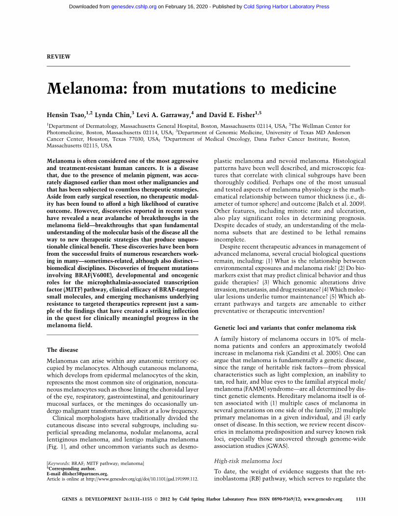
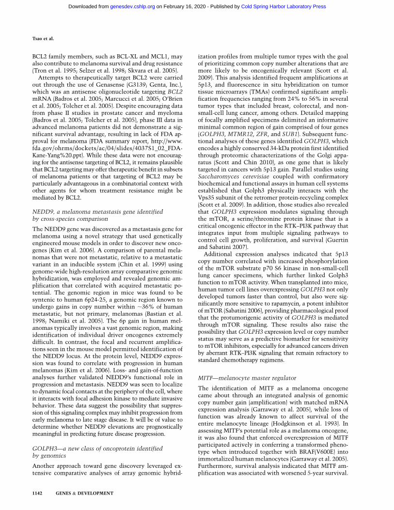
Figure 1. Clinical disease. (A) A patient with mul-tiple clinically atypical moles. Since ;80% of allacquired nevi harbor BRAF(V600E) mutations, hun-dreds of BRAF-activating events occur in such nevi-prone individuals without progression to melanoma.It is thought that melanocyte growth arrest resultsfrom OIS. Common forms of cutaneouos melanomainclude superficial spreading melanoma (B) andnodular melanoma (C). Both of these subtypes areassociated with BRAF or NRAS mutations. (D) Acrallentiginous melanoma is the most common subtypeamong darker-skinned individuals and is more oftenassociated with KIT aberrations. (E) Ocular mela-noma is rarer than cutaneous melanoma and is notassociated with BRAF, NRAS, or KIT changes, butrather with GNAQ or GNA11 alterations (picturecourtesy of Dr. Ivana Kim, Massachusetts Eye andEar Infirmary).
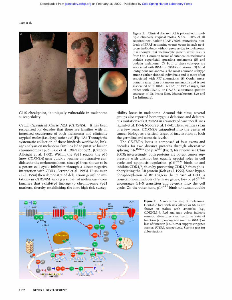
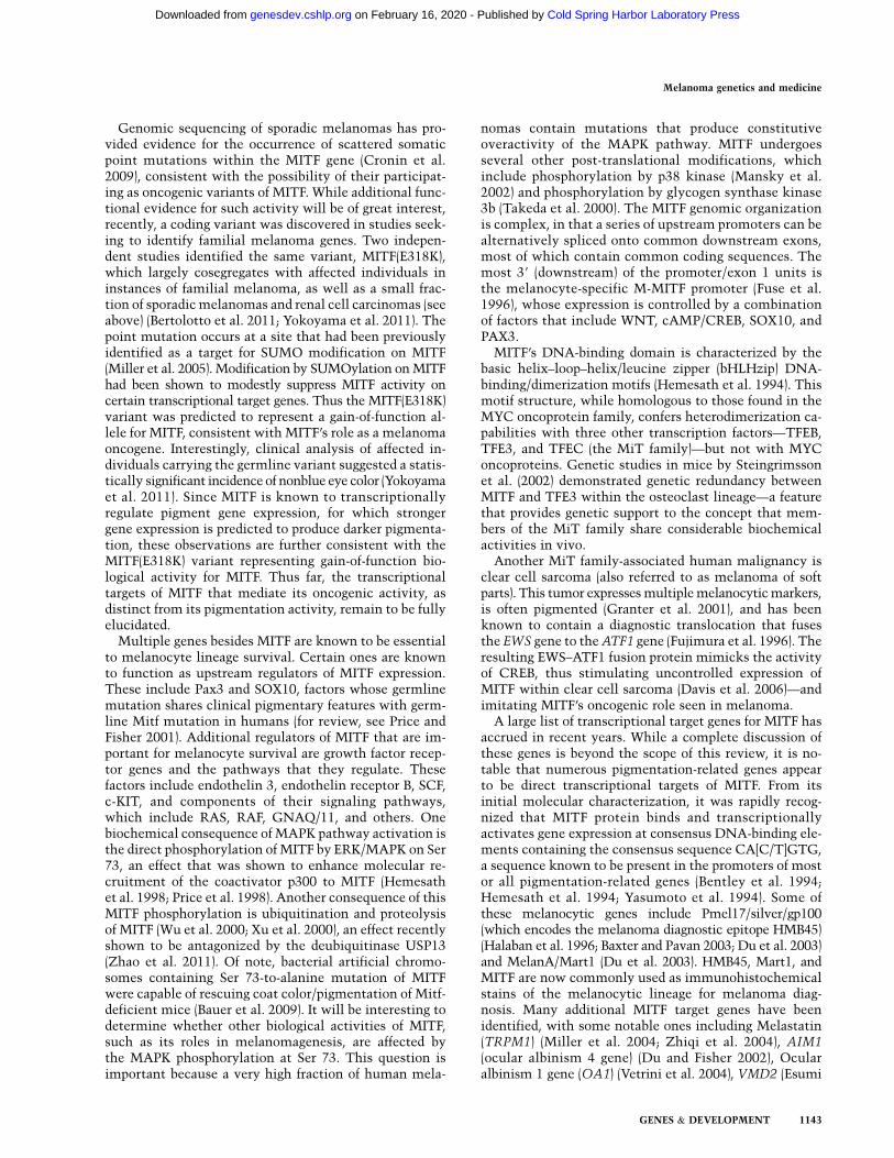
Figure 2. A molecular map of melanoma.Heritable loci with risk alleles or SNPs areshown in italics with asterisks (e.g.,CDKN2A*). Red and gray colors indicatesomatic alterations that result in gain offunction (i.e., oncogenes such as BRAF) orloss of function (i.e., tumor suppressor genessuch as PTEN), respectively. See the text forabbreviations.
Tsao et al.
1132 GENES & DEVELOPMENT
Cold Spring Harbor Laboratory Press on February 16, 2020 - Published by genesdev.cshlp.orgDownloaded from

minute-2 (HDM2) protein at its N terminus and promotesthe rapid degradation of HDM2. Since HDM2 in turnubiquitinates and condemns p53 to destruction, the neteffect of p14ARF loss is a destabilization of p53 (Kamijoet al. 1998; Stott et al. 1998; Zhang et al. 1998). Thus, intrue genetic economy, CDKN2A lesions eliminate boththe RB and p53 pathways through loss of p16INK4a andp14ARF, respectively (Lin et al. 2008).
Most germline mutations that confer melanoma riskoccur in exons 1a and 2, which suggests that p16INK4a isthe preferentially targeted, and functionally dominant,component of CDKN2A. For p14ARF, rare deletions anda 16-base-pair (bp) insertion at exon 1b with a mutationalhot spot at the exon 1b splice site (Hewitt et al. 2002;Harland et al. 2005b) have also been reported. The pres-ence of exon 1b-specific alterations suggest that p14ARFis a bona fide melanoma susceptibility gene independentfrom p16. Even rarer deep intronic mutations of CDKN2Ahave also been described, although these account for veryfew cases worldwide (Harland et al. 2001, 2005a).
Several studies now have estimated the risk of devel-oping cutaneous melanoma among CDKN2A mutationcarriers. For familial cases, the overall penetrance hasbeen calculated to be 30% by age 50, and 67% by age 80,although this risk is higher in sunnier climes. The risk ofmelanoma by age 50 reaches 13% in Europe, 50% in theUnited States, and 32% in Australia; by age 80, it is 58%in Europe, 76% in the United States, and 91% inAustralia (Bishop et al. 2002). Among sporadic CDKN2Acarriers, the risk appears lower: 14%, 24%, and 28% byages 50, 70, and 80, respectively (Begg et al. 2005). Othercoinherited modifiers (such as MC1R variants) (Demenaiset al. 2010) may enhance risk, although the familialascertainment itself may introduce bias and producea higher calculated penetrance. Absolute melanoma riskamong CDKN2A mutation carriers is clearly modulatedby pedigree structure and environmental input.
Several animal models have provided biological proofof CDKN2A’s involvement in carcinogenesis, particularlyin melanoma. These models demonstrated formation offibrosarcomas and lymphomas with high frequency inmice containing a targeted disruption of Cdkn2a (Serranoet al. 1996). Evidence for involvement of this locus inmelanoma has been shown using Cdkn2a�/� mice withalleles containing activating mutations in the oncogenesHRAS (Chin et al. 1997) and NRAS (VanBrocklin et al.2010). These mice were readily induced to form mela-noma-like cutaneous tumors. Within this family of on-cogenes, however, observations have revealed NRAS asthe most frequently mutated family member in humantumors (Hocker and Tsao 2007). Further proof of Cdkn2ainvolvement is shown in a gene exposure model throughultraviolet (UV) irradiation of Cdkn2a�/� mice. In thismodel, in the absence of DNA excision repair (e.g.,inactivation or loss of Xpc), UV irradiation of mice ledto rapid formation of cutaneous melanomas. It is interestingto note that the melanomas induced in this backgroundharbored activating mutations in KRAS (Yang et al. 2007).This model illustrates the importance of fidelity in DNArepair and that any UV-induced damage to the DNAwithout
this repair machinery in place may result in activation of theRAS pathway, thereby leading to melanomagenesis.
CDK4 Recurrent mutations of CDK4, which lead to cellcycle progression, have been reported in the germline ofmelanoma-prone families and in tumors (Wolfel et al.1995; Zuo et al. 1996; Soufir et al. 1998; Tsao et al. 1998a;Molven et al. 2005). These mutations were observed tooccur at a conserved arginine residue (Arg 24) thatabolished regulatory interactions with p16INK4a. Carcin-ogen treatment of a knock-in mouse model expressingthe R24C mutation in CDK4 had a marked increase inpotential to acquire melanomas after exposure. In con-cordance with these observations, some human melano-mas have been known to amplify and/or mutate CDK4(Muthusamy et al. 2006).
RB1 Hereditary retinoblastoma has been linked to inac-tivated copies of RB1 in the germline; however, suchcarriers have a fourfold to 80-fold elevated risk to developmelanoma (Draper et al. 1986; Sanders et al. 1989; Enget al. 1993; Fletcher et al. 2004). Development of sarco-mas in patients with loss of RB1 function decreasesproportionately with decreased use of radiotherapy. How-ever, the incidence of melanomas does not track withradiotherapy use, which is likely due to loss of heterozy-gosity (LOH) for the RB1 allele. This demonstrates theintricate linkages between the CDKN2A/CDK/RB path-ways of tumor suppression in humans.
Low-to-moderate-risk melanoma loci
In contrast to high-risk alleles, low-to-moderate diseasevariants often lack familial clustering and dictate cancer-susceptible traits, such as skin color, rather than canceritself. Although some of these variants are quite common inthe general population, some risk-conferring alleles haveminor allele frequencies (MAFs) below 1%. These low-to-moderate-risk loci are outlined in the following sections.
Determinants of pigmentation and melanoma risk
MC1R A host of epidemiological studies long establisheda direct positive connection between light skin colorand melanoma risk (for review, see Gandini et al. 2005).Among the plethora of genes known to regulate constitu-tive and facultative pigmentation, the melanocortin-1receptor (MC1R) has emerged as one of the leadingmoderate-risk loci for melanoma susceptibility (for review,see Miller and Tsao 2010).
MC1R is a seven-transmembrane G-protein-coupledreceptor that activates adenylate cyclase (Fig. 2) in re-sponse to a-MSH binding (Garcia-Borron et al. 2005).Subsequent increases in cAMP up-regulates the micro-phthalmia-associated transcription factor (MITF), whichconsequently induces the transcription of pigment syn-thetic genes and the production of eumelanin—the majorsource of UV attenuation in darkly pigmented skin.Germline variants of MC1R that disrupt this signalingcascade are present in ;80% of individuals with red haircolor (RHC), <20% of people with brown or black hair,
Melanoma genetics and medicine
GENES & DEVELOPMENT 1133
Cold Spring Harbor Laboratory Press on February 16, 2020 - Published by genesdev.cshlp.orgDownloaded from

and <4% of persons with a robust tanning response(Valverde et al. 1995). Association studies have found thatthe MC1R variants p.D84E, p.R151C, p.R160W, p.D294H,p.R142H, and p.I155T are strongly associated with theRHC phenotype (i.e., ‘‘R’’ variants), while the p.V60L,p.V92M, and p.R163Q variants seem to have weakerassociation with the RHC phenotype (i.e., ‘‘r’’ alleles)(Raimondi et al. 2008). A recent meta-analysis foundthat all but the p.V60L and p.V92M variants were as-sociated with melanoma risk with odds ratios (ORs) rang-ing from 1.42-fold for p.R163Q to 2.45-fold for p.I155T(Raimondi et al. 2008). Interestingly, some variants are asso-ciated with melanoma risk but not pigmentary phenotypes,suggesting that MC1R may harbor subtle cancer effects thatgo beyond hair and skin color (Kennedy et al. 2001).
MITF As discussed above, engagement of a-MSH andMC1R leads to increased cAMP and the induction ofMITF by the cAMP response element-binding protein(CREB) (Fig. 2). Although MITF’s role in melanoma riskwas thought to be purely responsive to MSH signaling,it now appears that variants in MITF itself are also in-structive of risk. Through a whole-genome sequencingeffort, a novel germline MITF variant (p.E318K) was dis-covered in a patient with melanoma. Among familiesharboring this variant, linkage analysis generated a log-of-odds (LOD) score of 2.7, implicating MITF(E318K) as apotential risk variant. In a large Australian/United King-dom case control series of 3940 melanoma patients and4036 controls, the p.E318K variant was found to confera 2.19-fold risk, which is the range calculated for MC1Rvariants, although the prevalence of the MITF(E318K)alteration is significantly lower (<1%) in the generalwhite population compared with MC1R RHC variants.Functional analysis (see ‘‘MITF—Melanocyte MasterRegulator’’ ) showed that the missense mutation at codon318 abrogated a conserved SUMOylation site, therebyaltering the transcription of several MITF targets, in-cluding hypoxia-inducible factor 1a (HIF1a), a regulatoryevent that appears to also confer renal cell carcinoma riskamong MITF(E318K) carriers (Bertolotto et al. 2011;Yokoyama et al. 2011).
Lessons from GWAS
Multiple GWAS have also yielded several risk-associatedsingle-nucleotide polymorphisms (SNPs) across the ge-nome (for review, see Chatzinasiou et al. 2011). The mostsubstantiated loci include MC1R (per allele OR = 1.83;95% confidence interval [CI] = 1.44–2.32), ASIP (per alleleOR = 1.35; 95% CI = 1.08–1.68), TYR (per allele OR = 1.34;95% CI = 1.14–1.58), IRF4 (per allele OR = 0.80; 95% CI =0.67–0.95), and SLC45A2 (per allele OR = 0.72; 95% CI =0.44–1.18). Additional loci that have recently been foundinclude loci at CASP8 (OR = 1.11; 95% CI = 1.06–1.18),CCND1 (OR = 1.07; 95% CI = 1.01–1.13), ATM (OR = 0.87;95% CI = 0.81–0.94), MX2 (OR = 0.91; 95% CI = 0.86–0.96), and chromosome 1q21.3 (OR = 0.89; 95% CI = 0.85–0.95), including a region containing the ARNT and SETDB1genes (Chatzinasiou et al. 2011; Macgregor et al. 2011). It is
clear from careful correlative analysis that some of theseSNPs define overlapping phenotypes such as sun sensitiv-ity, propensity for nevus formation, and skin cancer risk,including melanoma and nonmelanoma skin cancers.
In summary, a harvest of novel low–moderate mela-noma risk loci has now been discovered through candidateand systematic genome-wide approaches. At this point, itis unlikely that there are many undisclosed recurrent high-risk loci (e.g., CDKN2A). Rather, mutations in multiplehigh-penetrant genes or the presence of several moderate-risk alleles in a single kindred may explain the balance ofstrong melanoma pedigrees. On a population level, most ofthe attributable risk for melanoma may in fact result froman untoward conspiracy of moderate-risk SNPs.
Biological drivers and therapeutic targets in melanoma
Receptor tyrosine kinase (RTK) activation
Over the years, a considerable amount of evidence hasaccrued in support of the notion that RTKs may contrib-ute importantly to melanoma biology. Whereas earlierstudies focused primarily on receptor overexpression, re-cent genomic studies suggest that genetic dysregulationmay also play a role in some cases. These advances raisethe possibility that small-molecule therapeutics targetingcertain RTKs may prove clinically useful against specificsubsets of melanoma. An integrated diagram of signalingmolecules and biological drivers in melanoma is shownin Figure 2.
KIT The c-KIT gene encodes the RTK for stem cell fac-tor (SCF). Although early reports described a sequentialloss of c-KIT expression from benign to primary and met-astatic melanomas (Montone et al. 1997; Shen et al. 2003;Zhu and Fitzpatrick 2006), reconstitution of the RTK inmetastatic melanoma cells apparently restored sensitivityto SCF-induced apoptosis in vitro (S Huang et al. 1996).The role of c-KIT in melanoma biology had been uncertainuntil more recent genomic screens.
A survey of copy number imbalances in primary mela-nomas found the KIT locus (chromosome 4q11) to beamplified and/or mutated in 39% of mucosal, 36% of acral,and 28% of melanomas on chronically sun-damaged skin(CSD), respectively (Curtin et al. 2006). However, subse-quent large series from Australia (Handolias et al. 2010)and China (Kong et al. 2011) revealed much lower rates ofaberrations. It is notable that many of the activating KITmutations described in melanomas overlap with thosereported for gastrointestinal stromal tumors (GISTs)(Curtin et al. 2006; Antonescu et al. 2007; Rivera et al.2008; Smalley et al. 2008; Ashida et al. 2009), suggestingthat a similar trophic signaling pathway is shared be-tween the two tumor types.
Given the success of imatinib mesylate in KIT-mutatedGISTs, a pharmacological framework for targeting KIT-mutated melanomas became evident. Several early re-ports of dramatic responses using imatinib (Hodi et al.2008; Lutzky et al. 2008) led to the development of twophase II (open label) trials of imatinib for KIT-mutated
Tsao et al.
1134 GENES & DEVELOPMENT
Cold Spring Harbor Laboratory Press on February 16, 2020 - Published by genesdev.cshlp.orgDownloaded from

melanomas. In one trial (Carvajal et al. 2011), 28 patients(N = 25 evaluable) with metastatic KIT-altered melano-mas were treated, and two complete responses (CRs; 94and 95 wk), two durable partial responses (PRs; 53 and 89wk), and two transient PRs (12 and 18 wk) were reported.The median progression-free survival (PFS) and overallsurvival (OS) were 12 wk and 46.3 wk, respectively. Interms of the genetics, 23.4% of the cases harbored KITmutations and/or amplifications, with the most signifi-cant responses occurring in patients with c-KIT(K642E) orc-KIT(L576P) mutations and those with tumors thatenriched for the presence of a KIT mutation (i.e., muta-nt:allele ratio >1). In the second trial (Guo et al. 2011), 43patients with metastatic melanomas harboring KIT aber-rations (i.e., exons 9, 11, 13, 17, and 18 mutations and/orcopy number gains) were treated with imatinib. Partialresponses were observed in 10 patients (23.3%), withdisease stabilization observed in another 13 patients(30.2%), and progressive disease observed in an additional20 patients (46.5%). The median PFS and OS were 3.5 moand 14.0 mo, respectively, in this second trial. Genotype–phenotype correlations did not reveal any evident relation-ships between response and KIT mutations. It is clear thatimatinib exhibited only modest effects, although otherRTK inhibitors (e.g., sunitinib, nilotinib, and dasatanib)are being tested with hopes of being more efficacious.
Epidermal growth factor receptor (EGFR) The gene en-coding the EGFR is located on chromosome 7, which haslong been known to undergo frequent polysomy in ad-vanced melanoma (Koprowski et al. 1985; Bastian et al.1998; Udart et al. 2001). Whereas enforced activation ofEGFR may promote metastatic progression in cell linestudies (de Wit et al. 1992; TS Huang et al. 1996), neitheractivating EGFR mutations nor focal EGFR amplifica-tions have been observed in melanoma. Thus, most stud-ies linking EGFR activation to melanoma biology haverelied on protein expression or activation studies (Topcu-Yilmaz et al. 2010; Tworkoski et al. 2011). An assortmentof in vitro studies has suggested that ectopic EGFR ex-pression may enhance melanoma cell growth (Diaz et al.2007; Ueno et al. 2008) and that pharmacological block-ade of EGFR using small-molecule inhibitors or mono-clonal antibodies may suppress growth in melanoma celllines (Boone et al. 2011), either alone or in combinationwith other targeted agents (Ivanov and Hei 2005; Schicheret al. 2009). Also, preliminary results raise the possibilitythat EGFR activation may contribute to uveal melanomapathogenesis in some instances (Wu et al. 2012). How-ever, some studies found that the gefitinib GI50 valuesreached the 2–3 mM range and that melanoma cellscontinued to grow even at 10 mM gefitinib (Djerf et al.2009). Together with the paucity of driver EGFR muta-tions in melanoma, such results have dampened preclin-ical enthusiasm for EGFR as a target in this malignancy. Infact, one recent study found that EGFR expression maymodestly suppress melanoma growth in a B16 model (Diazet al. 2009).
The limited clinical experience with EGFR inhibitorshas been similarly disappointing. A phase II trial of the
EGFR inhibitor gefitinib in 46 metastatic melanomapatients resulted in a median PFS of only 1.4 mo and amedian OS of only 9.7 mo. During treatment, there wereno reproducible changes in tumoral p-ERK1/2, p-AKT,and PAK1 and serum vascular endothelial growth factor(VEGF) and IL-8 levels (Patel et al. 2011). Thus, despitethe aforementioned experimental evidence of EGFR’sinvolvement in melanoma progression, there are scantclinical data to support single-agent anti-EGFR therapy.
Despite the relatively poor support for either EGFRdependency or monotherapy in melanoma, recent resultsraise the possibility that the combination of EGFR andRAF inhibitors might prove beneficial in some cases of denovo resistance to RAF inhibition. Bernards and col-leagues (Prahallad et al. 2012) performed a synthetic-lethal RNAi screen in the setting of RAF inhibition inBRAF mutant colon cancer cells, which show limited sen-sitivity to RAF inhibitor monotherapy. They found thatEGFR knockdown and pharmacological blockade weresynergistic with vemurafenib in the suppression ofBRAF mutant cells that were intrinsically resistant tovemurafenib alone. In this study, the addition of EGFRinhibition appeared to interdict a feedback up-regula-tion engendered by vemurafenib treatment. Althoughthese results were seen in BRAF mutant colorectal cancer,they raise the intriguing possibility that at least someBRAF mutant melanomas that exhibit de novo resistanceto RAF inhibition may be candidates for concomitantEGFR inhibition in future clinical trials.
MET The RTK c-MET is normally activated by bindingof its ligand, hepatocyte growth factor/scatter factor(HGF). Autocrine activation of HGF-MET has been de-scribed in melanoma progression (for review, see VandeWoude et al. 1997; Li et al. 2001). Although increased c-MET expression has been observed in metastatic mela-noma (Natali et al. 1993), the MET locus also resideson chromosome 7, which commonly undergoes poly-somy in this setting (as described above). As with EGFR,MET amplifications and activating point mutations thusfar have not been described in melanoma. Thus, theevidence that MET activation constitutes a bona fidemelanoma dependency remains scant. However, severallines of experimental evidence suggest that MET signal-ing may enhance melanoma growth and metastasis. Forexample, HGF exposure may promote increased melano-cytic cell mobility (Damm et al. 2010), and HIF-1a maypromote MET-dependent invasion and vasculogenicmimicry in melanoma cells (Comito et al. 2011). Thus,METactivation may serve to augment rather than ‘‘drive’’melanomagenesis and progression per se.
A small-molecule inhibitor of c-MET (SU11274) is nowavailable for potential clinical use. In one preclinical anal-ysis, melanoma lines that exhibited constitutive c-METstimulation in the absence of MET alterations underwentdecreased proliferation and an increase in apoptosis whenexposed to SU11274. In a xenograft model, the compoundalso demonstrated significant anti-tumor activity (Kenesseyet al. 2010). Currently, the role of anti-c-MET treatmentin melanoma remains a theoretical promise at best.
Melanoma genetics and medicine
GENES & DEVELOPMENT 1135
Cold Spring Harbor Laboratory Press on February 16, 2020 - Published by genesdev.cshlp.orgDownloaded from

Ephrin receptors Ephrin receptors constitute the largestfamily of RTKs in the kinome. Physiologically, ephrinligands are membrane-bound, so forward (through Ephreceptors) and reverse (through ephrin molecules) signal-ing can occur upon cell–cell interaction (Genander andFrisen 2010). One specific Eph member, EphA2, has beendirectly implicated in tumor formation. Published stud-ies report an increased expression of EphA2 in multiplecancer types, including melanoma (Easty et al. 1999;Seftor et al. 2002; Hendrix et al. 2003; Kinch and Carles-Kinch 2003; Miyazaki et al. 2003; Herath et al. 2006;Genander and Frisen 2010). Furthermore, the EPHA2locus maps to chromosomal region 1p36, which is a regionfrequently altered in melanoma (Sulman et al. 1997).Mechanistically, EphA2 appears to participate in cross-talk between other cancer signaling circuits, such as theRAS–phosphatidylinositol 3-kinase (PI3K)–AKT and RAS–MAPK pathways (Menges and McCance 2008). Recentinvestigations revealed that some melanomas are, infact, ‘‘addicted’’ to EphA2 (Udayakumar et al. 2011).shRNA-mediated silencing of EphA2 led to a rapid apo-ptotic response along with tumor suppression in a xeno-graft model, while ectopic expression of EphA2 led toenhanced colony formation and migration. Interestingly,EphA2 appears to be essential for UV-mediated apoptosis,and acute introduction of EphA2 into normal and im-mortalized cells also elicits an apoptotic response (Zhanget al. 2008). These findings suggest that Eph receptorsplay a complex role in melanocytes and melanomas. It ispossible that early death purges the genetically suscepti-ble population, thereby leaving more aggressive tumorcells that come to depend on EphA2 signaling for suste-nance; this is compatible with an ‘‘overdose/addiction’’model whereby oncogene stress, which is a commonphysiological response, serves as a selection pressure fortumorigenic recruits.
Although mutagenic activation of EPHA2 is not com-monly observed (Udayakumar et al. 2011), other Eph re-ceptors have been shown to be mutated in melanoma. Inparticular, multiple lesions have been observed in EPHA6,EPHA10, EPHB1, EPHB2, and EPHB6 (Prickett et al. 2009).Given the various molecular systems that are impactedby these receptors, functional classification of the Eph re-ceptors into oncogenes or tumor suppressors is not yetpossible without further study.
ERBB4 In a sweeping analysis of the tyrosine kinome,Prickett et al. (2009) screened the coding exons of 86 pro-tein tyrosine kinases and identified 99 nonsynonymoussomatic mutations. Most prominent among these wasERBB4, which was mutated in 19% of melanoma cases.The missense mutations were oncogenic in several in vitroassays, such as NIH-3T3 transformation and soft agargrowth; a recent genotyping effort (Dutton-Regester et al.2012) points to a possible low-prevalence ERBB4(E452K)hot spot. Inhibition of ERBB4 by lapatinib also led toapoptosis in ERBB4-mutated cells. Given the broad-basednature of this kinome screen, it is unlikely that commonforms of melanoma harbor high rates of recurrent acti-vating mutations in any single RTK. It is possible that
melanoma cells have bypassed the need for more up-stream signaling, given the high rate of oncogenic changesin downstream molecules such as NRAS and BRAF (seebelow).
Glutamate receptor dysfunction
GRMs Although RTKs represent attractive biologicaltargets for oncogenic activation, other surface receptorsmay promote melanomagenesis in unsuspecting ways. Itis intriguing that several neurotransmitter receptors havenow been shown to participate in melanoma pathogene-sis. Since melanocytes are derived embryologically fromneural crest cells, it is possible that a shared mechanisticcircuitry between neural and melanocyte descendantspermits melanoma cells privileged access to neurophys-iological molecules. The earliest hints that glutamatereceptors contribute to melanoma formation were de-rived from mouse studies (Pollock et al. 2003a) in whichaberrant Grm1 expression was associated with a mela-noma-prone phenotype. GRM1 is expressed in humanmelanoma specimens but not benign melanocytic nevi.This surface molecule is a member of the metabotropicglutamate receptor family, which is comprised of G-protein-coupled receptors (GPCRs) that activate phospholipase Cupon ligand binding. More recently, Choi et al. (2011)showed that mice in which metabotropic glutamatereceptor 5 (mGluR5) expression was driven by the Trp1promoter also developed murine melanomas with highpenetrance. Furthermore, mGluR5 expression could bedocumented in human melanomas and may provide anoncogenic signal through ERK (Choi et al. 2011).
The role of metabotropic glutamate receptors remainedcircumstantial until mutations were uncovered in an-other metabotropic glutamate receptor, GRM3, duringa GPCR family-wide screen of human melanoma speci-mens (Prickett et al. 2011). Four of these mutations weresubjected to functional analysis (p.Gly561Glu, p.Ser610Leu,p.Glu767Lys, and p.Glu870Lys) and found to stimulateMEK1/2 in the presence of agonist and melanoma migra-tion even in the absence of agonists. Melanoma cells withGRM3 variants were also more sensitive to MEK inhi-bition by AZD6244. On the surface, activated GRM3appears to be an accessory to MAPK signaling in mela-nomas. However, since BRAF or NRAS is often mutatedin melanoma cells (see below), the precise contribution ofGRM3 to this pathway is unclear. It is known that evenamong BRAF-mutated melanomas, there is a range ofprimary sensitivity to BRAF and MEK inhibitors (Flahertyet al. 2010; Chapman et al. 2011). Thus, accessory MAPKsignal flux through proteins such as GRM3 may playa compensatory role.
GRIN2A Through an unbiased exome-wide sequencingeffort, mutations in GRIN2A have also been discovered(Wei et al. 2011). Unlike GRM3, GRIN2A is an ionotropicglutamate-gated ion channel that binds N-methyl-D-aspartate (NMDA). This ligand-gated channel is per-meable to cations, including Ca2+. Mutations in GRIN2Awas found in 34 out of 135 melanoma samples (25.2%).
Tsao et al.
1136 GENES & DEVELOPMENT
Cold Spring Harbor Laboratory Press on February 16, 2020 - Published by genesdev.cshlp.orgDownloaded from

Unlike GRM3, however, there were multiple missenseand several nonsense mutations scattered along GRIN2A,suggesting that this gene is not a canonical oncogene inmelanoma. Functional validation of GRIN2A as an on-cogene has yet to be performed.
Small G proteins, including RAS
The RAS family of small G proteins serves to transducesignals triggered by extracellular growth factors (Ji et al.2012a). Unlike other solid tumors, activating RASmutations occur in a relatively small fraction of mela-nomas (;10%–15%), with a higher frequency noted inamelanotic nodular melanoma subtypes (for review, seeChin et al. 1998). Among RAS genes, melanoma muta-tions are most common in NRAS, which is also thoughtto be mutated in the majority of congenital nevi (Pappet al. 1999) but rarely in dysplastic nevi (Albino et al. 1989;Jafari et al. 1995; Papp et al. 1999). HRAS mutation hasbeen associated with Spitz nevi, based on both genomicamplifications and mutations (Bastian et al. 2000). KRASand HRAS mutations have both been reported in ;2% ofmelanomas (http://www.sanger.ac.uk/genetics/CGP/cosmic).
Interestingly, differences between HRAS and NRAShave also been noted in relation to the consequences oftheir transgenic targeting to melanocytes. Whereas acti-vated HRAS together with loss-of-function mutations inCdkn2a and/or Trp53 produce nonmetastatic melanomasin mice (Chin et al. 1997; Bardeesy et al. 2001; Sharplesset al. 2003), activated NRAS together with Cdkn2a de-ficiency produce melanomas with major metastatic pro-pensity to both lymph nodes and distant sites (Ackermannet al. 2005).
Since oncogenic RAS proteins were among the firstoncogenes described in humans, potent pharmacologicalinhibitors of RAS proteins have been a source of aggres-sive development. Farnesyl transferase inhibitors (FTIs;e.g., R115777/tibifarnib) were first deployed as selectiveRAS inhibitors, given the drug’s ability to interfere withthe requisite lipid modification of RAS (James et al. 1993);despite initial enthusiasm, FTIs have not fared well inclinical trials (Caponigro et al. 2003). One hypothesisfor this failure is that FTIs impair other farnesylated pro-teins, which then lead to dose-limiting toxicities. Alterna-tively, RAS proteins may use geranylgeranyltransferases,thereby bypassing the block imposed by FTIs (Whyteet al. 1997). More recently, S-trans, trans-farnesylthiosali-cylic acid (FTS) was developed to mimic the C-terminalfarnesylcysteine (Weisz et al. 1999), thereby competingwith the active, GTP-bound forms of RAS for specific bindingsites on the cellular membrane (Aharonson et al. 1998).FTS appears to be effective in inhibiting melanoma growthboth in vitro and in animal models (Jansen et al. 1999;Smalley and Eisen 2002). Clinical studies in pancreaticcancer also showed some possible survival benefits (Johnsonet al. 2009; Laheru et al. 2009), although the efficacy of FTSas a single agent in melanoma awaits clinical testing.
GNAQ The story behind GNAQ’s involvement in mel-anoma unfolded through an exchange between develop-
mental biology and cancer genetics. In a forward geneticscreen, Barsh’s laboratory (Van Raamsdonk et al. 2004)identified hypermorphic mutations in GNAQ and GNA11as causative of diffuse hyperpigmentation and dermalmelanocytosis in mice. Among comparable human le-sions, blue nevi represent a benign proliferation of dermalmelanocytes. Given the phenotypical overlap, analyses ofGNAQ and GNA11 were performed in a collection ofbenign and malignant melanocytic tumors, and GNAQmutations were found in 83% of blue nevi (N = 29), 50% of‘‘malignant blue nevi’’ (N = 2), and 46% of uveal melano-mas (N = 48) (Van Raamsdonk et al. 2009). Like otheroncogenes, a single missense change (p.Q209L) accountsfor all identified GNAQ mutations. This GNAQ variantfully activates the MAPK pathway and is oncogenic inboth in vitro and in vivo assays. In a follow-up study (VanRaamsdonk et al. 2010), somatic mutations in exon 5(affecting Q209) and exon 4 (affecting R183) of bothGNA11 and GNAQ were seen in a mutually exclusivepattern. GNA11 mutations are present in 7% of blue nevi,32% of primary uveal melanomas, and 57% of uveal mel-anoma metastases, while GNAQ alterations were presentin 55% of blue nevi, 45% of uveal melanomas, and 22% ofuveal melanoma metastases. Both GNAQ and GNA11mutations activate the MAPK pathway. The epistaticrelationship between GNAQ and GNA11 implies thatboth fulfill overlapping functions in melanocytes or thatthe presence of both mutations creates a synthetic-lethalcondition. There is also early evidence that GNA11 alter-ations may be more predictive of metastases and thus mayrepresent a potentially crucial therapeutic target (VanRaamsdonk et al. 2010).
Other members of the heterotrimeric G-protein family,which includes GNAQ and GNA11, have also beenscreened in unselected metastatic melanomas (Cardenas-Navia et al. 2010). No other recurrent alterations weredetected, although the overall nonsynonymous somaticmutation rate was 17.5%.
BRAF activation—the heart of melanoma oncogenesis
The discovery of oncogenic BRAF mutations in mela-noma (Davies et al. 2002) stands as one of the mostpowerful affirmations of the transformative potential ofsystematic cancer genome characterization. In hindsight,it is all the more remarkable that BRAF mutations werediscovered by Stratton and colleagues (Davies et al. 2002)using a discovery set of only 15 tumor/normal pairs—onlyone of which was a melanoma sample! Since that seminaldiscovery, BRAF mutations—most commonly a valine-to-glutamic acid substitution at codon 600—have been ob-served in ;50% of melanomas (Maldonado et al. 2003;Pollock et al. 2003b; Uribe et al. 2003; Daniotti et al. 2004;Kumar et al. 2004; Shinozaki et al. 2004; Libra et al. 2005)and to a lesser extent in other cancers (Ciampi andNikiforov 2005; Young et al. 2005). BRAF mutations alsooccur at high frequencies (>80%) in melanocytic nevi(Pollock et al. 2003b; Yazdi et al. 2003; Kumar et al. 2004;Saldanha et al. 2004), suggesting that these somatic alter-ations occur early in melanomagenesis. Interestingly, in-
Melanoma genetics and medicine
GENES & DEVELOPMENT 1137
Cold Spring Harbor Laboratory Press on February 16, 2020 - Published by genesdev.cshlp.orgDownloaded from
dividuals with germline BRAF mutations develop cardio–facio–cutaneous syndrome but do not exhibit increasedcancer risk (Niihori et al. 2006; Rodriguez-Viciana et al.2006)—nor do they harbor the V600E mutation that is soprevalent in melanoma, colorectal cancer, and thyroidcancer.
Although it is tempting to speculate that the BRAF(V600E)mutation is induced by UV damage, the T / A trans-version that converts the valine to glutamic acid at aminoacid 600 (V600E) is not part of the ‘‘classic’’ UV-inducedmutational signature (Daya-Grosjean et al. 1995). None-theless, a durable epidemiological relationship betweenBRAF mutations and sun exposure has been noted. Inparticular, BRAF mutations are much more common inmelanomas arising on intermittent sun-exposed (solarelastosis of arms, trunk, etc.) than in acral melanomas—which arise on less sun-exposed glabrous skin—and mu-cosal melanomas (e.g., gastrointestinal, vaginal origin, etc.)(Maldonado et al. 2003; Edwards et al. 2004; Curtin et al.2005; Bauer et al. 2011). Strikingly, BRAF mutations areabsent in uveal melanoma (Cohen et al. 2003; Cruz et al.2003; Edmunds et al. 2003; Rimoldi et al. 2003; Weberet al. 2003). Conceivably, the substitution that undergirdsthe BRAF(V600E) mutation may reflect a secondary effectof UV damage, such as the generation of reactive oxygenspecies. Toward this end, recent results suggest that mel-anocytic cells may be deficient in repair of oxidative DNAdamage (Wang et al. 2010). Alternatively, this event mayarise as a result of ‘‘nonclassic’’ DNA lesions induced byUV (for review, see Besaratinia and Pfeifer 2008).
Given that melanocytic nevi rarely progress into mel-anoma, it stands to reason that BRAF(V600E)-inducedcheckpoint mechanisms may produce a senescence-likestate in the absence of additional genetic or molecularevents that promote tumorigenesis. Toward this end, con-genital nevi stain positively for senescence-associatedacidic b-galactosidase (SA-b-Gal) (Michaloglou et al. 2005),and BRAF(V600E) expression in primary human melano-cytes induces cell cycle arrest. Thus, oncogene-inducedsenescence (OIS) appears to constrain progression of pre-malignant melanocytic lesions (Sharpless and DePinho2005). Senescent melanocytes exhibit a mosaic p16-staining pattern, suggesting that melanocytic senes-cence is not invariably dependent on p16 up-regulation(Michaloglou et al. 2005).
While BRAF(V600E) mutation proved insufficient totransform human melanocytes by itself, multiple lines ofevidence showed that dysregulated MAPK activation wasnecessary for melanoma cell viability in this setting.Suppression of oncogenic BRAF by RNAi-mediated knock-down resulted in markedly reduced cell growth, dimin-ished ERK phosphorylation, and induction of apoptosisin some instances (Hingorani et al. 2003; Karasarideset al. 2004; Wellbrock et al. 2004). BRAF knockdown alsoreduced tumor formation in murine xenograft models(Hoeflich et al. 2006). Furthermore, selective small-mole-cule RAF inhibitors potently suppressed the growth ofBRAF mutant melanoma cell lines but had little effect onmelanoma cells that lacked these mutations (Joseph et al.2010). Thus, the presence of BRAF mutations conferred
a stringent tumor dependency on MAPK signaling ingeneral and ectopic BRAF activation in particular.
Several groups have identified genes whose proteinproducts may drive oncogenesis together with BRAF inmelanoma. The master melanocyte regulator MITF wasfound to cooperate with BRAF in melanoma tumor for-mation in vitro (Garraway et al. 2005) and in vivo (Jane-Valbuena et al. 2010). In zebrafish, BRAF activation aloneresulted in benign nevus formation, while malignanttransformation requires concurrent loss of p53 (Pattonet al. 2005). Expression of the BRAF(V600E) allele alonein TERT-immortalized RB–p53 mutant human melano-cytes produced only junctional moles in a human/mouseskin graft model, in contrast to activated NRAS or PI3Kp110a mutants, which generated invasive melanomalesions (Chudnovsky et al. 2005). Two very similarBraf(V600E) murine models have emerged, confirmingthe tumorigenic potential of the mutated Braf allele(Dankort et al. 2009; Dhomen et al. 2009). While bothmodels capitalize on a Tyr-cre-inducible ‘‘knock-in’’ ofthe oncogenic change (i.e., V600E), the phenotypic detailsdiffer in subtle but important ways. The Dankort model(Dankort et al. 2009) provided evidence for cooperativitybetween Braf activation and Pten loss—an observationfirst made in human melanoma cell lines (Tsao et al.2004). Furthermore, they found that Braf(V600E) expres-sion alone in skin melanocytes led to widespread benignmelanocytic neoplasia but very few, if any, frank mela-nomas. This is consistent with the idea that BRAF ac-tivation alone results in senescence and nevi. In contrast,the Dhomen model (Dhomen et al. 2009) generateda 60%–70% rate of melanoma tumor formation withBraf(V600E) expression alone, although abrogation ofCdkn2a enhanced tumor multiplicity and shortened thelatency period. Interestingly, loss of Cdkn2a did not con-strain nevus development, suggesting that senescencefrom Braf(V600E) is independent of Cdkn2a. The animalmodels offer substantive proof that BRAF(V600E) medi-ates melanoma growth in various melanocytic systems.
Biochemical and biophysical studies have recentlyunraveled the mechanism by which RAF molecules be-come catalytically primed to phosphorylate MEK. RAFenzymatic activity appears to be triggered by a specific‘‘side-to-side’’ mode of dimerization as either dimersbetween two RAF proteins or heterodimers with theRAF-related pseudo-kinase KSR (kinase suppressor ofRAS). This side-to-side mechanism is essential for sig-naling by oncogenic BRAF mutants (Rajakulendran et al.2009) and is also a critical determinant of resistance toanti-BRAF therapies (see below).
Sorafenib, a type II kinase inhibitor, was the first RAFinhibitor subjected to clinical trial. Sorafenib inhibitsseveral kinases, including BRAF, CRAF, and the VEGFand PDGF RTKs (Wilhelm et al. 2004). After failing todemonstrate a survival benefit in a phase III trial of patientswith advanced melanoma (Hauschild et al. 2009), sorafenibhas now been largely abandoned in melanoma, certainly asa single agent. Whittaker et al. (2010) provided evidenceagainst the dogma that sorafenib’s effects were primarilyagainst BRAF. They engineered cells with a BRAF ‘‘gate-
Tsao et al.
1138 GENES & DEVELOPMENT
Cold Spring Harbor Laboratory Press on February 16, 2020 - Published by genesdev.cshlp.orgDownloaded from

keeper’’ mutation (i.e., p.T529N) that rendered theBRAF(T529N/V600E) cells resistant to sorafenib in vitroand yet sensitive to sorafenib in vivo, a paradox that doesnot exist with more selective BRAF inhibitors (SBIs) suchas PLX4720 (see below). It is thus possible that sorafenibpossesses BRAF-independent cellular effects and mayalso explain why sorafenib causes toxicity at a lower dosethan needed for MEK inhibition (Whittaker et al. 2010).
SBIs are now available in both clinical and researchsettings. The first of these SBIs, vemurafenib, is 10-foldmore selective for mutated BRAF compared with its wild-type counterpart (Tsai et al. 2008), although its effective-ness against CRAF (IC50 = 48 nM) in vitro is oftenoverlooked (Poulikakos et al. 2010). The clinical efficacyof vemurafenib in the metastatic setting has now beenthoroughly validated in clinical trials (Flaherty et al. 2009,2010; Chapman et al. 2011). A total of 675 patients withdocumented BRAF(V600E) mutations enrolled in BRIM-3(the pivotal phase III study). Patients received either dacar-bazine or vemurafenib. The rates of OS were 84% in thevemurafenib (N = 336) group and 64% in the dacarbazine(N = 336) group (hazard ratio = 0.37; P < 0.001). This translatedto an estimated median PFS of 1.6 mo in the dacarbazinegroup and 5.3 mo in the vemurafenib group. The compellingoutcomes of BRIM-3 led to the approval of vemurafenib bythe Food and Drug Administration (FDA) in 2011.
There are several other BRAF inhibitors that are cur-rently undergoing clinical testing (Smalley and Flaherty2009). GSK2118436 is another promising BRAF inhibitor(Kefford et al. 2010), with potential benefits for patientswith brain metastasis. In wild-type BRAF tumors, CRAFmay also be an effective therapeutic target (Garnett et al.2005; Gray-Schopfer et al. 2005), especially since CRAFappears to play a key role in NRAS-mutated melanomas(Dumaz et al. 2006). Broader-spectrum pan-RAF inhibitors
may be developed for NRAS-mutated and NRAS/BRAFdual wild-type tumors.
Despite vemurafenib’s success as an SBI, several senti-nel questions have also surfaced as a result of deeperinvestigations: (1) How do SBIs paradoxically stimulateMEK–ERK signaling in BRAF wild-type (e.g., RAS-mu-tated) cells? (2) How do BRAF-mutated cells eventuallyescape SBI suppression? As alluded to above, RAF activa-tion involves a side-to-side dimerization, and two modelsthat invoke this mechanism have emerged to answer thefirst question (Heidorn et al. 2010; Poulikakos et al. 2010).
In response to growth factor receptor engagement or inthe face of an oncogenic mutation, RAS mobilizes to theplasma membrane and induces homodimers and hetero-dimers of BRAF and CRAF, which then leads to MEKphosphorylation and activation. In BRAF(V600E) cells,MEK is largely phosphorylated by the constitutively activeBRAF, and consequently, an SBI abrogates nearly alldownstream MEK–ERK signaling. So why is thereparadoxical activation of MAPK signaling in BRAF wild-type cells? In one model, low concentrations of any RAFinhibitor leads to inactivation of a single monomer inthe RAF dimer, thereby ‘‘transactivating’’ its partner RAFmolecule and triggering MAPK signaling (Poulikakoset al. 2010); increasing levels of the RAF inhibitor inhibitsboth RAF partners, and all signaling is thus abrogated. Inthe Heidorn model (Heidorn et al. 2010), wild-type BRAFtranslocates to the cell membrane upon SBI binding,dimerizes with CRAF, and further stimulates CRAFsignaling. A pan-RAF inhibitor suppresses both BRAFand CRAF, thereby effectively shutting down all MAPKsignaling. Gatekeeper CRAF mutations [e.g., CRAF(T421N)]that interfere with the binding of RAF inhibitor to CRAFcould restore signaling. The bottom line is that RAS-mutated cells are stimulated by a SBI (Fig. 3), which could
Figure 3. Pharmacological mechanismsunderlying selective BRAF inhibition. Incells dependent on BRAF(V600E) (BRAF*)signaling, binding of a SBI such as PLX4720/4032 leads to attenuation of downstreamMEK1/2 and ERK1/2 signaling. In RAS*-dependent cells (tan background shading),MEK and ERK are paradoxically stimulatedby RAF inhibitors. In one model (i), bindingof SBIs to BRAF leads to binding betweenBRAF/CRAF and increased stimulation ofCRAF by RAS*, since BRAF activity isinhibited by the SBI. In another model (ii),RAS* mediates dimerization between RAFpartners; when one RAF molecule withinthe dimer is inhibited by a RAF inhibitor(RAFi), there is transactivation of theuninhibited partner, thereby stimulatingdownstream signaling. In cells with ac-quired SBI resistance, several mechanismshave been described. BRAF gene amplifica-tion, a BRAF(V600E) splice variant(p61BRAF*), MEK1 mutation (MEK1*), sec-
ondary NRAS activation (NRAS*), and stimulation of PDGF-R or IGF-R have all been observed in tumors samples. Overexpression ofCRAF and COT1 has been shown to confer resistance in functional screens.
Melanoma genetics and medicine
GENES & DEVELOPMENT 1139
Cold Spring Harbor Laboratory Press on February 16, 2020 - Published by genesdev.cshlp.orgDownloaded from
explain poor response to vemurafenib among patientswith BRAF wild-type tumors and the observed HRAS-driven squamous cell carcinomas (SCCs) that developwhile on vemurafenib (Su et al. 2012) and other first-generation RAF inhibitors. Thus, the use of vemurafenibrequires absolute genetic precision in order to avoid accel-eration of disease and potentially untoward side effects.
How do cells escape SBI inhibition? Multiple mecha-nisms have now come to light to explain vemurafenibresistance (Fig. 3). The acquisition of a new NRAS orMEK1 mutation or the up-regulation of PDGFRb or IGFRresults in reactivation of MAPK signaling and acquiredresistance to vemurafenib (Nazarian et al. 2010; Villanueva2010; Wagle et al. 2011). Ectopic expression of both CRAFand COT/TPL2/MAP3K8 was also associated with greaterresistance to PLX4720, a tool compound of vemurafenib(Johannessen et al. 2010). Most recently, a 61-kDa variantform of BRAF(V600E) [p61BRAF(V600E)], which lacksthe RAS-binding domain, was found to enhance RAFdimerization as compared with full-length BRAF(V600E)(Poulikakos et al. 2011). Ectopic p61BRAF(V600E) expres-sion creates constitutive ERK signaling that is unalteredby the inhibitor. Splice variants that lack the RAS-binding domain were also detected in six out of 19 tumorsfrom patients with acquired resistance to vemurafenib.BRAF amplification has also been recently reported to beassociated with vemurafenib resistance (Shi et al. 2012). Itis clear at this moment that vemurafenib creates a highselection pressure for survival, and any genetic or epige-netic mechanism that allows for reactivation or bypass ofERK signaling will likely induce resistance. One clinicallyuntenable situation is the emergence of multiple distinctresistance mechanisms within different metastatic de-posits, thereby rendering a uniform secondary therapeuticattack nearly impossible.
Downstream MAPK effectors
Recent whole-exome sequencing approaches have yieldedother MAPK pathway components that are also mutatedin addition to NRAS and BRAF. In the first study fromAustralia, investigators performed exome-wide sequenc-ing of eight melanoma lines along with matched normalgermline DNA. In this screen, isolated mutations inMAP3K5 and MAP3K9 were identified (Stark et al.2011). A subsequently expanded screen found that eightout of 85 additional melanoma cell lines harbored non-synonymous changes in MAP3K5, and 13 out of 85 celllines had mutations in MAP3K9. Functional analysis ofseveral MAPK3K5 alterations (p.Glu663Lys, c.G1987A,and p.Ile780Phe) and MAP3K9 changes (p.Trp333* andp.Lys171Ala) led to a loss of kinase function. In a parallelset of studies, these investigators also found that sup-pression of MAP3K9 activity also contributed to temo-zolomide resistance in melanoma. Undertaking a similarapproach, a Swiss group (Nikolaev et al. 2011) also per-formed whole-exome sequencing of seven metastaticmelanoma specimens and identified two samples withsomatic mutations in MEK1 and MEK2. In a subsequentscreen of MEK1 and MEK2 in 127 additional melanomas,
10 (8%) samples harbored mutations in either MAP2K1or MAP2K2, although the presence of these mutationsdid not correlate with BRAF mutation status. Although itis possible that the MEK1/2 mutations activate ERK, thepresence of these alterations in the face of oncogenicBRAF(V600E) lesions suggests that other signaling effectsmay be occurring. Initial evidence has also accrued tosuggest that at least some MEK1/2 mutations may alsoconfer resistance to RAF inhibition (Wagle et al. 2011).
Although MEK mutations have only recently beendescribed in melanoma, MEK inhibition has long beenrecognized as an attractive therapeutic approach in tu-mors with BRAF(V600E) mutations. Before potent andselective RAF inhibitors were widely available, MEKinhibitors were found to exhibit exquisite potency againstBRAF(V600E) melanomas (Solit et al. 2006). Accordingly,multiple small-molecule MEK inhibitors are in develop-ment. Early clinical trials of first-generation MEK in-hibitors were confounded by suboptimal potency andpharmacodynamic parameters (e.g., CI-1040) (Rinehartet al. 2004). In hindsight, then, it is perhaps not surprisingthat these inhibitors showed only moderate effects inphase I trials of a limited number of patients with BRAF(V600E) melanomas (Adjei et al. 2008). However, new MEKinhibitors have shown clinical promise both as single agentsand in combination with RAF inhibitors (Gray-Schopferet al. 2007; Infante et al. 2010; Gilmartin et al. 2011). Thus,MEK inhibition may ultimately find a role in the treatmentof BRAF mutant melanoma.
PTEN, negative regulator of the PI3K–AKT pathway
The PI3K pathway is often dysregulated in melanoma.The PTEN tumor suppressor gene encodes a lipid and pro-tein phosphatase that regulates cell growth and survivalthrough PI3K/AKT signaling. PTEN negatively regulatessignal transduction that uses phosphatidylinositol phos-phate (PIP3) as a cytosolic second messenger. Upon cellsurface receptor (e.g., RTK) activation, growth factorsaugment intracellular PIP3 levels, which triggers down-stream events that typically converge on phosphorylationof the serine/threonine kinase AKT. AKT is a well-knownoncogene that sends many downstream signals to pro-mote cell growth and survival. In melanoma, elevatedphospho-AKT levels may correlate adversely with patientsurvival (Dai et al. 2005).
PTEN/PI3K pathway genomic alterations are consider-ably less prevalent than MAPK pathway alterations inmelanoma; however, recent results may suggest a largerdiversity of genetic events affecting this pathway—at leastindirectly—than was initially appreciated. Since PI3Kitself is rarely mutated in melanoma (Omholt et al. 2006)in contrast to other malignancies, the PTEN tumor sup-pressor gene has emerged as the dominant genetic targetin this pathway. The PTEN locus is situated on chromo-some 10q, which undergoes frequent hemizygous de-letion and LOH in melanoma (Bastian 2003; Wu et al.2003). In melanoma, allelic loss or altered expression ofPTEN comprises 20% and 40% of melanoma tumors,respectively (Tsao et al. 1998b, 2003; Pollock et al. 2002;
Tsao et al.
1140 GENES & DEVELOPMENT
Cold Spring Harbor Laboratory Press on February 16, 2020 - Published by genesdev.cshlp.orgDownloaded from

Mikhail et al. 2005; Slipicevic et al. 2005; Goel et al.2006). Somatic point mutations and homozygous de-letions are uncommon (Lin et al. 2008), although focaldeletions affecting the PTEN locus may be more com-mon than initially appreciated, based on recent genomesequencing studies (Berger et al. 2012). This same study,along with an earlier whole-genome study of a singlemelanoma cell line (Pleasance et al. 2010), also foundrecurrent MAGI2 mutations. MAGI2 encodes a proteinknown to interact with and stabilize PTEN (Tolkachevaet al. 2001; Vazquez et al. 2001); thus, MAGI2 disruptionmay offer an additional means of PTEN dysregulation inmelanoma. More recently, Karreth et al. (2011) describeda competitive endogenous RNA (ceRNA), designatedZEB2, which modulates PTEN levels and whose attenua-tion leads to increased PI3K/AKT pathway signaling.Phosphatase-independent function, such as direct bindingand transactivation of p53 (Tang and Eng 2006), may alsocontribute to its melanoma-suppressive effects. Forcedexpression of PTEN in PTEN-deficient melanoma cellssuppresses AKT phosphorylation, cell growth, and severalother tumorigenic phenotypes (Robertson et al. 1998;Stewart et al. 2002; Stahl et al. 2003; for review, seeRobertson 2005).
Several genetic studies have noted that hemizygousdeletions spanning chromosome 10q (which includes thePTEN locus) occur with high frequency in BRAF mutantmelanomas (Bastian 2003; Lin et al. 2008). In contrast,PTEN deletions are less common in NRAS mutant mel-anomas. These observations suggested that BRAF andPTEN may cooperate in melanoma tumorigenesis (Tsaoet al. 2004). Indeed, this notion has been borne out ingenetically engineered mouse models in which melano-cytes were engineered to undergo PTEN inactivation inthe setting of BRAF(V600E) expression (Dankort et al.2009). The melanomas that form are often highly aggres-sive, but their growth can be inhibited by combined MEK/target of rapamycin (TOR) inhibition (Dankort et al. 2009).In one mouse model, the mechanism of metastasis ap-pears to involve b-catenin signaling (Damsky et al. 2011).Together, these observations suggest that cooperationbetween dysregulated MAPK and PI3K signaling (governedby BRAF mutation and PTEN deficiency) may comprise anoncogenic driver for a substantial subset of melanomas.
Aberrant AKT activation can also promote melanocytetransformation (Chudnovsky et al. 2005), as predictedfrom the aforementioned studies. Some lines of evidencepoint to a possible role for AKT3 in this process. Forexample, copy gains spanning AKT3 have been observedin melanoma, and AKT3 activation may be common(Stahl et al. 2004). Similarly, point mutations in AKT3have occasionally been reported (Davies et al. 2008).Despite these observations, the role of AKT as a mela-noma oncogene remains incompletely understood. Forexample, AKT1 activation inhibits migration and inva-sion in some cancer models (Yoeli-Lerner et al. 2005),including MDA-MB435, a cell line previously annotatedas a breast cancer model but subsequently found by geneticstudies to be melanoma-derived (Ross et al. 2000; Garrawayet al. 2005). Thus, while the general importance of PTEN/
PI3K dysregulation in at least a subset of melanoma seemswell-established, the contribution of AKTas a downstreameffector remains a topic of active investigation.
There is substantial evidence that the mammalianTOR (mTOR) pathway acts as a major effector of theAKT oncogenic signal downstream from PI3K activation.Thus far, however, TOR inhibitors have proved mostlyineffective in melanoma, at least as single agents (Margolinet al. 2004; Xu et al. 2004). However, no trial has beencompleted that evaluates TOR inhibitors in combinationwith the selective RAF and MEK inhibitors that are eitherFDA-approved or in clinical trials. Conceivably, suchtrials might be further enhanced by the concomitantincorporation of genomic profiling to identify patientswhose tumors harbor genetic alterations predicted todysregulate PTEN/PI3K signaling.
Apoptosis regulators in melanoma
An abundance of mechanistic studies indicated that thecapacity to undergo efficient apoptosis may predict sen-sitivity to anti-cancer therapeutics (Johnstone et al. 2002).The melanocyte lineage uses several mechanisms thatappear to diminish the propensity to activate an apoptoticprogram. Synthesis of eumelanin provides UV protection.Yet melanocytes and melanoma cells exhibit greaterprotection against UV-induced apoptosis than adjacentkeratinocytes, suggesting that additional survival signalsmay protect the melanocyte lineage against cell death (forreview, see Soengas and Lowe 2003). Melanomas com-monly resist induction of apoptosis (Glinsky et al. 1997)and appear to be protected by multiple mechanisms,which include activation of the MAPK and PI3K/AKTpathways (Wada and Penninger 2004; Kharas and Fruman2005). In melanoma, the MAPK pathway antagonizes ap-optosis via multiple mechanisms that include suppres-sion of Smac/DIABLO release from mitochondria (Zhanget al. 2003), expression of anti-apoptotic BCL2 throughMITF (McGill et al. 2002), suppression of the proapop-totic protein BAD (Eisenmann et al. 2003), and phos-phorylation-sensitive degradation of proapoptotic BIM(Cartlidge et al. 2008). While a fully integrated under-standing of apoptosis regulation remains to be completedfor melanoma, it is likely that such information willbe mechanistically informative regarding therapeutic ef-ficacy and resistance for the new generation of mela-noma treatments.
BCL2 has long been known to be expressed in bothmelanocytes and melanoma cells (Plettenberg et al. 1995).Its expression has been seen to be up-regulated by a varietyof growth factors, including KIT ligand (SCF) (Zhai et al.1996; von Willebrand et al. 2005), NRAS (Borner et al.1999), and MITF (McGill et al. 2002). BCL2 expression hasbeen correlated with several poor prognostic features thatinclude presence of ulceration and patient survival (Leiteret al. 2000; Ilmonen et al. 2005). Genetic deletion of BCL2in the mouse germline results in a striking hair-grayingphenotype that was found to result from abrupt death ofhair follicle melanocyte stem cells at approximately post-natal day 8 (Nishimura et al. 2005). Other anti-apoptotic
Melanoma genetics and medicine
GENES & DEVELOPMENT 1141
Cold Spring Harbor Laboratory Press on February 16, 2020 - Published by genesdev.cshlp.orgDownloaded from
BCL2 family members, such as BCL-XL and MCL1, mayalso contribute to melanoma survival and drug resistance(Tron et al. 1995; Selzer et al. 1998; Skvara et al. 2005).
Attempts to therapeutically target BCL2 were carriedout through the use of Genasense (G3139; Genta, Inc.),which was an antisense oligonucleotide targeting BCL2mRNA (Badros et al. 2005; Marcucci et al. 2005; O’Brienet al. 2005; Tolcher et al. 2005). Despite encouraging datafrom phase II studies in prostate cancer and myeloma(Badros et al. 2005; Tolcher et al. 2005), phase III data inadvanced melanoma patients did not demonstrate a sig-nificant survival advantage, resulting in lack of FDA ap-proval for melanoma (FDA summary report, http://www.fda.gov/ohrms/dockets/ac/04/slides/4037S1_02_FDA-Kane-Yang%20.ppt). While these data were not encourag-ing for the antisense targeting of BCL2, it remains plausiblethat BCL2 targeting may offer therapeutic benefit in subsetsof melanoma patients or that targeting of BCL2 may beparticularly advantageous in a combinatorial context withother agents for whom treatment resistance might bemediated by BCL2.
NEDD9, a melanoma metastasis gene identifiedby cross-species comparison
The NEDD9 gene was discovered as a metastasis gene formelanoma using a novel strategy that used geneticallyengineered mouse models in order to discover new onco-genes (Kim et al. 2006). A comparison of parental mela-nomas that were not metastatic, relative to a metastaticvariant in an inducible system (Chin et al. 1999) usinggenome-wide high-resolution array comparative genomichybridization, was employed and revealed genomic am-plification that correlated with acquired metastatic po-tential. The genomic region in mice was found to besyntenic to human 6p24-25, a genomic region known toundergo gains in copy number within ;36% of humanmetastatic, but not primary, melanomas (Bastian et al.1998; Namiki et al. 2005). The 6p gain in human mel-anomas typically involves a vast genomic region, makingidentification of individual driver oncogenes extremelydifficult. In contrast, the focal and recurrent amplifica-tions seen in the mouse model permitted identification ofthe NEDD9 locus. At the protein level, NEDD9 expres-sion was found to correlate with progression in humanmelanomas (Kim et al. 2006). Loss- and gain-of-functionanalyses further validated NEDD9’s functional role inprogression and metastasis. NEDD9 was seen to localizeto dynamic focal contacts at the periphery of the cell, whereit interacts with focal adhesion kinase to mediate invasivebehavior. These data suggest the possibility that suppres-sion of this signaling complex may inhibit progression fromearly melanoma to late stage disease. It will be of value todetermine whether NEDD9 elevations are prognosticallymeaningful in predicting future disease progression.
GOLPH3—a new class of oncoprotein identifiedby genomics
Another approach toward gene discovery leveraged ex-tensive comparative analyses of array genomic hybrid-
ization profiles from multiple tumor types with the goalof prioritizing common copy number alterations that aremore likely to be oncogenically relevant (Scott et al.2009). This analysis identified frequent amplifications at5p13, and fluorescence in situ hybridization on tumortissue microarrays (TMAs) confirmed significant ampli-fication frequencies ranging from 24% to 56% in severaltumor types that included breast, colorectal, and non-small-cell lung cancer, among others. Detailed mappingof focally amplified specimens delimited an informativeminimal common region of gain comprised of four genes(GOLPH3, MTMR12, ZFR, and SUB1). Subsequent func-tional analyses of these genes identified GOLPH3, whichencodes a highly conserved 34-kDa protein first identifiedthrough proteomic characterizations of the Golgi appa-ratus (Scott and Chin 2010), as one gene that is likelytargeted in cancers with 5p13 gain. Parallel studies usingSaccharomyces cerevisiae coupled with confirmatorybiochemical and functional assays in human cell systemsestablished that Golph3 physically interacts with theVps35 subunit of the retromer protein-recycling complex(Scott et al. 2009). In addition, those studies also revealedthat GOLPH3 expression modulates signaling throughthe mTOR, a serine/threonine protein kinase that is acritical oncogenic effector in the RTK–PI3K pathway thatintegrates input from multiple signaling pathways tocontrol cell growth, proliferation, and survival (Guertinand Sabatini 2007).
Additional expression analyses indicated that 5p13copy number correlated with increased phosphorylationof the mTOR substrate p70 S6 kinase in non-small-celllung cancer specimens, which further linked Golph3function to mTOR activity. When transplanted into mice,human tumor cell lines overexpressing GOLPH3 not onlydeveloped tumors faster than control, but also were sig-nificantly more sensitive to rapamycin, a potent inhibitorof mTOR (Sabatini 2006), providing pharmacological proofthat the protumorigenic activity of GOLPH3 is mediatedthrough mTOR signaling. These results also raise thepossibility that GOLPH3 expression level or copy numberstatus may serve as a predictive biomarker for sensitivityto mTOR inhibitors, especially for advanced cancers drivenby aberrant RTK–PI3K signaling that remain refractory tostandard chemotherapy regimens.
MITF—melanocyte master regulator
The identification of MITF as a melanoma oncogenecame about through an integrated analysis of genomiccopy number gain (amplification) with matched mRNAexpression analysis (Garraway et al. 2005), while loss offunction was already known to affect survival of theentire melanocyte lineage (Hodgkinson et al. 1993). Inassessing MITF’s potential role as a melanoma oncogene,it was also found that enforced overexpression of MITFparticipated actively in conferring a transformed pheno-type when introduced together with BRAF(V600E) intoimmortalized human melanocytes (Garraway et al. 2005).Furthermore, survival analysis indicated that MITF am-plification was associated with worsened 5-year survival.
Tsao et al.
1142 GENES & DEVELOPMENT
Cold Spring Harbor Laboratory Press on February 16, 2020 - Published by genesdev.cshlp.orgDownloaded from
Genomic sequencing of sporadic melanomas has pro-vided evidence for the occurrence of scattered somaticpoint mutations within the MITF gene (Cronin et al.2009), consistent with the possibility of their participat-ing as oncogenic variants of MITF. While additional func-tional evidence for such activity will be of great interest,recently, a coding variant was discovered in studies seek-ing to identify familial melanoma genes. Two indepen-dent studies identified the same variant, MITF(E318K),which largely cosegregates with affected individuals ininstances of familial melanoma, as well as a small frac-tion of sporadic melanomas and renal cell carcinomas (seeabove) (Bertolotto et al. 2011; Yokoyama et al. 2011). Thepoint mutation occurs at a site that had been previouslyidentified as a target for SUMO modification on MITF(Miller et al. 2005). Modification by SUMOylation on MITFhad been shown to modestly suppress MITF activity oncertain transcriptional target genes. Thus the MITF(E318K)variant was predicted to represent a gain-of-function al-lele for MITF, consistent with MITF’s role as a melanomaoncogene. Interestingly, clinical analysis of affected in-dividuals carrying the germline variant suggested a statis-tically significant incidence of nonblue eye color (Yokoyamaet al. 2011). Since MITF is known to transcriptionallyregulate pigment gene expression, for which strongergene expression is predicted to produce darker pigmenta-tion, these observations are further consistent with theMITF(E318K) variant representing gain-of-function bio-logical activity for MITF. Thus far, the transcriptionaltargets of MITF that mediate its oncogenic activity, asdistinct from its pigmentation activity, remain to be fullyelucidated.
Multiple genes besides MITF are known to be essentialto melanocyte lineage survival. Certain ones are knownto function as upstream regulators of MITF expression.These include Pax3 and SOX10, factors whose germlinemutation shares clinical pigmentary features with germ-line Mitf mutation in humans (for review, see Price andFisher 2001). Additional regulators of MITF that are im-portant for melanocyte survival are growth factor recep-tor genes and the pathways that they regulate. Thesefactors include endothelin 3, endothelin receptor B, SCF,c-KIT, and components of their signaling pathways,which include RAS, RAF, GNAQ/11, and others. Onebiochemical consequence of MAPK pathway activation isthe direct phosphorylation of MITF by ERK/MAPK on Ser73, an effect that was shown to enhance molecular re-cruitment of the coactivator p300 to MITF (Hemesathet al. 1998; Price et al. 1998). Another consequence of thisMITF phosphorylation is ubiquitination and proteolysisof MITF (Wu et al. 2000; Xu et al. 2000), an effect recentlyshown to be antagonized by the deubiquitinase USP13(Zhao et al. 2011). Of note, bacterial artificial chromo-somes containing Ser 73-to-alanine mutation of MITFwere capable of rescuing coat color/pigmentation of Mitf-deficient mice (Bauer et al. 2009). It will be interesting todetermine whether other biological activities of MITF,such as its roles in melanomagenesis, are affected bythe MAPK phosphorylation at Ser 73. This question isimportant because a very high fraction of human mela-
nomas contain mutations that produce constitutiveoveractivity of the MAPK pathway. MITF undergoesseveral other post-translational modifications, whichinclude phosphorylation by p38 kinase (Mansky et al.2002) and phosphorylation by glycogen synthase kinase3b (Takeda et al. 2000). The MITF genomic organizationis complex, in that a series of upstream promoters can bealternatively spliced onto common downstream exons,most of which contain common coding sequences. Themost 39 (downstream) of the promoter/exon 1 units isthe melanocyte-specific M-MITF promoter (Fuse et al.1996), whose expression is controlled by a combinationof factors that include WNT, cAMP/CREB, SOX10, andPAX3.
MITF’s DNA-binding domain is characterized by thebasic helix–loop–helix/leucine zipper (bHLHzip) DNA-binding/dimerization motifs (Hemesath et al. 1994). Thismotif structure, while homologous to those found in theMYC oncoprotein family, confers heterodimerization ca-pabilities with three other transcription factors—TFEB,TFE3, and TFEC (the MiT family)—but not with MYConcoproteins. Genetic studies in mice by Steingrimssonet al. (2002) demonstrated genetic redundancy betweenMITF and TFE3 within the osteoclast lineage—a featurethat provides genetic support to the concept that mem-bers of the MiT family share considerable biochemicalactivities in vivo.
Another MiT family-associated human malignancy isclear cell sarcoma (also referred to as melanoma of softparts). This tumor expresses multiple melanocytic markers,is often pigmented (Granter et al. 2001), and has beenknown to contain a diagnostic translocation that fusesthe EWS gene to the ATF1 gene (Fujimura et al. 1996). Theresulting EWS–ATF1 fusion protein mimicks the activityof CREB, thus stimulating uncontrolled expression ofMITF within clear cell sarcoma (Davis et al. 2006)—andimitating MITF’s oncogenic role seen in melanoma.
A large list of transcriptional target genes for MITF hasaccrued in recent years. While a complete discussion ofthese genes is beyond the scope of this review, it is no-table that numerous pigmentation-related genes appearto be direct transcriptional targets of MITF. From itsinitial molecular characterization, it was rapidly recog-nized that MITF protein binds and transcriptionallyactivates gene expression at consensus DNA-binding ele-ments containing the consensus sequence CA[C/T]GTG,a sequence known to be present in the promoters of mostor all pigmentation-related genes (Bentley et al. 1994;Hemesath et al. 1994; Yasumoto et al. 1994). Some ofthese melanocytic genes include Pmel17/silver/gp100(which encodes the melanoma diagnostic epitope HMB45)(Halaban et al. 1996; Baxter and Pavan 2003; Du et al. 2003)and MelanA/Mart1 (Du et al. 2003). HMB45, Mart1, andMITF are now commonly used as immunohistochemicalstains of the melanocytic lineage for melanoma diag-nosis. Many additional MITF target genes have beenidentified, with some notable ones including Melastatin(TRPM1) (Miller et al. 2004; Zhiqi et al. 2004), AIM1(ocular albinism 4 gene) (Du and Fisher 2002), Ocularalbinism 1 gene (OA1) (Vetrini et al. 2004), VMD2 (Esumi
Melanoma genetics and medicine
GENES & DEVELOPMENT 1143
Cold Spring Harbor Laboratory Press on February 16, 2020 - Published by genesdev.cshlp.orgDownloaded from

et al. 2004), HIF1a (Busca et al. 2005), and Plasminogenactivator inhibitor-1 (Murakami et al. 2006); a variety ofmast cell genes including Prostaglandin D2; multiplemast cell proteases; adhesion molecules; and others (Itoet al. 2004; Morii et al. 2004; Takeda et al. 2006).
Another interesting set of MITF target genes includesthose involved in cell cycle regulation. This list includesthe CDK inhibitors (CDKi) p16INK4a (Loercher et al. 2005)and p21 (Carreira et al. 2005). In addition, the cell cyclekinase CDK2 was found to be a direct transcriptionaltarget of MITF selectively within the melanocyte lineage(Du et al. 2004). The CDK2 genomic locus was found toreside directly adjacent to the melanocyte-specific pmel17/gp100 locus. Via an enhancer element containing theconsensus MITF-binding sequence, MITF was seen tocoregulate these genes, with CDK2 expression beinguniquely regulated by MITF within melanocytes (itsexpression is constitutively regulated in other lineages).Slug has also been suggested to be a direct transcriptionaltarget of MITF (Sanchez-Martin et al. 2002), a notablefinding because Waardenburg syndrome type 2 patientswere also identified to carry deletions of Slug (eithergermline or somatic). Slug has also been shown to con-tribute invasive behavior to melanocytes (Gupta et al.2005). c-Met, the RTK activated by the ligand HGF, wasalso identified as a direct transcriptional target of MITF(McGill et al. 2006). It is anticipated that the comingyears will provide a comprehensive picture of target genesregulated by MITF in melanocytes and melanoma cellsthrough the use of genome-wide technologies such asChIP-seq (chromatin immunoprecipitation in combinationwith deep sequencing) and RNA-seq (RNA sequencing)(Strub et al. 2011). Moreover, in all cases, it is virtuallycertain that additional transcription factors beyond MITFplay similarly important roles in controlling target geneexpression and perhaps function as partners with MITF tocontrol specific biological pathways (such as pigmenta-tion). A key mechanistic question involves the means bywhich MITF induces melanomagenesis on the one handversus differentiation/pigmentation on the other. It is pos-sible that such activities relate to altered protein levels,distinct post-translational modifications, or other con-textual features within the melanocyte.
Cell cycle dysregulation as a melanomatherapeutic target
Deletion, mutation, or silencing of CDKN2A is commonin melanoma (Chin 2003) and, in combination withevidence for CDK2 dependency (Du et al. 2004), sug-gests that the RB pathway should represent a potentially‘‘druggable’’ dependency in melanoma cells. Activationof CDK2 and CDK4/6 results in RB hyperphosphoryla-tion in melanoma cells (Halaban 1999). In the case ofCDK2, there appears to be a lineage-selective melanomadependency because multiple nonmelanomas and normaltissues are resistant to CDK2 suppression (Tetsu andMcCormick 2003; Du et al. 2004). However, selectiveCDK2 antagonists have not yet appeared, and less-selec-tive CDK2 inhibitors (such as flavopiridol, which antag-
onizes CDK2, CDK1, CDK4, and CDK7) are the types ofsmall molecules that have been clinically tested for avariety of malignancies. Numerous additional CDKi arein various stages of development currently.
Circumventing p53 DNA damage signaling
Inactivation of the p53 pathway appears to be a rite ofpassage for virtually all tumor cells. While most humansolid tumors neutralize this tumor-suppressive pathwayat the level of p53 itself (e.g., mutations within the TP53-coding region), melanoma provides a notable exception tothis rule. In a recent meta-analytical survey of 645 un-cultured melanoma specimens, there were only 85(13.2%) TP53 mutations reported. Interestingly, morethan half of these (N = 43) were UV signature changes(Hocker and Tsao 2007), suggesting that TP53 is, in fact,a melanoma-promoting target of UV. With the recog-nition that p14ARF (an exon 1b-specific transcript ofCDKN2A) down-regulates HDM2 (Kamijo et al. 1998;Pomerantz et al. 1998; Stott et al. 1998; Zhang et al.1998), it became clear that functional inactivation of p53could be achieved by the melanoma cell throughCDKN2A/p14ARF loss. In fact, concomitant deletion ofthe CDKN2A locus does occur in the face of activatingCDK4 mutations (Yang et al. 2005), suggesting that ab-rogation of the RB pathway through a CDK4-mediatedmechanism is still insufficient and that secondary in-activation of p53 is needed and accomplished throughabrogation of p14ARF. If direct TP53 mutagenesis andp14ARF represent two possible mechanisms for p53pathway injury, one would expect HDM2 amplificationas a third potential mechanism; indeed, HDM2 amplifi-cation has been described in 3%–5% of human melano-mas (Muthusamy et al. 2006).
Since p53 is only rarely mutated but only functionallyconstrained by HDM2, restoration of p53 functionthrough small-molecule antagonists of HDM2 representsa viable therapeutic approach. Ji et al. (2012b) recentlydemonstrated that melanomas can be suppressed bynutlin-3, a canonical HDM2 antagonist, in a TP53-dependent fashion. Moreover, synergy with MAPKpathway inhibition suggests that optimization may bepossible with future development (Ji et al. 2012b).
Conclusion: future perspectives on melanoma
The recent approval of ipilimumab and vemurafenibmarks 2011 as a historical year for melanoma therapeu-tics. However, rather than celebrating the extraordinaryknowledge that has been uncovered in the last 20 years,perhaps it is also appropriate to conclude with the mostpressing and challenging questions in melanoma today.
(1) Is there a melanoma-initiating cell? The existence ofa rare melanoma stem cell population was suggestedby early studies that identified a CD20-enriched sub-population of cells from melanoma lines or metastasesthat grew as nonadherent spheroids when cultured inhuman embryonic stem cell-like medium and that couldbe induced to differentiate toward multiple lineages,
Tsao et al.
1144 GENES & DEVELOPMENT
Cold Spring Harbor Laboratory Press on February 16, 2020 - Published by genesdev.cshlp.orgDownloaded from
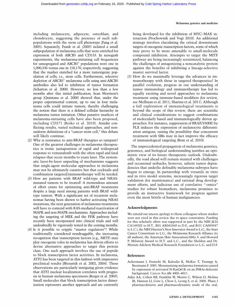
including melanocyte, adipocyte, osteoblast, andchondrocyte, suggesting the presence of such sub-populations with the stem cell phenotype (Fang et al.2005). Separately, Frank et al. (2005) isolated a smallsubpopulation of melanoma cells that were enriched forexpression of both ABCB5 and CD133. In xenograftexperiments, the melanoma-initiating cell frequenciesfor unsegregated and ABCB5+ populations were one in1,090,336 versus one in 158,170, respectively, suggestingthat the marker enriched for a more tumorigenic pop-ulation of cells; i.e., stem cells. Furthermore, selectivedepletion of ABCB5+ melanoma cells using anti-ABCB5antibodies also led to inhibition of tumor formation(Schatton et al. 2008). However, no less than a fewmonths after this initial publication, Sean Morrison’sgroup (Quintana et al. 2008) showed that, under theproper experimental context, up to one in four mela-noma cells could initiate tumors, thereby challengingthe notion that there is a defined cellular hierarchy inmelanoma tumor initiation. Other putative markers ofmelanoma-initiating cells have also been proposed,including CD271 (Boiko et al. 2010). Amidst con-flicting results, varied technical approaches, and non-uniform definitions of a ‘‘cancer stem cell,’’ this debatewill likely continue.
(2) Why is resistance to anti-BRAF therapies so rampant?One of the greatest challenges in melanoma therapeu-tics is ironic juxtaposition of rapid and widespreadresponse to vemurafenib with the often rapid and fatalrelapses that occur months to years later. The system-atic layer-by-layer unpeeling of mechanisms suggeststhat single-agent molecular approaches in melanomamay not be ultimately curative but that cocktails andcombination targeted/immunotherapy will be needed.
(3) How are patients with BRAF wild-type and NRAS-mutated tumors to be treated? A tremendous amountof effort exists for optimizing anti-BRAF treatmentsdespite a large need among patients with BRAF wild-type tumors. With a significant set of recurrent mela-nomas having been shown to harbor activating NRASmutations, the next generation of melanoma treatmentswill have to contend with RAS-mediated reactivation ofMAPK and non-MAPK mechanisms. Approaches includ-ing the targeting of MEK and the PI3K pathway haverecently been incorporated into clinical trials and willundoubtedly be vigorously tested in the coming months.
(4) Is it possible to cripple ‘‘master regulators’’? Whiletraditionally considered nondruggable, the increasingrecognition that transcription factors (e.g., MITF) mayplay oncogenic roles in melanoma has driven efforts todevise alternative approaches to target this proteinclass. One such approach involves the use of peptideto block transcription factor activities. In melanoma,ATF2 has been targeted in this fashion with impressivepreclinical results (Bhoumik et al. 2002, 2004). Theseobservations are particularly intriguing given evidencethat ATF2 nuclear localization correlates with progno-sis in human melanoma specimens (Berger et al. 2003).Small molecules that block transcription factor dimer-ization represents another approach and are currently
being developed for the inhibition of MYC–MAX in-teraction (Prochownik and Vogt 2010). An additionalstrategy involves elucidating the critical downstreamtargets of oncogenic transcription factors, some of whichmay prove to be more amenable to small-moleculecompound inhibition. Attempts to target the MITFpathway are being increasingly scrutinized, balancingthe challenges of antagonizing a noncatalytic proteinagainst the benefits of inhibiting a lineage-selectivemaster survival factor.
(5) How do we maximally leverage the advances in im-munotherapy with those in targeted therapeutics? Inparallel evolution, progress in our understanding oftumor immunology and immunotherapy has led toequally exciting and novel approaches to melanomatreatment using immune-based modifiers (for review,see Mellman et al. 2011; Sharma et al. 2011). Althougha full exploration of immunological treatments isbeyond the scope of this review, there are scientificand clinical considerations to suggest combinationsof molecularly based and immunologically driven ap-proaches. For instance, suppression of BRAF(V600E) byPLX induces the expression of melanocyte differenti-ation antigens, raising the possibility that concurrenttreatment with SBIs may in fact improve the efficacyof immunological targeting (Boni et al. 2010).
The unprecedented propagation of melanoma genetics,genomics, and biological understanding justifies an opti-mistic view of its future therapeutic outlook. Undoubt-edly, the road ahead will remain stunted with challengesand occasional setbacks; however, salient tumor depen-dencies that underlie definable melanoma subtypes havebegun to emerge. In partnership with versatile in vitroand in vivo model systems, increasingly rigorous targetvalidation (for maintenance) and rational drug develop-ment efforts, and judicious use of correlative ‘‘-omics’’studies for robust biomarkers, melanoma promises toprovide an instructive framework for progress againsteven the most brittle of human malignancies.
Acknowledgments
We extend our sincere apology to those colleagues whose studieswere not cited in this review due to space constraints. Fundingfor this scholarly effort was supported in part by the NIH (K24CA149202 to H.T., R01 AR043369 to D.F., and RO1 CA093947to L.C.), the NIH Director’s New Innovator Award to L.G., the StarrCancer Consortium to L.G., the Melanoma Research Alliance (toall authors), the American Skin Association/Abby S. and HowardP. Milstein Award to H.T. and L.C., and the Sheldon and Dr.Miriam Adelson Medical Research Foundation to L.G. and D.F.
References
Ackermann J, Frutschi M, Kaloulis K, McKee T, Trumpp A,Beermann F. 2005. Metastasizing melanoma formation causedby expression of activated N-RasQ61K on an INK4a-deficientbackground. Cancer Res 65: 4005–4011.
Adjei AA, Cohen RB, Franklin W, Morris C, Wilson D, MolinaJR, Hanson LJ, Gore L, Chow L, Leong S, et al. 2008. Phase Ipharmacokinetic and pharmacodynamic study of the oral,
Melanoma genetics and medicine
GENES & DEVELOPMENT 1145
Cold Spring Harbor Laboratory Press on February 16, 2020 - Published by genesdev.cshlp.orgDownloaded from

small-molecule mitogen-activated protein kinase kinase1/2 inhibitor AZD6244 (ARRY-142886) in patients withadvanced cancers. J Clin Oncol 26: 2139–2146.
Aharonson Z, Gana-Weisz M, Varsano T, Haklai R, Marciano D,Kloog Y. 1998. Stringent structural requirements for anti-Rasactivity of S-prenyl analogues. Biochim Biophys Acta 1406:40–50.
Albino AP, Nanus DM, Mentle IR, Cordon-Cardo C, McNuttNS, Bressler J, Andreeff M. 1989. Analysis of ras oncogenes inmalignant melanoma and precursor lesions: Correlation ofpoint mutations with differentiation phenotype. Oncogene
4: 1363–1374.Antonescu CR, Busam KJ, Francone TD, Wong GC, Guo T,
Agaram NP, Besmer P, Jungbluth A, Gimbel M, Chen CT,et al. 2007. L576P KIT mutation in anal melanomas corre-lates with KIT protein expression and is sensitive to specifickinase inhibition. Int J Cancer 121: 257–264.
Ashida A, Takata M, Murata H, Kido K, Saida T. 2009.Pathological activation of KIT in metastatic tumors of acraland mucosal melanomas. Int J Cancer 124: 862–868.
Badros AZ, Goloubeva O, Rapoport AP, Ratterree B, Gahres N,Meisenberg B, Takebe N, Heyman M, Zwiebel J, Streicher H,et al. 2005. Phase II study of G3139, a Bcl-2 antisense oligo-nucleotide, in combination with dexamethasone and thalid-omide in relapsed multiple myeloma patients. J Clin Oncol
23: 4089–4099.Balch CM, Gershenwald JE, Soong SJ, Thompson JF, Atkins MB,
Byrd DR, Buzaid AC, Cochran AJ, Coit DG, Ding S, et al.2009. Final version of 2009 AJCC melanoma staging andclassification. J Clin Oncol 27: 6199–6206.
Bale SJ, Dracopoli NC, Tucker MA, Clark WH Jr, Fraser MC,Stanger BZ, Green P, Donis-Keller H, Housman DE, GreeneMH. 1989. Mapping the gene for hereditary cutaneous malig-nant melanoma-dysplastic nevus syndrome to chromosome1p. N Engl J Med 320: 1367–1372.
Bardeesy N, Bastian BC, Hezel A, Pinkel D, DePinho RA, ChinL. 2001. Dual inactivation of RB and p53 pathways in RAS-induced melanomas. Mol Cell Biol 21: 2144–2153.
Bastian BC. 2003. Understanding the progression of melanocyticneoplasia using genomic analysis: From fields to cancer.Oncogene 22: 3081–3086.
Bastian BC, LeBoit PE, Hamm H, Brocker EB, Pinkel D. 1998.Chromosomal gains and losses in primary cutaneous mela-nomas detected by comparative genomic hybridization.Cancer Res 58: 2170–2175.
Bastian BC, LeBoit PE, Pinkel D. 2000. Mutations and copynumber increase of HRAS in Spitz nevi with distinctivehistopathological features. Am J Pathol 157: 967–972.
Bauer GL, Praetorius C, Bergsteinsdottir K, Hallsson JH,Gisladottir BK, Schepsky A, Swing DA, O’Sullivan TN,Arnheiter H, Bismuth K, et al. 2009. The role of MITFphosphorylation sites during coat color and eye develop-ment in mice analyzed by bacterial artificial chromosometransgene rescue. Genetics 183: 581–594.
Bauer J, Buttner P, Murali R, Okamoto I, Kolaitis NA, Landi MT,Scolyer RA, Bastian BC. 2011. BRAF mutations in cutaneousmelanoma are independently associated with age, anatomicsite of the primary tumor, and the degree of solar elastosis atthe primary tumor site. Pigment Cell Melanoma Res 24:345–351.
Baxter LL, Pavan WJ. 2003. Pmel17 expression is Mitf-depen-dent and reveals cranial melanoblast migration during mu-rine development. Gene Expr Patterns 3: 703–707.
Begg CB, Orlow I, Hummer AJ, Armstrong BK, Kricker A, MarrettLD, Millikan RC, Gruber SB, Anton-Culver H, Zanetti R,et al. 2005. Lifetime risk of melanoma in CDKN2A mutation
carriers in a population-based sample. J Natl Cancer Inst 97:1507–1515.
Bentley NJ, Eisen T, Goding CR. 1994. Melanocyte-specificexpression of the human tyrosinase promoter: Activationby the microphthalmia gene product and role of the initiator.Mol Cell Biol 14: 7996–8006.
Berger AJ, Kluger HM, Li N, Kielhorn E, Halaban R, Ronai Z,Rimm DL. 2003. Subcellular localization of activating tran-scription factor 2 in melanoma specimens predicts patientsurvival. Cancer Res 63: 8103–8107.
Berger MF, Hodis E, Heffernan TP, Lissanu Deribe Y, LawrenceMS, Protopopov A, Ivanova E, Stransky N, Watson IR,Nickerson E, et al. 2012. Melanoma genome sequencing revealsfrequent PREX2 mutations. Nature doi: 10.1038/nature11071.
Bertolotto C, Lesueur F, Giuliano S, Strub T, de Lichy M, Bille K,Dessen P, d’Hayer B, Mohamdi H, Remenieras A, et al. 2011.A SUMOylation-defective MITF germline mutation predis-poses to melanoma and renal carcinoma. Nature 480: 94–98.
Besaratinia A, Pfeifer GP. 2008. Sunlight ultraviolet irradiationand BRAF V600 mutagenesis in human melanoma. HumMutat 29: 983–991.
Bhoumik A, Huang TG, Ivanov V, Gangi L, Qiao RF, Woo SL,Chen SH, Ronai Z. 2002. An ATF2-derived peptide sensitizesmelanomas to apoptosis and inhibits their growth andmetastasis. J Clin Invest 110: 643–650.
Bhoumik A, Gangi L, Ronai Z. 2004. Inhibition of melanomagrowth and metastasis by ATF2-derived peptides. Cancer Res
64: 8222–8230.Bishop DT, Demenais F, Goldstein AM, Bergman W, Bishop JN,
Paillerets BB, Chompret A, Ghiorzo P, Gruis N, Hansson J,et al. 2002. Geographical variation in the penetrance ofCDKN2A mutations for melanoma. J Natl Cancer Inst 94:894–903.
Boiko AD, Razorenova OV, van de Rijn M, Swetter SM, JohnsonDL, Ly DP, Butler PD, Yang GP, Joshua B, Kaplan MJ, et al.2010. Human melanoma-initiating cells express neural crestnerve growth factor receptor CD271. Nature 466: 133–137.
Boni A, Cogdill AP, Dang P, Udayakumar D, Njauw CN, SlossCM, Ferrone CR, Flaherty KT, Lawrence DP, Fisher DE, et al.2010. Selective BRAFV600E inhibition enhances T-cell rec-ognition of melanoma without affecting lymphocyte func-tion. Cancer Res 70: 5213–5219.
Boone B, Jacobs K, Ferdinande L, Taildeman J, Lambert J, PeetersM, Bracke M, Pauwels P, Brochez L. 2011. EGFR in mela-noma: Clinical significance and potential therapeutic target.J Cutan Pathol 38: 492–502.
Borner C, Schlagbauer Wadl H, Fellay I, Selzer E, Polterauer P,Jansen B. 1999. Mutated N-ras upregulates Bcl-2 in humanmelanoma in vitro and in SCID mice. Melanoma Res 9: 347–350.
Busca R, Berra E, Gaggioli C, Khaled M, Bille K, Marchetti B,Thyss R, Fitsialos G, Larribere L, Bertolotto C, et al. 2005.Hypoxia-inducible factor 1a is a new target of microphthal-mia-associated transcription factor (MITF) in melanomacells. J Cell Biol 170: 49–59.
Cannon-Albright LA, Goldgar DE, Meyer LJ, Lewis CM,Anderson DE, Fountain JW, Hegi ME, Wiseman RW, PetyyEM, Bale AE, et al. 1992. Assignment of a locus for familialmelanoma, MLM, to chromosome 9p13-p22. Science 258:1148–1152.
Caponigro F, Casale M, Bryce J. 2003. Farnesyl transferaseinhibitors in clinical development. Expert Opin Investig
Drugs 12: 943–954.Cardenas-Navia LI, Cruz P, Lin JC, Rosenberg SA, Samuels Y.
2010. Novel somatic mutations in heterotrimeric G proteinsin melanoma. Cancer Biol Ther 10: 33–37.
Tsao et al.
1146 GENES & DEVELOPMENT
Cold Spring Harbor Laboratory Press on February 16, 2020 - Published by genesdev.cshlp.orgDownloaded from

Carreira S, Goodall J, Aksan I, La Rocca SA, Galibert MD, DenatL, Larue L, Goding CR. 2005. Mitf cooperates with Rb1 andactivates p21Cip1 expression to regulate cell cycle progres-sion. Nature 433: 764–769.
Cartlidge RA, Thomas GR, Cagnol S, Jong KA, Molton SA,Finch AJ, McMahon M. 2008. Oncogenic BRAF(V600E) in-hibits BIM expression to promote melanoma cell survival.Pigment Cell Melanoma Res 21: 534–544.
Carvajal RD, Antonescu CR, Wolchok JD, Chapman PB, RomanRA, Teitcher J, Panageas KS, Busam KJ, Chmielowski B,Lutzky J, et al. 2011. KIT as a therapeutic target in metastaticmelanoma. JAMA 305: 2327–2334.
Chapman PB, Hauschild A, Robert C, Haanen JB, Ascierto P,Larkin J, Dummer R, Garbe C, Testori A, Maio M, et al.2011. Improved survival with vemurafenib in melanomawith BRAF V600E mutation. N Engl J Med 364: 2507–2516.
Chatzinasiou F, Lill CM, Kypreou K, Stefanaki I, Nicolaou V,Spyrou G, Evangelou E, Roehr JT, Kodela E, Katsambas A,et al. 2011. Comprehensive field synopsis and systematicmeta-analyses of genetic association studies in cutaneousmelanoma. J Natl Cancer Inst 103: 1227–1235.
Chin L. 2003. The genetics of malignant melanoma: Lessonsfrom mouse and man. Nat Rev Cancer 3: 559–570.
Chin L, Pomerantz J, Polsky D, Jacobson M, Cohen C, Cordon-Cardo C, Horner JW II, DePinho RA. 1997. Cooperativeeffects of INK4a and ras in melanoma susceptibility in vivo.Genes Dev 11: 2822–2834.
Chin L, Merlino G, DePinho RA. 1998. Malignant melanoma:Modern black plague and genetic black box. Genes Dev 12:3467–3481.
Chin L, Tam A, Pomerantz J, Wong M, Holash J, Bardeesy N,Shen Q, O’Hagan R, Pantginis J, Zhou H, et al. 1999.Essential role for oncogenic Ras in tumour maintenance.Nature 400: 468–472.
Choi KY, Chang K, Pickel JM, Badger JD II, Roche KW. 2011.Expression of the metabotropic glutamate receptor 5 (mGluR5)induces melanoma in transgenic mice. Proc Natl Acad Sci 108:15219–15224.
Chudnovsky Y, Adams AE, Robbins PB, Lin Q, Khavari PA.2005. Use of human tissue to assess the oncogenic activity ofmelanoma-associated mutations. Nat Genet 37: 745–749.
Ciampi R, Nikiforov YE. 2005. Alterations of the BRAF gene inthyroid tumors. Endocr Pathol 16: 163–172.
Cohen Y, Goldenberg-Cohen N, Parrella P, Chowers I, Merbs SL,Pe’er J, Sidransky D. 2003. Lack of BRAF mutation in primaryuveal melanoma. Invest Ophthalmol Vis Sci 44: 2876–2878.
Comito G, Calvani M, Giannoni E, Bianchini F, Calorini L,Torre E, Migliore C, Giordano S, Chiarugi P. 2011. HIF-1a
stabilization by mitochondrial ROS promotes Met-depen-dent invasive growth and vasculogenic mimicry in mela-noma cells. Free Radic Biol Med 51: 893–904.
Cronin JC, Wunderlich J, Loftus SK, Prickett TD, Wei X, Ridd K,Vemula S, Burrell AS, Agrawal NS, Lin JC, et al. 2009.Frequent mutations in the MITF pathway in melanoma.Pigment Cell Melanoma Res 22: 435–444.
Cruz F III, Rubin BP, Wilson D, Town A, Schroeder A, Haley A,Bainbridge T, Heinrich MC, Corless CL. 2003. Absence ofBRAF and NRAS mutations in uveal melanoma. Cancer Res
63: 5761–5766.Curtin JA, Fridlyand J, Kageshita T, Patel HN, Busam KJ,
Kutzner H, Cho KH, Aiba S, Brocker EB, LeBoit PE, et al.2005. Distinct sets of genetic alterations in melanoma. N
Engl J Med 353: 2135–2147.Curtin JA, Busam K, Pinkel D, Bastian BC. 2006. Somatic
activation of KIT in distinct subtypes of melanoma. J ClinOncol 24: 4340–4346.
Dai DL, Martinka M, Li G. 2005. Prognostic significance ofactivated Akt expression in melanoma: A clinicopathologicstudy of 292 cases. J Clin Oncol 23: 1473–1482.
Damm S, Koefinger P, Stefan M, Wels C, Mehes G, Richtig E, Kerl H,Otte M, Schaider H. 2010. HGF-promoted motility in primaryhuman melanocytes depends on CD44v6 regulated via NF-k B,Egr-1, and C/EBP-b. J Invest Dermatol 130: 1893–1903.
Damsky WE, Curley DP, Santhanakrishnan M, Rosenbaum LE,Platt JT, Gould Rothberg BE, Taketo MM, Dankort D, RimmDL, McMahon M, et al. 2011. b-Catenin signaling controlsmetastasis in Braf-activated Pten-deficient melanomas. Can-
cer Cell 20: 741–754.Daniotti M, Oggionni M, Ranzani T, Vallacchi V, Campi V, Di
Stasi D, Torre GD, Perrone F, Luoni C, Suardi S, et al. 2004.BRAF alterations are associated with complex mutationalprofiles in malignant melanoma. Oncogene 23: 5968–5977.
Dankort D, Curley DP, Cartlidge RA, Nelson B, Karnezis AN,Damsky WE Jr, You MJ, DePinho RA, McMahon M,Bosenberg M. 2009. Braf(V600E) cooperates with Pten lossto induce metastatic melanoma. Nat Genet 41: 544–552.
Davies H, Bignell GR, Cox C, Stephens P, Edkins S, Clegg S,Teague J, Woffendin H, Garnett MJ, Bottomley W, et al. 2002.Mutations of the BRAF gene in human cancer. Nature 417:949–954.
Davies MA, Stemke-Hale K, Tellez C, Calderone TL, Deng W,Prieto VG, Lazar AJ, Gershenwald JE, Mills GB. 2008. Anovel AKT3 mutation in melanoma tumours and cell lines.Br J Cancer 99: 1265–1268.
Davis IJ, Kim JJ, Ozsolak F, Widlund HR, Rozenblatt-Rosen O,Granter SR, Du J, Fletcher JA, Denny CT, Lessnick SL, et al.2006. Oncogenic MITF dysregulation in clear cell sarcoma:Defining the MiT family of human cancers. Cancer Cell 9:473–484.
Daya-Grosjean L, Dumaz N, Sarasin A. 1995. The specificity ofp53 mutation spectra in sunlight induced human cancers.J Photochem Photobiol B 28: 115–124.
Demenais F, Mohamdi H, Chaudru V, Goldstein AM, NewtonBishop JA, Bishop DT, Kanetsky PA, Hayward NK, GillandersE, Elder DE, et al. 2010. Association of MC1R variants and hostphenotypes with melanoma risk in CDKN2A mutation car-riers: A GenoMEL study. J Natl Cancer Inst 102: 1568–1583.
de Wit PE, Moretti S, Koenders PG, Weterman MA, van MuijenGN, Gianotti B, Ruiter DJ. 1992. Increasing epidermalgrowth factor receptor expression in human melanocytictumor progression. J Invest Dermatol 99: 168–173.
Dhomen N, Reis-Filho JS, da Rocha Dias S, Hayward R, SavageK, Delmas V, Larue L, Pritchard C, Marais R. 2009. Onco-genic Braf induces melanocyte senescence and melanoma inmice. Cancer Cell 15: 294–303.
Diaz A, Suarez E, Blanco R, Badia T, Rivero D, Lopez-RequenaA, Lopez A, Montero E. 2007. Functional expression ofhuman-epidermal-growth-factor receptor in a melanoma cellline. Biotechnol Appl Biochem 48: 21–27.
Diaz A, Suarez E, Blanco R, Lopez A, Montero E. 2009.Epidermal growth factor receptor modulates the tumorigenicpotential of melanoma. Front Biosci 14: 159–166.
Djerf EA, Trinks C, Abdiu A, Thunell LK, Hallbeck AL, WalzTM. 2009. ErbB receptor tyrosine kinases contribute toproliferation of malignant melanoma cells: Inhibition bygefitinib (ZD1839). Melanoma Res 19: 156–166.
Draper GJ, Sanders BM, Kingston JE. 1986. Second primaryneoplasms in patients with retinoblastoma. Br J Cancer 53:661–671.
Du J, Fisher DE. 2002. Identification of Aim-1 as the underwhitemouse mutant and its transcriptional regulation by MITF.J Biol Chem 277: 402–406.
Melanoma genetics and medicine
GENES & DEVELOPMENT 1147
Cold Spring Harbor Laboratory Press on February 16, 2020 - Published by genesdev.cshlp.orgDownloaded from

Du J, Miller AJ, Widlund HR, Horstmann MA, Ramaswamy S,Fisher DE. 2003. MLANA/MART1 and SILV/PMEL17/GP100 are transcriptionally regulated by MITF in melano-cytes and melanoma. Am J Pathol 163: 333–343.
Du J, Widlund HR, Horstmann MA, Ramaswamy S, Ross K,Huber WE, Nishimura EK, Golub TR, Fisher DE. 2004. Criticalrole of CDK2 for melanoma growth linked to its melanocyte-specific transcriptional regulation by MITF. Cancer Cell 6:565–576.
Dumaz N, Hayward R, Martin J, Ogilvie L, Hedley D, Curtin JA,Bastian BC, Springer C, Marais R. 2006. In melanoma, RASmutations are accompanied by switching signaling fromBRAF to CRAF and disrupted cyclic AMP signaling. CancerRes 66: 9483–9491.
Dutton-Regester K, Irwin D, Hunt P, Aoude L, Tembe V, PupoGM, Lanagan C, Carter CD, O’Connor L, O’Rourke M, et al.2012. A high throughput panel for identifying clinically-relevant mutation profiles in melanoma. Mol Cancer Ther
11: 888–897.Easty DJ, Hill SP, Hsu MY, Fallowfield ME, Florenes VA, Herlyn
M, Bennett DC. 1999. Up-regulation of ephrin-A1 duringmelanoma progression. Int J Cancer 84: 494–501.
Edmunds SC, Cree IA, Di Nicolantonio F, Hungerford JL, HurrenJS, Kelsell DP. 2003. Absence of BRAF gene mutations inuveal melanomas in contrast to cutaneous melanomas. Br
J Cancer 88: 1403–1405.Edwards RH, Ward MR, Wu H, Medina CA, Brose MS, Volpe P,
Nussen-Lee S, Haupt HM, Martin AM, Herlyn M, et al. 2004.Absence of BRAF mutations in UV-protected mucosal mel-anomas. J Med Genet 41: 270–272.
Eisenmann KM, VanBrocklin MW, Staffend NA, Kitchen SM,Koo HM. 2003. Mitogen-activated protein kinase pathway-dependent tumor-specific survival signaling in melanomacells through inactivation of the proapoptotic protein bad.Cancer Res 63: 8330–8337.
Eng C, Li FP, Abramson DH, Ellsworth RM, Wong FL, GoldmanMB, Seddon J, Tarbell N, Boice JD Jr. 1993. Mortality fromsecond tumors among long-term survivors of retinoblas-toma. J Natl Cancer Inst 85: 1121–1128.
Esumi N, Oshima Y, Li Y, Campochiaro PA, Zack DJ. 2004.Analysis of the VMD2 promoter and implication of E-boxbinding factors in its regulation. J Biol Chem 279: 19064–19073.
Fang D, Nguyen TK, Leishear K, Finko R, Kulp AN, Hotz S, VanBelle PA, Xu X, Elder DE, Herlyn M. 2005. A tumorigenicsubpopulation with stem cell properties in melanomas.Cancer Res 65: 9328–9337.
Flaherty K, Puzanov I, Sosman J, Kim K, Ribas A, McArthur G,Lee RJ, Grippo JF, Nolop K, Chapman P. 2009. Phase I studyof PLX4032: Proof of concept for V600E BRAF mutation asa therapeutic target in human cancer. J Clin Oncol 27: 15s,Abstract 9000.
Flaherty KT, Puzanov I, Kim KB, Ribas A, McArthur GA,Sosman JA, O’Dwyer PJ, Lee RJ, Grippo JF, Nolop K, et al.2010. Inhibition of mutated, activated BRAF in metastaticmelanoma. N Engl J Med 363: 809–819.
Fletcher O, Easton D, Anderson K, Gilham C, Jay M, Peto J.2004. Lifetime risks of common cancers among retinoblas-toma survivors. J Natl Cancer Inst 96: 357–363.
Frank NY, Margaryan A, Huang Y, Schatton T, Waaga-GasserAM, Gasser M, Sayegh MH, Sadee W, Frank MH. 2005.ABCB5-mediated doxorubicin transport and chemoresistancein human malignant melanoma. Cancer Res 65: 4320–4333.
Fujimura Y, Ohno T, Siddique H, Lee L, Rao VN, Reddy ES.1996. The EWS-ATF-1 gene involved in malignant mela-noma of soft parts with t(12;22) chromosome translocation,
encodes a constitutive transcriptional activator. Oncogene
12: 159–167.Fuse N, Yasumoto K, Suzuki H, Takahashi K, Shibahara S. 1996.
Identification of a melanocyte-type promoter of the micro-phthalmia-associated transcription factor gene. BiochemBiophys Res Commun 219: 702–707.
Gandini S, Sera F, Cattaruzza MS, Pasquini P, Zanetti R, MasiniC, Boyle P, Melchi CF. 2005. Meta-analysis of risk factors forcutaneous melanoma: III. Family history, actinic damage andphenotypic factors. Eur J Cancer 41: 2040–2059.
Garcia-Borron JC, Sanchez-Laorden BL, Jimenez-Cervantes C.2005. Melanocortin-1 receptor structure and functional reg-ulation. Pigment Cell Res 18: 393–410.
Garnett MJ, Rana S, Paterson H, Barford D, Marais R. 2005.Wild-type and mutant B-RAF activate C-RAF through dis-tinct mechanisms involving heterodimerization. Mol Cell20: 963–969.
Garraway LA, Widlund HR, Rubin MA, Getz G, Berger AJ,Ramaswamy S, Beroukhim R, Milner DA, Granter SR, Du J,et al. 2005. Integrative genomic analyses identify MITF asa lineage survival oncogene amplified in malignant mela-noma. Nature 436: 117–122.
Genander M, Frisen J. 2010. Ephrins and Eph receptors in stemcells and cancer. Curr Opin Cell Biol 22: 611–616.
Gilmartin AG, Bleam MR, Groy A, Moss KG, Minthorn EA,Kulkarni SG, Rominger CM, Erskine S, Fisher KE, Yang J,et al. 2011. GSK1120212 (JTP-74057) is an inhibitor of MEKactivity and activation with favorable pharmacokineticproperties for sustained in vivo pathway inhibition. Clin
Cancer Res 17: 989–1000.Glinsky GV, Glinsky VV, Ivanova AB, Hueser CJ. 1997. Apoptosis
and metastasis: Increased apoptosis resistance of metastaticcancer cells is associated with the profound deficiency ofapoptosis execution mechanisms. Cancer Lett 115: 185–193.
Goel VK, Lazar AJ, Warneke CL, Redston MS, Haluska FG.2006. Examination of mutations in BRAF, NRAS, and PTENin primary cutaneous melanoma. J Invest Dermatol 126:154–160.
Granter SR, Weilbaecher KN, Quigley C, Fletcher CD, FisherDE. 2001. Clear cell sarcoma shows immunoreactivity formicrophthalmia transcription factor: Further evidence formelanocytic differentiation. Mod Pathol 14: 6–9.
Gray-Schopfer VC, da Rocha Dias S, Marais R. 2005. The role ofB-RAF in melanoma. Cancer Metastasis Rev 24: 165–183.
Gray-Schopfer VC, Karasarides M, Hayward R, Marais R. 2007.Tumor necrosis factor-a blocks apoptosis in melanoma cellswhen BRAF signaling is inhibited. Cancer Res 67: 122–129.
Guertin DA, Sabatini DM. 2007. Defining the role of mTOR incancer. Cancer Cell 12: 9–22.
Guo J, Si L, Kong Y, Flaherty KT, Xu X, Zhu Y, Corless CL, Li L,Li H, Sheng X, et al. 2011. Phase II, open-label, single-armtrial of imatinib mesylate in patients with metastatic mela-noma harboring c-Kit mutation or amplification. J Clin Oncol29: 2904–2909.
Gupta PB, Kuperwasser C, Brunet JP, Ramaswamy S, Kuo WL,Gray JW, Naber SP, Weinberg RA. 2005. The melanocytedifferentiation program predisposes to metastasis after neo-plastic transformation. Nat Genet 37: 1047–1054.
Halaban R. 1999. Melanoma cell autonomous growth: The Rb/E2F pathway. Cancer Metastasis Rev 18: 333–343.
Halaban R, Bohm M, Dotto P, Moellmann G, Cheng E, Zhang Y.1996. Growth regulatory proteins that repress differentiationmarkers in melanocytes also downregulate the transcriptionfactor microphthalmia. J Invest Dermatol 106: 1266–1272.
Handolias D, Salemi R, Murray W, Tan A, Liu W, Viros A,Dobrovic A, Kelly J, McArthur GA. 2010. Mutations in KIT
Tsao et al.
1148 GENES & DEVELOPMENT
Cold Spring Harbor Laboratory Press on February 16, 2020 - Published by genesdev.cshlp.orgDownloaded from

occur at low frequency in melanomas arising from anatom-ical sites associated with chronic and intermittent sunexposure. Pigment Cell Melanoma Res 23: 210–215.
Harland M, Mistry S, Bishop DT, Bishop JA. 2001. A deepintronic mutation in CDKN2A is associated with diseasein a subset of melanoma pedigrees. Hum Mol Genet 10:2679–2686.
Harland M, Taylor CF, Bass S, Churchman M, Randerson-MoorJA, Holland EA, Mann GJ, Bishop DT, Newton Bishop JA.2005a. Intronic sequence variants of the CDKN2A gene inmelanoma pedigrees. Genes Chromosomes Cancer 43: 128–136.
Harland M, Taylor CF, Chambers PA, Kukalizch K, Randerson-Moor JA, Gruis NA, de Snoo FA, ter Huurne JA, GoldsteinAM, Tucker MA, et al. 2005b. A mutation hotspot at thep14ARF splice site. Oncogene 24: 4604–4608.
Hauschild A, Agarwala SS, Trefzer U, Hogg D, Robert C, HerseyP, Eggermont A, Grabbe S, Gonzalez R, Gille J, et al. 2009.Results of a phase III, randomized, placebo-controlled studyof sorafenib in combination with carboplatin and paclitaxelas second-line treatment in patients with unresectable stageIII or stage IV melanoma. J Clin Oncol 27: 2823–2830.
Heidorn SJ, Milagre C, Whittaker S, Nourry A, Niculescu-DuvasI, Dhomen N, Hussain J, Reis-Filho JS, Springer CJ, PritchardC, et al. 2010. Kinase-dead BRAF and oncogenic RAS co-operate to drive tumor progression through CRAF. Cell 140:209–221.
Hemesath TJ, Steingrimsson E, McGill G, Hansen MJ, Vaught J,Hodgkinson CA, Arnheiter H, Copeland NG, Jenkins NA,Fisher DE. 1994. Microphthalmia, a critical factor in mela-nocyte development, defines a discrete transcription factorfamily. Genes Dev 8: 2770–2780.
Hemesath TJ, Price ER, Takemoto C, Badalian T, Fisher DE.1998. MAP kinase links the transcription factor Micro-phthalmia to c-Kit signalling in melanocytes. Nature 391:298–301.
Hendrix MJ, Seftor EA, Hess AR, Seftor RE. 2003. Molecularplasticity of human melanoma cells. Oncogene 22: 3070–3075.
Herath NI, Spanevello MD, Sabesan S, Newton T, CummingsM, Duffy S, Lincoln D, Boyle G, Parsons PG, Boyd AW. 2006.Over-expression of Eph and ephrin genes in advanced ovariancancer: ephrin gene expression correlates with shortenedsurvival. BMC Cancer 6: 144. doi: 10.1186/1471-2407-6-144.
Hewitt C, Lee Wu C, Evans G, Howell A, Elles RG, Jordan R,Sloan P, Read AP, Thakker N. 2002. Germline mutation ofARF in a melanoma kindred. Hum Mol Genet 11: 1273–1279.
Hingorani SR, Jacobetz MA, Robertson GP, Herlyn M, TuvesonDA. 2003. Suppression of BRAF(V599E) in human melanomaabrogates transformation. Cancer Res 63: 5198–5202.
Hocker T, Tsao H. 2007. Ultraviolet radiation and melanoma: Asystematic review and analysis of reported sequence vari-ants. Hum Mutat 28: 578–588.
Hodgkinson CA, Moore KJ, Nakayama A, Steingrimsson E,Copeland NG, Jenkins NA, Arnheiter H. 1993. Mutationsat the mouse microphthalmia locus are associated withdefects in a gene encoding a novel basic-helix–loop–helix-zipper protein. Cell 74: 395–404.
Hodi FS, Friedlander P, Corless CL, Heinrich MC, Mac Rae S,Kruse A, Jagannathan J, Van den Abbeele AD, Velazquez EF,Demetri GD, et al. 2008. Major response to imatinib mesy-late in KIT-mutated melanoma. J Clin Oncol 26: 2046–2051.
Hoeflich KP, Gray DC, Eby MT, Tien JY, Wong L, Bower J,Gogineni A, Zha J, Cole MJ, Stern HM, et al. 2006. Onco-genic BRAF is required for tumor growth and maintenance inmelanoma models. Cancer Res 66: 999–1006.
Huang S, Luca M, Gutman M, McConkey DJ, Langley KE,Lyman SD, Bar-Eli M. 1996. Enforced c-KIT expressionrenders highly metastatic human melanoma cells suscepti-ble to stem cell factor-induced apoptosis and inhibits theirtumorigenic and metastatic potential. Oncogene 13: 2339–2347.
Huang TS, Rauth S, Das Gupta TK. 1996. Overexpression ofEGF receptor is associated with spontaneous metastases ofa human melanoma cell line in nude mice. Anticancer Res16: 3557–3563.
Hussussian CJ, Struewing JP, Goldstein AM, Higgins PA, AllyDS, Sheahan MD, Clark WH Jr, Tucker MA, Dracopoli NC.1994. Germline p16 mutations in familial melanoma. NatGenet 8: 15–21.
Ilmonen S, Hernberg M, Pyrhonen S, Tarkkanen J, Asko-Seljavaara S. 2005. Ki-67, Bcl-2 and p53 expression in primaryand metastatic melanoma. Melanoma Res 15: 375–381.
Infante JR, Fecher LA, Nallapareddy S, Gordon MS, Flaherty KT,Cox DS, DeMarini DJ, Morris SR, Burris HA, MessersmithWA. 2010. Safety and efficacy results from the first-in-humanstudy of the oral MEK 1/2 inhibitor GSK1120212. J Clin Oncol
28: 15s, Abstract 2503.Ito A, Koma Y, Watabe K, Jippo T, Wakayama T, Iseki S, Kitamura
Y. 2004. Contribution of the SgIGSF adhesion molecule tosurvival of cultured mast cells in vivo. Biochem Biophys Res
Commun 319: 200–206.Ivanov VN, Hei TK. 2005. Combined treatment with EGFR
inhibitors and arsenite upregulated apoptosis in humanEGFR-positive melanomas: A role of suppression of thePI3K–AKT pathway. Oncogene 24: 616–626.
Jafari M, Papp T, Kirchner S, Diener U, Henschler D, Burg G,Schiffmann D. 1995. Analysis of ras mutations in humanmelanocytic lesions: Activation of the ras gene seems to beassociated with the nodular type of human malignantmelanoma. J Cancer Res Clin Oncol 121: 23–30.
James GL, Goldstein JL, Brown MS, Rawson TE, Somers TC,McDowell RS, Crowley CW, Lucas BK, Levinson AD,Marsters JC Jr. 1993. Benzodiazepine peptidomimetics: Po-tent inhibitors of Ras farnesylation in animal cells. Science260: 1937–1942.
Jane-Valbuena J, Widlund HR, Perner S, Johnson LA, Dibner AC,Lin WM, Baker AC, Nazarian RM, Vijayendran KG, SellersWR, et al. 2010. An oncogenic role for ETV1 in melanoma.Cancer Res 70: 2075–2084.
Jansen B, Schlagbauer-Wadl H, Kahr H, Heere-Ress E, Mayer BX,Eichler H, Pehamberger H, Gana-Weisz M, Ben-David E,Kloog Y, et al. 1999. Novel Ras antagonist blocks humanmelanoma growth. Proc Natl Acad Sci 96: 14019–14024.
Ji Z, Flaherty KT, Tsao H. 2012a. Targeting the RAS pathway inmelanoma. Trends Mol Med 18: 27–35.
Ji Z, Njauw CN, Taylor M, Neel V, Flaherty KT, Tsao H. 2012b.p53 rescue through HDM2 antagonism suppresses mela-noma growth and potentiates MEK inhibition. J InvestDermatol 132: 356–364.
Johannessen CM, Boehm JS, Kim SY, Thomas SR, Wardwell L,Johnson LA, Emery CM, Stransky N, Cogdill AP, Barretina J,et al. 2010. COT drives resistance to RAF inhibition throughMAP kinase pathway reactivation. Nature 468: 968–972.
Johnson ML, Rizvi NA, Ginsberg MS, Miller VA, Kris MG, PaoW, Riely GJ. 2009. A phase II trial of salirasib in patients withstage IIIB/IV lung adenocarcinoma enriched for KRAS muta-tions. J Clin Oncol 27: 15s, Abstract 8012.
Johnstone RW, Ruefli AA, Lowe SW. 2002. Apoptosis: A linkbetween cancer genetics and chemotherapy. Cell 108: 153–164.
Joseph EW, Pratilas CA, Poulikakos PI, Tadi M, Wang W, TaylorBS, Halilovic E, Persaud Y, Xing F, Viale A, et al. 2010. The
Melanoma genetics and medicine
GENES & DEVELOPMENT 1149
Cold Spring Harbor Laboratory Press on February 16, 2020 - Published by genesdev.cshlp.orgDownloaded from

RAF inhibitor PLX4032 inhibits ERK signaling and tumorcell proliferation in a V600E BRAF-selective manner. ProcNatl Acad Sci 107: 14903–14908.
Kamb A, Gruis NA, Weaver-Feldhaus J, Liu Q, Harshman K,Tavtigian SV, Stockert E, Day RS III, Johnson BE, SkolnickMH. 1994. A cell cycle regulator potentially involved ingenesis of many tumor types. Science 264: 436–440.
Kamijo T, Weber JD, Zambetti G, Zindy F, Roussel MF, Sherr CJ.1998. Functional and physical interactions of the ARF tumorsuppressor with p53 and Mdm2. Proc Natl Acad Sci 95:8292–8297.
Karasarides M, Chiloeches A, Hayward R, Niculescu-Duvaz D,Scanlon I, Friedlos F, Ogilvie L, Hedley D, Martin J, MarshallCJ, et al. 2004. B-RAF is a therapeutic target in melanoma.Oncogene 23: 6292–6298.
Karreth FA, Tay Y, Perna D, Ala U, Tan SM, Rust AG, DeNicolaG, Webster KA, Weiss D, Perez-Mancera PA, et al. 2011. Invivo identification of tumor-suppressive PTEN ceRNAs inan oncogenic BRAF-induced mouse model of melanoma.Cell 147: 382–395.
Kefford RAAH, Brown MP, Millward M, Infante JR, Long GV,Ouellet D, Curtis M, Lebowitz PF, Falchook GS. 2010. PhaseI/II study of GSK2118436, a selective inhibitor of oncogenicmutant BRAF kinase, in patients with metastatic melanomaand other solid tumors. J Clin Oncol 28: 15s, Abstract 8503.
Kenessey I, Keszthelyi M, Kramer Z, Berta J, Adam A, Dobos J,Mildner M, Flachner B, Cseh S, Barna G, et al. 2010. Inhibitionof c-Met with the specific small molecule tyrosine kinaseinhibitor SU11274 decreases growth and metastasis formationof experimental human melanoma. Curr Cancer Drug Targets
10: 332–342.Kennedy C, ter Huurne J, Berkhout M, Gruis N, Bastiaens M,
Bergman W, Willemze R, Bavinck JN. 2001. Melanocortin 1receptor (MC1R) gene variants are associated with an in-creased risk for cutaneous melanoma which is largely in-dependent of skin type and hair color. J Invest Dermatol 117:294–300.
Kharas MG, Fruman DA. 2005. ABL oncogenes and phospho-inositide 3-kinase: Mechanism of activation and downstreameffectors. Cancer Res 65: 2047–2053.
Kim M, Gans JD, Nogueira C, Wang A, Paik JH, Feng B, BrennanC, Hahn WC, Cordon-Cardo C, Wagner SN, et al. 2006.Comparative oncogenomics identifies NEDD9 as a mela-noma metastasis gene. Cell 125: 1269–1281.
Kinch MS, Carles-Kinch K. 2003. Overexpression and functionalalterations of the EphA2 tyrosine kinase in cancer. Clin ExpMetastasis 20: 59–68.
Koh J, Enders GH, Dynlacht BD, Harlow E. 1995. Tumour-derived p16 alleles encoding proteins defective in cell-cycleinhibition. Nature 375: 506–510.
Kong Y, Si L, Zhu Y, Xu X, Corless CL, Flaherty KT, Li L, Li H,Sheng X, Cui C, et al. 2011. Large-scale analysis of KITaberrations in Chinese patients with melanoma. Clin CancerRes 17: 1684–1691.
Koprowski H, Herlyn M, Balaban G, Parmiter A, Ross A, NowellP. 1985. Expression of the receptor for epidermal growthfactor correlates with increased dosage of chromosome 7 inmalignant melanoma. Somat Cell Mol Genet 11: 297–302.
Kumar R, Angelini S, Snellman E, Hemminki K. 2004. BRAFmutations are common somatic events in melanocytic nevi.J Invest Dermatol 122: 342–348.
Laheru D, Rudek M, Taylor G, Goldsweig H, Rajeshkumar NV,Linden S, Angenendt M, Le D, Donehower R, Jimeno A, et al.2009. Integrated development of S-trans, trans-farnesylthio-salicyclic acid (FTS, salirasib) in advanced pancreatic cancer.J Clin Oncol 27: 15s, Abstract 4529.
Leiter U, Schmid RM, Kaskel P, Peter RU, Krahn G. 2000.Antiapoptotic bcl-2 and bcl-xL in advanced malignant mel-anoma. Arch Dermatol Res 292: 225–232.
Li G, Schaider H, Satyamoorthy K, Hanakawa Y, Hashimoto K,Herlyn M. 2001. Downregulation of E-cadherin and Desmo-glein 1 by autocrine hepatocyte growth factor during mela-noma development. Oncogene 20: 8125–8135.
Libra M, Malaponte G, Navolanic PM, Gangemi P, BevelacquaV, Proietti L, Bruni B, Stivala F, Mazzarino MC, Travali S,et al. 2005. Analysis of BRAF mutation in primary andmetastatic melanoma. Cell Cycle 4: 1382–1384.
Lin WM, Baker AC, Beroukhim R, Winckler W, Feng W, MarmionJM, Laine E, Greulich H, Tseng H, Gates C, et al. 2008.Modeling genomic diversity and tumor dependency in malig-nant melanoma. Cancer Res 68: 664–673.
Loercher AE, Tank EM, Delston RB, Harbour JW. 2005. MITFlinks differentiation with cell cycle arrest in melanocytes bytranscriptional activation of INK4A. J Cell Biol 168: 35–40.
Lutzky J, Bauer J, Bastian BC. 2008. Dose-dependent, completeresponse to imatinib of a metastatic mucosal melanomawith a K642E KIT mutation. Pigment Cell Melanoma Res
21: 492–493.Macgregor S, Montgomery GW, Liu JZ, Zhao ZZ, Henders AK,
Stark M, Schmid H, Holland EA, Duffy DL, Zhang M, et al.2011. Genome-wide association study identifies a new mela-noma susceptibility locus at 1q21.3. Nat Genet 43: 1114–1118.
Maldonado JL, Fridlyand J, Patel H, Jain AN, Busam K, KageshitaT, Ono T, Albertson DG, Pinkel D, Bastian BC. 2003.Determinants of BRAF mutations in primary melanomas.J Natl Cancer Inst 95: 1878–1890.
Mansky KC, Sankar U, Han J, Ostrowski MC. 2002. Micro-phthalmia transcription factor is a target of the p38 MAPKpathway in response to receptor activator of NF-kB ligandsignaling. J Biol Chem 277: 11077–11083.
Marcucci G, Stock W, Dai G, Klisovic RB, Liu S, Klisovic MI,Blum W, Kefauver C, Sher DA, Green M, et al. 2005. Phase Istudy of oblimersen sodium, an antisense to Bcl-2, in un-treated older patients with acute myeloid leukemia: Phar-macokinetics, pharmacodynamics, and clinical activity.J Clin Oncol 23: 3404–3411.
Margolin KA, Longmate J, Baratta T, Synold T, Weber J, GajewskiT, Quirt I, Christensen S, Doroshow JH. 2004. CCI-779 inmetastatic melanoma: A phase II trial of the California CancerConsortium. ASCO Annual Meeting. Abstract no. 7523.
McGill GG, Horstmann M, Widlund HR, Du J, Motyckova G,Nishimura EK, Lin YL, Ramaswamy S, Avery W, Ding HF,et al. 2002. Bcl2 regulation by the melanocyte master regula-tor Mitf modulates lineage survival and melanoma cellviability. Cell 109: 707–718.
McGill GG, Haq R, Nishimura EK, Fisher DE. 2006. c-Metexpression is regulated by Mitf in the melanocyte lineage.J Biol Chem 281: 10365–10373.
Mellman I, Coukos G, Dranoff G. 2011. Cancer immunotherapycomes of age. Nature 480: 480–489.
Menges CW, McCance DJ. 2008. Constitutive activation of theRaf–MAPK pathway causes negative feedback inhibition ofRas–PI3K–AKT and cellular arrest through the EphA2 re-ceptor. Oncogene 27: 2934–2940.
Michaloglou C, Vredeveld LC, Soengas MS, Denoyelle C,Kuilman T, van der Horst CM, Majoor DM, Shay JW, MooiWJ, Peeper DS. 2005. BRAFE600-associated senescence-like cell cycle arrest of human naevi. Nature 436: 720–724.
Mikhail M, Velazquez E, Shapiro R, Berman R, Pavlick A,Sorhaindo L, Spira J, Mir C, Panageas KS, Polsky D, et al.2005. PTEN expression in melanoma: Relationship with
Tsao et al.
1150 GENES & DEVELOPMENT
Cold Spring Harbor Laboratory Press on February 16, 2020 - Published by genesdev.cshlp.orgDownloaded from

patient survival, Bcl-2 expression, and proliferation. Clin
Cancer Res 11: 5153–5157.Miller AJ, Tsao H. 2010. New insights into pigmentary path-
ways and skin cancer. Br J Dermatol 162: 22–28.Miller AJ, Du J, Rowan S, Hershey CL, Widlund HR, Fisher DE.
2004. Transcriptional regulation of the melanoma prognosticmarker melastatin (TRPM1) by MITF in melanocytes andmelanoma. Cancer Res 64: 509–516.
Miller AJ, Levy C, Davis IJ, Razin E, Fisher DE. 2005. Sumoy-lation of MITF and its related family members TFE3 andTFEB. J Biol Chem 280: 146–155.
Miyazaki T, Kato H, Fukuchi M, Nakajima M, Kuwano H. 2003.EphA2 overexpression correlates with poor prognosis inesophageal squamous cell carcinoma. Int J Cancer 103:657–663.
Molven A, Grimstvedt MB, Steine SJ, Harland M, Avril MF,Hayward NK, Akslen LA. 2005. A large Norwegian familywith inherited malignant melanoma, multiple atypical nevi,and CDK4 mutation. Genes Chromosomes Cancer 44: 10–18.
Montone KT, van Belle P, Elenitsas R, Elder DE. 1997. Proto-oncogene c-kit expression in malignant melanoma: Proteinloss with tumor progression. Mod Pathol 10: 939–944.
Morii E, Oboki K, Ishihara K, Jippo T, Hirano T, Kitamura Y.2004. Roles of MITF for development of mast cells in mice:Effects on both precursors and tissue environments. Blood
104: 1656–1661.Murakami M, Ikeda T, Saito T, Ogawa K, Nishino Y, Nakaya K,
Funaba M. 2006. Transcriptional regulation of plasminogenactivator inhibitor-1 by transforming growth factor-b, acti-vin A and microphthalmia-associated transcription factor.Cell Signal 18: 256–265.
Muthusamy V, Hobbs C, Nogueira C, Cordon-Cardo C, McKeePH, Chin L, Bosenberg MW. 2006. Amplification of CDK4and MDM2 in malignant melanoma. Genes Chromosomes
Cancer 45: 447–454.Namiki T, Yanagawa S, Izumo T, Ishikawa M, Tachibana M,
Kawakami Y, Yokozeki H, Nishioka K, Kaneko Y. 2005.Genomic alterations in primary cutaneous melanomasdetected by metaphase comparative genomic hybridiza-tion with laser capture or manual microdissection: 6pgains may predict poor outcome. Cancer Genet Cytogenet
157: 1–11.Natali PG, Nicotra MR, Di Renzo MF, Prat M, Bigotti A,
Cavaliere R, Comoglio PM. 1993. Expression of the c-Met/HGF receptor in human melanocytic neoplasms: Demon-stration of the relationship to malignant melanoma tumourprogression. Br J Cancer 68: 746–750.
Nazarian R, Shi H, Wang Q, Kong X, Koya RC, Lee H, Chen Z,Lee MK, Attar N, Sazegar H, et al. 2010. Melanomas acquireresistance to B-RAF(V600E) inhibition by RTK or N-RASupregulation. Nature 468: 973–977.
Niihori T, Aoki Y, Narumi Y, Neri G, Cave H, Verloes A,Okamoto N, Hennekam RC, Gillessen-Kaesbach G, WieczorekD, et al. 2006. Germline KRAS and BRAF mutations in cardio-facio-cutaneous syndrome. Nat Genet 38: 294–296.
Nikolaev SI, Rimoldi D, Iseli C, Valsesia A, Robyr D, Gehrig C,Harshman K, Guipponi M, Bukach O, Zoete V, et al. 2011.Exome sequencing identifies recurrent somatic MAP2K1and MAP2K2 mutations in melanoma. Nat Genet 44: 133–139.
Nishimura EK, Granter SR, Fisher DE. 2005. Mechanisms ofhair graying: Incomplete melanocyte stem cell maintenancein the niche. Science 307: 720–724.
Nobori T, Miura K, Wu DJ, Lois A, Takabayashi K, Carson DA.1994. Deletions of the cyclin-dependent kinase-4 inhibitorgene in multiple human cancers. Nature 368: 753–756.
O’Brien SM, Cunningham CC, Golenkov AK, Turkina AG,Novick SC, Rai KR. 2005. Phase I to II multicenter studyof oblimersen sodium, a Bcl-2 antisense oligonucleotide, inpatients with advanced chronic lymphocytic leukemia.J Clin Oncol 23: 7697–7702.
Omholt K, Krockel D, Ringborg U, Hansson J. 2006. Mutationsof PIK3CA are rare in cutaneous melanoma. Melanoma Res
16: 197–200.Papp T, Pemsel H, Zimmermann R, Bastrop R, Weiss DG,
Schiffmann D. 1999. Mutational analysis of the N-ras, p53,p16INK4a, CDK4, and MC1R genes in human congenitalmelanocytic naevi. J Med Genet 36: 610–614.
Patel SP, Kim KB, Papadopoulos NE, Hwu WJ, Hwu P, Prieto VG,Bar-Eli M, Zigler M, Dobroff A, Bronstein Y, et al. 2011. Aphase II study of gefitinib in patients with metastaticmelanoma. Melanoma Res 21: 357–363.
Patton EE, Widlund HR, Kutok JL, Kopani KR, Amatruda JF,Murphey RD, Berghmans S, Mayhall EA, Traver D, FletcherCD, et al. 2005. BRAF mutations are sufficient to promotenevi formation and cooperate with p53 in the genesis ofmelanoma. Curr Biol 15: 249–254.
Pleasance ED, Cheetham RK, Stephens PJ, McBride DJ, HumphraySJ, Greenman CD, Varela I, Lin ML, Ordonez GR, BignellGR, et al. 2010. A comprehensive catalogue of somaticmutations from a human cancer genome. Nature 463: 191–196.
Plettenberg A, Ballaun C, Pammer J, Mildner M, Strunk D,Weninger W, Tschachler E. 1995. Human melanocytes andmelanoma cells constitutively express the Bcl-2 proto-onco-gene in situ and in cell culture. Am J Pathol 146: 651–659.
Pollock PM, Walker GJ, Glendening JM, Que Noy T, Bloch NC,Fountain JW, Hayward NK. 2002. PTEN inactivation is rarein melanoma tumours but occurs frequently in melanomacell lines. Melanoma Res 12: 565–575.
Pollock PM, Cohen-Solal K, Sood R, Namkoong J, Martino JJ,Koganti A, Zhu H, Robbins C, Makalowska I, Shin SS, et al.2003a. Melanoma mouse model implicates metabotropicglutamate signaling in melanocytic neoplasia. Nat Genet
34: 108–112.Pollock PM, Harper UL, Hansen KS, Yudt LM, Stark M, Robbins
CM, Moses TY, Hostetter G, Wagner U, Kakareka J, et al.2003b. High frequency of BRAF mutations in nevi. NatGenet 33: 19–20.
Pomerantz J, Schreiber-Agus N, Liegeois NJ, Silverman A,Alland L, Chin L, Potes J, Chen K, Orlow I, Lee HW, et al.1998. The Ink4a tumor suppressor gene product, p19Arf,interacts with MDM2 and neutralizes MDM2’s inhibition ofp53. Cell 92: 713–723.
Poulikakos PI, Zhang C, Bollag G, Shokat KM, Rosen N. 2010.RAF inhibitors transactivate RAF dimers and ERK signallingin cells with wild-type BRAF. Nature 464: 427–430.
Poulikakos PI, Persaud Y, Janakiraman M, Kong X, Ng C,Moriceau G, Shi H, Atefi M, Titz B, Gabay MT, et al. 2011.RAF inhibitor resistance is mediated by dimerization ofaberrantly spliced BRAF(V600E). Nature 480: 387–390.
Prahallad A, Sun C, Huang S, Di Nicolantonio F, Salazar R,Zecchin D, Beijersbergen RL, Bardelli A, Bernards R. 2012.Unresponsiveness of colon cancer to BRAF(V600E) inhibitionthrough feedback activation of EGFR. Nature 483: 100–103.
Price ER, Fisher DE. 2001. Sensorineural deafness and pigmen-tation genes: Melanocytes and the Mitf transcriptionalnetwork. Neuron 30: 15–18.
Price ER, Ding HF, Badalian T, Bhattacharya S, Takemoto C, YaoTP, Hemesath TJ, Fisher DE. 1998. Lineage-specific signalingin melanocytes. C-kit stimulation recruits p300/CBP tomicrophthalmia. J Biol Chem 273: 17983–17986.
Melanoma genetics and medicine
GENES & DEVELOPMENT 1151
Cold Spring Harbor Laboratory Press on February 16, 2020 - Published by genesdev.cshlp.orgDownloaded from
Prickett TD, Agrawal NS, Wei X, Yates KE, Lin JC, Wunderlich JR,Cronin JC, Cruz P, Rosenberg SA, Samuels Y. 2009. Analysis ofthe tyrosine kinome in melanoma reveals recurrent mutationsin ERBB4. Nat Genet 41: 1127–1132.
Prickett TD, Wei X, Cardenas-Navia I, Teer JK, Lin JC, Walia V,Gartner J, Jiang J, Cherukuri PF, Molinolo A, et al. 2011.Exon capture analysis of G protein-coupled receptors iden-tifies activating mutations in GRM3 in melanoma. Nat
Genet 43: 1119–1126.Prochownik EV, Vogt PK. 2010. Therapeutic targeting of Myc.
Genes Cancer 1: 650–659.Quintana E, Shackleton M, Sabel MS, Fullen DR, Johnson TM,
Morrison SJ. 2008. Efficient tumour formation by singlehuman melanoma cells. Nature 456: 593–598.
Raimondi S, Sera F, Gandini S, Iodice S, Caini S, Maisonneuve P,Fargnoli MC. 2008. MC1R variants, melanoma and red haircolor phenotype: A meta-analysis. Int J Cancer 122: 2753–2760.
Rajakulendran T, Sahmi M, Lefrancois M, Sicheri F, Therrien M.2009. A dimerization-dependent mechanism drives RAFcatalytic activation. Nature 461: 542–545.
Rimoldi D, Salvi S, Lienard D, Lejeune FJ, Speiser D, Zografos L,Cerottini JC. 2003. Lack of BRAF mutations in uvealmelanoma. Cancer Res 63: 5712–5715.
Rinehart J, Adjei AA, Lorusso PM, Waterhouse D, Hecht JR,Natale RB, Hamid O, Varterasian M, Asbury P, Kaldjian EP,et al. 2004. Multicenter phase II study of the oral MEKinhibitor, CI-1040, in patients with advanced non-small-celllung, breast, colon, and pancreatic cancer. J Clin Oncol 22:4456–4462.
Rivera RS, Nagatsuka H, Gunduz M, Cengiz B, Gunduz E, SiarCH, Tsujigiwa H, Tamamura R, Han KN, Nagai N. 2008.C-kit protein expression correlated with activating muta-tions in KIT gene in oral mucosal melanoma. Virchows Arch
452: 27–32.Robertson GP. 2005. Functional and therapeutic significance of
Akt deregulation in malignant melanoma. Cancer Metasta-
sis Rev 24: 273–285.Robertson GP, Furnari FB, Miele ME, Glendening MJ, Welch
DR, Fountain JW, Lugo TG, Huang HJ, Cavenee WK. 1998. Invitro loss of heterozygosity targets the PTEN/MMAC1 genein melanoma. Proc Natl Acad Sci 95: 9418–9423.
Rodriguez-Viciana P, Tetsu O, Tidyman WE, Estep AL, CongerBA, Cruz MS, McCormick F, Rauen KA. 2006. Germlinemutations in genes within the MAPK pathway cause cardio-facio-cutaneous syndrome. Science 311: 1287–1290.
Ross P, Hall L, Haff LA. 2000. Quantitative approach to single-nucleotide polymorphism analysis using MALDI-TOF massspectrometry. Biotechniques 29: 620–629.
Sabatini DM. 2006. mTOR and cancer: Insights into a complexrelationship. Nat Rev Cancer 6: 729–734.
Saldanha G, Purnell D, Fletcher A, Potter L, Gillies A, PringleJH. 2004. High BRAF mutation frequency does not charac-terize all melanocytic tumor types. Int J Cancer 111: 705–710.
Sanchez-Martin M, Rodriguez-Garcia A, Perez-Losada J, SagreraA, Read AP, Sanchez-Garcia I. 2002. SLUG (SNAI2) deletionsin patients with Waardenburg disease. Hum Mol Genet 11:3231–3236.
Sanders BM, Jay M, Draper GJ, Roberts EM. 1989. Non-ocularcancer in relatives of retinoblastoma patients. Br J Cancer60: 358–365.
Schatton T, Murphy GF, Frank NY, Yamaura K, Waaga-GasserAM, Gasser M, Zhan Q, Jordan S, Duncan LM, Weishaupt C,et al. 2008. Identification of cells initiating human melano-mas. Nature 451: 345–349.
Schicher N, Paulitschke V, Swoboda A, Kunstfeld R, Loewe R,Pilarski P, Pehamberger H, Hoeller C. 2009. Erlotinib andbevacizumab have synergistic activity against melanoma.Clin Cancer Res 15: 3495–3502.
Scott KL, Chin L. 2010. Signaling from the Golgi: Mechanismsand models for Golgi phosphoprotein 3-mediated oncogene-sis. Clin Cancer Res 16: 2229–2234.
Scott KL, Kabbarah O, Liang MC, Ivanova E, Anagnostou V, WuJ, Dhakal S, Wu M, Chen S, Feinberg T, et al. 2009. GOLPH3modulates mTOR signalling and rapamycin sensitivity incancer. Nature 459: 1085–1090.
Seftor EA, Meltzer PS, Schatteman GC, Gruman LM, Hess AR,Kirschmann DA, Seftor RE, Hendrix MJ. 2002. Expression ofmultiple molecular phenotypes by aggressive melanomatumor cells: Role in vasculogenic mimicry. Crit Rev Oncol
Hematol 44: 17–27.Selzer E, Schlagbauer-Wadl H, Okamoto I, Pehamberger H,
Potter R, Jansen B. 1998. Expression of Bcl-2 family membersin human melanocytes, in melanoma metastases and inmelanoma cell lines. Melanoma Res 8: 197–203.
Serrano M, Hannon GJ, Beach D. 1993. A new regulatory motifin cell-cycle control causing specific inhibition of cyclinD/CDK4. Nature 366: 704–707.
Serrano M, Lee H, Chin L, Cordon-Cardo C, Beach D, DePinhoRA. 1996. Role of the INK4a locus in tumor suppression andcell mortality. Cell 85: 27–37.
Sharma P, Wagner K, Wolchok JD, Allison JP. 2011. Novelcancer immunotherapy agents with survival benefit: Recentsuccesses and next steps. Nat Rev Cancer 11: 805–812.
Sharpless NE, DePinho RA. 2005. Cancer: Crime and punish-ment. Nature 436: 636–637.
Sharpless NE, Kannan K, Xu J, Bosenberg MW, Chin L. 2003.Both products of the mouse Ink4a/Arf locus suppress mela-noma formation in vivo. Oncogene 22: 5055–5059.
Shen SS, Zhang PS, Eton O, Prieto VG. 2003. Analysis of proteintyrosine kinase expression in melanocytic lesions by tissuearray. J Cutan Pathol 30: 539–547.
Shi H, Moriceau G, Kong X, Lee MK, Lee H, Koya RC, Ng C,Chodon T, Scolyer RA, Dahlman KB, et al. 2012. Melanomawhole-exome sequencing identifies (V600E)B-RAF amplifica-tion-mediated acquired B-RAF inhibitor resistance. Nat
Commun 3: 724. doi: 10.1038/ncomms1727.Shinozaki M, Fujimoto A, Morton DL, Hoon DS. 2004. In-
cidence of BRAF oncogene mutation and clinical relevancefor primary cutaneous melanomas. Clin Cancer Res 10:1753–1757.
Skvara H, Thallinger C, Wacheck V, Monia BP, Pehamberger H,Jansen B, Selzer E. 2005. Mcl-1 blocks radiation-inducedapoptosis and inhibits clonogenic cell death. Anticancer
Res 25: 2697–2703.Slipicevic A, Holm R, Nguyen MT, Bohler PJ, Davidson B,
Florenes VA. 2005. Expression of activated Akt and PTENin malignant melanomas: Relationship with clinical out-come. Am J Clin Pathol 124: 528–536.
Smalley KS, Eisen TG. 2002. Farnesyl thiosalicylic acid inhibitsthe growth of melanoma cells through a combination ofcytostatic and pro-apoptotic effects. Int J Cancer 98: 514–522.
Smalley KS, Flaherty KT. 2009. Integrating BRAF/MEK inhibi-tors into combination therapy for melanoma. Br J Cancer
100: 431–435.Smalley KS, Contractor R, Nguyen TK, Xiao M, Edwards R,
Muthusamy V, King AJ, Flaherty KT, Bosenberg M, HerlynM, et al. 2008. Identification of a novel subgroup of melano-mas with KIT/cyclin-dependent kinase-4 overexpression.Cancer Res 68: 5743–5752.
Tsao et al.
1152 GENES & DEVELOPMENT
Cold Spring Harbor Laboratory Press on February 16, 2020 - Published by genesdev.cshlp.orgDownloaded from

Soengas MS, Lowe SW. 2003. Apoptosis and melanoma chemo-resistance. Oncogene 22: 3138–3151.
Solit DB, Garraway LA, Pratilas CA, Sawai A, Getz G, Basso A,Ye Q, Lobo JM, She Y, Osman I, et al. 2006. BRAF mutationpredicts sensitivity to MEK inhibition. Nature 439: 358–362.
Soufir N, Avril MF, Chompret A, Demenais F, Bombled J, SpatzA, Stoppa-Lyonnet D, Benard J, Bressac-de Paillerets B. 1998.Prevalence of p16 and CDK4 germline mutations in 48melanoma-prone families in France. The French FamilialMelanoma Study Group. Hum Mol Genet 7: 209–216.
Stahl JM, Cheung M, Sharma A, Trivedi NR, Shanmugam S,Robertson GP. 2003. Loss of PTEN promotes tumor develop-ment in malignant melanoma. Cancer Res 63: 2881–2890.
Stahl JM, Sharma A, Cheung M, Zimmerman M, Cheng JQ,Bosenberg MW, Kester M, Sandirasegarane L, Robertson GP.2004. Deregulated Akt3 activity promotes development ofmalignant melanoma. Cancer Res 64: 7002–7010.
Stark MS, Woods SL, Gartside MG, Bonazzi VF, Dutton-Regester K, Aoude LG, Chow D, Sereduk C, Niemi NM,Tang N, et al. 2011. Frequent somatic mutations in MAP3K5and MAP3K9 in metastatic melanoma identified by exomesequencing. Nat Genet 44: 165–169.
Steingrimsson E, Tessarollo L, Pathak B, Hou L, Arnheiter H,Copeland NG, Jenkins NA. 2002. Mitf and Tfe3, twomembers of the Mitf–Tfe family of bHLH-Zip transcriptionfactors, have important but functionally redundant roles inosteoclast development. Proc Natl Acad Sci 99: 4477–4482.
Stewart AL, Mhashilkar AM, Yang XH, Ekmekcioglu S, Saito Y,Sieger K, Schrock R, Onishi E, Swanson X, Mumm JB, et al.2002. PI3 kinase blockade by Ad-PTEN inhibits invasion andinduces apoptosis in RGP and metastatic melanoma cells.Mol Med 8: 451–461.
Stott FJ, Bates S, James MC, McConnell BB, Starborg M, BrookesS, Palmero I, Ryan K, Hara E, Vousden KH, et al. 1998. Thealternative product from the human CDKN2A locus,p14(ARF), participates in a regulatory feedback loop withp53 and MDM2. EMBO J 17: 5001–5014.
Strub T, Giuliano S, Ye T, Bonet C, Keime C, Kobi D, Le Gras S,Cormont M, Ballotti R, Bertolotto C, et al. 2011. Essentialrole of microphthalmia transcription factor for DNA repli-cation, mitosis and genomic stability in melanoma. Onco-
gene 30: 2319–2332.Su F, Viros A, Milagre C, Trunzer K, Bollag G, Spleiss O, Reis-
Filho JS, Kong X, Koya RC, Flaherty KT, et al. 2012. RASmutations in cutaneous squamous-cell carcinomas in patientstreated with BRAF inhibitors. N Engl J Med 366: 207–215.
Sulman EP, Tang XX, Allen C, Biegel JA, Pleasure DE, BrodeurGM, Ikegaki N. 1997. ECK, a human EPH-related gene, mapsto 1p36.1, a common region of alteration in human cancers.Genomics 40: 371–374.
Takeda K, Takemoto C, Kobayashi I, Watanabe A, Nobukuni Y,Fisher DE, Tachibana M. 2000. Ser298 of MITF, a mutationsite in Waardenburg syndrome type 2, is a phosphorylationsite with functional significance. Hum Mol Genet 9: 125–132.
Takeda K, Yokoyama S, Aburatani H, Masuda T, Han F,Yoshizawa M, Yamaki N, Yamamoto H, Eguchi N, UradeY, et al. 2006. Lipocalin-type prostaglandin D synthase asa melanocyte marker regulated by MITF. Biochem Biophys
Res Commun 339: 1098–1106.Tang Y, Eng C. 2006. PTEN autoregulates its expression by
stabilization of p53 in a phosphatase-independent manner.Cancer Res 66: 736–742.
Tetsu O, McCormick F. 2003. Proliferation of cancer cellsdespite CDK2 inhibition. Cancer Cell 3: 233–245.
Tolcher AW, Chi K, Kuhn J, Gleave M, Patnaik A, Takimoto C,Schwartz G, Thompson I, Berg K, D’Aloisio S, et al. 2005. A
phase II, pharmacokinetic, and biological correlative study ofoblimersen sodium and docetaxel in patients with hormone-refractory prostate cancer. Clin Cancer Res 11: 3854–3861.
Tolkacheva T, Boddapati M, Sanfiz A, Tsuchida K, KimmelmanAC, Chan AM. 2001. Regulation of PTEN binding to MAGI-2 by two putative phosphorylation sites at threonine 382 and383. Cancer Res 61: 4985–4989.
Topcu-Yilmaz P, Kiratli H, Saglam A, Soylemezoglu F, HascelikG. 2010. Correlation of clinicopathological parameters withHGF, c-Met, EGFR, and IGF-1R expression in uveal mela-noma. Melanoma Res 20: 126–132.
Tron VA, Krajewski S, Klein-Parker H, Li G, Ho VC, Reed JC.1995. Immunohistochemical analysis of Bcl-2 proteinregulation in cutaneous melanoma. Am J Pathol 146:643–650.
Tsai J, Lee JT, Wang W, Zhang J, Cho H, Mamo S, Bremer R,Gillette S, Kong J, Haass NK, et al. 2008. Discovery ofa selective inhibitor of oncogenic B-Raf kinase with potentantimelanoma activity. Proc Natl Acad Sci 105: 3041–3046.
Tsao H, Benoit E, Sober AJ, Thiele C, Haluska FG. 1998a. Novelmutations in the p16/CDKN2A binding region of the cyclin-dependent kinase-4 gene. Cancer Res 58: 109–113.
Tsao H, Zhang X, Benoit E, Haluska FG. 1998b. Identification ofPTEN/MMAC1 alterations in uncultured melanomas andmelanoma cell lines. Oncogene 16: 3397–3402.
Tsao H, Mihm MC Jr, Sheehan C. 2003. PTEN expression innormal skin, acquired melanocytic nevi, and cutaneousmelanoma. J Am Acad Dermatol 49: 865–872.
Tsao H, Goel V, Wu H, Yang G, Haluska FG. 2004. Geneticinteraction between NRAS and BRAF mutations and PTEN/MMAC1 inactivation in melanoma. J Invest Dermatol 122:337–341.
Tworkoski K, Singhal G, Szpakowski S, Zito CI, Bacchiocchi A,Muthusamy V, Bosenberg M, Krauthammer M, Halaban R,Stern DF. 2011. Phosphoproteomic screen identifies potentialtherapeutic targets in melanoma. Mol Cancer Res 9: 801–812.
Udart M, Utikal J, Krahn GM, Peter RU. 2001. Chromosome 7aneusomy. A marker for metastatic melanoma? Expressionof the epidermal growth factor receptor gene and chromo-some 7 aneusomy in nevi, primary malignant melanomasand metastases. Neoplasia 3: 245–254.
Udayakumar D, Zhang G, Ji Z, Njauw CN, Mroz P, Tsao H.2011. Epha2 is a critical oncogene in melanoma. Oncogene
30: 4921–4929.Ueno Y, Sakurai H, Tsunoda S, Choo MK, Matsuo M, Koizumi
K, Saiki I. 2008. Heregulin-induced activation of ErbB3 byEGFR tyrosine kinase activity promotes tumor growth andmetastasis in melanoma cells. Int J Cancer 123: 340–347.
Uribe P, Wistuba II, Gonzalez S. 2003. BRAF mutation: Afrequent event in benign, atypical, and malignant melano-cytic lesions of the skin. Am J Dermatopathol 25: 365–370.
Valverde P, Healy E, Jackson I, Rees JL, Thody AJ. 1995. Variantsof the melanocyte-stimulating hormone receptor gene areassociated with red hair and fair skin in humans. Nat Genet
11: 328–330.VanBrocklin MW, Robinson JP, Lastwika KJ, Khoury JD, Holmen
SL. 2010. Targeted delivery of NRASQ61R and Cre-recombi-nase to post-natal melanocytes induces melanoma in Ink4a/Arflox/lox mice. Pigment Cell Melanoma Res 23: 531–541.
Vande Woude GF, Jeffers M, Cortner J, Alvord G, Tsarfaty I,Resau J. 1997. Met-HGF/SF: Tumorigenesis, invasion andmetastasis. Ciba Found Symp 212: 119–130.
Van Raamsdonk CD, Fitch KR, Fuchs H, de Angelis MH, BarshGS. 2004. Effects of G-protein mutations on skin color. NatGenet 36: 961–968.
Melanoma genetics and medicine
GENES & DEVELOPMENT 1153
Cold Spring Harbor Laboratory Press on February 16, 2020 - Published by genesdev.cshlp.orgDownloaded from

Van Raamsdonk CD, Bezrookove V, Green G, Bauer J, Gaugler L,O’Brien JM, Simpson EM, Barsh GS, Bastian BC. 2009.Frequent somatic mutations of GNAQ in uveal melanomaand blue naevi. Nature 457: 599–602.
Van Raamsdonk CD, Griewank KG, Crosby MB, Garrido MC,Vemula S, Wiesner T, Obenauf AC, Wackernagel W, GreenG, Bouvier N, et al. 2010. Mutations in GNA11 in uvealmelanoma. N Engl J Med 363: 2191–2199.
Vazquez F, Grossman SR, Takahashi Y, Rokas MV, NakamuraN, Sellers WR. 2001. Phosphorylation of the PTEN tail actsas an inhibitory switch by preventing its recruitment intoa protein complex. J Biol Chem 276: 48627–48630.
Vetrini F, Auricchio A, Du J, Angeletti B, Fisher DE, Ballabio A,Marigo V. 2004. The microphthalmia transcription factor(Mitf) controls expression of the ocular albinism type 1 gene:Link between melanin synthesis and melanosome biogene-sis. Mol Cell Biol 24: 6550–6559.
Villanueva J. 2010. Acquired resistance to BRAF inhibitorsmediated by a RAF kinase switch in melanoma can beovercome by cotargeting MEK and IGF-1R/PI3K. CancerCell 18: 683–695.
von Willebrand M, Kohler K, Alanko T, Laiho M, Saksela O.2005. FGF-2 blocks TGF-b1-mediated suppression of Bcl-2 innormal melanocytes. Exp Dermatol 14: 202–208.
Wada T, Penninger JM. 2004. Mitogen-activated protein kinasesin apoptosis regulation. Oncogene 23: 2838–2849.
Wagle N, Emery C, Berger MF, Davis MJ, Sawyer A, PochanardP, Kehoe SM, Johannessen CM, Macconaill LE, Hahn WC,et al. 2011. Dissecting therapeutic resistance to RAF in-hibition in melanoma by tumor genomic profiling. J Clin
Oncol 29: 3085–3096.Wang HT, Choi B, Tang MS. 2010. Melanocytes are deficient in
repair of oxidative DNA damage and UV-induced photoprod-ucts. Proc Natl Acad Sci 107: 12180–12185.
Weber A, Hengge UR, Urbanik D, Markwart A, MirmohammadsaeghA, Reichel MB, Wittekind C, Wiedemann P, Tannapfel A.2003. Absence of mutations of the BRAF gene and constitu-tive activation of extracellular-regulated kinase in malignantmelanomas of the uvea. Lab Invest 83: 1771–1776.
Wei X, Walia V, Lin JC, Teer JK, Prickett TD, Gartner J, Davis S,Stemke-Hale K, Davies MA, Gershenwald JE, et al. 2011.Exome sequencing identifies GRIN2A as frequently mutatedin melanoma. Nat Genet 43: 442–446.
Weisz B, Giehl K, Gana-Weisz M, Egozi Y, Ben-Baruch G,Marciano D, Gierschik P, Kloog Y. 1999. A new functionalRas antagonist inhibits human pancreatic tumor growth innude mice. Oncogene 18: 2579–2588.
Wellbrock C, Ogilvie L, Hedley D, Karasarides M, Martin J,Niculescu-Duvaz D, Springer CJ, Marais R. 2004. V599EB-RAF is an oncogene in melanocytes. Cancer Res 64: 2338–2342.
Whittaker S, Kirk R, Hayward R, Zambon A, Viros A, CantarinoN, Affolter A, Nourry A, Niculescu-Duvaz D, Springer C,et al. 2010. Gatekeeper mutations mediate resistance to BRAF-targeted therapies. Sci Transl Med 2: 35ra41. doi: 10.1126/scitranslmed.3000758.
Whyte DB, Kirschmeier P, Hockenberry TN, Nunez-Oliva I,James L, Catino JJ, Bishop WR, Pai JK. 1997. K- and N-Ras aregeranylgeranylated in cells treated with farnesyl proteintransferase inhibitors. J Biol Chem 272: 14459–14464.
Wilhelm SM, Carter C, Tang L, Wilkie D, McNabola A, Rong H,Chen C, Zhang X, Vincent P, McHugh M, et al. 2004. BAY43-9006 exhibits broad spectrum oral antitumor activity andtargets the RAF/MEK/ERK pathway and receptor tyrosinekinases involved in tumor progression and angiogenesis.Cancer Res 64: 7099–7109.
Wolfel T, Hauer M, Schneider J, Serrano M, Wolfel C, Klehmann-Hieb E, De Plaen E, Hankeln T, Meyer zum Buschenfelde KH,Beach D. 1995. A p16INK4a-insensitive CDK4 mutant tar-geted by cytolytic T lymphocytes in a human melanoma.Science 269: 1281–1284.
Wu M, Hemesath TJ, Takemoto CM, Horstmann MA, WellsAG, Price ER, Fisher DZ, Fisher DE. 2000. c-Kit triggers dualphosphorylations, which couple activation and degradationof the essential melanocyte factor Mi. Genes Dev 14: 301–312.
Wu H, Goel V, Haluska FG. 2003. PTEN signaling pathways inmelanoma. Oncogene 22: 3113–3122.
Wu X, Zhou J, Rogers AM, Janne PA, Benedettini E, Loda M,Hodi FS. 2012. c-Met, epidermal growth factor receptor, andinsulin-like growth factor-1 receptor are important forgrowth in uveal melanoma and independently contributeto migration and metastatic potential. Melanoma Res 22:123–132.
Xu W, Gong L, Haddad MM, Bischof O, Campisi J, Yeh ET,Medrano EE. 2000. Regulation of microphthalmia-associatedtranscription factor MITF protein levels by association withthe ubiquitin-conjugating enzyme hUBC9. Exp Cell Res 255:135–143.
Xu G, Zhang W, Bertram P, Zheng XF, McLeod H. 2004.Pharmacogenomic profiling of the PI3K/PTEN–AKT-mTORpathway in common human tumors. Int J Oncol 24: 893–900.
Yang G, Rajadurai A, Tsao H. 2005. Recurrent patterns of dualRB and p53 pathway inactivation in melanoma. J Invest
Dermatol 125: 1242–1251.Yang G, Curley D, Bosenberg MW, Tsao H. 2007. Loss of
xeroderma pigmentosum C (Xpc) enhances melanoma pho-tocarcinogenesis in Ink4a–Arf-deficient mice. Cancer Res 67:5649–5657.
Yasumoto K, Yokoyama K, Shibata K, Tomita Y, Shibahara S.1994. Microphthalmia-associated transcription factor as aregulator for melanocyte-specific transcription of the humantyrosinase gene. Mol Cell Biol 14: 8058–8070.
Yazdi AS, Palmedo G, Flaig MJ, Puchta U, Reckwerth A, RuttenA, Mentzel T, Hugel H, Hantschke M, Schmid-WendtnerMH, et al. 2003. Mutations of the BRAF gene in benign andmalignant melanocytic lesions. J Invest Dermatol 121: 1160–1162.
Yoeli-Lerner M, Yiu GK, Rabinovitz I, Erhardt P, Jauliac S, TokerA. 2005. Akt blocks breast cancer cell motility and invasionthrough the transcription factor NFAT. Mol Cell 20: 539–550.
Yokoyama S, Woods SL, Boyle GM, Aoude LG, MacGregor S,Zismann V, Gartside M, Cust AE, Haq R, Harland M, et al.2011. A novel recurrent mutation in MITF predisposes tofamilial and sporadic melanoma. Nature 480: 99–103.
Young J, Barker MA, Simms LA, Walsh MD, Biden KG,Buchanan D, Buttenshaw R, Whitehall VL, Arnold S, JacksonL, et al. 2005. Evidence for BRAF mutation and variablelevels of microsatellite instability in a syndrome of famil-ial colorectal cancer. Clin Gastroenterol Hepatol 3: 254–263.
Zhai S, Yaar M, Doyle SM, Gilchrest BA. 1996. Nerve growthfactor rescues pigment cells from ultraviolet-induced apopto-sis by upregulating BCL-2 levels. Exp Cell Res 224: 335–343.
Zhang Y, Xiong Y, Yarbrough WG. 1998. ARF promotes MDM2degradation and stabilizes p53: ARF–INK4a locus deletionimpairs both the Rb and p53 tumor suppression pathways.Cell 92: 725–734.
Zhang XD, Borrow JM, Zhang XY, Nguyen T, Hersey P. 2003.Activation of ERK1/2 protects melanoma cells from TRAIL-
Tsao et al.
1154 GENES & DEVELOPMENT
Cold Spring Harbor Laboratory Press on February 16, 2020 - Published by genesdev.cshlp.orgDownloaded from

induced apoptosis by inhibiting Smac/DIABLO release frommitochondria. Oncogene 22: 2869–2881.
Zhang G, Njauw CN, Park JM, Naruse C, Asano M, Tsao H.2008. EphA2 is an essential mediator of UV radiation-in-duced apoptosis. Cancer Res 68: 1691–1696.
Zhao X, Fiske B, Kawakami A, Li J, Fisher DE. 2011. Regulationof MITF stability by the USP13 deubiquitinase. Nat Com-
mun 2: 414. doi: 10.1038/ncomms1421.Zhiqi S, Soltani MH, Bhat KM, Sangha N, Fang D, Hunter JJ,
Setaluri V. 2004. Human melastatin 1 (TRPM1) is regulatedby MITF and produces multiple polypeptide isoforms inmelanocytes and melanoma. Melanoma Res 14: 509–516.
Zhu YI, Fitzpatrick JE. 2006. Expression of c-kit (CD117) in Spitznevus and malignant melanoma. J Cutan Pathol 33: 33–37.
Zuo L, Weger J, Yang Q, Goldstein AM, Tucker MA, Walker GJ,Hayward NK, Dracopoli NC. 1996. Germline mutations inthe p16INK4a binding domain of CDK4 in familial mela-noma. Nat Genet 12: 97–99.
Melanoma genetics and medicine
GENES & DEVELOPMENT 1155
Cold Spring Harbor Laboratory Press on February 16, 2020 - Published by genesdev.cshlp.orgDownloaded from
10.1101/gad.191999.112Access the most recent version at doi: 26:2012, Genes Dev.
Hensin Tsao, Lynda Chin, Levi A. Garraway, et al. Melanoma: from mutations to medicine
References
http://genesdev.cshlp.org/content/26/11/1131.full.html#ref-list-1
This article cites 303 articles, 88 of which can be accessed free at:
License
ServiceEmail Alerting
click here.right corner of the article or
Receive free email alerts when new articles cite this article - sign up in the box at the top
Copyright © 2012 by Cold Spring Harbor Laboratory Press
Cold Spring Harbor Laboratory Press on February 16, 2020 - Published by genesdev.cshlp.orgDownloaded from