Embed Size (px)
Citation preview

G
T
Md
Da
Ub
c
d
a
ARRAA
KAFGHMM
1
at2cbRiph
MU
(
0d
ARTICLE IN PRESSModel
OX-50568; No. of Pages 13
Toxicology xxx (2010) xxx–xxx
Contents lists available at ScienceDirect
Toxicology
journa l homepage: www.e lsev ier .com/ locate / tox ico l
elatonin: Action as antioxidant and potential applications in humanisease and aging
ominique Bonnefont-Rousselota,b,∗, Fabrice Collinc,d
EA 3617, Département de Biologie Expérimentale, Métabolique et Clinique, Faculté des Sciences Pharmaceutiques et Biologiques,niversité Paris Descartes, 4 avenue de l’Observatoire, 75006 Paris, FranceService de Biochimie Métabolique, Groupe Hospitalier Pitié-Salpêtrière (AP-HP), 47-83 boulevard de l’Hôpital, 75651 Paris Cedex 13, FranceUFR Biomédicale des Saints-Pères, Université Paris Descartes, 45 rue des Saints-Pères, 75006 Paris, FranceLaboratoire DCMR, CNRS UMR 7651, Ecole Polytechnique, 91128 Palaiseau Cedex, France
r t i c l e i n f o
rticle history:eceived 31 January 2010eceived in revised form 9 April 2010ccepted 16 April 2010vailable online xxx
eywords:gingree radicalsamma Radiolysisuman diseaseass spectrometryelatonin
a b s t r a c t
This review aims at describing the beneficial properties of melatonin related to its antioxidant effects.Oxidative stress, i.e., an imbalance between the production of reactive oxygen species and antioxidantdefences, is involved in several pathological conditions such as cardiovascular or neurological disease,and in aging. Therefore, research for antioxidants has developed. However, classical antioxidants oftenfailed to exhibit beneficial effects, especially in metabolic diseases. Melatonin has been shown as a spe-cific antioxidant due to its amphiphilic feature that allows it to cross physiological barriers, therebyreducing oxidative damage in both lipid and aqueous cell environments. Studies on the antioxidantaction of melatonin are reported, with a special mention to water gamma radiolysis as a method toproduce oxygen-derived free radicals, and on structure–activity relationships of melatonin derivatives.Mass spectrometry-based techniques have been developed to identify melatonin oxidation products.Besides its ability to scavenge several radical species, melatonin regulates the activity of antioxidantenzymes (indirect antioxidant properties). Efficient detection methods confirmed the presence of mela-
tonin in several plant products. Therapeutic potential of melatonin relies either on increasing melatonindietary intake or on supplementation with supraphysiological dosages. Clinical trials showed that mela-tonin could be efficient in preventing cell damage, as well under acute (sepsis, asphyxia in newborns) asunder chronic (metabolic and neurodegenerative diseases, cancer, inflammation, aging). Its global actionon oxidative stress, together with its rhythmicity that plays a role in several metabolic functions, leadntere
melatonin to be of great i. Oxidative stress involvement in human disease and aging
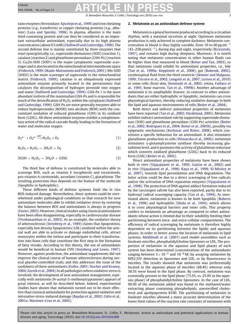
Reactive oxygen species (ROS) are continuously produced undererobic conditions; they are involved in several processes, i.e.,ransformation, regulation or degradation (Gardès-Albert et al.,003; Favier, 2003). Nevertheless, ROS concentration is strictlyontrolled by endogenous antioxidants, thereby protecting humaneings against the potentially deleterious effects of ROS (Fig. 1).
Please cite this article in press as: Bonnefont-Rousselot, D., Collin, F., Medisease and aging. Toxicology (2010), doi:10.1016/j.tox.2010.04.008
OS include superoxide anion radical (O2•−) -especially produced
n cytosol, mitochondria and endoplasmic reticulum-, hydrogeneroxide (H2O2) produced in peroxysomes, the highly reactiveydroxyl radical (•OH) and singlet oxygen (1O2). Reactive nitro-
∗ Corresponding author at: EA 3617, Département de Biologie Expérimentale,étabolique et Clinique, Faculté des Sciences Pharmaceutiques et Biologiques,niversité Paris Descartes, 4 avenue de l’Observatoire, 75006 Paris, France.
E-mail address: [email protected]. Bonnefont-Rousselot).
300-483X/$ – see front matter © 2010 Elsevier Ireland Ltd. All rights reserved.oi:10.1016/j.tox.2010.04.008
st for future clinical research in order to improve public health.© 2010 Elsevier Ireland Ltd. All rights reserved.
gen species (RNS) can also be involved in cell damage, such asnitric oxide (•NO) that is produced by the NO-synthases, andthat reacts with O2
•− to form peroxynitrite (ONOO−), an effi-cient oxidative and nitrosative agent (Halliwell and Gutteridge,1999). An oxidative imbalance, i.e., a disturbance in the balancebetween the production of ROS (especially free radicals) and antiox-idant defenses, has been described as an oxidative stress status(Halliwell and Gutteridge, 1999). This can result in several kindsof cell damage, leading to a loss of function and integrity. Oxy-gen radical-mediated tissue damage (Betteridge, 2000) has beeninvolved in a large number of pathological conditions (Dalle-Donneet al., 2006; Fisher-Wellman et al., 2009) including cardiovas-cular diseases (Bolli et al., 1989; Charniot et al., 2007; Sawyeret al., 2002), neurological disorders (Kontush, 2001; Dexter et
latonin: Action as antioxidant and potential applications in human
al., 1998), cancer (Jung-Hynes et al., 2010) and aging process(Calabrese et al., 2010). As an example, the oxidative stress the-ory of aging proposed by Harman suggested that ROS formed asby-products from metabolic processes can play a key role in aging(Harman, 1956). Mitochondria are specifically involved in these

ARTICLE IN PRESSG Model
TOX-50568; No. of Pages 13
2 D. Bonnefont-Rousselot, F. Collin / Toxicology xxx (2010) xxx–xxx
F trogenp S: oxidg nucleop
p2
sa.reiciectartT
ig. 1. The main cellular sources of reactive oxygen species (ROS) and reactive niolyunsaturated fatty acid; ROOH: lipid hydroperoxide; SH2: reductive substrate;lutathione reductase; NADH + H+: reduced form of the nicotinamide adenine dihosphate; Vit. E: vitamin E; Vit. C: vitamin C.
rocesses (Harman, 1972) and in related diseases (Salmon et al.,010).
ROS play a physiological role by acting at low concentrations asecond messengers able to regulate apoptosis processes (Curtin etl., 2002), to activate transcription factors (NF-kB, p38-MAP kinase,. .) responsible for the activation of genes involved in immuneesponse (Owuor and Kong, 2002), or genes coding antioxidantnzymes (Holgrem, 2003). The role of oxidative stress is crucialn the modulation of cellular functions, notably for neurons astro-ytes and microglia, such as apoptosis and excitotoxicity, bothnvolved in neuronal death. Mitochondrial dysfunction, i.e. cellnergy impairment, apoptosis and overproduction of ROS, is a finalommon pathogenic mechanism in aging and in neurodegenera-
Please cite this article in press as: Bonnefont-Rousselot, D., Collin, F., Medisease and aging. Toxicology (2010), doi:10.1016/j.tox.2010.04.008
ive disease such as Alzheimer’s disease, Parkinson’s disease andmyotrophic lateral sclerosis. Nitric oxide is also a highly diffusibleadical and biological messenger, that plays a prominent role inhe physiology of the central nervous system (Yun et al., 1996).hree isoforms account for •NO production and include neuronal
species (RNS) and the interrelationships between the antioxidant defenses. RH:ized substrate; SOD: superoxide dismutase; GSH-Px: glutathione peroxidase; GR:tide phosphate; NAD+: oxidized form of the nicotinamide adenine dinucleotide
NO synthase (nNOS; type I), inducible NO synthase (iNOS; type II)which is produced in very large amounts by activated microglia(macrophages), and endothelial NO synthase (eNOS; type III), Inthe central nervous system, nNOS, whose expression is regulatedby both physiological and pathophysiological stimuli, accounts formost •NO production, and allows a further formation of the highlyreactive peroxynitrite, via the rapid reaction of O2
•− with •NO(Emerit et al., 2004).
Antioxidant defense systems help the organism to fight againsta ROS excess (Fig. 1). These systems comprise non enzymaticproteins (transferrin, ferritin, ceruleoplasmin), enzymes (superox-ide dismutases (Cu, Zn-SOD and Mn-SOD), catalase, glutathioneperoxidase, . . .), oxidizable molecules (glutathione, vitamins E, A,
latonin: Action as antioxidant and potential applications in human
C, carotenoids, flavonoids. . .) and trace elements (copper, zinc,selenium). Some of them are endogenous whereas others are pro-vided by alimentation. These antioxidants constitute three lines ofdefense: a first line of defense limits an overproduction of ROS byinactivating endogenous cations such as Fe2+ or Cu+; this line con-
ING
T
lin / T
tpmttcsrc3e((macaamarcftw
O
H
2
spr(
Rwat(h(oermtowHieu2icgstn2
ARTICLEModel
OX-50568; No. of Pages 13
D. Bonnefont-Rousselot, F. Col
ains enzymes (ferroxidase; Epsztejn et al., 1999) and iron chelatingroteins (e.g., transferrin) or copper chelating proteins (e.g., albu-in) (Luza and Speisky, 1996). In plasma, albumin is the main
hiol-containing protein and can thus be considered as an impor-ant extracellular antioxidant, especially due to its high plasmaoncentration (about 0.5 mM) (Halliwell and Gutteridge, 1990). Theecond defense line is mainly constituted by three enzymes thateact synergistically, i.e., superoxide dismutases (SOD) (reaction 1),atalase (reaction 2) and glutathione peroxidase (GSH-Px) (reaction). Cu,Zn-SOD (SOD1) is the major cytoplasmic superoxide scav-nger and is also located in the mitochondrial intermembrane spaceFridovich, 1999; Okado-Matsumoto and Fridovich, 2001); Mn-SODSOD2) is the main scavenger of superoxide in the mitochondrial
atrix (Fridovich, 1999); catalase is an ubiquitously expressedntioxidant enzyme primarily located in the peroxisomes thatatalyses the decomposition of hydrogen peroxide into oxygennd water (Halliwell and Gutteridge, 1999); GSH-Px 1 is the mostbundant isoform of the mammalian GSH-Px and is responsible foruch of the detoxification of H2O2 within the cytoplasm (Halliwell
nd Gutteridge, 1999). GSH-Px are more generally enzymes able toeduce hydroperoxides (ROOH) into alcohols (ROH) with the con-omitant oxidation of reduced glutathione (GSH) in its oxidizedorm (GSSG). All these antioxidant enzymes exhibit a complemen-ary action of the radical cascade finally leading to the formation ofater and molecular oxygen
2•− + O2
•− 2H+−→H2O2 + O2 (1)
2O2 + H2O2 → 2H2O + O2 (2)
GSH + H2O2 → 2H2O + GSSG (3)
The third line of defense is constituted by molecules able tocavenge ROS, such as vitamin E tocopherols and tocotrienols,ro-vitamin A carotenoids, ascorbate (vitamin C), glutathione. Theesulting protection thus depends on the feature of the moleculelipophilic or hydrophilic).
These different kinds of defense systems limit the in vivoOS-induced damage. Nevertheless, these systems could be over-helmed under pathological conditions so that research for new
ntioxidant molecules able to inhibit oxidative stress by restoringhe balance between ROS and antioxidants is always in progressFavier, 2003). However, clinical studies using classical antioxidantsave been often disappointing, especially in cardiovascular diseaseVivekananthan et al., 2003). As an example, the oxidative theoryf atherosclerosis (Steinberg et al., 1989) claims that lipoproteins,specially low density lipoproteins (LDL) oxidized within the arte-ial wall are able to activate or damage endothelial cells, attractonocytes within the intima and participate to their transforma-
ion into foam cells that constitute the first step in the formationf fatty streaks. According to this theory, the use of antioxidantsould be beneficial in human CVD (Steinberg and Lewis, 1997).owever, against prediction, the antioxidant supplements did not
mprove the clinical course of human atherosclerosis during sev-ral placebo-controlled trials, and this addresses the issue of thesefulness of these antioxidants (Kuller, 2001; Stocker and Keaney,004; Zureik et al., 2004). In all pathologies where oxidative stress is
nvolved, the development of new antioxidant management, espe-ially with melatonin (N-acetyl-5-methoxytryptamine) appears of
Please cite this article in press as: Bonnefont-Rousselot, D., Collin, F., Medisease and aging. Toxicology (2010), doi:10.1016/j.tox.2010.04.008
reat interest, as will be described below. Indeed, experimentaltudies have shown that melatonin turned out to be more effec-ive than classical antioxidants for protection against oxidative anditrosative stress-induced damage (Baydas et al., 2002; Gitto et al.,001a; Martinez-Cruz et al., 2002).
PRESSoxicology xxx (2010) xxx–xxx 3
2. Melatonin as an antioxidant defense system
Melatonin is a pineal hormone produced according to a circadianrhythm, with a maximal secretion at night. Optimum melatoninproduction is only achieved in complete darkness. Melatonin con-centration in blood is thus highly variable, from 10 to 60 pg mL−1
(43–258 pmol L−1), during day and night, respectively (Brzezinski,1997), and remains high during sleepness. Moreover, it is worthnoting that melatonin concentration in other human fluids canbe higher than that measured in blood (Reiter and Tan, 2003), sothat melatonin could exhibit its antioxidant properties, i.e., bile(Tan et al., 1999a; Koppisetti et al., 2008), gut (Bubenik, 2002),cerebrospinal fluid from the third ventricle (Skinner and Malpaux,1999; Tricoire et al., 2002; Longatti et al., 2007; Leston et al., 2010)or some cells (from skin, Slominski et al., 2002; retina, Faillace etal., 1995; bone marrow, Tan et al., 1999b). Another advantage ofmelatonin is its amphiphilic feature: in contrast to other antioxi-dants that are either hydrophilic or lipophilic, melatonin can crossphysiological barriers, thereby reducing oxidative damage in boththe lipid and aqueous environments of cells (Reiter et al., 2004).
Both direct and indirect antioxidant properties of melatoninhave been reported (Tan et al., 2002; Hardeland, 2005). Melatoninexhibits indirect antioxidant role by supporting superoxide dismu-tase (SOD) and glutathione peroxidase (GSH-Px) activities (Reiteret al., 2002; Rodriguez et al., 2004; Reiter et al., 2005b), possibly viaepigenetic mechanisms (Korkmaz and Reiter, 2008), which con-stitutes a specific behaviour for an antioxidant. It also stimulatesglutathione production in cells (Winiarska et al., 2006); melatoninstimulates �-glutamylcysteine synthase thereby increasing glu-tathione level, and it promotes the activity of glutathione reductasewhich converts oxidized glutathione (GSSG) back to its reducedform (GSH) (Reiter et al., 2002).
Direct antioxidant properties of melatonin have been shownboth in vitro (Vijayalaxmi et al., 1995; Gulcin et al., 2002) andin vivo (Vijayalaxmi et al., 1996; Kaya et al., 1999; El-Missiry etal., 2007), towards lipid peroxidation and DNA degradation. Thelatter action could be due to a direct scavenging of free radicalsand/or the activation of DNA reparation enzymes (Vijayalaxmi etal., 1998). The protection of DNA against adduct formation inducedby the carcinogen safrole has also been reported, partly due to itshydroxyl radical scavenging capacity (Tan et al., 1994). As men-tioned above, melatonin is known to be both lipophilic (Robertset al., 1998) and hydrophilic (Shida et al., 1994), which allowsit to cross physiological barriers and enter cells. This propertyconfers to melatonin an advantage as compared to other antiox-idants whose action is limited due to their solubility limiting theirpartitioning between intra- and extra-cellular compartments. Theefficiency of radical scavenging by melatonin is therefore highlydependent on its partitioning between the lipidic and aqueousphases. In order to better assess the location of melatonin in lipidsystems, Mekhloufi et al. (2007) used model assemblies such aslinoleate micelles, phosphatidylcholine liposomes or LDL. The pro-portion of melatonin in the aqueous and lipid phases of eachsystem has been determined (concentrations of the antioxidantsranging between 3 × 10⁻⁵ and 10−4 M) by assaying melatonin byHPLC/UV detection in liposomes and LDL, or by fluorescence inmicelles. The results showed that melatonin was preferentiallylocated in the aqueous phase of micelles (68.4%) whereas only30.5% were found in the lipid phase. By contrast, melatonin wasessentially present in the lipid phase (73.5%, vs. 25.9% in the aque-ous phase) of phosphatidylcholine liposomes. In the case of LDL,
latonin: Action as antioxidant and potential applications in human
99.9% of the melatonin added was found in the methanol/waterextracting phase containing phospholipids, unesterified choles-terol and apolipoprotein B100. The partitioning of melatonin inlinoleate micelles allowed a more accurate determination of thelower limit values of the reaction rate constants of melatonin with

ING
T
4 llin / T
liooto
ea(taoo2ebiled
ARTICLEModel
OX-50568; No. of Pages 13
D. Bonnefont-Rousselot, F. Co
ipid peroxyl radicals than previously done (Mekhloufi et al., 2005),.e. k(LOO• + melatonin) ≥ 9.0 × 10⁴ M−1 s−1. The partitioning databtained in these lipid systems could thus help explain the abilityf melatonin to scavenge free radicals according to their produc-ion site, and thereby to protect lipids and/or proteins against thexidation process.
Several studies demonstrated the ability of melatonin to scav-nge oxygen-derived free radicals, such as hydroxyl (Poeggeler etl., 2002; Roberts et al., 1998; Matuszak et al., 1997) and peroxylLivrea et al., 1997; Pieri et al., 1994) radicals. By contrast, mela-onin seems poorly able to react with superoxide radicals (Zang etl., 1998; Walters-Laporte et al., 1998). Moreover, protective effectsf melatonin have been reported in the case of the in vitro oxidationf LDL by copper or by macrophages (Bonnefont-Rousselot et al.,002, 2003; Walters-Laporte et al., 1998; Duell et al., 1998; Livreat al., 1997; Abuja et al., 1997). This protection has been explainedy the ability of melatonin to directly scavenge the free radicals
Please cite this article in press as: Bonnefont-Rousselot, D., Collin, F., Medisease and aging. Toxicology (2010), doi:10.1016/j.tox.2010.04.008
nvolved in these oxidation processes (Allegra et al., 2003). Simi-arly, models of melatonin-leaded nanoparticles have shown theirfficiency to protect liposomes or microsomes against lipid peroxi-ation (Schaffazick et al., 2005). Melatonin is also able to neutralize
Fig. 2. Chemical structures of mela
PRESSoxicology xxx (2010) xxx–xxx
singlet oxygen, peroxynitrite anion, and nitric oxide (Allegra et al.,2003; Reiter et al., 2004; Gilad et al., 1997; Ucar et al., 2007; Topalet al., 2005). Although some metabolites have been identified invivo (e.g., 3-cyclohydroxymelatonin, Tan et al., 1998), the mecha-nisms by which melatonin displays its direct antioxidant propertieshave not totally been unravelled. As previously specified, mela-tonin is known to exhibit antioxidant properties towards a widerange of biological systems, as for example linoleate model system(Mekhloufi et al., 2005) or LDL (Pieri et al., 1996; Kelly and Loo,1997; Bonnefont-Rousselot et al., 2002). In the latter model, mela-tonin exhibited a relatively low activity. This could be explainedin term of lipophilicity since replacing acetyl by nonanoyl groupdramatically increases the antioxidant activity (Gozzo et al., 1999).Other functional groups play an important role in the beneficialeffect of melatonin in the prevention of oxidation. If the amidefunction is not an absolute requirement for the antioxidant activityof melatonin, it was suggested that the presence of the acyl ter-
latonin: Action as antioxidant and potential applications in human
minal group could prevent a possible prooxidant behaviour (Tanet al., 1993). This was confirmed when 5-methoxytryptamine wasfound to shorten the lag phase duration during oxidation of the LDLmodel (Gozzo et al., 1999). However, the acyl terminal group had
tonin and some derivatives.

ARTICLE IN PRESSG Model
TOX-50568; No. of Pages 13
D. Bonnefont-Rousselot, F. Collin / Toxicology xxx (2010) xxx–xxx 5
•−/HO
nst2
a(avi(bimigpi
3a
mtfohdopRii
sWpqp
Fig. 3. Selective production of •OH/O2•− , •OH, O2
o influence on the scavenging capacity of hydroxyl radicals in vitroince rate constants for the reaction of hydroxyl radicals with mela-onin and 5-methoxytryptamine were similar (respectively 2.7 and.3 × 1010 M−1 s−1; Matuszak et al., 1997).
The methoxy group has also a beneficial effect in antioxidantctivity of melatonin, whatever its position on the indole ring5- or 6-methoxy). However, the methoxyl function is not anbsolute requirement since phenolic derivatives of melatonin areery active. In particular, N-acetyl-5-hydroxytryptamine activitys increased by one order of magnitude comparing to melatoninGozzo et al., 1999) (Fig. 2). This latter compound seems to haveeneficial effects in preventing both lipid and protein oxidation
n synaptosomal membranes (Millán-Plano et al., 2010). Thus,ethoxy and acyl groups are essential substituents for the antiox-
dant properties of melatonin. The compound lacking both theseroups (namely 5-hydroxytryptamine) was found to efficientlyromote the generation of •OH radicals in the presence of ferric
ron and hydrogen peroxide (Matuszak et al., 1997) (Fig. 2).
. Gamma radiolysis as a tool to study molecularntioxidant properties of melatonin
In order to improve the knowledge of melatonin antioxidantode of action, a study of the direct antioxidant properties of mela-
onin has been performed in vitro against different oxygen-derivedree radical species generated in aqueous solution by gamma radi-lysis. Gamma radiolysis of water is a well known method, thatas many advantages, such as the homogeneous production ofefined concentrations of free radicals (as superoxide anion O2
•−
r hydroxyl radical •OH), as well as the possibility to selectivelyroduce one specific radical to be studied at a time (Bonnefont-ousselot, 2005). Free radicals thus generated have been used to
nitiate one-electron oxidation reaction(s) on melatonin dissolvedn water.
Radiolysis can be defined as the chemical transformations of a
Please cite this article in press as: Bonnefont-Rousselot, D., Collin, F., Medisease and aging. Toxicology (2010), doi:10.1016/j.tox.2010.04.008
olvent due to the absorption of ionizing radiations (Spinks andoods, 1990). These radiations can be of several kinds (electrons,
hotons, neutrons, heavy charged particles, but the more fre-uently used is the � radiation emitted by 60Co (1.17 and 1.33 MeVhotons) or by 137Cs (≈600 keV photons). Those photons ionize the
2• radical species upon water gamma radiolysis.
aqueous (or ethanolic) solvent, leading to the very rapid production(within a few nanoseconds) of a homogeneous solution of radicalspecies. Radiolytically generated free radicals are independent ofthe nature and the concentration of the dissolved compound aslong as its concentration remains lower or equal to 10−2 mol L−1
(Spinks and Woods, 1990). Under these conditions, the compounddissolved into the irradiated solvent is not directly subjected to theionizing radiations, but to the action of the free radicals producedby the solvent radiolysis.
Briefly, water �-radiolysis leads within a few nanoseconds tothe production of a homogeneous solution of radicals and molecu-lar species whose nature depends on the experimental conditions(Fig. 3). The mechanisms of formation of the primary species (•OH,e−
aq, •H, H2, H2O2) are well known (Spinks and Woods, 1990).The latter are produced with the following radiolytic yields (G)expressed as the number of molecules or radicals produced perunit of energy absorbed, i.e. per Joule (Spinks and Woods, 1990;Draganic and Draganic, 1971).
This method has been used to evaluate antioxidant propertiesof melatonin, firstly when melatonin was included in lipid sys-tems, then with a molecular approach with analysis of melatoninoxidation products by mass spectrometry.
Regarding lipid systems, low density lipoproteins (LDL) areof special interest since oxidized LDL are involved in the patho-genesis of atherosclerosis (Rizzo et al., 2009). In a comparativestudy, LDL oxidation was initiated in vitro either by defined freeradicals [i.e., superoxide anion (O2
•−) and ethanol-derived peroxylradicals (RO2
•)] produced by gamma radiolysis or by copper ions.The antioxidant effect of melatonin was evaluated at 100 �M.Under these experimental conditions, melatonin did not protectendogenous LDL alpha-tocopherol from oxidation by RO2
•/O2•−
free radicals. By contrast, it protected �-carotene from the attackof those radicals and partially inhibited the formation of productsderived from lipid peroxidation (conjugated dienes and thiobar-bituric acid-reactive substances or TBARS) (Bonnefont-Rousselot
latonin: Action as antioxidant and potential applications in human
et al., 2002, 2003). As previously reported (Walters-Laporte etal., 1998), melatonin inhibited copper-induced LDL oxidationby increasing 1.60-fold the lag phase duration of conjugateddiene formation over the 8 h of the experimental procedure.This study also showed a stronger antioxidant activity of some

ING
T
6 llin / T
m3(cswtppo(
4t
mientpdst(afieupiie(aeie
tsctAba9(dsvfmapswaphhsip
ARTICLEModel
OX-50568; No. of Pages 13
D. Bonnefont-Rousselot, F. Co
elatonin derivatives (Fig. 2), i.e., N-[2-(5-methoxy-1H-indol--yl)ethyl]-3,5-di-tert-butyl-4-hydroxybenzamide (DTBHB) andR,S)-1-(3-methoxyphenyl)-2-propyl-1,2,3,4-tetrahydro-beta-arboline (GWC20) which is a pinoline derivative, so that iteemed of interest to test in vivo whether DTBHB and GWC20,hich exhibited a strong capacity to inhibit in vitro LDL oxida-
ion, would reduce atherosclerosis in animals susceptible to thisathology. Nevertheless, it is worth remembering that an in vitrorotective effect of molecules is not always related to a predictionf their capacity to inhibit in vivo atherosclerosis developmentTailleux et al., 2005).
. Melatonin analysis by mass spectrometry-basedechniques
Mass spectrometry (MS)-based techniques are among theost efficient and versatile analytical techniques that are used
ncreasingly to determine, both quantitatively and qualitatively,ndogenous substances in biological samples. Melatonin determi-ation by mass spectrometry makes identity confirmation possible,hus increasing the validity of the results. In addition, when cou-led to liquid chromatography, mass spectrometry allows workingirectly on complex samples such as saliva or serum withoutample preparation step. This is made possible by the use ofhe common discriminative working modes of mass spectrometryselected ion monitoring (SIM), selected reaction monitoring (SRM)nd multiple reaction monitoring (MRM)) that enhance the speci-city, and thus the sensitivity, of the analytical method. Motoyamat al. (2004) have determined endogenous melatonin in saliva bysing a simple quadrupole mass spectrometer coupled to reverse-hase liquid chromatography and working in the SIM mode with
n-source fragmentation focused on the ion at m/z 174. This latters usually one of the most intense fragments of melatonin, gen-rated by the fragmentation of the aliphatic chain of melatoninformal loss of CH3CONH2, −59 Da). It has been used both in SIMnd SRM mode for the quantitative determination of melatonin bylectrospray ionisation (ESI) triple quadrupole mass spectrometryn complex plant samples (Cao et al., 2006) or human serum (Yangt al., 2002).
Separative methods are widely used for qualitative and quan-itative analysis of melatonin in complex samples prior to masspectrometric detection. Among them, enzyme immunoaffinityhromatography (IAC) is successfully applied to determine mela-onin in biological sample such as human serum, plasma and saliva.s tandem MS techniques separate specifically single ion that cane assigned to single analyte, IAC is able to separate one specificnalyte among myriads of compounds, with recovery varying from2 to 98% for amounts of melatonin ranging from 50 to 400 fmolRolcik et al., 2002). Enzyme immunoassay is also employed toetermine melatonin in food samples such as olive oil or grapekin (De La Puerta et al., 2007; Iriti et al., 2006). Other more inno-ative methods are tentatively developed for melatonin separationrom complex samples. One method, based on the use of copoly-
ers of N-isopropylacrylamide (PNIPAM), N-tert-butylacrylamidend acrylic acid, exhibiting both pH- and temperature-dependentroperties, was proposed for the qualitative and quantitative analy-is of melatonin (Ayano et al., 2007). The copolymers were modifiedith cross-linked hydrogel applied onto aminopropyl silica beds
nd packed as an HPLC column. The lower critical solution tem-erature of PNIPAM is 32 ◦C: below, the polymer hydrates and has
Please cite this article in press as: Bonnefont-Rousselot, D., Collin, F., Medisease and aging. Toxicology (2010), doi:10.1016/j.tox.2010.04.008
ydrophilic properties, while above, it dehydrates to get a compactydrophobic conformation. This property was successfully used toeparate melatonin from L-tryptophan and serotonin by workingsocratically with aqueous mobile phase and by varying the tem-erature.
PRESSoxicology xxx (2010) xxx–xxx
As melatonin possesses several reaction sites for oxidationby HO• radicals, such as the indole ring, one pair of electronsof nitrogen atoms or the hydrogen on the aliphatic chain, theprediction of structures for oxidation products remain difficult.Further information on the molecular mechanisms of the in vitroradical-induced oxidation of melatonin and the identification ofthe resulting products is often given by mass spectrometry anal-ysis. In an investigation on the possible role of melatonin inmelanogenesis, melatonin was submitted to the attack of HO•,generated by the decomposition of hydrogen peroxide underUV irradiation (Rizzi et al., 2006). Oxidation of melatonin wasfound to lead to several products, including 2-hydroxymelatonin,N1-acetyl-N2-formyl-5-methoxykynurenin (AFMK) and cyclic 3-hydroxymelatonin (c3HO-MLT), identified by Matrix AssistedLaser Desorption Ionization—Time-Of-Flight (MALDI-TOF) massspectrometry, fluorescence and UV–visible spectrophotometry.However, proposals of chemical structures are often based on thededuction of the molecular mechanism of HO• attack towardsmelatonin or on the comparison to known in vivo metabolites ofmelatonin (as AFMK, Harthe et al., 2003 or c3HO-MLT, Tan et al.,1998), and are not supported accurately by the experimental spec-tra because of the lack of specific information for identification.
Advanced mass spectrometric and chromatography-coupledmethodologies are required to identify the oxidation products ofmelatonin with a better accuracy. Liquid chromatography coupledto tandem mass spectrometry with hydrogen–deuterium exchange(LC-HDX-MS/MS) is one of them and is susceptible to providemore valuable information for identification. HDX of protonatedmelatonin and its in vitro oxidation end-products have been exam-ined by liquid chromatography coupled to electrospray ionization(ESI)/ion-trap mass spectrometry (Collin et al., 2009). In deuter-ated medium (D2O/MeOD, 1/1 v/v), melatonin is able to exchange3 times and is thus detected at m/z 236 as a pseudo-molecularmonocharged ion. In these conditions, an hydrogen–deuteriumscrambling was found to occur between deuterium and hydrogenon position C2 and C4 of the indole ring during the fragmenta-tion process. The spectrum showed multiple consecutive fragmentions corresponding to the loss of variably deuterated neutralmolecules. This characteristic, also observed for tryptophan (Lioeet al., 2004), was used to identify the hydroxylated oxidation prod-ucts of melatonin in vitro, after a chromatographic separation ona reverse-phase C18 column by eluting with deuterated solvents(D2O/acetonitrile, 80/20 v/v). Two chromatographic peaks weredetected and further fragmented: by looking at the multiplic-ity of product ions, HDX methodology showed that the hydroxylgroup of radiolytically hydroxylated melatonin was located onposition C6 or C7 for the first product and on position C2 orC4 for the second (Fig. 4). The same strategy was used for theidentification of the other radio-induced oxidation products ofmelatonin in vitro: N1-acetyl-5-methoxykynurenin (AMK), N1-acetyl-N2-formyl-5-methoxykynurenin (AFMK), known as minormetabolites of melatonin in vivo (Harthe et al., 2003; Reiter et al.,2000; Silva et al., 2004), and dehydro-AFMK.
5. Results from melatonin analyses
Efficient detection methods of melatonin, together with opti-mized extraction protocols from plant samples, allowed to confirmthe presence of melatonin in several plant products, but alsoshowed that its concentration varies widely on the species and on
latonin: Action as antioxidant and potential applications in human
plant parts (Paredes et al., 2009). The role of melatonin in plantsmay be a protection against oxidative stress (Tan et al., 2002), espe-cially in seeds where melatonin has been hypothesized to protectgerm from oxidative damage due to UV light, drought, tempera-ture variations, and environmental toxins (Manchester et al., 2000).

ARTICLE IN PRESSG Model
TOX-50568; No. of Pages 13
D. Bonnefont-Rousselot, F. Collin / Toxicology xxx (2010) xxx–xxx 7
Fig. 4. Hydrogen Deuterium Exchange of hydroxylated melatonin (HO-MLT). During MS fragmentation, H/D scrambling occurs between deuterium of the amide terminalgroup and protons on positions C2 and C4 of the indole ring. The replacement of one of these protons by the deuterated hydroxyl group is responsible for quenching one ofthe possible H/D back exchange during fragmentation, and thus leads to a lower multiplicity for consecutive fragment ions. After chromatographic separation, two peaksw ultiplio
Tmbgiepma
bhAww(taaaAtceoiano12imhs
ere detected for HO-MLT (at tr = 6.1 min and tr = 6.87) and fragmented: the peak mn the indole ring of HO-MLT (adapted from Collin et al., 2009).
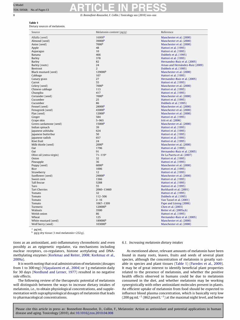
able 1 presents some dietary sources of melatonin, with a specialention to Graminae that exhibit high contents of melatonin (rice,
arley, sweet corn, oat, tall fescue) (Hattori et al., 1995). This sug-ests that it is possible to ingest sufficient quantities of melatoninn edible plants to perhaps influence physiological processes. As anxample, food-derived melatonin could also be involved in post-randial sleepiness after ingestion of melatonin-rich food, sinceelatonin is considered as a sleep inducing molecule (Dollins et
l., 1994).The fate of melatonin in circulation is still poorly understood,
ut intra-arterial infusion of melatonin in rats showed that thealf-life of melatonin was only 20 min (Gibbs and Vriend, 1981).fter ingestion of 1 g melatonin in human, about 90% melatoninas converted into metabolites (especially 6-hydroxymelatonin)ith a renal elimination of sulphated or glucuronidated conjugates
Leone et al., 1987). A recent study in critically ill patients showedhat oral melatonin supplementation resulted in a rapid enteralbsorption, with pharmacological levels reached within 5 min andserum peak (11,040 pg mL−1, or 47,582 pmol L−1) after 16 min
nd a half-elimination time of about 1.5 h (Mistraletti et al., 2010).pharmacokinetic study in rats showed that about 500 ng mela-
onin should be continuously infused per hour to freely movingatheterized rats to maintain a 10-fold elevation of their plasma lev-ls (Huether et al., 1992); under these conditions, concentrationsf melatonin (plus melatonin metabolites) have been monitoredn several tissues in order to appreciate partitioning of melatoninnd the results showed large amounts in small intestine (duode-um, jejunum, ileum) and lower gut (caecum, colon) at the endf a 2h-infusion, and in feces after 6 h (Table 2) (Messner et al.,998). Indeed, about 45% of the melatonin administered during the
Please cite this article in press as: Bonnefont-Rousselot, D., Collin, F., Medisease and aging. Toxicology (2010), doi:10.1016/j.tox.2010.04.008
h infusion period were found in the urine and 20% in the smallntestine whereas 6 h after the end of the infusion, the majority of
elatonin and its metabolites initially present in the small intestinead been eliminated in the feces. The gut so appears not only as aite of melatonin synthesis (Lee et al., 1995) but also as a site where
city was used as a reliable information regarding the position of the hydroxyl group
melatonin concentrates, possibly suggesting a role of melatonin inthe gastrointestinal tract, especially for ensuring protection againstinflammation and ulceration of the intestinal mucosa (Melchiorriet al., 1997; Bubenik et al., 1998). The study of Messner et al. (1998)also demonstrated that a certain proportion of circulating mela-tonin must enter tissues and become covalently bound to aminoacids of tissue proteins. Apart from this non oral supplementationin rats, Table 2 shows melatonin amounts in the body resulting fromoral melatonin intakes. It clearly shows that increased amountsof ingested melatonin (at low doses, i.e., 0.1–10 mg) resulted inenhanced serum melatonin concentration, as performed in healthyvolunteers (Dollins et al., 1994); under these conditions, this orallyadministrated melatonin has been shown to be a potent hypnoticagent; this study also suggested that the physiological increase ofmelatonin level at night may initiate the normal sleep onset.
6. Therapeutic potential of melatonin
As melatonin is produced according to a circadian rhythm,it has been used mostly to correct insomnia and jet lag (Petrieet al., 1993; Dahlitz et al., 1991; Haimov and Lavie, 1997) as itplays a major role in the synchronization of the sleep/wake cycle(Reiter, 1980). Indeed, circulating melatonin concentration is sig-nificantly decreased in elderly insomniacs than in age-matchedcontrols and their onset and peak times are delayed (Haimov andLavie, 1997). Moreover, the regulation of central and peripheralclocks in humans has been shown to be based on clock genes(Cermakian and Boivin, 2009), and circadian clock gene expres-sion in peripheral tissues could be uncoupled from the activityof the central clock during periods of acute systemic inflamma-
latonin: Action as antioxidant and potential applications in human
tion, as recently reported in blood leukocytes (Haimovich et al.,2010). Under these conditions, the realignment of the centraland peripheral clocks could also be of help for recovery fromdisease in humans. Increasing evidence suggests that melatoninrhythmicity plays by itself key roles in several metabolic func-
ARTICLE IN PRESSG Model
TOX-50568; No. of Pages 13
8 D. Bonnefont-Rousselot, F. Collin / Toxicology xxx (2010) xxx–xxx
Table 1Dietary sources of melatonin.
Source Melatonin content (pg/g) Reference
Alfalfa (seed) 16000b Manchester et al. (2000)Almond (seed) 39000b Manchester et al. (2000)Anise (seed) 7000b Manchester et al. (2000)Apple 48 Hattori et al. (1995)Asparagus 10 Hattori et al. (1995)Banana 466 Dubbels et al. (1995)Barley 378 Hattori et al. (1995)Barley 82 Hernandez-Ruiz et al. (2005)Barley (roots) 24 Arnao and Hernández-Ruiz (2009)Beetroot 2 Dubbels et al. (1995)Black mustard (seed) 129000b Manchester et al. (2000)Cabbage 107 Hattori et al. (1995)Canary grass 27 Hernandez-Ruiz et al. (2005)Carrot 55 Hattori et al. (1995)Celery (seed) 7000b Manchester et al. (2000)Chinese cabbage 113 Hattori et al. (1995)Chungiku 417 Hattori et al. (1995)Coriander (seed) 7000b Manchester et al. (2000)Cucumber 25 Hattori et al. (1995)Cucumber 86 Dubbels et al. (1995)Fennel (seed) 28000b Manchester et al. (2000)Fenugreek (seed) 43000b Manchester et al. (2000)Flax (seed) 12000b Manchester et al. (2000)Ginger 584 Hattori et al. (1995)Grape skin 5–965 Iriti et al. (2006)Green cardamone (seed) 15000b Manchester et al. (2000)Indian spinach 39 Hattori et al. (1995)Japanese ashitaba 624 Hattori et al. (1995)Japanese butterbur 50 Hattori et al. (1995)Japanese radish 657 Hattori et al. (1995)Kiwi fruit 24 Hattori et al. (1995)Milk thistle (seed) 2000b Manchester et al. (2000)Oat 1796 Hattori et al. (1995)Oat 91 Hernandez-Ruiz et al. (2005)Olive oil (extra virgin) 71–119a De La Puerta et al. (2007)Onion 32 Hattori et al. (1995)Pineapple 36 Hattori et al. (1995)Poppy (seed) 6000b Manchester et al. (2000)Rice 1006 Hattori et al. (1995)Strawberry 12 Hattori et al. (1995)Sunflower (seed) 29000b Manchester et al. (2000)Sweet corn 1366 Hattori et al. (1995)Tall fescue 5288 Hattori et al. (1995)Taro 55 Hattori et al. (1995)Tart Cherries 2060–13460 Burkhardt et al. (2001)Tomato 32 Hattori et al. (1995)Tomato 112–506 Dubbels et al. (1995)Tomato 2–16 Van Tassel et al. (2001)Tomato 1067–1399 Pape and Lüning (2006)Turmeric 120000b Chen et al. (2003)Walnuts 3500 Reiter et al. (2005a,b)Welsh onion 86 Hattori et al. (1995)Wheat 125 Hernandez-Ruiz et al. (2005)White mustard (seed) 189000b Manchester et al. (2000)
tpnm2
ffs
wmmt
Wolf berry (seed) 103000b
a pg/mL.b pg/g dry tissue (1 mol melatonin = 232 g).
ions as an antioxidant, anti-inflammatory chronobiotic and evenossibly as an epigenetic regulator, via mechanisms includinguclear receptors, co-regulators, histone acetylating and DNA-ethylating enzymes (Korkmaz and Reiter, 2008; Korkmaz et al.,
009a).It is worth noting that oral administration of melatonin (dosages
rom 1 to 300 mg) (Vijayalaxmi et al., 2004) or 1 g melatonin dailyor 30 days (Nordlund and Lerner, 1977) resulted in no negativeide effects.
Please cite this article in press as: Bonnefont-Rousselot, D., Collin, F., Medisease and aging. Toxicology (2010), doi:10.1016/j.tox.2010.04.008
The following review of the therapeutic potential of melatoninill distinguish between the ways to increase dietary intakes ofelatonin, i.e., to obtain physiological concentrations, and supple-entation with supraphysiological dosages of melatonin that leads
o pharmacological concentrations.
Manchester et al. (2000)
6.1. Increasing melatonin dietary intakes
As mentioned above, relevant amounts of melatonin have beenfound in many roots, leaves, fruits and seeds of several plantspecies, although the concentration of melatonin is greatly vari-able in species and plant tissues (Table 1) (Paredes et al., 2009).It may be of great interest to identify beneficial plant propertiesrelated to the presence of melatonin, and whether the positivehealth effects observed in humans could be due to melatonin
latonin: Action as antioxidant and potential applications in human
consumed in the diet, and whether melatonin may be workingsynergistically with other antioxidant molecules present in plants.An efficient uptake of melatonin from food should be expected toinfluence blood plasma concentration, which is basically very low(200 pg mL−1 (862 pmol L−1) at the maximal night level, and below

ARTICLE IN PRESSG Model
TOX-50568; No. of Pages 13
D. Bonnefont-Rousselot, F. Collin / Toxicology xxx (2010) xxx–xxx 9
Table 2Examples of exogenous melatonin intakes and resulting amounts found in the body.
Species Body part Physiologicalvalue (pg/mL)
Oral intakeamounts
Concentration (pg/mL) Delay after intake Reference
European see bass Plasma 18 2.5 mg/kga 464 45 min Rubio et al. (2004)0.1 mg/kga 170
Human Saliva – 5 mg 4199 30 min Wirz-Justice et al. (2002)Serum 3 to 20 – – – Reiter (1991)
15 0.1 mg 50 4 h Dollins et al. (1994)0.3 mg 1201 mg 41010 mg 6825
Chick Plasma 19 3.5 ng/g plantfoodb
33 90 min Hattori et al. (1995)
Wistar rat Small intestine – 1 �gc 13900d immediately after a2 h-infusion period
Messner et al. (1998)Lower gut 720d
Feces 9400d 6 h
a mg/kg BW (Body Weight).b
1Pitnte
6m
uss
ia(atarImsawbohoiataM3taated
Corn, milo, beans, rice.c 500 ng/h continuous infusion, for 2 h.d Melatonin + metabolites, pg/g wet weight tissue (1 mol melatonin = 232 g).
0 pg mL−1 (43 pmol L−1) during the day) (Hardeland and Pandi-erumal, 2005). The question arises whether the amounts presentn the food can be sufficient to increase the plasma level of mela-onin, as previously proposed by Hattori et al. (1995). It is worthoting that gastrointestinal tract plays a role not only in mela-onin intake, participates in enterohepatic cycling, but also as anxtrapineal site of melatonin biosynthesis (Konturek et al., 2007).
.2. Supplementation with supraphysiological dosages ofelatonin
Supplemental melatonin (i.e., use at supra-physiological doses),nlike that derived from natural sources (e.g. “greens” productsuch as wheatgrass or ryegrass), needs a pre-market authorizationince it is regarded as a medicinal product.
To focus on the antioxidant effects of melatonin, several clin-cal studies have been carried out with patients in an attempt tossess the activity of melatonin towards radical-induced damageGitto et al., 2001b; Fulia et al., 2001; Ochoa et al., 2003; Pappolla etl., 2000, 2003). Melatonin turned out to be a modulator of oxida-ive stress under acute conditions such as surgery (Kücükakin etl., 2009). Under these conditions, there is a massive inflammatoryesponse with production of cytokines (interleukins: IL-1, IL-2, IL-6,L-8) that play a role in cell activation and in the subsequent for-
ation of ROS and RNS (Gitto et al., 2004). Several clinical trialshowed that melatonin could be efficient in preventing cell dam-ge. As an example, Fulia et al. (2001) supplemented 10 newbornsith asphyxia with 80 mg melatonin (8 doses of 10 mg, separated
y 2 h intervals) per os, within the first 6 h of life, and monitoredxidative and nitrosative stress markers by assaying malondialde-yde (end-product of lipid peroxidation) and nitrite/nitrate (nitricxide-derived products) levels, before and after melatonin admin-stration. Whereas these markers were elevated in newborns withsphyxia compared with newborns without asphyxia, the patientsreated with melatonin exhibited lower levels of malondialdehydend nitrate/nitrite than in the placebo group without melatonin.oreover, no death was observed in the group with melatonin, vs.deaths in the placebo group. These results indicate that mela-
onin may be beneficial in the treatment of newborn infants with
Please cite this article in press as: Bonnefont-Rousselot, D., Collin, F., Medisease and aging. Toxicology (2010), doi:10.1016/j.tox.2010.04.008
sphyxia, and these beneficial effects may be related both to thentioxidant properties of melatonin and to its ability to increasehe efficiency of mitochondrial electron transport. Similarly, Gittot al. (2001b) determined the serum concentration of lipid peroxi-ation products (malondialdehyde, 4-hydroxyalkenals) in 10 septic
newborns given 20 mg melatonin (two oral doses of 10 mg each,with a 1-h interval) or placebo; the concentrations of these oxi-dation products in newborns with sepsis were significantly higherthan those in healthy infants without sepsis. In contrast, there wasa significant reduction of these concentrations in septic newbornstreated with melatonin to the levels observed in the healthy con-trols. Melatonin also improved the clinical outcome of the septicnewborns, as 10 out of all septic children who were not treated withmelatonin died within 72 h after diagnosis of sepsis whereas noneof the 10 septic newborns treated with melatonin died. Finally,in neonates undergoing surgery, where clinical outcomes seemsto depend on oxidative stress-induced damage, ten newborns(group 1), 5 newborns with surgical malformations and respiratorydistress (group 1a) and 5 with isolated abdominal surgical mal-formations (group 1b) received a total of 10 doses of melatonin(10 mg/kg) at defined times interval for 72 h, within 3 h after theend of the operation. Ten surgical neonates (group 2) did not receivemelatonin, and twenty healthy neonates (group 3) served as con-trols (Gitto et al., 2004). Surgical stress markers (IL-6, IL-8, TNF-�,nitrite/nitrate levels) were measured at the end of the operation,before treatment (melatonin or placebo) and respectively 24 h, 72 h,and 7 days after treatment. Postoperative value of cytokines andnitrite/nitrate levels of groups 1 and 2 were significantly higherthan in group 3. Compared with group 1b, group 2 displayed sig-nificantly higher cytokines and nitrite/nitrate levels at 24 h, 72 h,and at 7 days intervals. In group 1a the immediate postoperativevalues of cytokines were significantly higher than in groups 1b and2, but a significant improvement was observed after administra-tion of melatonin with significantly lower levels of IL-6 and IL-8with respect to group 2. This study showed that melatonin reducedcytokines and nitrate/nitrite levels, showing potent antioxidantproperties with improvement in clinical outcome. Melatonin, via itsantioxidant properties, could thus modulate oxidative stress, whichmay improve organ function and reduce morbidity and mortality,at least as tested in newborns where the drug was able to decreaseoxidative stress induced by surgery (Kücükakin et al., 2009). Mela-tonin has been investigated in a wide range of diseases, such asheart disease, Alzheimer’s disease, AIDS, diabetes, depression, can-cer (Beyer et al., 1998).
latonin: Action as antioxidant and potential applications in human
As an example of use of melatonin in chronic diseases, it has beenshown that melatonin administration in patients with Alzheimerdisease significantly delayed the progression of the disease anddecreased brain atrophy as assessed by MRI (Brusco et al., 1998).It is now admitted that the neuronal loss in Alzheimer disease

IN PRESSG
T
1 llin / Toxicology xxx (2010) xxx–xxx
cttnd(tnhnPs
2onbTi(cstt2dasiimosdwpm2amoiStmmt2
ii(mea
aataK(lmaa
ARTICLEModel
OX-50568; No. of Pages 13
0 D. Bonnefont-Rousselot, F. Co
ould result from radical-induced apoptosis of neuronal cells. Ithus seems that the antioxidant properties of melatonin could pro-ect neurons from the degeneration leading to cell death. Moreover,o toxicity or adverse effect has been reported consecutively to theaily administration of melatonin (1–300 mg, or 4.3–1291.5 �mol)Jan et al., 2000; Seabra et al., 2000), which could encouragehe long-term administration of melatonin in patients suffering aeurodegenerative disease such as Alzheimer disease. Melatoninas been reported to inhibit the intrinsic apoptotic pathways ineurodegenerative diseases including stroke, Alzheimer disease,arkinson disease, Huntington disease, and amyotrophic lateralclerosis (Wang, 2009).
Experimental studies in rodents showed that melatonin (5 mg or1.5 �mol/kg body weight, 5 day/week, for 50 days) decreased thexidative stress induced by D-galactose (DG) treatment to mimicatural aging; melatonin thus lowered the formation of protein car-onyls in the liver, kidney and brain of DG treated mice, decreasedBARS production in serum and brain, prevented DG-inducedncrease of soluble receptors for advanced glycation endproductssRAGE), decreased the expression of the pro-apoptotic bax andaspase-3 proteins in splenocytes, and lowered A� protein expres-ion in the brain (Hsieh et al., 2009). It has been suggested thathe anti-aging effect of melatonin may be related to the reduc-ion of A�-induced lipid peroxidation in vitro (Feng and Zhang,004). More specifically, melatonin (10 mg or 43 �mol/kg in therinking water, for 9 months, starting 1 month after birth) seemedble to modulate the expression of alpha-secretase in a model ofenescence-accelerated prone mice (SAMP8), thereby contribut-ng to the decrease of the high levels of aggregating A� proteinn this model (Gutierrez-Cuesta et al., 2008). Moreover, in this
odel exhibiting a marked acceleration of aging in relation withxidative stress (Caballero et al., 2009), melatonin improved pro-urvival signals and reduced pro-death signals, as shown by theecrease of Bid and the increase of Bcl-2 levels, in comparisonith non-treated SAMP8. Melatonin also exhibited antiapoptoticroperties after ischemic neuronal injury in the rat, via enhance-ent of Bcl-2 induction and DNA repair capacity (Sun et al.,
002). The influence of melatonin on the antioxidant role of Bcl-2nd more generally against the oxidative stress-related impair-ents has been demonstrated, paving the way for the treatment
f age-induced neural processes. Moreover, a significant decreasen the fluidity of synaptosomal and mitochondrial membranes ofAMP8 mice was observed, as assessed by fluorescent polariza-ion after incorporation of a probe (TMA-DPH). In this model,
elatonin could prevent the rigidity observed in mitochondrialembranes, and it could slow down the aging process by main-
aining membrane fluidity and structural pathways (García et al.,010).
More generally, there is a search for any therapeutic agentmproving the quality of life of the elderly, and melatonin admin-stration may improve the temporal organization during agingKarasek, 2004). Although recommendations of melatonin supple-
entation in elderly should be considered, there is a need forxtensive studies on the use of melatonin in order to accede tobetter quality of life in advanced age.
Beneficial antioxidant effects of low doses of melatonin havelso been shown in several chronic diseases, such as rheumatoidrthritis (10 mg/day) (Forrest et al., 2007), primary essential hyper-ension in elderly patients (5 mg/day) (Kedziora-Kornatowska etl., 2008), type 2 diabetes in elderly patients (5 mg/day) (Kedziora-ornatowska et al., 2009) or females suffering infertility (3 mg/day)
Please cite this article in press as: Bonnefont-Rousselot, D., Collin, F., Medisease and aging. Toxicology (2010), doi:10.1016/j.tox.2010.04.008
Tamura et al., 2008). Melatonin attenuates molecular and cel-ular damages resulting from cardiac ischemia/reperfusion (a
assive release of free radicals is involved in the tissue dam-ge following the reperfusion process); anti-inflammatory andntioxidative properties of melatonin also seem to be involved
Fig. 5. Melatonin: action on oxidative stress and potential applications in humandisease and aging. GSH-Px: glutathione peroxidase; ROS: reactive oxygen species;SOD: superoxide dismutase.
in the protection against vascular disease and atherosclerosisdevelopment (Dominguez-Rodriguez et al., 2009). Its protectiveaction in ischemia/reperfusion processes could also be beneficialin limiting damage following organ transplantation (Fildes et al.,2009).
Melatonin may be useful for the treatment of inflammatory dis-ease, as it reduces inflammatory injury by blocking transcriptionfactors and NF�B (Li et al., 2005), thereby decreasing further ROSformation within cells. In the same way, melatonin seems ableto inhibit the activation of cyclooxygenase 2 (COX-2) and of theinducible NO synthase (iNOS), both activated in chronic inflam-mation disorders (Deng et al., 2006). Physiologic data suggest thatmelatonin is an important regulator of both inflammation andmotility in the gastrointestinal tract, and some studies in humanssuggest that supplemental melatonin may have an ameliorativeeffect on ulcerative colitis (Terry et al., 2009).
There has also been evidence of suppressive effects of mela-tonin on carcinogenesis (Anisimov et al., 2006). Indeed, the positiveeffects of melatonin treatment have been demonstrated in patientswith advanced cancer. Mills et al. (2005) conducted a systematicreview of ten randomized controlled trials of melatonin in 643 solidtumor cancer patients and its effect on survival at 1 year. Melatoninreduced the risk of death after 1 year (relative risk: 0.66, 95% con-fidence interval: 0.59–0.73), without any severe adverse events. Apilot phase II study (Lissoni et al., 1995), conducted in women withmetastatic breast cancer who had progressed in response to tamox-ifen alone, would suggest that the concomitant administration ofmelatonin, which was given orally at 20 mg/day in the evening, mayinduce objective tumour regressions in metastatic breast cancerpatients refractory to tamoxifen alone, and this effect was observedwith a good tolerance.
More specifically, a very recent study reported a beneficialaction of high doses of melatonin (20 mg/kg) for inhibiting apopto-sis and liver damage resulting from the oxidative stress in malaria,which could be a novel approach in the treatment of this disease(Srinivasan et al., 2010).
To conclude, a growing body of evidence suggests that mela-tonin could have significant potential for beneficial properties,especially on chronic diseases, as recently reported (Reiter et al.,2006; Korkmaz et al., 2009b; Jung-Hynes et al., 2010). By contrastwith classical antioxidants that often failed to exhibit beneficialeffects in metabolic disease and aging (Vivekananthan et al., 2003;
latonin: Action as antioxidant and potential applications in human
Miller et al., 2005; Bjelakovic et al., 2007), melatonin displaysprotective effects, especially related to its antioxidant and anti-inflammatory properties (Fig. 5). This should lead to future clinicalresearch with the aim to improve public health.

ING
T
lin / T
A
Es
R
A
A
A
A
A
B
BB
B
B
B
B
B
B
B
B
B
B
C
C
C
C
C
C
C
C
ARTICLEModel
OX-50568; No. of Pages 13
D. Bonnefont-Rousselot, F. Col
cknowledgements
Grateful acknowledgements for proofreading and correcting thenglish edition go to Philippe Bardy (Maison des Langues, Univer-ité Paris Descartes).
eferences
buja, P.M., Liebmann, P., Hayn, M., Schauenstein, K., Esterbauer, H., 1997. Antiox-idant role of melatonin in lipid peroxidation of human LDL. FEBS Lett. 413,289–293.
llegra, M., Reiter, R.J., Tan, D.X., Gentile, C., Tesoriere, L., Livrea, M.A., 2003. Thechemistry of melatonin’s interaction with reactive species. J. Pineal Res. 34, 1–10.
nisimov, V.N., Popovich, I.G., Zabezhinski, M.A., Anisimov, S.V., Vesnushkin, G.M.,Vinogradova, I.A., 2006. Melatonin as antioxidant, geroprotector and anticar-cinogen. Biochim. Biophys. Acta 1757, 573–589.
rnao, M.B., Hernández-Ruiz, J., 2009. Assessment of different sample processingprocedures applied to the determination of melatonin in plants. Phytochem.Anal. 20, 14–18.
yano, E., Suzuki, Y., Kanezawa, M., Sakamoto, C., Morita-Murase, Y., Nagata, Y.,Kanazawa, H., Kikuchi, A., Okano, T., 2007. Analysis of melatonin using a pH-and temperature-responsive aqueous chromatography system. J. Chromatogr.A 1156, 213–219.
aydas, G., Canatan, H., Turkoglu, A., 2002. Comparative analysis of the protectiveeffects of melatonin and vitamin E on streptozocin-induced diabetes mellitus. J.Pineal Res. 32, 225–230.
etteridge, D.J., 2000. What is oxidative stress? Metabolism 49, 3–8.eyer, C.E., Steketee, J.D., Saphier, D., 1998. Antioxidant properties of melatonin—an
emerging mystery. Biochem. Pharmacol. 56, 1265–1272.jelakovic, G., Nikolova, D., Gluud, L.L., Simonetti, R.G., Gluud, C., 2007. Mortality
in randomized trials of antioxidant supplements for primary and secondaryprevention: systematic review and meta-analysis. JAMA 297, 842–857.
olli, R., Jeroudi, M., Patel, B., Aruoma, O., Halliwell, B., Lai, E., McCay, P., 1989. Markedreduction of free radical generation and contractile dysfunction by antioxidanttherapy begun at the time of reperfusion. Evidence that myocardial “stunning”is a manifestation of reperfusion injury. Circ. Res. 65, 607–622.
onnefont-Rousselot, D., Chevé, G., Gozzo, A., Tailleux, A., Guilloz, V., Caisey, S.,Teissier, E., Fruchart, J.C., Delattre, J., Jore, D., Lesieur, D., Duriez, P., Gardès-Albert,M., 2002. Melatonin related compounds inhibit lipid peroxidation during copperor free radical-induced LDL oxidation. J. Pineal Res. 33, 109–117.
onnefont-Rousselot, D., Guilloz, V., Lepage, S., Bizard, C., Duriez, P., Lesieur,D., Delattre, J., Jore, D., Gardès-Albert, M., 2003. Protection of endogenous�-carotene in LDL oxidized by oxygen free radicals in the presence of supra-physiological concentrations of melatonin. Redox Report 8, 95–104.
onnefont-Rousselot, D., 2005. Gamma radiolysis as a tool to study lipoprotein oxi-dation mechanisms. Biochimie 86, 903–911.
rusco, L.I., Marquez, M., Cardinali, D.P., 1998. Monozygotic twins with Alzheimer’sdisease treated with melatonin: case report. J. Pineal Res. 25, 260–263.
rzezinski, A., 1997. Mechanism of disease: melatonin in humans. N. Engl. J. Med.336, 186–195.
ubenik, G.A., Ayles, H.L., Friedship, M., Ball, R.O., Brown, G.M., 1998. Relationshipbetween melatonin levels in plasma and gastrointestinal tissues and the inci-dence and severity of gastric ulcers in pigs. J. Pineal Res. 24, 62–66.
ubenik, G.A., 2002. Gastrointestinal melatonin: localization, function, and clinicalrelevance. Dig. Dis. Sci. 47, 2336–2338.
urkhardt, S., Tan, D.X., Manchester, L.C., Hardeland, R., Reiter, R.J., 2001. Detectionand quantification of the antioxidant melatonin in Montmorency and Balatontart cherries (Prunus cerasus). J. Agric. Food Chem. 49, 4898–4902.
aballero, B., Vega-Naredo, I., Sierra, V., Huidobro-Fernàndez, C., Soria-Valles, C., DeGonzalo-Calvo, D., Tolivia, D., Pallàs, M., Camins, A., Rodriguez-Colunga, M.J.,Coto-Montes, A., 2009. Melatonin alters cell death processes in response to age-related oxidative stress in the brain of senescence-accelerated mice. J. PinealRes. 46, 106–114.
alabrese, V., Cornelius, C., Mancuso, C., Lentile, R., Stella, A.M., Butterfield, D.A.,2010. Redox homeostasis and cellular stress response in aging and neurodegen-eration. Methods Mol. Biol. 610, 285–308.
ao, J., Murch, S.J., O’Brien, R., Saxena, P.K., 2006. Rapid method for accu-rate analysis of melatonin, serotonin and auxin in plant samples usingliquid chromatography-tandem mass spectrometry. J. Chromatogr. A 1134,333–337.
ermakian, N., Boivin, D.B., 2009. The regulation of central and peripheral circadianclocks in humans. Obes. Rev. 2, 25–36.
harniot, J.C., Bonnefont-Rousselot, D., Marchand, C., Zerhouni, K., Vignat, N., Peynet,J., Plotkine, M., Legrand, A., Artigou, J.Y., 2007. Oxidative stress implication in anew phenotype of amyotrophic quadricipital syndrome with cardiac involve-ment due to lamin A/C mutation. Free Radic. Res. 41, 424–431.
hen, G., Huo, Y., Tan, D.-X., Liang, Z., Zhang, W., Zhang, Y., 2003. Melatonin in Chinesemedicinal herbs. Life Sci. 73, 19–26.
Please cite this article in press as: Bonnefont-Rousselot, D., Collin, F., Medisease and aging. Toxicology (2010), doi:10.1016/j.tox.2010.04.008
ollin, F., Bonnefont-Rousselot, D., Yous, S., Marchetti, C., Jore, D., Gardès-Albert,M., 2009. Online H/D exchange liquid chromatography as a support for themass spectrometric identification of the oxidation products of melatonin. J. MassSpectrom. 44, 318–329.
urtin, J.F., Donovan, M., Cotter, T.G., 2002. Regulation and measurement of oxidativestress in apoptosis. J. Immunol. Methods 265, 49–72.
PRESSoxicology xxx (2010) xxx–xxx 11
Dahlitz, M., Alvarez, B., Vignau, J., English, J., Arendt, J., Parkes, J.D., 1991. Delayedsleep phase syndrome response to melatonin. Lancet 337, 1121–1124.
Dalle-Donne, I., Rossi, R., Colombo, R., Giustarini, D., Milzani, A., 2006. Biomarkers ofoxidative damage in human disease. Clin. Chem. 52, 601–623.
De La Puerta, C., Carrascosa-Salmoral, M.P., Garcia-Luna, P.P., Lardone, P.J., Herrera,J.L., Fernandez-Montesinos, R., Guerrero, J.M., Pozo, D., 2007. Melatonin is aphytochemical in olive oil. Food Chem. 104, 609–612.
Deng, W.G., Tang, S.T., Tseng, H.P., Wu, K.K., 2006. Melatonin suppresses macrophagecyclooxygenase-2 and inducible nitric oxide synthase expression by inhibitingp52 acetylation and binding. Blood 108, 518–524.
Dexter, D.T., Carter, C.J., Wells, F.R., Javoy-Agid, Y., Lees, A.J., Jenner, P., Marsden, C.D.,1998. Basal lipid peroxidation in substantia nigra is increased in Parkinson’sdisease. J. Neurochem. 52, 381–389.
Dominguez-Rodriguez, A., Abreu-Gonzalez, P., Reiter, R.J., 2009. Clinical aspectsof melatonin in the acute coronary syndrome. Curr. Vasc. Pharmacol. 7, 367–373.
Dollins, A.B., Zhdanova, I.V., Wurtman, R.J., Lynch, H.J., Deng, M.H., 1994. Effect ofinducing nocturnal serum melatonin concentrations in daytime on sleep, mood,body temperature, and performance. Proc. Natl. Acad. Sci. 91, 1824–1828.
Draganic, I.G., Draganic, Z.D., 1971. The Radiation Chemistry of Water. AcademicPress, New York, p. 142.
Dubbels, R., Reiter, R.J., Klenke, E., Goebel, A., Schnakenberg, E., Ehlers, C., Schi-wara, H.W., Schloot, W., 1995. Melatonin in edible plants identified byradioimmunoassay and by high performance liquid chromatography-massspectrometry. J. Pineal Res. 18, 28–31.
Duell, P.B., Wheaton, D.L., Shultz, A., Nguyen, H., 1998. Inhibition of LDL oxida-tion by melatonin requires supraphysiologic concentrations. Clin. Chem. 44,1931–1936.
El-Missiry, M.A., Fayed, T.A., El-Sawy, M.R., El-Sayed, A.A., 2007. Ameliorative effectof melatonin against gamma-irradiation-induced oxidative stress and tissueinjury. Ecotoxicol. Environ. Saf. 66, 278–286.
Emerit, J., Edeas, M., Bricaire, F., 2004. Neurodegenerative diseases and oxidativestress. Biomed. Pharmacother. 58, 39–46.
Epsztejn, S., Glickstein, H., Picard, V., Slotki, I.N., Breuer, W., Beaumont, C.,Cabantchik, Z.I., 1999. H-ferritin subunit overexpression in erythroid cellsreduces the oxidative stress response and induces multidrug resistance proper-ties. Blood 94, 3593–3603.
Faillace, M.P., Cutrera, R., Sarmiento, M.I., Rosenstein, R.E., 1995. Evidence forlocal synthesis of melatonin in golden hamster retina. Neurol. Rep. 6,2093–2095.
Favier, A., 2003. Le stress oxydant. Intérêt conceptuel et expérimental dans la com-préhension des mécanismes des maladies et potentiel thérapeutique. L’actualitéchimique (November–December), 108–115.
Feng, Z., Zhang, J.T., 2004. Protective effect of melatonin on �-amyloid induced apop-tosis in rat astroglioma C6 cells and its mechanism. Free Radic. Biol. Med. 37,1790–1801.
Fildes, J.E., Yonan, N., Keevil, B.G., 2009. Melatonin—a pleiotropic molecule involvedin pathophysiological processes following organ transplantation. Immunology127, 443–449.
Fisher-Wellman, K., Bell, H.K., Bloomer, R.J., 2009. Oxidative stress and antioxidantdefense mechanisms linked to exercise during cardiopulmonary and metabolicdisorders. Oxid. Med. Cell Longev. 2, 43–51.
Forrest, C.M., Mackay, G.M., Stoy, N., Stone, T.W., Darlington, L.G., 2007. Inflamma-tory status and kynurenine metabolism in rheumatoid arthritis treated withmelatonin. Br. J. Clin. Pharmacol. 64, 517–526.
Fridovich, I., 1999. Fundamental aspects of reactive oxygen species, or what’s thematter with oxygen? Ann. N. Y. Acad. Sci. 893, 13–18.
Fulia, F., Gitto, E., Cuzzocrea, S., Reiter, R.J., Dugo, L., Gitto, P., Barberi, S., Cordaro,S., Barber, I., 2001. Increased levels of malondialdehyde and nitrite/nitrate inthe blood of asphyxiated newborns: reduction by melatonin. J. Pineal Res. 31,343–349.
García, J.J., Pinol-Ripoll, G., Martínez-Ballarín, E., Fuentes-Broto, L., Miana-Mena, F.J.,Venegas, C., Caballero, B., Escames, G., Coto-Montes, A., Acuna-Castroviejo, D.,2010. Melatonin reduces membrane rigidity and oxidative damage in the brainof SAMP(8) mice. Neurobiol. Aging Jan 20 (Epub ahead of print).
Gardès-Albert, M., Bonnefont-Rousselot, D., Abedinzadeh, Z., Jore, D., 2003. Espècesréactives de l’oxygène. Comment l’oxygène peut-il devenir toxique? L’actualitéchimique November–December, 91–96.
Gibbs, F.P., Vriend, J., 1981. The half-life of melatonin elimination from rat plasma.Endocrinology 109, 1796–1798.
Gilad, E., Cuzzocrea, S., Zingarelli, B., Salzman, A.L., Szabo, C., 1997. Melatonin is ascavenger of peroxynitrite. Life Sci. 60, PL169–PL174.
Gitto, E., Tan, D.X., Reiter, R.J., Karbownik, M., Manchester, L.C., Cuzzocera, S., Fula, F.,Barberi, I., 2001a. Individual and synergistic actions of melatonin: studies withvitamin E, vitamin C, glutathione and desferrioxamine (desferoxamine) in liverhomogenates. J. Pharm. Pharmacol. 53, 1393–1401.
Gitto, E., Karbownik, M., Reiter, R.J., Tan, D.X., Cuzzocrea, S., Chiurazzi, P., Cordaro,S., Corona, G., Trimarchi, G., Barberi, I., 2001b. Effects of melatonin treatment inseptic newborns. Pediatr. Res. 50, 756–760.
Gitto, E., Reiter, R.J., Cordaro, S.P., La Rosa, M., Chiurazzi, P., Trimarchi, G., Gitto, P.,
latonin: Action as antioxidant and potential applications in human
Calabrò, M.P., Barberi, I., 2004. Oxidative and inflammatory parameters in respi-ratory distress syndrome of preterm newborns: beneficial effects of melatonin.Am. J. Perinatol. 21, 209–216.
Gozzo, A., Lesieur, D., Duriez, P., Fruchart, J.-C., Teissier, E., 1999. Structure-activityrelationships in a series of melatonin analogues with the low-density lipoproteinoxidation model. Free Radic. Biol. Med. 26, 1538–1543.

ING
T
1 llin / T
G
G
H
H
H
H
HH
H
H
H
H
H
H
H
H
I
J
J
K
K
K
K
K
K
K
K
K
K
K
K
K
L
ARTICLEModel
OX-50568; No. of Pages 13
2 D. Bonnefont-Rousselot, F. Co
ulcin, I., Buyukokuroglu, M.E., Oktay, M., Kufrevioglu, O.I., 2002. On the in vitroantioxidative properties of melatonin. J. Pineal Res. 33, 167–171.
utierrez-Cuesta, J., Tajes, M., Jiménez, A., Coto-Montes, A., Camins, A., Pallàs, M.,2008. Evaluation of potential pro-survival pathways regulated by melatonin ina murine senescence model. J. Pineal Res. 45, 497–505.
aimov, I., Lavie, P., 1997. Melatonin—a chronobiotic and soporific hormone. Arch.Gerontol. Geriatr. 24, 167–173.
aimovich, B., Calvano, J., Haimovich, A.D., Calvano, S.E., Coyle, S.M., Lowry S.F., 2010.In vivo endotoxin synchronizes and suppresses clock gene expression in humanperipheral blood leukocytes. Crit. Care Med. January 14 (Epub ahead of print).
alliwell, B., Gutteridge, J.M.C., 1990. The antioxidants of the human extracellularfluids. Arch. Biochem. Biophys. 280, 1–8.
alliwell, B., Gutteridge, J.M.C., 1999. Free Radicals in Biology and Medicine, thirded. Oxford University Press.
ardeland, R., 2005. Antioxidative protection by melatonin. Endocrine 27, 119–130.ardeland, R., Pandi-Perumal, S.R., 2005. Melatonin, a potent agent in antioxidative
defense: actions as a natural food constituent, gastrointestinal factor, drug andprodrug. Nutr. Metab. (Lond.) 2, 22.
arman, D., 1956. Aging: a theory based on free radical and radiation chemistry. J.Gerontol. 11, 298–300.
arman, D., 1972. The biologic clock: the mitochondria? J. Am. Geriatr. Soc. 20,145–147.
arthe, C., Claudy, D., Dechaud, H., Vivien-Roels, B., Pevet, P., Claustrat, B., 2003.Radioimmunoassay of N-acetyl-N-formyl-5-methoxykynuramine (AFMK): amelatonin oxidative metabolite. Life Sci. 73, 1587–1597.
attori, A., Migitaka, H., Iigo, M., Itoh, M., Yamamoto, K., Ohtani-Kaneko, R., Hara, M.,Suzuki, T., Reiter, R.J., 1995. Identification of melatonin in plants and its effectson plasma melatonin levels and binding to melatonin receptors in vertebrates.Biochem. Mol. Biol. Int. 35, 627–634.
ernandez-Ruiz, J., Cano, A., Arnao, M.B., 2005. Melatonin acts as a growth-stimulating compound in some monocot species. J. Pineal Res. 39, 137–142.
olgrem, A., 2003. Redox regulation of genes and cell function. In: Cutler, R.G.,Rodriguez, H. (Eds.), Critical Review of Oxidative Stress and Aging, vol. II. WorldScientific, pp. 102–111.
sieh, H.-M., Wu, W.-M., Hu, M.-L., 2009. Soy isoflavones attenuate oxidative stressand improve parameters related to aging and Alzheimer’s disease in C57BL/6Jmice treated with D-galactose. Food Chem. Toxicol. 47, 625–632.
uether, G., Poeggeler, B., Reimer, A., George, A., 1992. Effect of tryptophanadministration on circulating melatonin levels in chicks and rats: evidence forstimulation of melatonin synthesis and release in the gastrointestinal tract. LifeSci. 52, 945–953.
riti, M., Rossoni, M., Faoro, F., 2006. Melatonin content in grape: myth or panacea?J. Sci. Food Agric. 86, 1432–1438.
an, J.E., Hamilton, D., Seward, N., Fast, D.K., Freeman, R.D., Landon, M., 2000. Clinicaltrials of controlled-release melatonin in children with sleep-wake disorders. J.Pineal Res. 29, 34–39.
ung-Hynes, B., Reiter, R.J., Ahmad, N., 2010. Sirtuins, melatonin and circadianrhythms: building a bridge between aging and cancer. J. Pineal Res. 48, 9–19.
arasek, M., 2004. Melatonin, human aging, and age-related diseases. Exp. Gerontol.89, 1723–1729.
aya, H., Oral, B., Ozguner, F., Tahan, V., Babar, Y., Delibas, N., 1999. The effect ofmelatonin application on lipid peroxidation during cyclophosphamide therapyin female rats. Zentralbl. Gynakol. 12, 499–502.
edziora-Kornatowska, K., Szewczyk-Golec, K., Czuczejko, J., Pawluk, H., van Markede Lumen, K., Kozakiewicz, M., Bartosz, G., Kedziora, J., 2008. Antioxidativeeffects of melatonin administration in elderly primary essential hypertensionpatients. J. Pineal Res. 45, 312–317.
edziora-Kornatowska, K., Szewczyk-Golec, K., Kozakiewicz, M., Pawluk, H.,Czuczejko, J., Kornatowski, T., Bartosz, G., Kedziora, J., 2009. Melatonin improvesoxidative stress parameters measured in the blood of elderly type 2 diabeticpatients. J. Pineal Res. 46, 333–337.
elly, M.R., Loo, G., 1997. Melatonin inhibits oxidative modification of human low-density lipoprotein. J. Pineal Res. 22, 203–209.
onturek, S.J., Konturek, P.C., Brzozowski, T., Bubenik, G.A., 2007. Role of melatoninin upper gastrointestinal tract. J. Physiol. Pharmacol. 58 (Suppl. 6), 23–52.
ontush, A., 2001. Amyloid-�: an antioxidant that becomes a pro-oxidant and crit-ically contributes to Alzheimer’s disease. Free Radic. Biol. Med. 31, 1120–1131.
oppisetti, S., Jenigiri, B., Terron, M.P., Tengattini, S., Tamura, H., Flores, L.J., Tan,D.X., Reiter, R.J., 2008. Reactive oxygen species and the hypomotility of the gallbladder as targets for the treatment of gallstones with melatonin: a review. Dig.Dis. Sci. 53, 2592–2603.
orkmaz, A., Reiter, R.J., 2008. Epigenetic regulation: a new research area for mela-tonin? J. Pineal Res. 44, 41–44.
orkmaz, A., Topal, T., Tan, D.X., Reiter, R.J., 2009a. Role of melatonin in metabolicregulation. Rev. Endocr. Metab. Disord. 10, 261–270.
orkmaz, A., Reiter, R.J., Topal, T., Manchester, L.C., Oter, S., Tan, D.X., 2009b. Mela-tonin: an established antioxidant worthy of use in clinical trials. Mol. Med. 15,43–50.
ücükakin, B., Gögenur, I., Reiter, R.J., Rosenberg, J., 2009. Oxidative stress in rela-tion to surgery: is there a role for the antioxidant melatonin? J. Surg. Res. 152,
Please cite this article in press as: Bonnefont-Rousselot, D., Collin, F., Medisease and aging. Toxicology (2010), doi:10.1016/j.tox.2010.04.008
338–347.uller, L.H., 2001. A time to stop prescribing antioxidant vitamins to prevent and
treat heart diseases? Arterioscler. Thromb. Vasc. Biol. 21, 1253.ee, P.P.N., Shin, S.Y.W., Chow, P.H., Pang, S.F., 1995. Regional and diurnal stud-
ies of melatonin binding sites in the duck gastrointestinal tract. Biol. Signals4, 212–224.
PRESSoxicology xxx (2010) xxx–xxx
Leone, A.M., Francis, P.L., Silman, R.E., 1987. The isolation, purification, and char-acterisation of the principal urinary metabolites of melatonin. J. Pineal Res. 4,253–266.
Leston, J., Harthé, C., Brun, J., Mottolese, C., Mertens, P., Sindou, M., Claustrat, B., 2010.Melatonin is released in the third ventricle in humans. A study in movementdisorders. Neurosci. Lett. 469, 294–297.
Li, J.H., Yu, J.P., Yu, H.G., Xu, X.M., Yu, L.L., Liu, J., Luo, H.S., 2005. Melatonin reducesinflammatory injury through inhibiting NF-kappaB activation in rats with colitis.Mediators Inflamm. 2005, 185–193.
Lioe, H., O’Hair, R.A.J., Reid, G.E., 2004. Gas-phase reactions of tryptophan. J. Am. Soc.Mass. Spectrom. 15, 65–76.
Lissoni, P., Barni, S., Meregalli, S., Fossati, V., Cazzaniga, M., Esposti, D., Tancini, G.,1995. Modulation of cancer endocrine therapy by melatonin: a phase II studyof tamoxifen plus melatonin in metastatic breast cancer patients progressingunder tamoxifen alone. Br. J. Cancer 71, 854–856.
Livrea, M.A., Tesoriere, L., D’Arpa, D., Morreale, M., 1997. Reaction of melatonin withlipoperoxyl radicals in phospholipid bilayers. Free Radic. Biol. Med. 23, 706–711.
Longatti, P., Perin, A., Rizzo, V., Comai, S., Giusti, P., Costa, C.V., 2007. Ventricularcerebrospinal fluid melatonin concentrations investigated with an endoscopictechnique. J. Pineal Res. 42, 113–118.
Luza, S., Speisky, H., 1996. Liver copper storage and transport during development:implications for cytotoxicity. Am. J. Clin. Nutr. 63, 812S–820S.
Manchester, L.C., Tan, D.X., Reiter, R.J., Park, W., Monis, K., Qi, W., 2000. High lev-els of melatonin in the seeds of edible plants: possible function in germ tissueprotection. Life Sci. 67, 3023–3029.
Martinez-Cruz, F., Pozo, D., Osuna, C., Espinar, A., Marchante, C., Guerrero, J.M.,2002. Oxidative stress induced by phenylketonuria in the rat: Prevention bymelatonin, vitamin E, and vitamin C. J. Neurosci. Res. 69, 550–558.
Matuszak, Z., Reszka, K.J., Chignell, C.F., 1997. Reaction of melatonin and relatedindoles with hydroxyl radicals: EPR and spin trapping investigations. Free Radic.Biol. Med. 23, 367–372.
Mekhloufi, J., Bonnefont-Rousselot, D., Yous, S., Lesieur, D., Couturier, M., Thérond, P.,Legrand, A., Jore, D., Gardès-Albert, M., 2005. Antioxidant activity of melatoninand a pinoline derivative on linoleate model system. J. Pineal Res. 39, 27–33.
Mekhloufi, J., Vitrac, H., Yous, S., Duriez, P., Jore, D., Gardès-Albert, M., Bonnefont-Rousselot, D., 2007. Quantification of the water/lipid affinity of melatonin and apinoline derivative in lipid models. J. Pineal Res. 42, 330–337.
Melchiorri, D., Sewerynek, E., Reiter, R.J., Ortiz, G.G., Poeggeler, B., Nistico, G., 1997.Suppressive effect of melatonin administration on ethanol-induced gastroin-testinal injury in rats in vivo. Br. J. Pharmacol. 121, 264–270.
Messner, M., Hardeland, R., Rodenbeck, A., Huether, G., 1998. Tissue retention andsubcellular distribution of continuously infused melatonin in rats under nearphysiological conditions. J. Pineal Res. 25, 251–259.
Millán-Plano, S., Piedrafita, E., Miana-Mena, F.J., Fuentes-Broto, L., Martínez-Ballarín,E., López-Pingarrón, L., Sáenz, M.A., García, J.J., 2010. Melatonin and structurally-related compounds protect synaptosomal membranes from free radical damage.Int. J. Mol. Sci. 11, 312–328.
Miller 3rd, E.R., Pastor-Barriuso, R., Dalal, D., Riemersma, R.A., Appel, L.J., Guallar,E., 2005. Meta-analysis: high-dosage vitamin E supplementation may increaseall-cause mortality. Ann. Intern. Med. 142, 37–46.
Mills, E., Wu, P., Seely, D., Guyatt, G., 2005. Melatonin in the treatment of cancer: asystematic review of randomized controlled trials and meta-analysis. J. PinealRes. 39, 360–366.
Mistraletti, G., Sabbatini, G., Taverna, M., Figini, M.A., Magni, P., Ruscica, M., Dozio,E., Esposti, R., Demartini, G., Fraschini, F., Rezzani, R., Reiter, R.J., Iapichino, G.,2010. Pharmackinetics of orally administered melatonin in critically ill patients.J. Pineal Res. January 8 (Epub ahead of print).
Motoyama, A., Taketoshi, K., Namba, R., 2004. Direct determination of endoge-nous melatonin in human saliva by column-switching semi-microcolumn liquidchromatography/mass spectrometry with on-line analyte enrichment. RapidCommun. Mass Spectrom. 18, 1250–1258.
Nordlund, J.J., Lerner, A.B., 1977. The effects of oral melatonin on skin color and onthe release of pituitary hormones. J. Clin. Endocrinol. Metab. 45, 768–774.
Ochoa, J.J., Vilchez, M.J., Palacios, M.A., Garcia, J.J., Reiter, R.J., Munoz-Hoyos, A., 2003.Melatonin protects against lipid peroxidation and membrane rigidity in erythro-cytes from patients undergoing cardiopulmonary bypass surgery. J. Pineal Res.35, 104–108.
Okado-Matsumoto, A., Fridovich, I., 2001. Subcelllar distribution of superoxide dis-mutases (SOD) in rat liver. Cu,Zn-SOD in mitochondria. J. Biol. Chem. 296,38388–38393.
Owuor, E.D., Kong, A.N., 2002. Antioxidants and oxidants regulated signal transduc-tion pathways. Biochem. Pharmacol. 64, 765–770.
Pape, C., Lüning, K., 2006. Quantification of melatonin in phototrophic organisms. J.Pineal Res. 41, 157–165.
Pappolla, M.A., Chyan, Y.J., Poeggler, B., Frangione, B., Wilson, G., Chiso, J., Reiter,R.J., 2000. An assessment of the antioxidant and antimyloidogenic proper-ties of melatonin: implications for Alzheimer’s disease. J. Neural Transm. 107,203–231.
Pappolla, M.A., Reiter, R.J., Bryant-Thomas, T.K., Poeggeler, B., 2003. Oxidativemediated neurodegeneration in Alzheimer’s disease: Melatonin and related
latonin: Action as antioxidant and potential applications in human
antioxidants as neuroprotective agents. Curr. Med. Chem. 3, 233–243.Paredes, S.D., Korkmaz, A., Manchester, L.C., Tan, D.-X., Reiter, R.J., 2009. Phytome-
latonin: a review. J. Exp. Bot. 60, 57–69.Petrie, K., Dawson, A.G., Thompson, L., Brook, R., 1993. A double-blind trial of mela-
tonin as a treatment for jet lag in international cabin crew. Biol. Psychiatry 33,526–530.

ING
T
lin / T
P
P
P
R
R
R
R
R
R
R
R
R
R
R
R
R
R
R
S
S
S
S
S
S
S
S
S
S
S
S
S
Zang, L.Y., Cosma, G., Gardner, H., Vallyathan, V., 1998. Scavenging of reactive oxygen
ARTICLEModel
OX-50568; No. of Pages 13
D. Bonnefont-Rousselot, F. Col
ieri, C., Marra, M., Moroni, F., Recchioni, R., Marcheselli, F., 1994. Melatonin: a per-oxyl radical scavenger more effective than vitamin E. Life Sci. 55, PL271–PL276.
ieri, C., Marra, M., Gaspar, R., Damjanovich, S., 1996. Melatonin protects LDL fromoxidation but does not prevent the apolipoprotein derivatization. Biochem. Bio-phys. Res. Commun. 222, 256–260.
oeggeler, B., Thuermann, S., Dose, A., Schoenke, M., Burkhardt, S., Haderland, R.,2002. Melatonin’s unique radical scavenging properties-roles of its functionalsubstituents as revealed by a comparison with its structural analogs. J. PinealRes. 33, 20–30.
eiter, R.J., 1980. The pineal and its hormones in the control of reproduction inmammals. Endocr. Rev. 1, 109–131.
eiter, R.J., 1991. Pineal melatonin: cell biology of its synthesis and of its physiolog-ical interactions. Endocr. Rev. 12, 151–180.
eiter, R.J., Tan, D.X., Osuna, C., Gitto, E., 2000. Actions of melatonin in the reductionof oxidative stress: a review. J. Biomed. Sci. 7, 444–458.
eiter, R.J., Tan, D.X., Sainz, R.M., Mayo, J.C., Lopez-Burillo, S., 2002. Melatonin: reduc-ing the toxicity and increasing the efficacy of drugs. J. Pharm. Pharmacol. 54,1299–1321.
eiter, J.R., Tan, D.X., 2003. What constitutes a physiological concentration of mela-tonin? J. Pineal Res. 34, 79–80.
eiter, R.J., Tan, D.X., Gitto, E., Sainz, R.M., Mayo, J.C., Leon, J., Manchester, L.C., Vijay-alaxmi, Kilic, E., Kilic, U., 2004. Pharmacological utility of melatonin in reducingoxidative cellular and molecular damage. Pol. J. Pharmacol. 56, 159–170.
eiter, R.J., Manchester, L.C., Tan, D.-X., 2005a. Melatonin in walnuts: Influenceon levels of melatonin and total antioxidant capacity of blood. Nutrition 21,920–924.
eiter, R.J., Tan, D.X., Maldonado, M.D., 2005b. Melatonin as an antioxidant: physi-ology versus pharmacology. J. Pineal Res. 39, 215–216.
eiter, R., Gultekin, F., Flores, L.J., Terron, M.P., Tan, D.X., 2006. Melatonin: potentialutility for improving public health. TAF Prev. Med. Bull. 5, 131–158.
izzi, A., Comai, S., Bertazzo, A., Costa, C.V.L., Allegri, G., Traldi, P., 2006. An investi-gation on the possible role of melatonin in melanogenesis. J. Mass Spectrom. 41,517–526.
izzo, M., Kotur-Stevuljevic, J., Berneis, K., Spinas, G., Rini, G.B., Jelic-Ivanovic,Z., Spasojevic-Kalimanovska, V., Vekic, J., 2009. Atherogenic dyslipidemia andoxidative stress: a new look. Transl. Res. 153, 217–223.
oberts, J.E., Hu, D.H., Wishart, J.F., 1998. Pulse radiolysis studies of melatonin andchloromelatonin. J. Photochem. Photobiol. B 42, 125–132.
odriguez, C., Mayo, J.C., Sainz, R.M., Antolín, I., Herrera, F., Martín, V., Reiter, R.J.,2004. Regulation of antioxidant enzymes: a significant role for melatonin. J.Pineal Res. 36, 1–9.
olcik, J., Lenobel, R., Siglerova, V., Strnad, M., 2002. Isolation of melatonin byimmunoaffinity chromatography. J. Chromatogr. B 775, 9–15.
ubio, V.C., Sanchez-Vazquez, F.J., Madrid, J.A., 2004. Oral administration of mela-tonin reduces food intake and modifies macronutrient in European see bass(Dicentrarchus labrax, L.). J. Pineal Res. 37, 42–47.
almon, A.B., Richardson, A., Pérez, V.L., 2010. Update on the oxidative stress theoryof aging: does oxidative stress play a role in aging or healthy aging? Free RadicalBiol. Med. 48, 642–655.
awyer, D.B., Siwik, D.A., Xiao, L., Pimentel, D.R., Singh, K., Colucci, W.S., 2002. Roleof oxidative stress in myocardial hypertrophy and failure. J. Mol. Cell. Cardiol.34, 379–388.
chaffazick, S.R., Pohlmann, A.R., de Cordora, C.A.S., Creczynski-Pasa, T.B., Guterres,S.S., 2005. Protective properties of melatonin-leaded nanoparticles against lipidperoxidation. Int. J. Pharm. 289, 209–213.
eabra, M.L., Bignotto, M., Pinto Jr., L.R., Tufik, S., 2000. Randomized double blindclinical trial, controlled with placebo, of the toxicology of chronic melatonintreatment. J. Pineal Res. 29, 193–200.
hida, C.S., Castrucci, A.M.L., Lamy-Freund, M.T., 1994. High melatonin solubility inaqueous medium. J. Pineal Res. 16, 198–201.
ilva, S.O., Rodrigues, M.R., Carvalho, S.R.Q., Catalani, L.H., Campa, A., Ximenes,V.F., 2004. Oxidation of melatonin and its catabolites, N1-acetyl-N2-formyl-5-methoxykynuramine and N1-acetyl-5-methoxykynuramine, by activatedleukocytes. J. Pineal Res. 37, 171.
kinner, D.C., Malpaux, B., 1999. High melatonin concentrations in third ventricularcerebrospinal fluid are not due to Galen Vein blood recirculating through thechoroids plexus. Endocrinology 140, 4399–4405.
lominski, A., Pisarchik, A., Semak, I., Sweatman, T., Wortsman, J., Szczesniewski,A., Slugocki, G., McNulty, J., Kauser, S., Tobin, D.J., Jing, C., Johansson, O., 2002.Serotonergic and melatoninergic systems are fully expressed in human skin.FASEB J. 30, 243–247.
pinks, J.W.T., Woods, R.J., 1990. Water and inorganic aqueous systems, in: Introduc-tion to Radiation Chemistry, John Wiley & Sons, Inc. (Eds.), third ed., pp. 243–313(Chapter 7).
rinivasan, V., Spence, D.W., Moscovitch, A., Pandi-Perumal, S.R., Trakht, I., Brown,G.M., Cardinali, D.P., 2010. Malaria: therapeutic implications of melatonin. J.Pineal Res. 48, 1–8.
teinberg, D., Parthasarathy, S., Carew, T.E., Khoo, J.C., Witztum, J.L., 1989. Beyondcholesterol. Modification of low-density lipoprotein that increases its athero-genicity. N. Engl. J. Med. 320, 915–924.
Please cite this article in press as: Bonnefont-Rousselot, D., Collin, F., Medisease and aging. Toxicology (2010), doi:10.1016/j.tox.2010.04.008
teinberg, D., Lewis, A., 1997. Conner memorial lecture. Oxidative modifica-tion of LDL and atherogenesis. Circulation and atherogenesis. Circulation 95,1062–1071.
tocker, R., Keaney Jr., J.F., 2004. Role of oxidative modifications in atherosclerosis.Physiol. Rev. 84, 1381–1478.
PRESSoxicology xxx (2010) xxx–xxx 13
Sun, F.Y., Lin, X., Mao, L.Z., Ge, W.H., Zhang, L.M., Huang, Y.L., Gu, J., 2002. Neu-roprotection by melatonin against ischemic neuronal injury associated withmodulation of DNA damage and repair in the rat following a transient cerebralischemia. J. Pineal Res. 33, 48–56.
Tailleux, A., Gozzo, A., Torpier, G., Martin-Nizard, F., Bonnefont-Rousselot, D., Lem-dani, M., Furman, C., Foricher, R., Chevé, G., Yous, S., Micard, F., Bordet, R.,Gardès-Albert, M., Lesieur, D., Teissier, E., Fruchart, J.C., Fiévet, C., Duriez, P.,2005. Increased susceptibility of low-density lipoprotein to ex vivo oxidation inmice transgenic for human apolipoprotein B treated with 1 melatonin-relatedcompound is not associated with atherosclerosis progression. J. Cardiovasc.Pharmacol. 46, 241–249.
Tamura, H., Takasaki, A., Miwa, I., Taniguchi, K., Maekawa, R., Asada, H., Taketani, T.,Matsuoka, A., Yamagata, Y., Shimamura, K., Morioka, H., Ishikawa, H., Reiter, R.J.,Sugino, N., 2008. Oxidative stress impairs oocyte quality and melatonin protectsoocytes from free radical damage and improves fertilization rate. J. Pineal Res.44, 280–287.
Tan, D.X., Chen, L.D., Poeggeler, B., Manchester, L.D., Reiter, R.J., 1993. Melatonin: Apotent, endogenous hydroxyl radical scavenger. Endocr. J. 1, 57–60.
Tan, D., Reiter, R.J., Chen, L.D., Poeggeler, B., Manchester, L.C., Barlow-Walden,L.R., 1994. Both physiological and pharmacological levels of melatonin reduceDNA adduct formation induced by the carcinogen safrole. Carcinogenesis 15,215–218.
Tan, D.X., Manchester, L.C., Reiter, R.J., Plummer, B.F., Hardies, L.J., Weintraub, S.T.,Vijayalaxmi, Shepard, A.M.M., 1998. A novel melatonin metabolite, cyclic 3-hydroxymelatonin: a biomarker of in vivo hydroxyl radical generation. Biochem.Biophys. Res. Commun. 253, 614–620.
Tan, D.X., Manchester, L.C., Reiter, R.J., Qi, W., Hanes, M.A., Farley, N.J., 1999a.High physiological levels of melatonin in bile of mammals. Life Sci. 65, 2523–2529.
Tan, D.X., Manchester, L.C., Reiter, R.J., Qi, W.B., Zhang, M., Weintraub, S.T., Cabrera,J., Sainz, R.M., Mayo, J.C., 1999b. Identification of high elevated levels of mela-tonin in bone marrow: its origin and significance. Biochim. Biophys. Acta 1472,206–214.
Tan, D.X., Reiter, R.J., Manchester, L.C., Yan, M.T., El Sawi, M., Sainz, R.M., Mayo, J.C.,Kohen, R., Allegra, M., Hardeland, R., 2002. Chemical and physical propertiesand potential mechanisms: melatonin as a broad spectrum antioxidant and freeradical scavenger. Curr. Top. Med. Chem. 2, 181–197.
Terry, P.D., Villinger, F., Bubenik, G.A., Sitaraman, S.V., 2009. Melatonin and ulcerativecolitis: evidence, biological mechanisms, and future research. Inflamm. BowelDis. 15, 134–140.
Topal, T., Oztas, Y., Korkmaz, A., Sadir, S., Oter, S., Coskun, O., Bilgic, H., 2005. Mela-tonin ameliorates bladder damage induced by cyclophosphamide in rats. J.Pineal Res. 38 (4), 272–277.
Tricoire, H., Loatelli, A., Chemineau, P., Malpaux, B., 2002. Melatonin enters cere-brospinal fluid through the pineal recess. Endocrinology 143, 184–190.
Ucar, M., Korkmaz, A., Reiter, R.J., Yaren, H., Oter, S., Kurt, B., Topal, T., 2007. Melatoninalleviates lung damage induced by the chemical warfare agent nitrogen mustard.Toxicol. Lett. 173, 124–131.
Van Tassel, D.L., Roberts, N., Lewy, A., O’Neill, S.D., 2001. Melatonin in plant organs.J. Pineal Res. 31, 8–15.
Vijayalaxmi, Reiter, R.J., Meltz, M.L., 1995. Melatonin protects human bloodlymphocytes from radiation induced chromosome damage. Mutat. Res. 34,623–631.
Vijayalaxmi, Reiter, R.J., Herman, T.S., Meltz, M.L., 1996. Melatonin and radioprotec-tion from genetic damage: in vivo/in vitro studies with human volunteers. Mutat.Res. 37, 221–228.
Vijayalaxmi, Reiter, R.J., Meltz, M.L., 1998. Melatonin: possible mechanisms involvedin its “radioprotective” effect. Mutat. Res. 404, 187–189.
Vijayalaxmi, Reiter, R.J., Tan, D.X., Herman, T.S., Thomas Jr., C.R., 2004. Melatonin asa radioprotective agent: a review. Int. J. Radiat. Oncol. Biol. Phys. 59, 639–653.
Vivekananthan, D.P., Penn, M.S., Sapp, S.K., Hsu, A., Topol, E.J., 2003. Use of antiox-idant vitamins fort he prevention of cardiovascular disease: meta-analysis ofrandomized trials. Lancet 361, 2017–2023.
Walters-Laporte, E., Furman, C., Fouquet, S., Martin-Nizard, F., Lestavel, S., Gozzo,A., Lesieur, D., Fruchart, J.C., Duriez, P., Teissier, E., 1998. A high concentrationof melatonin inhibits in vitro LDL peroxidation but not oxidized LDL toxicitytoward cultured endothelial cells. J. Cardiovasc. Pharm. 32, 582–592.
Wang, X., 2009. The antiapoptotic activity of melatonin in neurodegenerative dis-eases. CNS Neurosci. Ther. 15, 345–357.
Winiarska, K., Fraczyk, T., Malinska, D., Drosaz, J., Bryla, J., 2006. Melatonin attenuatesdiabetes-induced oxidative stress in rabbits. J. Pineal Res. 40, 168–176.
Wirz-Justice, A., Werth, E., Renz, C., Müller, S., Kräuchi, K., 2002. No evidence fora phase delay in human circadian rhythms after a single morning melatoninadministration. J. Pineal Res. 32, 1–5.
Yang, S., Zheng, X., Xu, Y., Zhou, X., 2002. Rapid determination of serum melatoninby ESI-MS-MS with direct sample injection. J. Pharm. Biomed. Anal. 30, 781–790.
Yun, H.Y., Dawson, V.L., Dawson, T.M., 1996. Neurobiology of nitric oxide. Crit. Rev.Neurobiol. 10, 291–316.
latonin: Action as antioxidant and potential applications in human
species by melatonin. Biochim. Biophys. Acta 1425, 469–477.Zureik, M., Galan, P., Bertrais, S., Mennen, L., Czernichow, S., Blacher, J., Ducimetiere,
P., Hercberg, S., 2004. Effects of long-term daily low-dose supplementation withantioxidant vitamins and minerals on structure and function of large arteries.Arterioscler. Thromb. Vasc. Biol., 1485–1491.