Embed Size (px)
Citation preview

801
http://journals.tubitak.gov.tr/biology/
Turkish Journal of Biology Turk J Biol(2015) 39: 801-812© TÜBİTAKdoi:10.3906/biy-1503-12
Melatonin, mitochondria, and Ca2+ homeostasis in the exocrine pancreas: an overview
Antonio GONZALEZ*, Patricia SANTOFIMIA-CASTAÑO, Gines M. SALIDO Cell Physiology Research Group (FICEL), Department of Physiology, University of Extremadura, Caceres, Spain
* Correspondence: [email protected]
1. IntroductionThe pancreas is a gland composed by two functionally distinct parts, which are closely associated: the endocrine and the exocrine pancreas. The exocrine pancreas is an important part of the digestive system. The major component of the pancreas is represented by the exocrine cells, which synthesize and secrete enzymes that participate in the digestion of food in the gut. The duct system comprises the main pancreatic duct (duct of Wirsung) and interlobular and intralobular ducts. Centroacinar cells are a kind of cell located at the junction of an acinus or acinar tubule with a small ductule, but they may be interspersed within an acinar tubule. The pancreatic acinar cell is the functional unit of the exocrine pancreas. It is responsible for the synthesis, storage, and secretion of the digestive enzymes. Pancreatic acinar cells display a pyramidal shape and a polarized distribution of subcellular components. Areas of specialization can be distinguished in the cytoplasm: an apical (luminal) side where the granule (containing zymogens) area is located and the nuclear side, located at the basal pole of the acinar cell. The endoplasmic reticulum is mainly observed in the basal region. Finally, between the luminal and the basal poles the Golgi complex can be found (Leung and Ip, 2006; Longnecker, 2014).
Mitochondria are the organelles responsible for the aerobic metabolism in eukaryotic cells. They carry out a major role in the generation of energy that the cells use for survival, function, and proliferation (Galloway et al., 2012). Within the acinar cells, the mitochondria can be observed as punctuate and filamentous subcellular components (Gonzalez and Salido, 2001). It has been pointed out that mitochondria are preferentially located surrounding the granule region in a belt-shaped manner (Tinel et al., 1999). However, mitochondria can be also observed beneath the basolateral plasma membrane and in between the zymogen granules in the luminal cell pole (Gonzalez et al., 2000).
An increase in intracellular free-Ca2+ concentration ([Ca2+]i) is the primary driver of digestive enzymes by pancreatic acinar cells (Williams, 2010). Ca2+ signals are the sum of different events that coordinately lead to controlled changes in [Ca2+]i. An increase of [Ca2+]i is the result of Ca2+ release from intracellular stores (Streb et al., 1983), the coordinated influx from the extracellular space (Putney, 1988), Ca2+ extrusion across the plasma membrane (Carafoli, 1991), and Ca2+ uptake into intracellular organelles (González et al., 1997; González et al., 2010). Mitochondria behave as organelles that contribute to the control of [Ca2+]i following its release from the intracellular nonmitochondrial Ca2+ pools, or following its influx from
Abstract: Melatonin, a derivative of the amino acid tryptophan, was initially thought to be mainly produced and secreted by the pineal gland; with time, it was also found in other tissues and organs, and even in plants. Since its discovery, the study of the role of the indole in cellular homeostasis has generated impressive data, which have led researchers to a common idea regarding the positive actions of melatonin in health. The uncontrolled production of free radicals in cellular systems leads to the situation termed oxidative stress, which has been signaled as the basis of disease and aging. In the exocrine pancreas, as in other parts of the body, a daily confrontation takes place against oxidative stress. The major signaling mechanisms controlling the physiology of the gland are affected under this situation. Because a love-hate relationship involving mitochondria and oxidative stress has been considered the basis of disease, any physiological regulator that sets hands on the system as a pacemaker will be determinant for the health’s fate. Here we present an overview of the recent findings regarding the protective role of melatonin on the function of the exocrine pancreas, paying attention to the role of Ca2+ signaling and the involvement of mitochondria.
Key words: Pancreas, melatonin, calcium, mitochondria
Received: 04.03.2015 Accepted/Published Online: 09.05.2015 Printed: 31.12.2015
Review Article

GONZALEZ et al. / Turk J Biol
802
the extracellular milieu (Gonzalez and Salido, 2001). It has been proposed that mitochondria exhibit a finite capacity to accumulate and retain Ca2+, and that they exert a critical role as “firewalls” of elevated [Ca2+]i (Walsh et al., 2009).
The major clinical disorder associated with pancreatic acinar cells is the intraglandular activation of digestive enzymes. This nonphysiological process has been regarded as the critical process responsible for the damage of the gland. Upon activation of the digestive enzymes, inflammation can occur (Sah et al., 2013). A considerable body of evidence indicates that the primary event initiating the disease process is the excessive release of Ca2+ from intracellular stores, followed by excessive entry of the ion from the interstitial fluid (Gerasimenko et al., 2014). In addition, it is assumed that acute pancreatitis involves Ca2+ overload in the cytosol and the overproduction of reactive oxygen species (ROS) in pancreatic acinar cells. Both factors significantly interfere with mitochondrial function (Gerasimenko and Gerasimenko, 2012).
A close relationship that involves Ca2+ signals, mitochondria, oxidative stress, and zymogens has been considered the basis of pancreatic disease. Therefore, any physiological regulator of the system acting as a pacemaker will have profound effects on the regulation of the pancreatic function and will be a determinant of the gland’s health fate.
Melatonin is produced in the mammalian pineal gland and retina following a circadian rhythm with high levels being released from the pineal in the blood at night (McArthur et al., 1997; Hunt et al., 2001). Melatonin synchronizes clock-generated circadian rhythms and thus is implicated in the regulation of many physiological processes (Mühlbauer et al., 2009). Melatonin is also released from the gastrointestinal tract, where its amount is greater than the content of melatonin in the nervous system (Jaworek, 2006). The indole is additionally present in plant foods (Paredes et al., 2009).
The plasmatic levels reported for circulating melatonin reach concentrations of up to 0.5 nmol in mammals. However, extrapineal concentrations may vary depending on the tissue and could reach micromolar levels (Acuña-Castroviejo et al., 2014). In the gastrointestinal tract (GIT) melatonin can reach levels higher than those present in serum. In this line, the total amount of melatonin in the GIT was calculated to be up to 400 times more than in the pineal gland (Huether, 1994; Bubenik et al., 1996; Vician et al., 1999). Melatonin has also been found in high concentrations in the GIT of several nonmammalian vertebrates, with the highest content (>1000 pg/g) reported in the stomach of the garter snake (Bubenik and Pang, 1997). The exclusive source of melatonin in the GIT is still a matter of discussion. Conversely to plasma levels, melatonin amounts in the digestive tract generally
do not show daily fluctuations; therefore, it is thought that melatonin in the gut has an intestinal origin. Regarding these observations, it has been found that certain foods contain melatonin and when they are consumed, the indoleamine is absorbed by the gut. Moreover, melatonin is also produced by microorganisms in large amounts. Thus, an alternative source of melatonin may involve the intestinal flora (Manchester et al., 1995).
The receptors for melatonin are distributed throughout the entire body, and their expression varies among different tissues and organs (Slominski et al., 2012). Melatonin receptors are present in the pancreas, and existing evidence indicates that melatonin plays a major role in the function of this organ (Mühlbauer et al., 2009; Gonzalez et al., 2011; Jaworek et al., 2014). Understanding of the mechanisms that regulate pancreatic acinar cell function and the comprehension of the participation of melatonin in pancreatic physiology are contributing to the knowledge of how this indole contributes to normal pancreatic function and its role in the prevention of disease.
2. Oxidative stress in the exocrine pancreasROS are derived from the metabolism of oxygen as by-products of cell respiration and are continuously produced in all aerobic organisms. Importantly, ROS are generated by the respiratory chain and other enzymatic components within the mitochondrion. Oxidative stress is a condition of imbalance between ROS formation and the antioxidant capacity of the cells, as a result of dysfunction of the cellular antioxidant system. Increased oxidative stress has been suggested to mediate the associated cell injury, mainly inflammation and cancer (Bruce and Elliot, 2007). Pancreatitis is an inflammatory disease of pancreatic tissue whereby intracellular Ca2+ signaling and enzyme secretion are impaired. In addition, a relationship between pancreatitis and cancer exists (Gong et al., 2014). It has been suggested that ROS have a role as second messengers at low or moderate concentrations, while excessive amounts of ROS could evolve towards an oxidative stress condition and would damage macromolecules like proteins, DNA, and lipids, finally leading to cell damage (Reczek and Chandel, 2014; Yu and Kim, 2014). Variations in the levels of these macromolecules are involved in various pathological changes and progression of diseases, including those affecting the exocrine pancreas (Armstrong et al., 2013; Peinado et al., 2014). Among others, the major inducers of oxidative stress in the pancreas include overstimulation of the gland and/or sensitization of the gland to agonists by dietary components, as for example alcohol intake. In this line a plethora of studies exist and coincide to a major extent regarding the “round-trip ticket” that Ca2+ bears between health and disease under oxidative stress.
GONZALEZ et al. / Turk J Biol
803
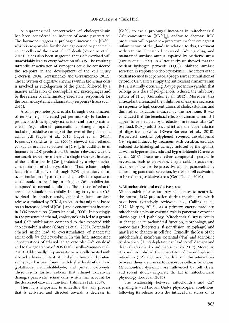
A supramaximal concentration of cholecystokinin has been considered an inducer of acute pancreatitis. The hormone triggers a prolonged increase in [Ca2+]i, which is responsible for the damage caused to pancreatic acinar cells and the eventual cell death (Voronina et al., 2015). It has also been suggested that Ca2+ overload will unavoidably lead to overproduction of ROS. The resulting intracellular activation of zymogens could be considered the set-point in the development of the cell injury (Petersen, 2004; Gerasimenko and Gerasimenko, 2012). The activation of digestive enzymes within the acinar cells is involved in autodigestion of the gland, followed by a massive infiltration of neutrophils and macrophages and by the release of inflammatory mediators, responsible for the local and systemic inflammatory response (Irrera et al., 2014).
Alcohol promotes pancreatitis through a combination of remote (e.g., increased gut permeability to bacterial products such as lipopolysaccharide) and more proximal effects (e.g., altered pancreatic cholinergic inputs), including oxidative damage at the level of the pancreatic acinar cell (Tapia et al., 2010; Lugea et al., 2011). Fernandez-Sanchez et al. (2009) showed that ethanol evoked an oscillatory pattern in [Ca2+]i, in addition to an increase in ROS production. Of major relevance was the noticeable transformation into a single transient increase of the oscillations in [Ca2+]i induced by a physiological concentration of cholecystokinin. Thus, ethanol might lead, either directly or through ROS generation, to an overstimulation of pancreatic acinar cells in response to cholecystokinin, resulting in a higher Ca2+ mobilization compared to normal conditions. The actions of ethanol created a situation potentially leading to cytosolic Ca2+ overload. In another study, ethanol reduced amylase release stimulated by CCK-8, an action that might be based on an increased level of [Ca2+]i and a concomitant increase in ROS production (Gonzalez et al., 2006). Interestingly, in the presence of ethanol, cholecystokinin led to a greater total Ca2+ mobilization compared to that expected with cholecystokinin alone (Gonzalez et al., 2008). Potentially, ethanol might lead to overstimulation of pancreatic acinar cells by cholecystokinin. In this line, intoxicating concentrations of ethanol led to cytosolic Ca2+ overload and to the generation of ROS (Del Castillo-Vaquero et al., 2010). Additionally, in pancreatic acinar cells treated with ethanol a lower content of total glutathione and protein sulfhydryls has been found, with higher levels of oxidized glutathione, malondialdehyde, and protein carbonyls. These results further indicate that ethanol oxidatively damages pancreatic acinar cells, which may account for the decreased exocrine function (Palmieri et al., 2007).
Thus, it is important to underline that any process that is activated and directed towards a decrease in
[Ca2+]i, to avoid prolonged increases in mitochondrial Ca2+ concentration ([Ca2+]m), and/or to decrease ROS production will represent a protective mechanism against inflammation of the gland. In relation to this, treatment with vitamin C restored impaired Ca2+ signaling and maintained amylase output impaired by oxidative stress (Sweiry et al., 1999). In a later study, we showed that the oxidant hydrogen peroxide (H2O2) inhibited amylase secretion in response to cholecystokinin. The effects of the oxidant seemed to depend on a progressive accumulation of cytosolic Ca2+. Interestingly, the antioxidant cinnamtannin B-1, a naturally occurring A-type proanthocyanidin that belongs to a class of polyphenols, reduced the inhibitory action of H2O2 (Gonzalez et al., 2012). Moreover, this antioxidant attenuated the inhibition of enzyme secretion in response to high concentrations of cholecystokinin and diminished oxidation induced by the hormone. It was concluded that the beneficial effects of cinnamtannin B-1 appear to be mediated by a reduction in intracellular Ca2+ overload, ROS production, and intracellular accumulation of digestive enzymes (Rivera-Barreno et al., 2010). Resveratrol, another polyphenol, reversed the abnormal Ca2+ signal induced by treatment with cerulein, and also reduced the histological damage induced by the agonist, as well as hyperamylasemia and hyperlipidemia (Carrasco et al., 2014). These and other compounds present in beverages, such as quercetin, ellagic acid, or catechins, have been shown to be protective against pancreatitis by controlling pancreatic secretion, by stellate cell activation, or by reducing oxidative stress (Gerloff et al., 2010).
3. Mitochondria and oxidative stressMitochondria possess an array of defenses to neutralize the normal ROS production during metabolism, which have been extensively reviewed (e.g., Collins et al., 2012; Murphy, 2012). As a primary energy producer, mitochondria play an essential role in pancreatic exocrine physiology and pathology. Mitochondrial stress results in changes in mitochondrial function, morphology, and homeostasis (biogenesis, fission/fusion, mitophagy) and may lead to changes in cell fate. Critically, the loss of the mitochondrial membrane potential (Ψm) and adenosine triphosphate (ATP) depletion can lead to cell damage and death (Gerasimenko and Gerasimenko, 2012). Moreover, it is well established that the status of the endoplasmic reticulum (ER) and mitochondria and the interactions between them are crucial to numerous cellular functions. Mitochondrial dynamics are influenced by cell stress, and recent studies implicate the ER in mitochondrial physiology (Lee et al., 2013).
The relationship between mitochondria and Ca2+ signaling is well known. Under physiological conditions, following its release from the intracellular stores or its

GONZALEZ et al. / Turk J Biol
804
influx from the extracellular space, Ca2+ accumulates in mitochondria. Once in the matrix, Ca2+ would regulate the activity of dehydrogenases, especially the pyruvate, isocitrate, and alpha-ketoglutarate dehydrogenases, therefore leading to a modulation of cellular energetic metabolism (Denton and McCormack, 1980). However, in the presence of oxidative stress, cytosolic Ca2+ responses in the exocrine pancreas are abnormally elevated and usually accompanied by elevation of Ca2+ in mitochondria, mitochondrial depolarization, and mitochondrial permeability transition pore opening. Thus, achievement of high mitochondrial Ca2+ levels under oxidative stress has been pointed out as the major factor determining mitochondria-dependent cell fate (Baumgartner et al., 2009). The implication of mitochondria in the impairment of the normal spatiotemporal pattern of intracellular Ca2+ signaling and, in particular, the transition to an irreversible intracellular Ca2+ overload has also been suggested (Baggaley et al., 2008).
In relation to this, stimulation of pancreatic acinar cells with cholecystokinin and/or the sarcoendoplasmic reticulum Ca2+ pump inhibitor, thapsigargin, both induced changes in the cellular oxidative status. The results indicated that ROS were generated via accumulation of Ca2+ in the mitochondria (Granados et al., 2004). Another study showed that the pancreatitis inducer taurolithocholate induced prolonged increases in [Ca2+]i, which were accompanied by an increase in the mitochondrial Ca2+ content. In turn, a dose-dependent increase ROS was observed, which impaired the production of ATP and resulted in cell death (Booth et al., 2011). A further study showed that stimulation of pancreatic acinar cells with a supraphysiological concentration of cholecystokinin caused a decrease in the mitochondrial membrane potential and ATP concentration, whereas the mitochondrial dehydrogenase activity was significantly increased. The antioxidant quercetin increased the AMP/ATP ratio, suggesting that the AMP-activated protein kinase system might be activated. In addition, quercetin strongly inhibited cholecystokinin-induced trypsin activity. This antioxidant might reduce the severity of pancreatitis by its modulation of mitochondrial physiology (Weber et al., 2014). Another study showed that ethanol decreased basal mitochondrial membrane potential and converted a transient depolarization, induced by physiologic concentrations of cholecystokinin, into a sustained decrease in mitochondrial membrane potential, resulting in reduced cellular ATP and increased cell death (Shalbueva et al., 2013). Finally, in addition to energy supply, mitochondria oxidative stress induced posttranslational histone modifications, promoter DNA methylation, and impairment of micro-RNA expression patterns (Mishra et al., 2014). Taking into account the existing evidence to
date, we can asseverate that pathological Ca2+ signaling is an important contributor to the initiation of cell injury. Cell damage may be caused by or acting through mitochondrial inhibition. The role of ROS in this process is also clear. However, establishing whether Ca2+ or ROS is the first one initiating the chain of events that finally ends in the disease remains controversial (Mukherjee et al., 2008). Preservation of mitochondria activity is thus an important challenge that the cell must accomplish in order to survive and prevent the development of diseases.
4. Melatonin and calcium in the exocrine pancreasAs we have mentioned above, impairment of Ca2+ signaling and excessive ROS production are pivotal in the induction of pancreatic disease. The antioxidant and radical scavenging actions of melatonin have been clearly demonstrated; however, the effect of the indole on Ca2+ handling by the cells is a matter of actual research. We have shown that the total Ca2+ mobilization evoked by cholecystokinin was attenuated in the presence of melatonin compared with the responses observed in the presence of the secretagogue alone. The effect of melatonin could be explained on the basis of a stimulated Ca2+ transport out of the cell through the plasma membrane and by a stimulation of Ca2+ reuptake into the ER (Santofimia-Castaño et al., 2014). In relation to this study, it has been suggested that melatonin stimulates an upregulation of sarco/endoplasmic reticulum Ca2+-ATPase and Na+/Ca2+ exchanger expression, which can alleviate Ca2+ overload in pancreatic acinar cells (Huai et al., 2012).
In a parallel study, we demonstrated that treatment of pancreatic acinar cells with melatonin also resulted in a smaller total Ca2+ mobilization in response to supramaximal concentrations of cholecystokinin. Basically, the evidence suggests that the indoleamine modulates Ca2+ handling by the cell, which is based on a more efficient transport of Ca2+ across cell membranes. The consequence would be that melatonin might alleviate a cytosolic Ca2+ overload following stimulation of cells with Ca2+-mobilizing agonists. Additionally, our studies show that melatonin reduced the oscillatory pattern of Ca2+ mobilization evoked by a physiological concentration of the secretagogue and completely inhibited Ca2+ mobilization induced by low picomolar concentrations of cholecystokinin. Melatonin improved cell viability in the presence of supraphysiological concentrations of cholecystokinin. Additionally, melatonin attenuated enzyme secretion in response to the high concentrations of cholecystokinin (Santofimia-Castaño et al., 2013). Therefore, all these observations suggest that melatonin alleviates intracellular Ca2+ accumulation, ROS production, and overstimulation of enzyme secretion, which are considered situations potentially leading to cell damage in the exocrine pancreas.

GONZALEZ et al. / Turk J Biol
805
Finally, our evidence further shows that accumulation of Ca2+ into mitochondria was not affected in the presence of melatonin; therefore, we think that Ca2+ handling by mitochondria might not be a major mechanism stimulated by melatonin to avoid cytosolic Ca2+ overload (Santofimia-Castaño et al., 2014).
5. Melatonin and pancreatic functionMany different receptors and signaling pathways govern the complex biology of pancreatic secretion (Chandra and Liddle, 2012). In the body, melatonin receptors include membrane and intracellular binding sites. Two types of melatonin receptors have been described at the plasma membrane, termed MT1 and MT2. These receptors belong to the family of guanine nucleotide-binding regulatory protein (G protein)-coupled receptors. A third melatonin binding site has been identified, formerly known as MT3. Quinone reductase 2, related to the xenobiotic metabolism of the cell, has been pointed out as the MT3. However, it has been stated that quinone reductase 2 does not comply with the criteria for being considered a melatonin receptor. Additionally, melatonin binding sites in the nucleus have been also reported. Binding of the indole within the nucleus has been reported to occur at the latter, identified as ROR orphan receptors. Finally, melatonin also interacts with cytosolic proteins, including calmodulin and calreticulin (Acuña-Castroviejo et al., 2014).
In general, it has been suggested that melatonin exerts a regulatory action on enzyme secretion. Given intraperitoneally, melatonin or its precursor L-tryptophan produced significant and dose-dependent increases in pancreatic amylase secretion under basal conditions or following stimulation of enzyme secretion by diversion of bile-pancreatic juice (Jaworek et al., 2004). Because bilateral vagotomy completely abolished the increase of amylase output caused by melatonin, the effects of the indole may be explained, at least in part, by stimulation of vagal nerves (Nawrot-Porąbka et al., 2013). Moreover, it has been shown that intraperitoneally administered melatonin or L-tryptophan, and also when delivered into the gut lumen, stimulated pancreatic amylase secretion. This effect was paralleled by significant increases of the plasma levels of cholecystokinin; therefore, it could be possible that melatonin stimulated pancreatic exocrine function via mechanisms involving enteropancreatic reflexes and cholecystokinin release (Jaworek et al., 2007; Nawrot-Porabka et al., 2007). However, when administered in vitro, melatonin attenuated enzyme secretion in response to high concentrations of cholecystokinin (Santofimia-Castaño et al., 2013). Thus, a controversy exists regarding the effect of the indole in in vivo vs. in vitro studies. Melatonin also exerts an action on acid/base movements. Aust et al. (2006) showed a stimulatory effect of melatonin
on bicarbonate secretion, which may help to prevent duodenal damage.
Nevertheless, a common agreement exists that clearly suggests protective actions of melatonin towards the exocrine pancreatic function. In this line, treatment with melatonin has shown beneficial effects on Ca2+ deregulations that are related to pancreatitis and the aging of the pancreas, therefore suggesting that melatonin could be an adequate therapeutic approach (Gomez-Pinilla et al., 2009). Melatonin protected against the induction of oxidative stress and tissue injury and restored cell function in experimental ischemia/reperfusion injury of the pancreas following obstruction of gastroduodenal or inferior splenic arteries in rats (Jaworek et al., 2003; Muñoz-Casares et al., 2006). Melatonin also exerts antioxidant effects on the gland. Evidence was given in the work by Jaworek et al. (2010) that showed that the pancreatic content of the lipid peroxidation products malondialdehyde and 4-hydroxynonenal increased, whereas the activity of the antioxidant enzyme glutathione peroxidase was decreased in pinealectomized rats. Similar observations were obtained in a previous work employing luzindole, an antagonist of melatonin MT2 receptors (Jaworek et al., 2002). Additionally, melatonin possesses antiinflammatory properties. Gülben et al. (2010) showed that, in the presence of melatonin, the edema, inflammation, perivascular infiltrate, acinar necrosis, fat necrosis, and hemorrhage were decreased in taurocholate-induced acute pancreatitis. Similarly, treatment with melatonin resulted in a significant reduction in pancreatic edema and the levels of lipid peroxidation, as well as a decrease of serum amylase activity, in pancreatitis evoked by cerulein (Qi et al., 1999). Recently it was suggested that melatonin protects the pancreatic damage via the decrease of oxidative stress and an increase of the activities of antioxidant enzymes (Carrasco et al., 2014). Last, but not least, melatonin also has direct free radicals scavenging action (Barlas et al., 2004; Jaworek et al., 2014).
On the other hand, it has been shown that the hormones ghrelin, leptin, and melatonin are able to modulate the immune function of the organism and to protect the pancreas against inflammatory damage. The protective mechanisms have been related to the stimulation of nonspecific immune defense and to the modulation of cytokine production, as well as to the activation of the antioxidant system in the acinar cells (Jaworek and Konturek, 2014). Moreover, melatonin and L-tryptophan activate mechanisms that involve direct scavenging of ROS, activation of antioxidant enzymes, reduction of proinflammatory cytokines and prostaglandins, activation of heat shock protein, and a decrease of necrosis and increase of regeneration in the pancreas (Jaworek et al., 2003; Jaworek et al., 2014). In this line, it has been

GONZALEZ et al. / Turk J Biol
806
shown that the indoleamine ameliorates cerulein-induced pancreatitis involving the activation of nuclear factor 2 erythroid related factor 2 (Nrf2) and nuclear factor kappa B (NFκB) (Jung et al., 2010).
6. Melatonin and mitochondriaPreventing mitochondrial dysfunction could be an effective therapeutic strategy against cellular degenerative processes, because mitochondrial dysfunction is considered an important contributing factor in a variety of physiopathological situations, including pancreatic diseases. Apoptosis, also known as programmed cell death, is an essential natural process that eliminates damaged, unneeded, or dangerous cells from the body. This process can be activated in several ways, and its impairment might be the basis of a long list of illnesses. Mitochondria play a pivotal role in the so called “intrinsic pathway” of apoptosis. This physiological mechanism relies on mitochondrial membrane permeabilization (MMP) to liberate proapoptotic factors from the intermembrane space of this organelle. Destabilization and/or decreased expression of antiapoptotic members of the Bcl-2 family of proteins (e.g., Bcl-2 and Bcl-xL) or induction of proapoptotic Bcl-2 family members (e.g., Bax, Bad, and Bak) can occur. In a later step, the permeability of the mitochondrial membrane is compromised, allowing the formation of outer membrane channels. As a consequence, proapoptotic factors from the intermembrane space of mitochondria are release into the cytoplasm. Among these, cytochrome c plays a critical role. In addition, proteases, ROS, and Ca2+ are also capable of promoting the MMP needed in order to release proapoptotic factors. Upon release of proapoptotic factors from mitochondria, activation of effector caspases (3, 6, and 7) takes place (Campo, 2009).
The potential mechanisms or approaches by which melatonin interacts with mitochondria may involve direct detoxification of free radicals, through its nonreceptor-mediated scavenging capacity, and an increase of the activity of mitochondrial electron transport chain complexes. It is expected that the indole thus improves mitochondrial respiration and ATP production. With regard to the mitochondrial electron transport chain, it has been suggested that melatonin interacts with complexes I and IV to promote electron flux. The indoleamine thus controls ROS production, thereby reducing electron leakage and ROS generation. Moreover, melatonin lowers damage to mitochondrial proteins and DNA, increases the activity of antioxidant enzymes, stabilizes the mitochondrial inner membrane, and regulates mitochondrial gene expression (Zhang and Zhang, 2014).
There is limited evidence showing the effect of melatonin on mitochondrial physiology in the healthy
exocrine pancreas. In a recent study by Santofimia-Castaño et al. (2014), which was carried out on pancreatic acinar cells stimulated with supraphysiological concentrations of cholecystokinin, the protective effects of melatonin on Ca2+ signaling could not be related to the involvement of Ca2+
accumulation into mitochondria. Interestingly, though, it has been suggested that melatonin improves Ψm of aged mice (Camello-Almaraz et al., 2008).
Nevertheless, the role of melatonin in preventing mitochondrial dysfunction in other tissues has been reported. To cite just some of the more recent research, we know that the indole protected the structural integrity of mitochondria in myocardial cells, promoted ATP synthesis, and avoided heart injury. The protection mechanism was related with antioxidative damage (Liu et al., 2014). The level of oxidative stress-related markers of mitochondrial apoptosis (Bax, cleaved caspase-3, and PARP, cytochrome c release into the cytosol) decreased in the presence of melatonin in acute respiratory distress syndrome (Sun et al., 2015). Melatonin abolished destructive demyelination effects induced by the drug cuprizone in the corpus callosum, by decreasing oxidative stress and by restoring mitochondrial respiratory enzyme activity and fusion and fission processes (Kashani et al., 2014). Melatonin administration also reduced Bax translocation to the mitochondria and the release of cytochrome c into the cytosol, preventing brain injury following a subarachnoid hemorrhage (Chen et al., 2014). The indoleamine also showed neuroprotective effects in transgenic mice of amyotrophic lateral sclerosis. Melatonin significantly delayed disease onset and neurological deterioration; its effects were explained by an inhibition of Rip2/caspase-1 pathway activation, by the release of mitochondrial cytochrome c, and by a decreased overexpression and activation of caspase-3 (Zhang et al., 2013). In hepatocytes, melatonin normalized the levels of ROS formation, intracellular reduced glutathione, cellular oxidized glutathione, lipid peroxidation, and mitochondrial depolarization induced by the antiepileptic drug phenytoin (Eghbal et al., 2014). In smooth muscle cells, aging induced a partial mitochondrial depolarization in resting conditions and reduced the depolarizing response to cellular stimulation, and melatonin treatment prevented all these changes (Martin-Cano et al., 2014).
Conversely to the study of its effects on intact tissue, the action of melatonin on pancreatic tumors has been an object of deepest research. It has been revealed that night work might influence cancer risk, possibly via suppression of melatonin release (Parent et al., 2012). In fact, changes of the levels of melatonin and tumor necrosis factor alpha (TNFα) in the serum of advanced cancer patients have been detected. It is noteworthy that the circadian rhythm of melatonin was altered (Muc-Wierzgon et al.,

GONZALEZ et al. / Turk J Biol
807
2003). In relation to cancer development, different studies have suggested that melatonin reduces proliferation in different tumor cell lines. The reported evidence indicates that melatonin could modulate the process of pancreatic oncogenesis and this coincides with the observation that melatonin, at high doses, could be potentially considered as a supportive treatment in the therapy of pancreatic cancer (Jaworek and Leja-Szpak, 2014). The major effects seem to be related to the apoptotic process, with a basis on the mitochondria. In this line, melatonin promotes apoptosis in pancreatic cancerous cells. Gonzalez et al. (2011) reported that melatonin induced transient changes in [Ca2+]i in AR42J tumor cells. This was accompanied by an increase in [Ca2+]m and depolarization of Ψm. A change
in the oxidative status of mitochondria was also noted. In addition, activation of caspase-3 was detected. Altogether, melatonin reduced AR42J cell viability. Formerly it was shown that melatonin exerted changes in the intrinsic pathway of apoptosis at the mitochondrial level and the cascade of caspases in human pancreatic carcinoma (PANC-1) cells. Interestingly, a stimulation of the protein levels of the proapoptotic factors Bcl-2/Bax and caspase-9 was observed. Of major relevance was the observation that the proapoptotic effect of melatonin was induced by a low (physiological) concentration of the indole (Leja-Szpak et al., 2010). In another study carried out in the same cell line, melatonin inhibited cellular proliferation, probably via reduction of vascular endothelial growth
Figure. Possible molecular pathways involved in the actions of melatonin in the exocrine pancreas. Melatonin regulates exocrine pancreatic function by either nonreceptor- or receptor-mediated effects. Melatonin might turn on an enteropancreatic reflex via release of intestinal hormones (like cholecystokinin, CCK) or of neurotransmitters at the nerve endings (such as, for example, acetylcholine, ACh, or vasoactive intestinal peptide, VIP) that will, in turn, regulate the pancreatic function. The indoleamine might also act on cellular plasma membrane receptors (MT1/MT2) and/or on intracellular receptors (MT3 or ROR/RZR). Acting on the latter, melatonin could promote antioxidant defenses. Other intracellular points of action of melatonin include calmodulin (Cmdlin) or calreticulin (Crtlin). An intracellular messenger involved in the actions of melatonin on the exocrine pancreas is calcium (Ca2+), by which the indole could modulate enzyme secretion. Finally, melatonin acts on the mitochondrial electron transport chain (ETC) to efficiently handle the oxidative status of these organelles, improving mitochondrial respiration and ATP production. Moreover, the indoleamine may regulate the mitochondrial pathway of apoptosis.
GONZALEZ et al. / Turk J Biol
808
factor (VEGF) concentrations in the extracellular medium and also intracellularly (Lv et al., 2012). The effect of melatonin on tumor angiogenesis was also investigated in a parallel study by Cui et al. (2012), who showed that the indole inhibited the cell proliferation and migration of human umbilical vein endothelial cells cocultured with PANC-1 cells, and also decreased the VEGF protein secreted to the cultured medium. Melatonin additionally stimulated phosphorylation of heat shock protein-27 (HSP27), a cytoprotective chaperone (Leja-Szpak et al., 2007). Another study carried out in the pancreatic cancer cell line SW-1990 revealed a growth inhibition after treatment of cells with high doses of melatonin; this action might be explained by a modulation of Bcl-2/Bax balance in the presence of the indole (Xu et al., 2013). Melatonin also enhanced chemotherapy-induced cytotoxicity and apoptosis in rat pancreatic tumor AR42J cells. The treatment with the indoleamine elevated mitochondrial membrane depolarization and augmented ROS production evoked by chemotherapeutic drugs (Uguz et al., 2012). An evaluation of pathological tumor evolution and oxidative stress markers in pancreatic tissue was carried out in Syrian hamsters. In that study, melatonin exerted beneficial effects that were associated with a decrease in lipoperoxide levels and increased antioxidant activity in the pancreatic tissue (Ruiz-Rabelo et al., 2011). Previously it had been shown that celecoxib, a specific inhibitor of cyclooxygenase-2,
induced a reduction in tumor nodules, oxidative stress, and death of Syrian hamsters with developed experimental pancreatic cancer. The combined treatment with melatonin exerted a synergistic beneficial effect. It is worth noting that melatonin alone had significant beneficial actions in improving the survival of hamsters. The antitumoral actions involved a modulation of the levels of lipoperoxides, reduced glutathione, superoxide dismutase, catalase, and glutathione peroxidase (Padillo et al., 2010).
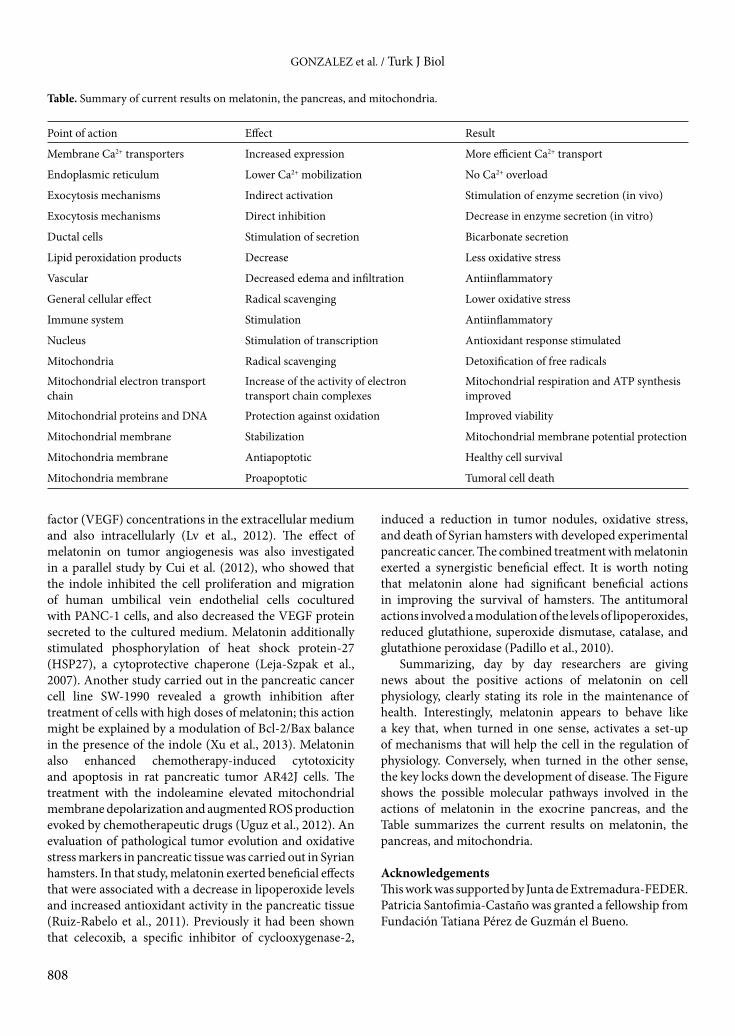
Summarizing, day by day researchers are giving news about the positive actions of melatonin on cell physiology, clearly stating its role in the maintenance of health. Interestingly, melatonin appears to behave like a key that, when turned in one sense, activates a set-up of mechanisms that will help the cell in the regulation of physiology. Conversely, when turned in the other sense, the key locks down the development of disease. The Figure shows the possible molecular pathways involved in the actions of melatonin in the exocrine pancreas, and the Table summarizes the current results on melatonin, the pancreas, and mitochondria.
AcknowledgementsThis work was supported by Junta de Extremadura-FEDER. Patricia Santofimia-Castaño was granted a fellowship from Fundación Tatiana Pérez de Guzmán el Bueno.
Table. Summary of current results on melatonin, the pancreas, and mitochondria.
Point of action Effect Result
Membrane Ca2+ transporters Increased expression More efficient Ca2+ transport
Endoplasmic reticulum Lower Ca2+ mobilization No Ca2+ overload
Exocytosis mechanisms Indirect activation Stimulation of enzyme secretion (in vivo)
Exocytosis mechanisms Direct inhibition Decrease in enzyme secretion (in vitro)
Ductal cells Stimulation of secretion Bicarbonate secretion
Lipid peroxidation products Decrease Less oxidative stress
Vascular Decreased edema and infiltration Antiinflammatory
General cellular effect Radical scavenging Lower oxidative stress
Immune system Stimulation Antiinflammatory
Nucleus Stimulation of transcription Antioxidant response stimulated
Mitochondria Radical scavenging Detoxification of free radicals
Mitochondrial electron transport chain
Increase of the activity of electrontransport chain complexes
Mitochondrial respiration and ATP synthesis improved
Mitochondrial proteins and DNA Protection against oxidation Improved viability
Mitochondrial membrane Stabilization Mitochondrial membrane potential protection
Mitochondria membrane Antiapoptotic Healthy cell survival
Mitochondria membrane Proapoptotic Tumoral cell death

GONZALEZ et al. / Turk J Biol
809
References
Acuña-Castroviejo D, Escames G, Venegas C, Díaz-Casado ME, Lima-Cabello E, López LC, Rosales-Corral S, Tan DX, Reiter RJ (2014). Extrapineal melatonin: sources, regulation, and potential functions. Cell Mol Life Sci 71: 2997–3025.
Armstrong JA, Cash N, Soares PM, Souza MH, Sutton R, Criddle DN (2013). Oxidative stress in acute pancreatitis: lost in translation? Free Radic Res 47: 917–933.
Aust S, Brucker B, Graf J, Klimpfinger M, Thalhammer T (2006). Melatonin modulates acid/base transport in human pancreatic carcinoma cells. Cell Physiol Biochem 18: 91–102.
Baggaley EM, Elliott AC, Bruce JI (2008). Oxidant-induced inhibition of the plasma membrane Ca2+-ATPase in pancreatic acinar cells: role of the mitochondria. Am J Physiol Cell Physiol 295: C1247–1260.
Barlas A, Cevik H, Arbak S, Bangir D, Sener G, Yeğen C, Yeğen BC (2004). Melatonin protects against pancreaticobiliary inflammation and associated remote organ injury in rats: role of neutrophils. J Pineal Res 37: 267–275.
Baumgartner HK, Gerasimenko JV, Thorne C, Ferdek P, Pozzan T, Tepikin AV, Petersen OH, Sutton R, Watson AJ, Gerasimenko OV (2009). Calcium elevation in mitochondria is the main Ca2+ requirement for mitochondrial permeability transition pore (mPTP) opening. J Biol Chem 284: 20796–20803.
Booth DM, Murphy JA, Mukherjee R, Awais M, Neoptolemos JP, Gerasimenko OV, Tepikin AV, Petersen OH, Sutton R, Criddle DN (2011). Reactive oxygen species induced by bile acid induce apoptosis and protect against necrosis in pancreatic acinar cells. Gastroenterology 140: 2116–2125.
Bruce JI, Elliott AC (2007). Oxidant-impaired intracellular Ca2+ signaling in pancreatic acinar cells: role of the plasma membrane Ca2+-ATPase. Am J Physiol Cell Physiol 293: C938–950.
Bubenik GA, Pang SF (1997). Melatonin levels in the gastrointestinal tissues of fish, amphibians, and a reptile. Gen Comp Endocrinol 106: 415–419.
Bubenik GA, Pang SF, Hacker RR, Smith PS (1996). Melatonin concentrations in serum and tissues of porcine gastrointestinal tract and their relationship to the intake and passage of food. J Pineal Res 21: 251–256.
Camello-Almaraz C, Gomez-Pinilla PJ, Pozo MJ, Camello PJ (2008). Age-related alterations in Ca2+ signals and mitochondrial membrane potential in exocrine cells are prevented by melatonin. J Pineal Res 45: 191–198.
Campo ML (2009). Apoptosis. In: Salido GM, Rosado JA, editors. Apoptosis: Involvement of Oxidative Stress and Intracellular Ca2+ Homeostasis. Dordrecht, the Netherlands: Springer, pp. 17–55.
Carafoli E (1991). Calcium pump of the plasma membrane. Physiol Rev 71: 129–153.
Carrasco C, Rodriguez AB, Pariente JA (2014). Effects of melatonin on the oxidative damage and pancreatic antioxidant defenses in cerulein-induced acute pancreatitis in rats. Hepatobiliary Pancreat Dis Int 13: 442–446.
Chandra R, Liddle RA (2012). Neurohormonal regulation of pancreatic secretion. Curr Opin Gastroenterol 28: 483–487.
Chen J, Wang L, Wu C, Hu Q, Gu C, Yan F, Li J, Yan W, Chen G (2014). Melatonin-enhanced autophagy protects against neural apoptosis via a mitochondrial pathway in early brain injury following a subarachnoid hemorrhage. J Pineal Res 56: 12–19.
Collins Y, Chouchani ET, James AM, Menger KE, Cochemé HM, Murphy MP (2012). Mitochondrial redox signalling at a glance. J Cell Sci 125: 801–806.
Cui P, Yu M, Peng X, Dong L, Yang Z (2012). Melatonin prevents human pancreatic carcinoma cell PANC-1-induced human umbilical vein endothelial cell proliferation and migration by inhibiting vascular endothelial growth factor expression. J Pineal Res 52: 236–243.
Del Castillo-Vaquero A, Salido GM, Gonzalez A (2010). Increased calcium influx in the presence of ethanol in mouse pancreatic acinar cells. Int J Exp Pathol 91: 114–124.
Denton RM, McCormack JG (1980). On the role of the calcium transport cycle in heart and other mammalian mitochondria. FEBS Lett 119: 1–8.
Eghbal MA, Taziki S, Sattari MR (2014). Mechanisms of phenytoin-induced toxicity in freshly isolated rat hepatocytes and the protective effects of taurine and/or melatonin. J Biochem Mol Toxicol 28: 111–118.
Fernandez-Sanchez M, del Castillo-Vaquero A, Salido GM, Gonzalez A (2009). Ethanol exerts dual effects on calcium homeostasis in CCK-8-stimulated mouse pancreatic acinar cells. BMC Cell Biol 10: 77.
Galloway CA, Lee H, Yoon Y (2012). Mitochondrial morphology-emerging role in bioenergetics. Free Radic Biol Med 53: 2218–2228.
Gerasimenko JV, Gerasimenko OV, Petersen OH (2014). The role of Ca2+ in the pathophysiology of pancreatitis. J Physiol 592: 269–280.
Gerasimenko OV, Gerasimenko JV (2012). Mitochondrial function and malfunction in the pathophysiology of pancreatitis. Pflugers Arch 464: 89–99.
Gerloff A, Singer MV, Feick P (2010). Beer and its non-alcoholic compounds: role in pancreatic exocrine secretion, alcoholic pancreatitis and pancreatic carcinoma. Int J Environ Res Public Health 7: 1093–1104.
Gomez-Pinilla PJ, Camello PJ, Pozo MJ (2009). Pancreatic calcium signaling: role in health and disease. Pancreatology 9: 329–333.
Gong J, Robbins LA, Lugea A, Waldron RT, Jeon CY, Pandol SJ (2014). Diabetes, pancreatic cancer, and metformin therapy. Front Physiol 5: 426.

GONZALEZ et al. / Turk J Biol
810
Gonzalez A, Camello PJ, Pariente JA, Salido GM (1997). Free cytosolic calcium levels modify intracellular pH in rat pancreatic acini. Biochem Biophys Res Commun 230: 652–656.
Gonzalez A, del Castillo-Vaquero A, Miro-Moran A, Tapia JA, Salido GM (2011). Melatonin reduces pancreatic tumor cell viability by altering mitochondrial physiology. J Pineal Res 50: 250–260.
Gonzalez A, Nuñez AM, Granados MP, Pariente JA, Salido GM (2006). Ethanol impairs CCK-8-evoked amylase secretion through Ca2+-mediated ROS generation in mouse pancreatic acinar cells. Alcohol 38: 51–57.
Gonzalez A, Pariente JA, Salido GM (2008). Ethanol impairs calcium homeostasis following CCK-8 stimulation in mouse pancreatic acinar cells. Alcohol 42: 565–573.
Gonzalez A, Salido GM (2001). Participation of mitochondria in calcium signalling in the exocrine pancreas. J Physiol Biochem 57: 331–339.
Gonzalez A, Santofimia-Castaño P, Rivera-Barreno R, Salido GM (2012). Cinnamtannin B-1, a natural antioxidant that reduces the effects of H2O2 on CCK-8-evoked responses in mouse pancreatic acinar cells. J Physiol Biochem 68: 181–191.
Gonzalez A, Schulz I, Schmid A (2000). Agonist-evoked mitochondrial Ca2+ signals in mouse pancreatic acinar cells. J Biol Chem 275: 38680–38686.
Granados MP, Salido GM, Pariente JA, Gonzalez A (2004). Generation of ROS in response to CCK-8 stimulation in mouse pancreatic acinar cells. Mitochondrion 3: 285–296.
Gülben K, Özdemir H, Berberoğlu U, Mersin H, Ýrkin F, Cakýr E, Aksaray S (2010). Melatonin modulates the severity of taurocholate-induced acute pancreatitis in the rat. Dig Dis Sci 55: 941–946.
Huai J, Shao Y, Sun X, Jin Y, Wu J, Huang Z (2012). Melatonin ameliorates acute necrotizing pancreatitis by the regulation of cytosolic Ca2+ homeostasis. Pancreatology 12: 257–263.
Huether G (1994). Melatonin synthesis in the gastrointestinal tract and the impact of nutritional factors on circulating melatonin. Ann NY Acad Sci 719: 146–158.
Hunt AE, Al-Ghoul WM, Gillette MU, Dubocovich ML (2001). Activation of MT2 melatonin receptors in rat suprachiasmatic nucleus phase advances the circadian clock. Am J Physiol Cell Physiol 280: C110–118.
Irrera N, Bitto A, Interdonato M, Squadrito F, Altavilla D (2014). Evidence for a role of mitogen-activated protein kinases in the treatment of experimental acute pancreatitis. World J Gastroenterol 20: 16535–16543.
Jaworek J (2006). Ghrelin and melatonin in the regulation of pancreatic exocrine secretion and maintaining of integrity. J Physiol Pharmacol 57: 83–96.
Jaworek J, Konturek SJ (2014). Hormonal protection in acute pancreatitis by ghrelin, leptin and melatonin. World J Gastroenterol 20: 16902–16912.
Jaworek J, Konturek SJ, Leja-Szpak A, Nawrot K, Bonior J, Tomaszewska R, Stachura J, Pawlik WW (2002). Role of endogenous melatonin and its MT2 receptor in the modulation of caerulein-induced pancreatitis in the rat. J Physiol Pharmacol 53: 791–804.
Jaworek J, Leja-Szpak A (2014). Melatonin influences pancreatic cancerogenesis. Histol Histopathol 29: 423–431.
Jaworek J, Leja-Szpak A, Bonior J, Nawrot K, Tomaszewska R, Stachura J, Sendur R, Pawlik W, Brzozowski T, Konturek SJ (2003). Protective effect of melatonin and its precursor L-tryptophan on acute pancreatitis induced by caerulein overstimulation or ischemia/reperfusion. J. Pineal Res 34: 40–52.
Jaworek J, Leja-Szpak A, Kot M, Jaworek A, Nawrot-Porbka K, Bonior J, Szklarczyk J (2014). The role of melatonin in pancreatic protection: could melatonin be used in the treatment of acute pancreatitis? Curr Pharm Des 20: 4834–4840.
Jaworek J, Nawrot K, Konturek SJ, Leja-Szpak A, Thor P, Pawlik WW (2004). Melatonin and its precursor, L-tryptophan: influence on pancreatic amylase secretion in vivo and in vitro. J Pineal Res 36: 155–164.
Jaworek J, Nawrot-Porabka K, Leja-Szpak A, Bonior J, Szklarczyk J, Kot M, Konturek SJ, Pawlik WW (2007). Melatonin as modulator of pancreatic enzyme secretion and pancreatoprotector. J Physiol Pharmacol 58: 65–80.
Jaworek J, Zwirska-Korczala K, Szklarczyk J, Nawrot-Porąbka K, Leja-Szpak A, Jaworek AK, Tomaszewska R (2010). Pinealectomy aggravates acute pancreatitis in the rat. Pharmacol Rep 62: 864–873.
Jung KH, Hong SW, Zheng HM, Lee HS, Lee H, Lee DH, Lee SY, Hong SS (2010). Melatonin ameliorates cerulein-induced pancreatitis by the modulation of nuclear erythroid 2-related factor 2 and nuclear factor-kappaB in rats. J Pineal Res 48: 239–250.
Kashani IR, Rajabi Z, Akbari M, Hassanzadeh G, Mohseni A, Eramsadati MK, Rafiee K, Beyer C, Kipp M, Zendedel A (2014). Protective effects of melatonin against mitochondrial injury in a mouse model of multiple sclerosis. Exp Brain Res 232: 2835–2846.
Lee JS, Hou X, Bishop N, Wang S, Flack A, Cho WJ, Chen X, Mao G, Taatjes DJ, Sun F et al. (2013). Aquaporin-assisted and ER-mediated mitochondrial fission: a hypothesis. Micron 47: 50–58.
Leja-Szpak A, Jaworek J, Pierzchalski P, Reiter RJ (2010). Melatonin induces pro-apoptotic signaling pathway in human pancreatic carcinoma cells (PANC-1). J Pineal Res 49: 248–255.
Leja-Szpak A, Jaworek J, Szklarczyk J, Konturek SJ, Pawlik WW (2007). Melatonin stimulates HSP27 phosphorylation in human pancreatic carcinoma cells (PANC-1). J Physiol Pharmacol 58: 177–188.
Leung PS, Ip SP (2006). Pancreatic acinar cell: its role in acute pancreatitis. Int J Biochem Cell Biol 38: 1024–1030.

GONZALEZ et al. / Turk J Biol
811
Liu LF, Qin Q, Qian ZH, Shi M, Deng QC, Zhu WP, Zhang H, Tao XM, Liu Y (2014). Protective effects of melatonin on ischemia-reperfusion induced myocardial damage and hemodynamic recovery in rats. Eur Rev Med Pharmacol Sci 18: 3681–3686.
Longnecker DS (2014). Anatomy and Histology of the Pancreas, 2014. 10.3998/panc.2014.3.
Lugea A, Waldron RT, French SW, Pandol SJ (2011). Drinking and driving pancreatitis: links between endoplasmic reticulum stress and autophagy. Autophagy 7: 783–785.
Lv D, Cui PL, Yao SW, Xu YQ, Yang ZX (2012). Melatonin inhibits the expression of vascular endothelial growth factor in pancreatic cancer cells. Chin J Cancer Res 24: 310–316.
Manchester LC, Poeggeler B, Alvares FL, Ogden GB, Reiter RJ (1995). Melatonin immunoreactivity in the photosynthetic prokaryote Rhodospirillum rubrum: implications for an ancient antioxidant system. Cell Mol Biol Res 41: 391–395.
Martin-Cano FE, Camello-Almaraz C, Acuña-Castroviejo D, Pozo MJ, Camello PJ (2014). Age-related changes in mitochondrial function of mouse colonic smooth muscle: beneficial effects of melatonin. J Pineal Res 56: 163–174.
McArthur AJ, Hunt AE, Gillette MU (1997). Melatonin action and signal transduction in the rat suprachiasmatic circadian clock: activation of protein kinase C at dusk and dawn. Endocrinology 138: 627–634.
Mishra PK, Raghuram GV, Jain D, Jain SK, Khare NK, Pathak N (2014). Mitochondrial oxidative stress-induced epigenetic modifications in pancreatic epithelial cells. Int J Toxicol 33: 116–129.
Muc-Wierzgon M, Nowakowska-Zajdel E, Zubelewicz B, Wierzgon J, Kokot T, Klakla K, Szkilnik R, Wiczkowski A (2003). Circadian fluctuations of melatonin, tumor necrosis factor-alpha and its soluble receptors in the circulation of patients with advanced gastrointestinal cancer. J Exp Clin Cancer Res 22: 171–178.
Mühlbauer E, Gross E, Labucay K, Wolgast S, Peschke E (2009). Loss of melatonin signalling and its impact on circadian rhythms in mouse organs regulating blood glucose. Eur J Pharmacol 606: 61–71.
Mukherjee R, Criddle DN, Gukovskaya A, Pandol S, Petersen OH, Sutton R (2008). Mitochondrial injury in pancreatitis. Cell Calcium 44: 14–23.
Muñoz-Casares FC, Padillo FJ, Briceño J, Collado JA, Muñoz-Castañeda JR, Ortega R, Cruz A, Túnez I, Montilla P, Pera C et al. (2006). Melatonin reduces apoptosis and necrosis induced by ischemia/reperfusion injury of the pancreas. J Pineal Res 40: 195–203.
Murphy MP (2012). Mitochondrial thiols in antioxidant protection and redox signaling: distinct roles for glutathionylation and other thiol modifications. Antioxid Redox Signal 16: 476–495.
Nawrot-Porąbka K, Jaworek J, Leja-Szpak A, Szklarczyk J, Konturek SJ, Reiter RJ (2013). Luminal melatonin stimulates pancreatic enzyme secretion via activation of serotonin-dependent nerves. Pharmacol Rep 65: 494–504.
Nawrot-Porabka K, Jaworek J, Leja-Szpak A, Szklarczyk J, Kot M, Mitis-Musioł M, Konturek SJ, Pawlik WW (2007). Involvement of vagal nerves in the pancreatostimulatory effects of luminal melatonin, or its precursor L-tryptophan. Study in the rats. J Physiol Pharmacol 58: 81–95.
Padillo FJ, Ruiz-Rabelo JF, Cruz A, Perea MD, Tasset I, Montilla P, Túnez I, Muntané J (2010). Melatonin and celecoxib improve the outcomes in hamsters with experimental pancreatic cancer. J Pineal Res 49: 264–270.
Palmieri VO, Grattagliano I, Palasciano G (2007). Ethanol induces secretion of oxidized proteins by pancreatic acinar cells. Cell Biol Toxicol 23: 459–464.
Paredes SD, Korkmaz A, Manchester LC, Tan DX, Reiter RJ (2009). Phytomelatonin: a review. J Exp Bot 60: 57–69.
Parent ME, El-Zein M, Rousseau MC, Pintos J, Siemiatycki J (2012). Night work and the risk of cancer among men. Am J Epidemiol 76: 751–759.
Peinado JR, Diaz-Ruiz A, Frühbeck G, Malagon MM (2014). Mitochondria in metabolic disease: getting clues from proteomic studies. Proteomics 14: 452–466.
Petersen OH (2004). Local and global Ca2+ signals: physiology and pathophysiology. Biol Res 37: 661–664.
Putney JW (1988). A model for receptor-regulated calcium entry. Cell Calcium 7: 1–12.
Qi W, Tan DX, Reiter RJ, Kim SJ, Manchester LC, Cabrera J, Sainz RM, Mayo JC (1999). Melatonin reduces lipid peroxidation and tissue edema in cerulein-induced acute pancreatitis in rats. Dig Dis Sci 44: 2257–2262.
Reczek CR, Chandel NS (2014). ROS-dependent signal transduction. Curr Opin Cell Biol 33: 8–13.
Rivera-Barreno R, del Castillo-Vaquero A, Salido GM, Gonzalez A (2010). Effect of cinnamtannin B-1 on cholecystokinin-8-evoked responses in mouse pancreatic acinar cells. Clin Exp Pharmacol Physiol 37: 980–988.
Ruiz-Rabelo J, Vazquez R, Arjona A, Perea D, Montilla P, Tunez I, Muntane J, Padillo J (2011). Improvement of capecitabine antitumoral activity by melatonin in pancreatic cancer. Pancreas 40: 410–414.
Sah RP, Dawra RK, Saluja AK (2013). New insights into the pathogenesis of pancreatitis. Curr Opin Gastroenterol 29: 523–530.
Santofimia-Castaño P, Ruy DC, Fernandez-Bermejo M, Salido GM, Gonzalez A (2014). Pharmacological dose of melatonin reduces cytosolic calcium load in response to cholecystokinin in mouse pancreatic acinar cells. Mol Cell Biochem 397: 75–86.
Santofimia-Castaño P, Ruy DC, Salido GM, González A (2013). Melatonin modulates Ca2+ mobilization and amylase release in response to cholecystokinin octapeptide in mouse pancreatic acinar cells. J Physiol Biochem 69: 897–908.
GONZALEZ et al. / Turk J Biol
812
Shalbueva N, Mareninova OA, Gerloff A, Yuan J, Waldron RT, Pandol SJ, Gukovskaya AS (2013). Effects of oxidative alcohol metabolism on the mitochondrial permeability transition pore and necrosis in a mouse model of alcoholic pancreatitis. Gastroenterology 144: 437–446.
Slominski RM, Reiter RJ, Schlabritz-Loutsevitch N, Ostrom RS, Slominski AT (2012). Melatonin membrane receptors in peripheral tissues: distribution and functions. Mol Cell Endocrinol 351: 152–166.
Streb H, Irvine RF, Berridge MJ, Schulz I (1983). Release of Ca2+ from a nonmitochondrial intracellular store in pancreatic acinar cells by inositol-1,4,5-trisphosphate. Nature 306: 67–69.
Sun CK, Lee FY, Kao YH, Chiang HJ, Sung PH, Tsai TH, Lin YC, Leu S, Wu YC, Lu HI et al. (2015). Systemic combined melatonin-mitochondria treatment improves acute respiratory distress syndrome in the rat. J Pineal Res 58: 137–150.
Sweiry JH, Shibuya I, Asada N, Niwa K, Doolabh K, Habara Y, Kanno T, Mann GE (1999). Acute oxidative stress modulates secretion and repetitive Ca2+ spiking in rat exocrine pancreas. Biochim Biophys Acta 1454: 19–30.
Tapia JA, Salido GM, González A (2010). Ethanol consumption as inductor of pancreatitis. World J Gastrointest Pharmacol Ther 1: 3–8.
Tinel H, Cancela JM, Mogami H, Gerasimenko JV, Gerasimenko OV, Tepikin AV, Petersen OH (1999). Active mitochondria surrounding the pancreatic acinar granule region prevent spreading of inositol trisphosphate-evoked local cytosolic Ca2+ signals. EMBO J 18: 4999–5008.
Uguz AC, Cig B, Espino J, Bejarano I, Naziroglu M, Rodríguez AB, Pariente JA (2012). Melatonin potentiates chemotherapy-induced cytotoxicity and apoptosis in rat pancreatic tumor cells. J Pineal Res 53: 91–98.
Vician M, Zeman M, Herichova I, Jurani M, Blazicek P, Matis P (1999). Melatonin content in plasma and large intestine of patients with colorectal carcinoma before and after surgery. J Pineal Res 27: 164–169.
Voronina S, Collier D, Chvanov M, Middlehurst B, Beckett AJ, Prior IA, Criddle DN, Begg M, Mikoshiba K, Sutton R et al. (2015). The role of Ca2+ influx in endocytic vacuole formation in pancreatic acinar cells. Biochem J 465: 405–412.
Walsh C, Barrow S, Voronina S, Chvanov M, Petersen OH, Tepikin A (2009). Modulation of calcium signalling by mitochondria. Biochim Biophys Acta 1787: 1374–1382.
Weber H, Jonas L, Wakileh M, Krüger B (2014). Beneficial effect of the bioflavonoid quercetin on cholecystokinin-induced mitochondrial dysfunction in isolated rat pancreatic acinar cells. Can J Physiol Pharmacol 92: 215–225.
Williams JA (2010). Regulation of acinar cell function in the pancreas. Curr Opin Gastroenterol 26: 478–483.
Xu C, Wu A, Zhu H, Fang H, Xu L, Ye J, Shen J (2013). Melatonin is involved in the apoptosis and necrosis of pancreatic cancer cell line SW-1990 via modulating of Bcl-2/Bax balance. Biomed Pharmacother 67: 133–139.
Yu JH, Kim H (2014). Oxidative stress and inflammatory signaling in cerulein pancreatitis. World J Gastroenterol 20: 17324–17329.
Zhang HM, Zhang Y (2014). Melatonin: a well-documented antioxidant with conditional pro-oxidant actions. J Pineal Res 57: 131–146.
Zhang Y, Cook A, Kim J, Baranov SV, Jiang J, Smith K, Cormier K, Bennett E, Browser RP, Day AL et al. (2013). Melatonin inhibits the caspase-1/cytochrome c/caspase-3 cell death pathway, inhibits MT1 receptor loss and delays disease progression in a mouse model of amyotrophic lateral sclerosis. Neurobiol Dis 55: 26–35.